Article

Evaporation of Saline Droplets on a Superhydrophobic Substrate: Formation of Crystal Shell and “Legs”

1
MOE Key Laboratory of Material Physics and Chemistry under Extraordinary Conditions, School of Physical Science and Technology, Northwestern Polytechnical University, Xi’an 710129, China
2
State Key Laboratory of Solidification Processing, Northwestern Polytechnical University, Xi’an 710072, China
*
Authors to whom correspondence should be addressed.
Materials 2023, 16(14), 5168; https://doi.org/10.3390/ma16145168
Submission received: 24 May 2023 / Revised: 14 July 2023 / Accepted: 19 July 2023 / Published: 22 July 2023

Abstract

:
We studied the evaporation-driven crystallization in the droplets of sodium acetate anhydrous (CH3COONa) aqueous solution, which were deposited on superhydrophobic substrates. The results reveal distinct crystallization behaviors between saturated and unsaturated droplets under identical experimental conditions. Specifically, unsaturated droplets could form a quasi-spherical crystal shell on the superhydrophobic substrate, while saturated droplets could develop crystal legs between the droplet and substrate when the crystal shell formed. Subsequently, the saturated droplet was lifted off the substrate by the growing crystal legs. The formation of crystal shell was closely associated with the evaporation from the droplet surface and the internal convection inside the droplet. The formation of crystal legs was induced by the heterogeneous nucleation effect caused by the substrate of SiO2 nanoparticles. These findings provide valuable insights into regulating the morphology of salt crystallization through adjustments in salt solution concentration and substrate surface structure.

1. Introduction

The evaporation of salt solution droplets plays an important role in many applications, such as analytical chemistry [1], coatings [2], biology [3], medical diagnosis [4], and food science [5]. The evaporation process of salt solution droplets involves many physical phenomena, such as contact line movements [6,7], natural and evaporation-induced convection [8,9], and crystallization [10,11]. Subjects related to droplet evaporation have been investigated for years, including the influence of the external environment (temperature, humidity and pressure) on droplet evaporation, the crystallization and phase transition inside the droplets, and the physical or chemical process at the contact lines.
The study of the evaporation of salt droplets under different environmental conditions provides insights into evaporation models and crystallization mechanisms. The flash evaporation process of saltwater droplets in a vacuum chamber showed that the level of salt in solution decreased the intensity of droplet evaporation. With the increase in salt concentration in water, the evaporation rate decreased [12,13]. The evaporation and crystallization processes in the pendant droplet showed that when the evaporation rate was low, a salt shell formed outside the droplet. Once the salt shell formed, the evaporation rate decreased and changed from linear to nonlinear [14]. In contrast, the evaporation of sessile salt droplets is closely related to the contact angle and contact line. G.V. Kuznetsov et al. reported that crystalline hydrates grew near the contact line and the droplet profile was distorted [15]. Virginie Soulié showed that low salt concentrations (≥10−6 mol/L NaCl) lead to pinning of the three-phase contact line. Droplets with salt concentration of ≤10−7 mol/L NaCl evaporate in a constant contact angle model [16]. These reports indicate that the contact line plays an essential role in the evaporation-caused crystallization of sessile salt droplets.
To minimize the influence of the contact line on droplet evaporation and crystallization, we investigated the process of CH3COONa crystallization during droplet evaporation on superhydrophobic substrates. Currently, the superhydrophobic surfaces can be prepared by several different techniques ranging from templating [17], etching [18], and deposition [19] to anodic oxidation [20], etc. Here, we adopted a facile dip-coating method [21]. Our findings indicate that evaporating CH3COONa solution droplets on a superhydrophobic substrate leads to the formation of crystal shells and legs. Notably, the temperature required for crystal leg growth in this study (20~30 °C) was significantly lower than that reported in previous research (60~100 °C) for similar phenomena [22,23]. The formation of CH3COONa crystal shells and legs, approximately at room temperature, holds significant value in various applications, including the preparation of heteromorphic crystals, corrosion mitigation on solid substrates, and self-transport during the crystallization of salt droplets.

2. Materials and Methods

2.1. Preparation of Superhydrophobic Substrate

The superhydrophobic substrates were fabricated by immersing a glass slide into a sol containing nano-sized silica (SiO2) particles which were synthesized via the Stöber process and doped with hexamethyl disilazane (HMDS) [24].
A certain volume of ethanol (C2H5OH, ≥99.5%, AR) and tetraethyl orthosilicate (TEOS, C8H20O4Si, 99.99% metals basis) was transferred into a beaker and stirred using a magnetic stirrer (HJ-2B, SHUANGXU, Beijing, China) at a speed of 600~700 r/min for 20~30 min at room temperature. Ammonium hydroxide aqueous solution (NH4OH, AR) was subsequently introduced into the mixture and stirred for 20~30 min. The beaker was then sealed and allowed to settle at room temperature for 5~7 days. Finally, hexamethyl disilazane (HMDS, C6H19NSi2, 98%, AR) was added to the prepared mixture and stirred for another 20–30 min. The final mixture was settled at room temperature for 1~2 days, yielding a sol suitable for coating substrates. The volume ratio of reagents used in sol preparation was C2H5OH:TEOS:NH4OH:HMDS = 10:1:0.36:0.56, with all reagents sourced from Aladdin Industrial Corporation, China.
To fabricate the superhydrophobic substrate, a glass slide was impregnated into the sol and stirred for 20~30 s using a dip coater (HTDC-300M, HTLAB, Shanghai, China) with a descending speed of 2 mm/s and an ascending speed of 4 mm/s. After being soaked for 20~30 s and naturally air-dried for 10~15 min at room temperature, the substrate was coated. The surface microstructure of the superhydrophobic substrate was characterized using a field emission scanning electron microscope (FESEM, Zeiss Supra 55, Jena, Germany). Image-Pro Plus 6.0 was used to analyze the SEM images for the quantitative characterization of the substrate.

2.2. Preparation of CH3COONa Solution

The CH3COONa salt solution was prepared by dissolving sodium acetate anhydrous salt (CH3COONa, ≥99.0%, AR, Aladdin Industrial Corporation, Shanghai, China) in water and stirring the mixture with a magnetic stirrer at a speed of 600~700 r/min for approximately 30 min. To obtain a saturated sodium acetate salt solution, excess sodium acetate salt was added to water until it could not completely dissolve. After being stirred for 2 h, and with the remaining solid residue filtered out, the saturated solution was obtained. The water used in this study was purified using an ultrapure water system (UPTA-20L, China).

2.3. Experimental Procedure

The experimental setup is illustrated in Figure 1. A saline droplet (10 μL) was deposited onto the superhydrophobic substrate using a microsyringe. The evaporation and crystallization process of the droplet were monitored in real time using a high-definition CCD video microscope (GP-640S, GaoPin, Guangzhou, China). The evaporation-induced change in droplet mass was monitored using a high-precision electronic balance (FA1004, Sunny Hengping Instrument, Shanghai, China) within a closed acrylic chamber to minimize the impact of external perturbations such as air flow and contamination. The surface tension of CH3COONa aqueous solution with various concentrations was measured using a surface-tension meter (JK99C, Powereach, Shanghai, China). The morphology of crystals and the thickness of crystal shell were characterized using a field emission scanning electron microscope. The experimental temperature was maintained at 25 ± 2 °C while the relative humidity (RH) was kept between approximately 40 and 45%.

3. Results and Discussion

3.1. Characterization of the Superhydrophobic Substrate

The wettability of the coated substrates was characterized by apparent contact angle (CA) and sliding angle (SA), which were measured using a contact angle meter (SDC-200S, Powereach, China). The results show that a water droplet (10 μL) had a CA of 160 ± 2° (Figure 2a) and an SA of 5° (Figure 2b) on this substrate, indicating that the prepared substrates had an excellent superhydrophobicity. The SEM image showed that SiO2 nanoparticles were randomly stacked on the substrate to form a hierarchical micro-nano porous surface (Figure 2c). The SiO2 particles on the substrate surface ranged from approximately 100 to 300nm, with an average diameter of 225 nm, as calculated using Gauss fitting (Figure 2c). The density of the SiO2 particles was also quantified by Image-Pro Plus 6.0 to be 1.05 × 10¹⁰/cm2. Based on the number (4~5 layers) of accumulated layers of particles and the average diameter (225 nm), the thickness of silica coating measures approximately 1.0 μm. The superhydrophobic coating exhibits a surface roughness of ~0.10 μm, as determined by the formula Ra = 1 n Z i [25], where Ra is the average roughness, and Z i represents the vertical distance from the particles located on the top layer to the mean height.
The superhydrophobicity of the substrate was determined via both the surface chemistry of SiO2 nanoparticles and the surface microstructure of the substrate. On the one hand, SiO2 nanoparticles were silanized during the preparation of the sol. The introduction of alkyl chains on the surface of SiO2 nanoparticles reduces their surface energy significantly. On the other hand, the hierarchical micro-nano porous structures enable the trapping of air pockets which greatly reduce the solid–liquid contact area [26], eventually leading to the Cassie–Baxter state of the deposited droplets [27], as shown in Figure 2d.

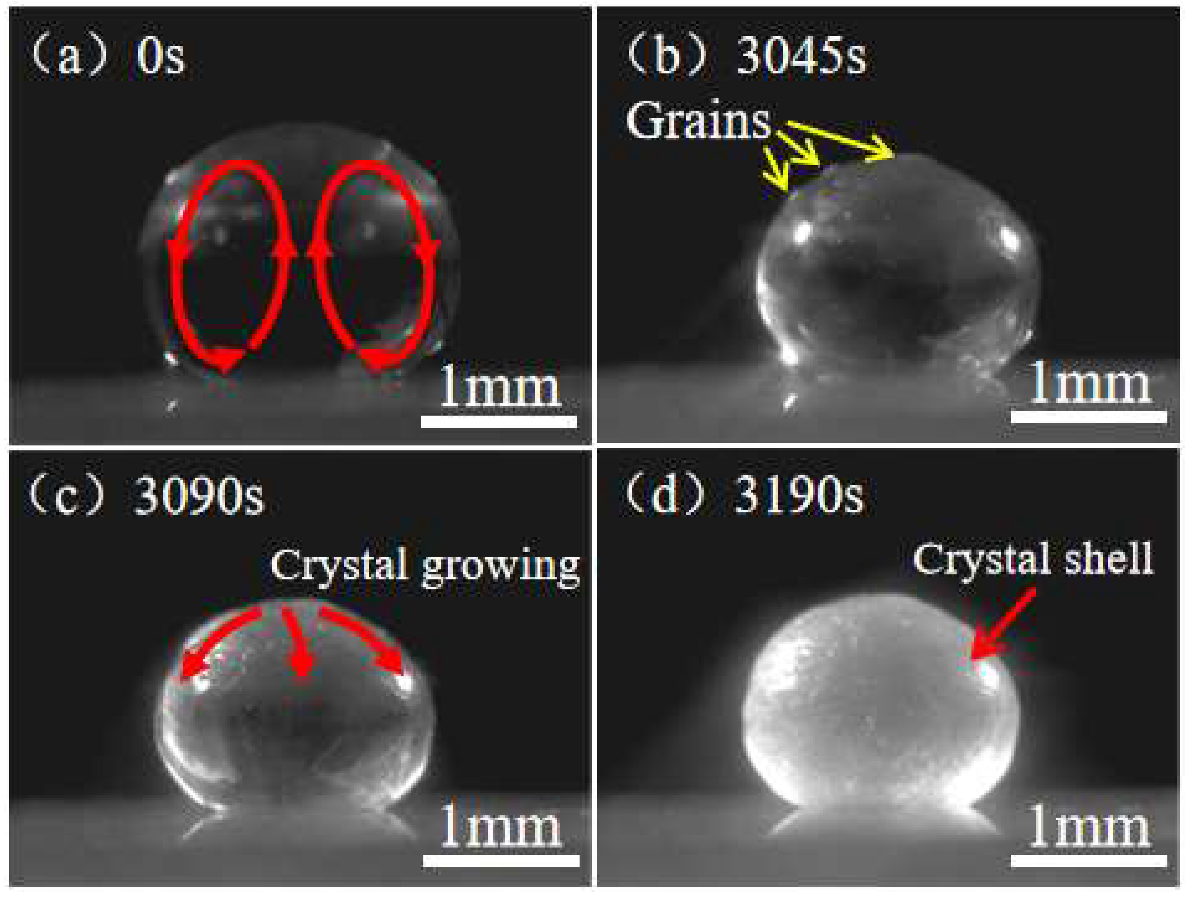
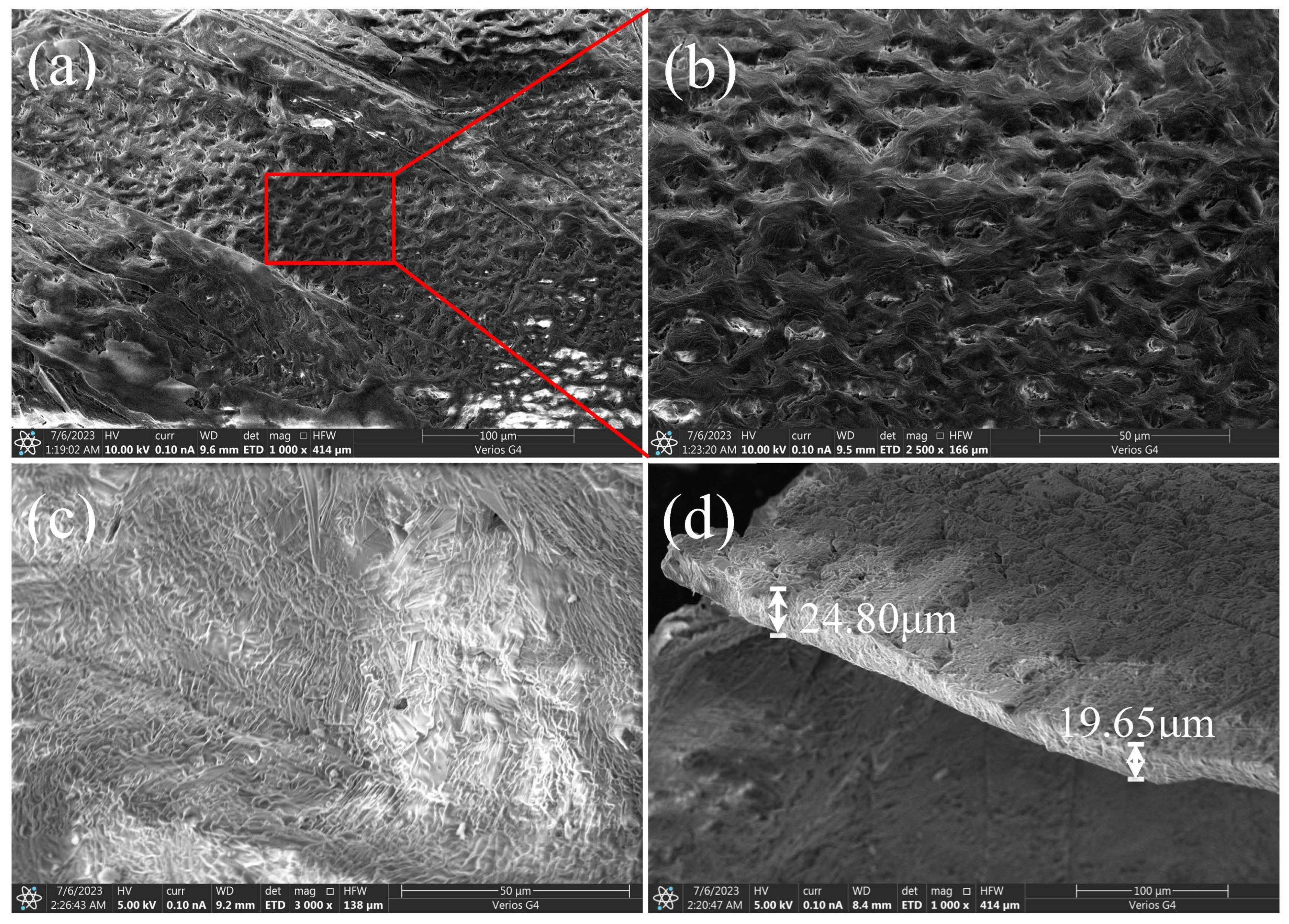
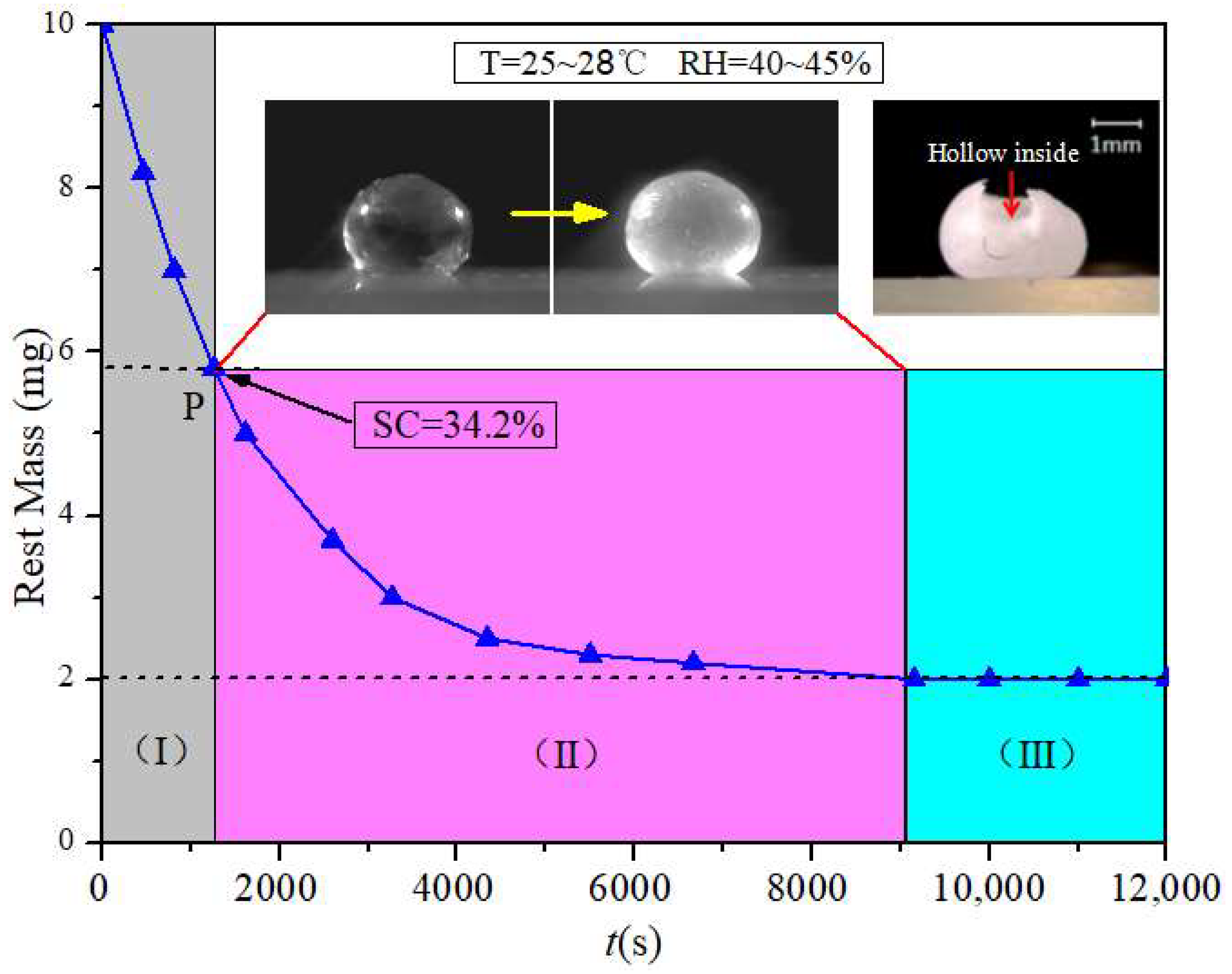
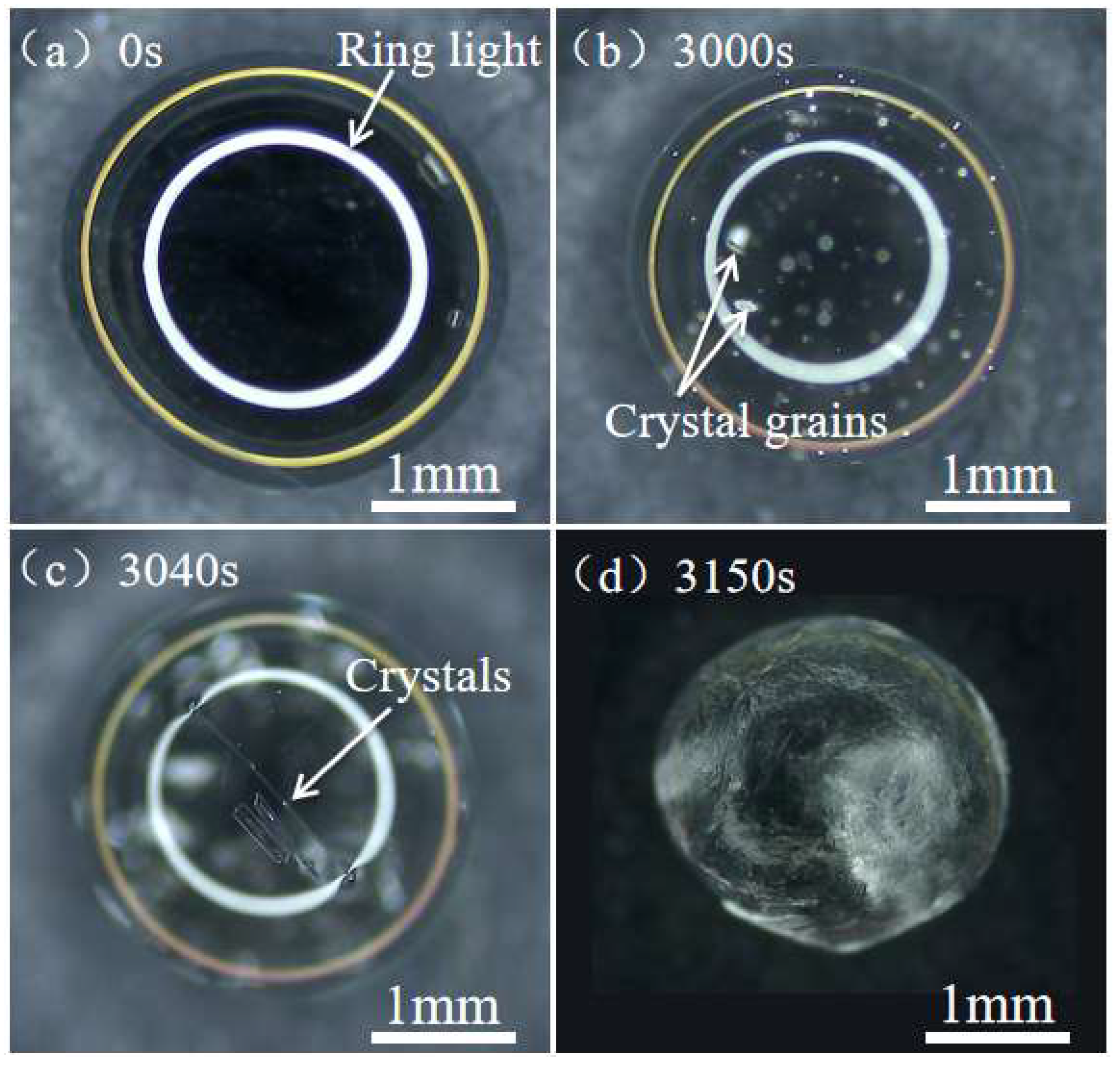
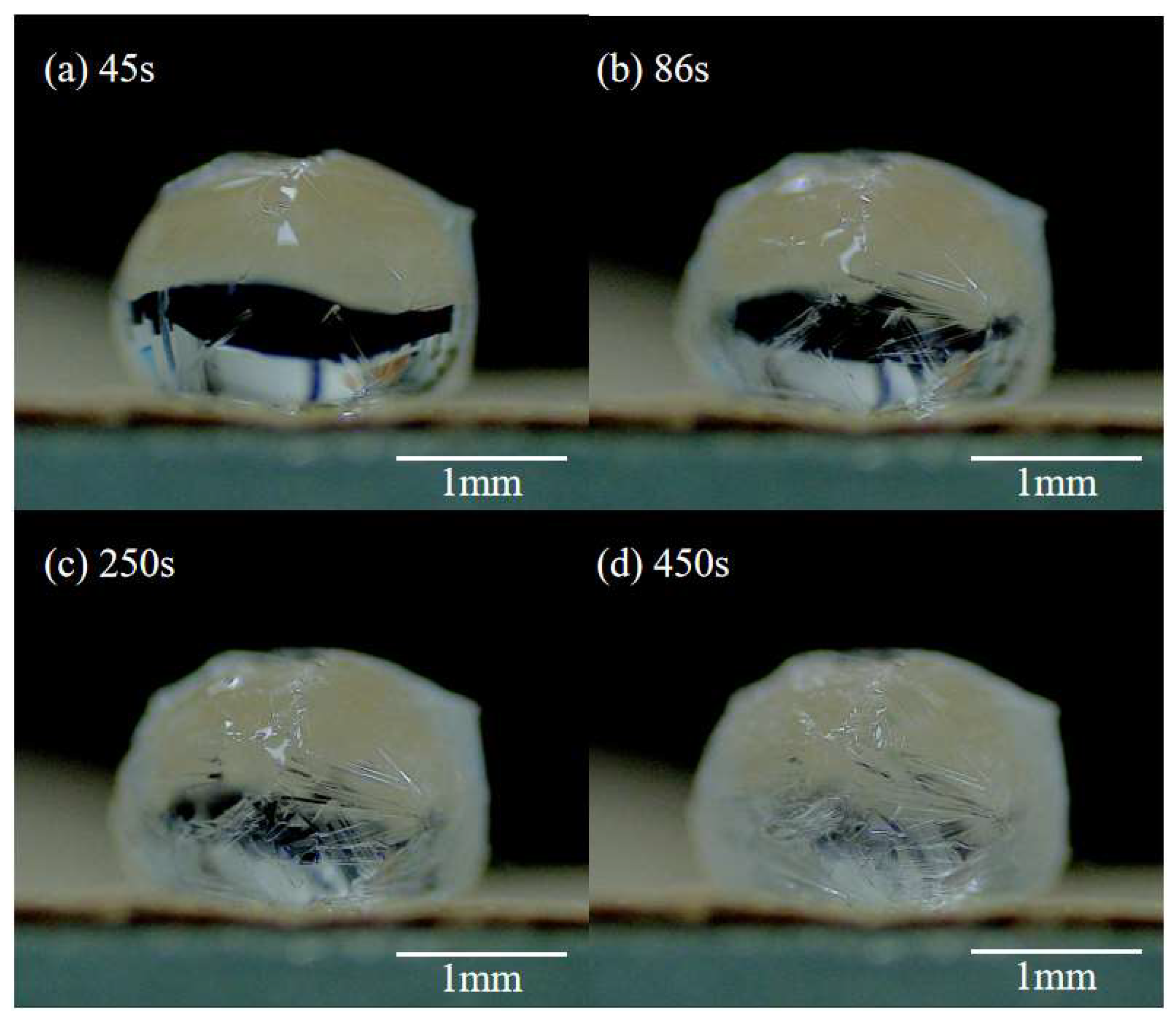
3.2. Formation of Crystal Shell

As shown in Figure 3a, when a droplet of unsaturated CH3COONa solution (10 μL, 20 wt.%) is deposited on the superhydrophobic substrate, it has an approximate spherical shape due to its high contact angle of 160 ± 2° on the superhydrophobic surface. In addition, the size of the deposited droplet is smaller than the capillary length: l c = γ e f f ρ g [28] (γeff is surface tension, ρ is the density of droplet, g is gravitational acceleration). For a 10 μL CH3COONa solution droplet, R~1.34 mm and l c ~2.00 mm; thus, the effect of gravity can be ignored in the whole process of droplet evaporation.
Development of internal natural convection. Being deposited on the substrate, there is a convection inside the droplet which flows upward along the central axis of the droplet and downward along the droplet surface (Figure 3a). On a superhydrophobic substrate, the evaporation rate is highest at the top of the droplet [29,30]. A higher evaporation rate leads to more solvent loss. As a result, a decreasing concentration gradient from the top to the contact line was observed on the droplet surface. Our experimental results (Figure 4) demonstrate that the surface tension of a CH3COONa salt solution decreases with the increasing concentration. Therefore, this decreasing concentration gradient results in an increasing surface tension gradient, which induces a Marangoni flow along the air–liquid interface of the droplet. The Marangoni flow flowed from the top of the droplet to the contact line along the droplet surface. Under the restriction of the substrate, the Marangoni flow increased in the center of the droplet, ultimately generating a symmetrical annular vortex, as shown in Figure 3a and the inset of Figure 4.
Crystallization on droplet surface. As evaporation proceeded, there was a significant volumetric loss of the droplet. Some small crystal grains were observed at the top of the droplet surface (Figure 3b). With further evaporation, small crystal grains grew from the top of the droplet downwards along the surface to the contact line (Figure 3c). Eventually, the growing crystal formed a spherical shell that coated the entire droplet surface (Figure 3d). The SEM image of the surface morphology of the spherical crystal shell is shown in Figure 5a–c, which indicates that the crystal shell resulting from the evaporation of CH3COONa solution droplet completely differs from the long rod-like structure formed in the CH3COONa solution. Figure 5d shows that the thickness of the crystal shell is about 20 μm. Figure 6 shows the mass of the droplet as a function of time during evaporation. After 41.5% of the water in the droplet was evaporated, the concentration of droplet became saturated (34.2 wt.%) at t ≈ 1300 s (P point in Figure 6). However, crystal grains at the top of the droplet were actually observed at t ≈ 3000 s (Figure 3b and Figure 7b). This can be explained by the Kelvin equation, which elucidates the relationship between concentration of grain-saturated solution and crystal radius via the following equation [31,32,33]:
R T ln c 2 c 1 = 2 σ M ρ ( 1 r 2 1 r 1 )
where c1 and c2 represent the solubility of grains with radii r1 and r2, respectively. Σ is the interfacial tension between crystal and solution, M is the molar mass, ρ is the density of crystal, R is the thermodynamic gas constant, and T is the absolute temperature.
It should be noted that even the droplet concentration reached critical saturation (34.2 wt.%), no evidenced crystal nuclei formed. As the concentration of the droplet increases to 58.8 wt.%, which is much higher than the critical saturation concentration of 34.2 wt.%, stable grains are observed (Figure 7b,c).
The growth of crystals on the droplet surface is attributed to a combination of factors, including droplet evaporation, droplet surface tension, and internal convection. The evaporation rate at the top of the droplet was the fastest when it was on a superhydrophobic surface. The evaporation rate was lower at a greater distance away from the top of the droplet surface [34,35]. When the surface concentration of the droplet reaches saturation, nucleation occurs first at the top of the droplet. With further evaporation, the nuclei gradually grow to form needle-shaped CH3COONa crystals that are buoyant on the droplet surface due to its surface tension. The needle-shaped CH3COONa crystals adhere to each other during their growth process, forming a localized crystal shell that acts as a “cap” covering the top surface of the droplet. During this period, the droplet provides support for the crystal “cap” to float on its surface due to surface tension. Furthermore, the internal convection within the droplet would also prevent the crystal shell from sinking until it grows to fully cover the surface of the droplet. The internal convection within the droplet not only generated an upward lifting force, but also provided a constant supply of CH3COO- and Na+ for the growth of crystals at the droplet surface. Consequently, the CH3COONa crystals tend to grow exclusively on the surface of droplets and eventually form a crystal shell, as shown in the corresponding inset of part (Ⅲ) in Figure 5.

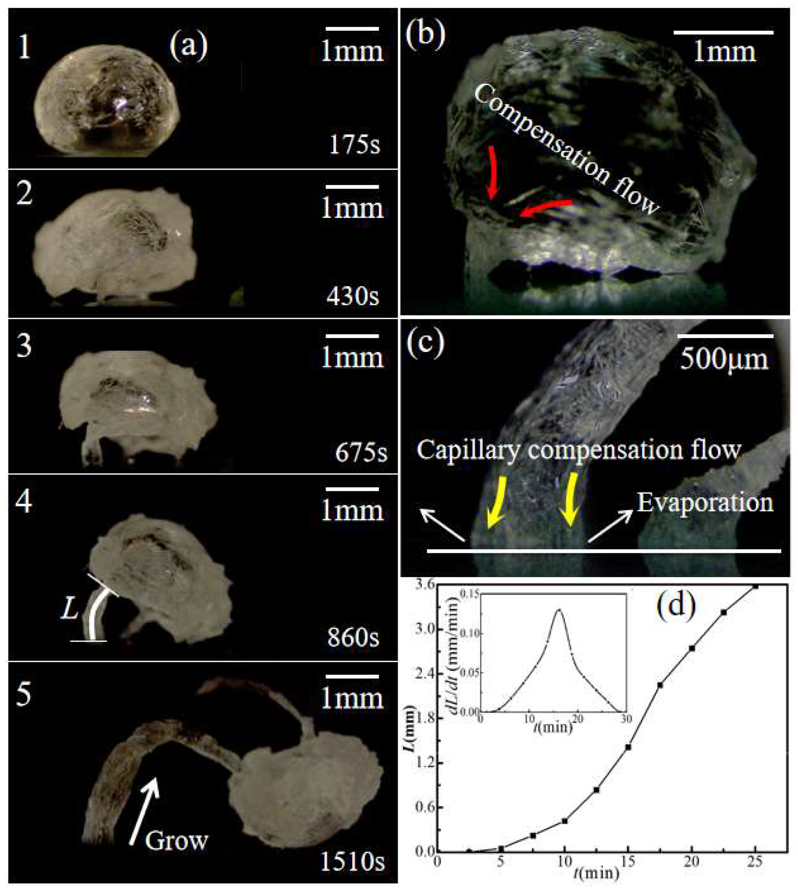
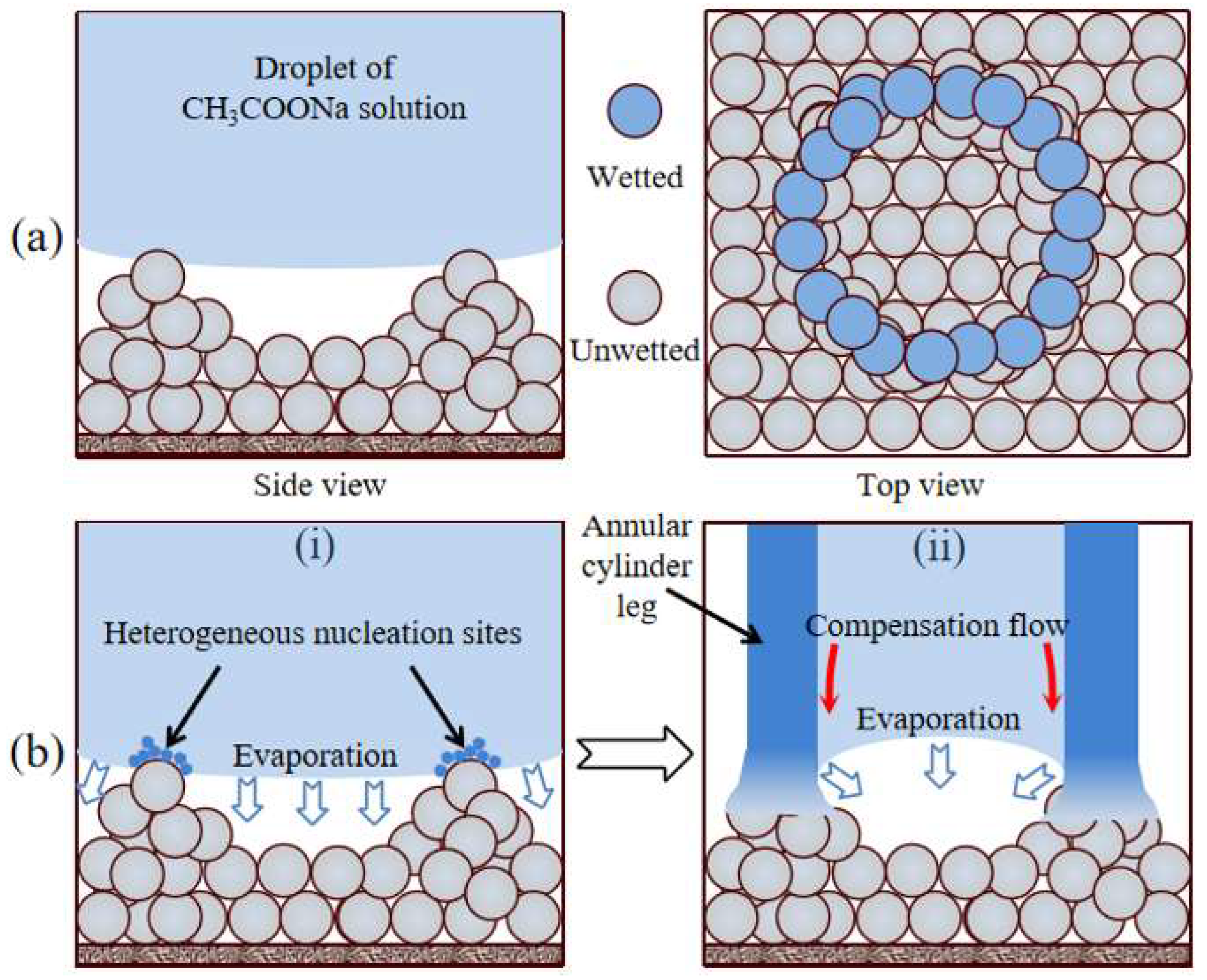
3.3. Formation of “Legs” for Saturated Droplet

By contrast, the saturated droplet had a distinct phenomenon after the formation of the crystal shell. For the saturated droplet, the substrate acted as heterogeneous nucleation sites immediately once the droplet was deposited on it. The crystals generated via heterogeneous nucleation on the substrate are constantly growing. As a result, when the saturated droplet forms a crystal shell, several crystal legs were observed between the crystal globe and the substrate (Figure 8a(1),a(2)). With further evaporation, the growing crystal legs gradually lifted the crystal globe away from the substrate until the evaporation was completed. It can be observed that the growth of crystal legs was initiated, and their size continuously increased at the points where they contacted the substrate (Figure 8a(3)–a(5)). The peak value of growth rate for a crystal leg reached 0.13mm/min (Figure 8d). The salt solution containing tiny crystals was transported from the crystal globe to the substrate via the growing tubular crystal “leg” (Figure 8b,c). This indicates that the salt crystals carried by the evaporative flow of water inside the hollow crystal legs are critical for their growth.
The emergence of crystal legs is the result of heterogeneous nucleation on the surface of the substrate, which differs from the homogeneous nucleation occurring on the droplet surface. When the droplet contacted the substrate, many annular contact lines formed at the solid–liquid interface due to the rough stacking of silanized SiO2 nanoparticles (Figure 9a). These annular contact lines provided heterogeneous nucleation sites for crystallization. For a saturated CH3COONa solution droplet, the crystal nuclei inside the droplet preferentially adhere to the annular contact line on the solid–liquid interface due to the heterogeneous nucleation effect (Figure 9b(i)). The continuous accumulation of crystals on the annular contact lines promotes the growth of hollow crystal legs (Figure 9b(ii)).
During the growth of crystal legs, the evaporation at the annular contact lines where the crystal legs contacted the substrate led to capillary compensation flow in the crystal legs [36], as shown in Figure 8c and Figure 9b(ii). This capillary compensation flow continuously transported the salt solution from crystal globe to the substrate through the hollow crystal legs, which provided a constant supply of raw materials (CH3COONa grains) for the growth of crystal legs.
The superhydrophobic substrate has a rough surface with lots of micro- and nano-sized porous structures formed by silanized SiO2 nanoparticles. Crystal growth in porous media can induce crystallization pressure [22]. Therefore, crystallization pressure is a crucial factor during the growth of crystal legs. Crystallization pressure is described by the following equation [22,37,38]:
P c r y s = v R T V m ln ( S ) + ln γ ± γ ± , 0 + v 0 v ln a w a w , 0
where v is the number of different ions released upon the complete dissociation of the salt (for CH3COONa, v = 2), R is the thermodynamic gas constant, T is the absolute temperature, Vm is the molar volume of the solution, S is the supersaturation, γ± is the mean activity coefficient, and aw is the water activity. The activity coefficient reflects the difference between the effective concentration and the actual concentration. The higher the concentration of the solution, the lower the activity. For saturated CH3COONa solution, its activity can be ignored. In the practical calculation, Equation (2) can be simplified as [39,40]:
P c r y s v R T V m ln ( S )
In Equation (3), the crystallization pressure Pcrys was calculated to be 447.9~457.2 Pa, which is much higher than the hydrostatic pressure of the droplet (~29.02 Pa) This suggested that the crystallization pressure was large enough to overcome the gravity of crystal globe and lifted it off the substrate. Therefore, the crystallization pressure generated during crystallization of sodium acetate crystals is large enough to act as the driving force for the growth of crystal legs.

3.4. Discussion

The emergence of crystal legs is mainly due to the heterogeneous nucleation effect of the superhydrophobic substrate on the crystallization of saturated CH3COONa droplets. However, the final crystallization results of saturated and unsaturated CH3COONa solution droplets on the same substrate dramatically differed. This is because, at the beginning of saturated CH3COONa solution droplet evaporation, both the surface and interior of the droplets are already saturated. Due to the lower nucleation barrier of heterogeneous nucleation compared to that of homogeneous nucleation, crystal nuclei tend to preferentially attach and grow on rough surfaces. Consequently, during the evaporation of a saturated droplet to form a crystal shell, the heterogeneous nucleation effect results in the formation of crystal legs between the droplet and the substrate, which subsequently lift it off from the substrate.
For the droplet of unsaturated solution, the crystallization was indeed dominated by homogeneous nucleation, which occurred at the top surface of the droplet [41]. The heterogeneous effect of substrate was inhibited because the evaporation-driven natural convection also promoted the enrichment of solute at the top surface. Finally, when the evaporation was finished, only a crystal shell formed with a hollow inside.
To demonstrate that the emergence of CH3COONa crystal legs was induced by a heterogeneous nucleation effect on crystals via the superhydrophobic substrate consisting of SiO2 nanoparticles, similar evaporation experiments were conducted on lotus leaves. The results show that both unsaturated and saturated CH3COONa solution droplets formed crystal shells after evaporation, but neither grew crystal legs on the surface of lotus leaves (Figure 10). This indicates that there was no heterogeneous nucleation on the surface of lotus leaf during the evaporation and crystallization of CH3COONa solution droplets.
The heterogeneous nucleation effect results in diverse crystal morphologies on two distinct surfaces, primarily attributed to the coherent relationship between the nascent phase and the interface of the substrate [42,43,44]. The crystal structure of anhydrous sodium acetate adopts a hexagonal closest-packing arrangement, featuring four tetrahedral voids within each unit cell, while silica exhibits a centroid regular tetrahedron in its spatial arrangement. These two crystal structures are spatially complementary to some extent. Considering this, the nucleation and crystallization of CH3COONa are more likely to occur on the surface of SiO2 nanoparticles. Additionally, the accumulation of SiO2 nanoparticles on the superhydrophobic surface results in the formation of granular layers that resemble the arrangement of unit cell during CH3COONa crystal formation. Thus, the CH3COONa crystals are inclined to nucleate and crystallize on the protruding annular contact lines, which were formed by SiO2 nanoparticles, and eventually form tubular crystal legs, whereas the surface structure of the lotus leaf consists of three parts, including papillae, papillae cover, and wax layer [45,46]. The epicuticular wax is primarily composed of a mixture of aliphatic compounds, particularly nonacosanol and nonacosanediols [47]. These aliphatic compounds exhibit an amorphous structure that is distinct from the crystal structure of CH3COONa crystal, which significantly deviates from the regular cubic crystal structure of CH3COONa salt. Consequently, the CH3COONa crystal nuclei would not attach to the surface of lotus leaf for formation and growth. This indicates that the surface of the lotus leaf has no heterogeneous nucleation effect on the crystallization of CH3COONa salt in small droplets.

4. Conclusions

In this study, we investigated the process of evaporative crystallization of CH3COONa solution droplets on a superhydrophobic substrate formed by silanized SiO2 nanoparticles. With the completion of evaporation, unsaturated CH3COONa solution droplets formed a spherical crystal shell. Meanwhile, a saturated CH3COONa solution droplet was lifted off the substrate by the growing crystal legs underneath the already formed crystal shell. The droplet evaporation, droplet surface tension, and internal convection of droplets are the main reasons for the formation of CH3COONa crystal shells. The heterogeneous nucleation effect of the substrate is responsible for the formation of crystal legs for the saturated CH3COONa solution droplets. The crystallization pressure of saturated CH3COONa solution droplets (447.9~457.2 Pa) was large enough to overcome its hydrostatic pressure (~29.02 Pa) and lift the crystal globe off the superhydrophobic substrate. Evaporation at the annular contact lines led to capillary compensation flow inside the tubular crystal legs, which brought liquid from crystal globe to the contact lines. These results may be reproducible for the morphological control of crystallization associated with droplet evaporation.

Author Contributions

X.Z.: Investigation (equal); Conceptualization (equal); Data curation (equal); Formal analysis (equal); Writing—original draft (equal). H.C.: Formal analysis (equal); Writing—review. Z.W.: Formal analysis (equal). N.W.: Formal analysis (equal). D.Z.: Conceptualization (equal); Supervision (equal); Writing—review and editing (equal). All authors have read and agreed to the published version of the manuscript.

Funding

We thank the National Natural Science Foundation of China (Grant Nos. 11972303 and 12272314) and the fund of the State Key Laboratory of Solidifification Processing, Northwestern Polytechnical University (Grant Nos. SKLSP202001).

Institutional Review Board Statement

Not applicable.

Informed Consent Statement

Not applicable.

Data Availability Statement

All the research data have been presented in the paper.

Conflicts of Interest

The authors declare no conflict of interest.

References

  1. Wang, J.W.; Gao, J.; Wang, H.F.; Jin, Q.H.; Rao, B.; Deng, W.; Cao, Y.; Lei, M.; Ye, S.; Fang, Q. Miniaturization of the Whole Process of Protein Crystallographic Analysis by a Microfluidic Droplet Robot: From Nanoliter-Scale Purified Proteins to Diffraction-Quality Crystals. Anal. Chem. 2019, 91, 10132–10140. [Google Scholar] [CrossRef] [PubMed]
  2. Xu, Z.C.; Dong, X.J.; Shen, J.N.; He, Y.J. Integrated Experimental and Modeling Approach to Evaluate Surface Crystallization on Polymer Coatings. Ind. Eng. Chem. Res. 2023, 62, 556–570. [Google Scholar] [CrossRef]
  3. Candoni, N.; Grossier, R.; Lagaize, M.; Veesler, S. Advances in the Use of Microfluidics to Study Crystallization Fundamentals. Annu. Rev. Chem. Biomol. 2019, 10, 59–83. [Google Scholar] [CrossRef] [PubMed] [Green Version]
  4. Wajman, R. The concept of 3D ECT system with increased border area sensitivity for crystallization processes diagnosis. Sensor Rev. 2021, 41, 35–45. [Google Scholar] [CrossRef]
  5. Liu, C.H.; Zheng, Z.J.; Meng, Z.; Chai, X.H.; Cao, C.; Liu, Y.F. Beeswax and carnauba wax modulate the crystallization behavior of palm kernel stearin. LWT-Food Sci. Technol. 2019, 115, 108446. [Google Scholar] [CrossRef]
  6. Yu, H.T.; Kant, P.; Dyett, B.; Lohse, D.; Zhang, X.H. Splitting droplets through coalescence of two different three-phase contact lines. Soft Matter 2019, 15, 6055–6061. [Google Scholar] [CrossRef] [Green Version]
  7. Jannati, K.; Rahimian, M.H.; Moradi, M. Pinning-depinning of the contact line during drop evaporation on textured surfaces: A lattice Boltzmann study. Phys. Rev. E 2020, 102, 033106. [Google Scholar] [CrossRef]
  8. Dgheim, J.; Chahine, A.; Nahed, J. Investigation on the droplet combustion in rotatory natural convection. J. King Saud Univ. Sci. 2019, 31, 937–945. [Google Scholar] [CrossRef]
  9. Wang, T.S.; Shi, W.Y. Transition of Marangoni convection instability patterns during evaporation of sessile droplet at constant contact line mode. Int. J. Heat. Mass Tran. 2020, 148, 119138. [Google Scholar] [CrossRef]
  10. Li, Y.X.; Salvator, V.; Wijshoff, H.; Versluis, M.; Lohse, D. Evaporation-Induced Crystallization of Surfactants in Sessile Multicomponent Droplets. Langmuir 2020, 36, 7545–7552. [Google Scholar] [CrossRef]
  11. Misyura, S.Y. Different modes of heat transfer and crystallization in a drop of NaCl solution: The influence of key factors on the crystallization rate and the heat transfer coefficient. Int. J. Therm. Sci. 2021, 159, 106602. [Google Scholar] [CrossRef]
  12. Liu, L.; Bi, Q.C.; Li, H.X. Experimental Investigation on Flash Evaporation of Saltwater Droplets Released into Vacuum. Microgravity Sci. Tec. 2009, 21, 255–260. [Google Scholar] [CrossRef]
  13. Liu, L.; Mi, M.L. Theoretical Investigation on Rapid Evaporation of a Saline Droplet During Depressurization. Microgravity Sci. Tec. 2014, 25, 295–302. [Google Scholar] [CrossRef]
  14. Qu, J.; Escobar, L.; Li, J.Z.; Rao, Z.H.; Xu, B. Experimental study of evaporation and crystallization of brine droplets under different temperatures and humidity levels. Int. Commun. Heat Mass 2020, 110, 104427. [Google Scholar] [CrossRef]
  15. Kuznetsov, G.V.; Feoktistov, D.V.; Orlova, E.G.; Misyura, S.Y.; Morozov, V.S.; Islamova, A.G. Evaporation modes of LiBr, CaCl2, LiCl, NaCl aqueous salt solution droplets on aluminum surface. Int. J. Heat Mass Tran. 2018, 126, 161–168. [Google Scholar] [CrossRef]
  16. Soulié, V.; Karpitschka, S.; Lequien, F.; Prené, P.; Zemb, T.; Moehwald, H.; Riegler, H. The evaporation behavior of sessile droplets from aqueous saline solutions. Phys. Chem. Chem. Phys. 2015, 17, 22296–22303. [Google Scholar] [CrossRef] [PubMed]
  17. Peng, P.P.; Ke, Q.P.; Zhou, G.; Tang, T.D. Fabrication of microcavity-array superhydrophobic surfaces using. an improved template method. J. Colloid Interf. Sci. 2013, 395, 326–328. [Google Scholar] [CrossRef] [PubMed]
  18. Ge-Zhang, S.; Yang, H.; Ni, H.M.; Mu, H.B.; Zhang, M.M. Biomimetic superhydrophobic metal/nonmetal surface manufactured by etching methods: A mini review. Front. Bioeng. Biotech. 2022, 10, 958095. [Google Scholar] [CrossRef]
  19. Cao, L.; Lu, X.Q.; Pu, F.; Yin, X.L.; Xia, Y.; Huang, W.; Li, Z.L. Facile fabrication of superhydrophobic Bi/Bi2O3 surfaces with hierarchical micro-nanostructures by electroless deposition or electrodeposition. Appl. Surf. Sci. 2014, 288, 558–563. [Google Scholar] [CrossRef]
  20. Tang, X.N.; Yan, X. Dip-coating for fibrous materials: Mechanism, methods and applications. J. Sol-Gel Sci. Techn. 2017, 81, 378–404. [Google Scholar] [CrossRef]
  21. Saji, V.S. Superhydrophobic surfaces and coatings by electrochemical anodic oxidation and plasma electrolytic oxidation. Adv. Colloid Interfac. 2020, 283, 102245. [Google Scholar] [CrossRef] [PubMed]
  22. Salim, H.; Kolpakov, P.; Bonn, D.; Shahidzadeh, N. Self-lifting NaCl crystals. J. Phys. Chem. Lett. 2020, 11, 7388–7393. [Google Scholar] [CrossRef] [PubMed]
  23. McBride, S.A.; Girard, H.L.; Varanasi, K.K. Crystal critters: Self-ejection of crystals from heated, superhydrophobic surfaces. Sci. Adv. 2021, 7, 6960. [Google Scholar] [CrossRef]
  24. Li, X.G.; Shen, J. A facile two-step dipping process based on two silica systems for a superhydrophobic surface. Chem. Commun. 2011, 47, 10761–10763. [Google Scholar] [CrossRef] [PubMed]
  25. Kónya, J.; Hargitai, H.; Jaber, H.; Pinke, P.; Kovács, T.A. Effect of Surface Modifications on Surface Roughness of Ti6Al4V Alloy Manufactured by 3D Printing, Casting, and Wrought. Materials 2023, 16, 3989. [Google Scholar] [CrossRef]
  26. Yang, C.W.; He, F.; Hao, P.F. The apparent contact angle of water droplet on the micro-structured hydrophobic surface. Sci. China Chem. 2010, 53, 912–916. [Google Scholar] [CrossRef]
  27. Lin, K.J.; Zang, D.Y.; Geng, X.G.; Chen, Z. Revisiting the effect of hierarchical structure on the superhydrophobicity. Eur. Phys. J. E 2016, 39, 15. [Google Scholar] [CrossRef] [PubMed]
  28. Quéré, D.; Aussillous, P. Properties of liquid marbles. Proc. R. Soc. A 2006, 462, 973–999. [Google Scholar]
  29. Shin, D.H.; Lee, S.H.; Jung, J.Y.; Yoo, J.Y. Evaporating characteristics of sessile droplet on hydrophobic and hydrophilic surfaces. Microelectron. Eng. 2009, 86, 1350–1353. [Google Scholar] [CrossRef]
  30. Dash, S.; Garimella, S.V. Droplet Evaporation Dynamics on a Superhydrophobic Surface with Negligible Hysteresis. Langmuir 2013, 29, 10785–10795. [Google Scholar] [CrossRef]
  31. Nelson, K.G. The Kelvin equation and solubility of small particles. J. Pharm. Sci. 1972, 61, 479–485. [Google Scholar] [CrossRef] [PubMed]
  32. Skinner, K.M.; Sambles, J.R. The Kelvin equation—A review. J. Aerosol Sci. 1972, 3, 199–210. [Google Scholar] [CrossRef]
  33. Wilcox, W.R. The relation between classical nucleation theory and the solubility of small particles. J. Cryst. Growth 1974, 26, 153–154. [Google Scholar] [CrossRef]
  34. Hu, H.; Larson, R.G.; Phys, J. Evaporation of a Sessile Droplet on a Substrate. Chem. B 2002, 106, 1334–1344. [Google Scholar] [CrossRef]
  35. Song, H.; Lee, Y.; Jin, S.; Kim, H.Y.; Yoo, J.Y. Prediction of sessile drop evaporation considering surface wettability. Microelectron. Eng. 2011, 88, 3249–3255. [Google Scholar] [CrossRef]
  36. Wang, F.S.; Tian, S.H.; Yuan, Q.Z. Evaporation-induced crystal self-assembly (EICSA) of salt drops regulated by trace of polyacrylamide. Colloids Surf. A 2022, 644, 128856. [Google Scholar] [CrossRef]
  37. Scherer, G.W. Crystallization in pores. Cem. Concr. Res. 1999, 29, 1347–1358. [Google Scholar] [CrossRef]
  38. Steiger, M. Crystal growth in porous materials-I: The crystallization pressure of large crystals. J. Cryst. Growth 2005, 282, 455–469. [Google Scholar] [CrossRef]
  39. Bormashenko, E. Liquid Marbles, Elastic Nonstick Droplets: From Minireactors to Self-Propulsion. Langmuir 2017, 33, 663–669. [Google Scholar] [CrossRef]
  40. Desarnaud, J.; Bonn, D.; Shahidzadeh, N. The pressure induced by salt crystallization in confinement. Sci. Rep. 2016, 6, 30856. [Google Scholar] [CrossRef] [Green Version]
  41. Bahri, K.; Eslami, H.; Muller-Plathe, F. Self-Assembly of Model Triblock Janus Colloidal Particles in Two Dimensions. J. Chem. Theory Comput. 2022, 18, 1870–1882. [Google Scholar] [CrossRef]
  42. Zhang, Y.; Wang, M.; Lin, X.; Huang, W.D. Effect of Substrate Wettability and Surface Structure on Nucleation of Crystal. J. Mater. Sci. Technol. 2012, 28, 859–864. [Google Scholar] [CrossRef]
  43. Lo, C.W.; Wang, C.C.; Lu, M.C. Spatial Control of Heterogeneous Nucleation on the Superhydrophobic Nanowire Array. Adv. Funct. Mater. 2014, 24, 1211–1217. [Google Scholar] [CrossRef]
  44. Yan, D.M.; Zeng, Q.; Xu, S.L.; Zhang, Q.; Wang, J.Y. Heterogeneous Nucleation on Concave Rough Surfaces: Thermodynamic Analysis and Implications for Nucleation Design. J. Phys. Chem. C 2016, 120, 10368–10380. [Google Scholar] [CrossRef]
  45. Hans, J.E.; Petra, D.K.; Christoph, N.; Wilhelm, B. Superhydrophobicity in perfection: The outstanding properties of the lotus leaf. Beilstein J. Nanotechnol. 2011, 2, 152–161. [Google Scholar]
  46. Feng, L.; Li, S.; Li, Y.; Li, H.; Zhang, L.; Zhai, J.; Song, Y.; Liu, B.; Jiang, L.; Zhu, D. Super-Hydrophobic Surfaces: From Natural to Artificial. Adv. Mater. 2002, 14, 1857–1860. [Google Scholar] [CrossRef]
  47. Koch, K.; Dommisse, A.; Barthlott, W. Chemistry and Crystal Growth of Plant Wax Tubules of Lotus (Nelumbo nucifera) and Nasturtium (Tropaeolum majus) Leaves on Technical Substrates. Cryst. Growth Des. 2006, 6, 2571–2578. [Google Scholar] [CrossRef]
Figure 1. Illustration of the experimental setup for droplet evaporation.
Materials 16 05168 g001
Figure 2. Characterization of the superhydrophobic substrate. (a) The apparent contact angle of a water droplet, 160 ± 2°; (b) The sliding angle, 5°; (c) SEM image of the superhydrophobic surface and SiO2 particle size distribution; (d) Illustration showing the contact between the droplet and the substrate.
Materials 16 05168 g002
Figure 3. Evaporation-induced crystallization of CH3COONa in an unsaturated droplet (20 wt.%) on nanoparticle superhydrophobic substrate.
Materials 16 05168 g003
Figure 4. Surface tension of sodium acetate solution as a function of concentration and the illustration of internal flow of a CH3COONa solution droplet.
Materials 16 05168 g004
Figure 5. Surface morphology of spherical crystal shell. (a) External surface and (b) its local magnification; (c) internal surface; (d) the cross section of the crystal shell.
Materials 16 05168 g005
Figure 6. Rest mass–time curve of the droplet during the evaporation and crystallization of a droplet from an unsaturated CH3COONa solution to form a crystal shell.
Materials 16 05168 g006
Figure 7. Top view of a CH3COONa solution droplet (20 wt.%) evaporating and crystallizing on a nanoparticle superhydrophobic substrate.
Materials 16 05168 g007
Figure 8. Evaporation-induced crystallization of CH3COONa from a saturated droplet (34.2 wt.%) on nanoparticle superhydrophobic substrate. (a) Formation of crystal globe and growing legs; (b) compensating flow from the droplet to the crystal leg; (c) liquid flow inside the crystal leg and evaporation at the annular contact line; (d) growth rate of a crystal leg.
Materials 16 05168 g008
Figure 9. The mechanism responsible for the formation of CH3COONa crystal “legs”. (a) Illustration showing the annular contact line formed due to the rough stacking of nanoparticles; (b) The annular contact line, which led to the heterogeneous nucleation sites provided by the formation of annular cylinder crystal “legs”.
Materials 16 05168 g009
Figure 10. Evaporation and crystallization process of saturated CH3COONa solution droplet on lotus leaf.
Materials 16 05168 g010
Disclaimer/Publisher’s Note: The statements, opinions and data contained in all publications are solely those of the individual author(s) and contributor(s) and not of MDPI and/or the editor(s). MDPI and/or the editor(s) disclaim responsibility for any injury to people or property resulting from any ideas, methods, instructions or products referred to in the content.

