Article

Influence of Low Temperature Plasma Oxidizing on the Bioactivity of NiTi Shape Memory Alloy for Medical Applications

1
Faculty of Materials Science and Engineering, Warsaw University of Technology, 02-507 Warsaw, Poland
2
Pathology Department, Children’s Memorial Health Institute, 04-730 Warsaw, Poland
3
Institute of Metallurgy and Materials Science, Polish Academy of Sciences, 30-059 Krakow, Poland
*
Author to whom correspondence should be addressed.
Materials 2023, 16(18), 6086; https://doi.org/10.3390/ma16186086
Submission received: 2 August 2023 / Revised: 24 August 2023 / Accepted: 31 August 2023 / Published: 6 September 2023
(This article belongs to the Special Issue Surface Engineering in Materials)

Abstract

:
The present study elucidates the impact of glow discharge oxidation within a low-temperature plasma environment on the bioactivity characteristics of an NiTi shape memory alloy. The properties of the produced surface layers, such as structure (TEM observations), surface morphology (SEM observations), chemical and phase composition (EDS and XRD measurements), wettability (optical gonimeter), and the biological response of osteoblasts and platelets to the oxidized surface compared with the NiTi alloy without a surface layer are presented. The presented surface modification of the NiTi shape memory alloy, achieved through oxidizing in a low-temperature plasma environment, led to the creation of a continuous surface layer composed of nanocrystalline titanium oxide TiO2 (rutile). The findings obtained from this study provide evidence that the oxidized layer augments the bioactivity of the shape memory alloy. This augmentation was substantiated through the spontaneous biomimetic deposition of apatite from a simulated body fluid (SBF) solution. Furthermore, the modified surface exhibited improved osteoblast proliferation, and enhanced platelet adhesion and activation. This proposed surface modification strategy holds promise as a prospective solution to enhance the biocompatibility and bioactivity of NiTi shape memory alloy intended for prolonged use in bone implant applications.

1. Introduction

NiTi alloy, with a chemical composition close to equiatomic, is categorized as a smart material and has growing prominence within the medical field. This prominence is due to its distinctive attributes, notably shape memory and superelasticity [1,2]. These properties enable the design of innovative bone or cardiac implants and medical instruments [3,4,5]. The biocompatibility of NiTi alloys is held in high regard, primarily thanks to a self-passivation mechanism wherein a thin and enduring oxide layer spontaneously forms on the alloy’s surface, which is related to the strong chemical affinity of titanium to oxygen [6]. It is the passive layers that are responsible for the corrosion resistance of NiTi alloy, which is comparable [7] or even higher [6] than that of stainless steels. However, as indicated in the literature, the passive layers formed on the NiTi alloy are thin (5–8 nm) [8], and their rate of self-rebuilding in case of damage may be slower than that of dissolution in the environment of the human body [9,10]. As demonstrated by Toker et al. [11], the efficiency of NiTi alloy surface passivation, and thus the degree of its biocompatibility, also depends on the geometry of the implant and the place of implantation in the body. Damage to the passive layer, e.g., as a result of prolonged exposure to physiological fluids, intensifies the process of releasing Ni ions from the surface [8]. In addition, the spontaneously formed passivated layer may contain small amounts of nickel oxides (NiO and Ni2O3) and metallic Ni [12], which is released from the surface more easily than in the form of oxides. It should be noted that the rate of nickel release to the surrounding environment decreases over time [13].
Therefore, the main problem in the application of NiTi alloy in medicine still remains its high nickel content (over 50% Ni), which although present in trace amounts in the human body, can cause allergies when the safe daily dose (approx. 200–300 μg) is exceeded, and exhibit cytotoxic and even carcinogenic effects [14,15,16,17,18,19]. Chrzanowski described changes in the osteoblast cytoskeleton after contact with metal ions, including nickel, of various concentrations [19]. He found that even low concentrations (0.1–0.5 mM) of nickel, cobalt and vanadium ions had a negative effect on the biological response of cells, which means that they are more cytotoxic than, for example, aluminum, chromium, molybdenum and iron, of which only a higher concentration (10 mM) translated into changes in the cytoskeleton of cells [19]. In his works [16,17], Plant showed the negative effect of nickel ions on human vascular endothelial cells, in which increased oxidative stress and impaired function were observed. Wataha et al. [20] studied the secretion of inflammatory cytokines by macrophages exposed to various metal ions, including nickel. It was found that although there were no clear changes in the level of inflammatory cytokines (IL-1 β and TNF-α) after 24 h, after 72 h the levels of nickel ions had significantly increased. It is worth noting that in in vitro and in vivo studies, inflammatory reactions appeared during exposure to concentrations of metal ions that were previously released from alloys used in dentistry. Similar observations were also noted during the study of cells exposed directly to NiTi alloy and other alloys used in medicine [21].
In pursuit of improving resistance against corrosion and tailoring the biological characteristics of the NiTi alloy to suit specific applications, various surface engineering methods are employed, such as oxidation in air and steam, electrochemical and laser methods, ion implantation, glow discharge treatments, PVD and CVD methods, and deposition of carbon, ceramic or polymer coatings [22,23,24,25,26,27]. Prospective processes are those that enable the treatment of complexly shaped implants generally constituting bone implants made of shape memory alloys, and that guarantee good adhesion of the surface layers, full control of microstructure, phase composition, surface topography, and thus enable the shaping of such properties as wettability, corrosion resistance and biological properties to ensure their compatibility with the intended use of the implants.
Our prior investigations have demonstrated that enhancing corrosion resistance, biocompatibility, and diminishing the release of nickel ions into the biological milieu can be accomplished through the application of low-temperature plasma treatment techniques under glow discharge conditions [22,23,24,25,26,27]. Nonetheless, the chemical bioactivity of layers produced during glow discharge processes, denoting the capacity to deposit calcium phosphates, specifically hydroxyapatite, in a biomimetic manner, remains inadequately understood [28]. This facet assumes great significance in the potential utilization of shape memory alloys for fabricating long-term bone implants as heightened bioactivity correlates with expedited osseointegration of the implant with the patient’s bone structure. An example of such a potential application is the TKR (total knee replacement) knee implant. The choice of an optimal metal alloy for the femoral and tibial parts of the implant plays a significant role in prolonging the life of TKR implants and preventing the need for revision surgery. Recently, a multiple-criteria decision-making analysis (MCDM) [29] has been used to help find a suitable TKR implant material, and it has been shown that NiTi shape memory alloys are the most prospective material for this purpose (porous NiTi alloy was ranked first, followed by solid NiTi alloy). Currently used materials, such as stainless steels, CoCr alloys, or other titanium alloys were ranked lower [30]. However, the slow process of osteogenesis, as well as the production of fibrous tissue at the interface of NiTi alloy implants with the bone, leading to poor bonding of the prosthesis with the surrounding bone and to micro-movements of the implant, make it necessary to use surface modifications that increase its bioactivity. Hence, the production of thin apatite layers on NiTi alloy implants are seen as the solution to increasing and accelerating osseointegration [31]. However, due to the porous structure of apatite layers and their poor adhesion to the substrate [32], they alone do not provide sufficient protection against metallosis. This could be ensured, for example, by prior oxidation of the alloy.
In light of the above, this work aims to investigate the impact of the glow discharge oxidizing process in low-temperature plasma on the bioactive properties of a shape memory alloy. The assessment is performed via incubation in SBF (simulated body fluid) solution and examination of the resulting apatite layers. A method of producing layers at temperatures up to 300 °C is presented, which guarantees the preservation of the specific properties of NiTi alloy, i.e., the effect of shape memory and superelasticity [33]. In addition, the properties of this layer, such as structure, morphology, chemical composition or wettability, as well as the analysis of the biological reactions elicited from osteoblasts and platelets in response to the oxidized surface, in relation to the NiTi alloy in its initial state are also presented. The results of the study can be used as the basis for the development of new material solution concepts for bone implants with shape memory for long-term use.

2. Materials and Methods

The researched material consisted of an NiTi shape memory alloy with a chemical composition of 50.8 at.% Ni (Ti—rest). Disc-shaped samples with a diameter of ø8 mm and a thickness of 1 mm were used in the tests. The samples were mechanically ground with up to 1200-grit sandpaper and degreased in acetone. The glow discharge oxidizing process was conducted at a temperature of 290 °C for 60 min in oxygen plasma at a chamber operating pressure of 1.6 mbar. The temperature was controlled by a thermocouple. Before oxidizing, the specimens were heated to 290 °C in nitrogen plasma (N2 atmosphere with 99.999% purity).
NiTi samples in the initial state and with an oxidized surface layer were incubated in SFB solution prepared according to Kokubo [34]. The composition is given in the table below (Table 1). The samples were incubated for 14 and 30 days in an incubator at 37 °C. The solution was changed every 5 days.
The microstructure investigations were carried out using a TECNAI SuperTWIN (200 kV) FEG transmission electron microscope (TEM) (FEI, Eindhoven, The Netherlands) capable of working in scanning mode (STEM). The nanoscale local chemical analysis was performed with an integrated EDAX energy dispersive microanalysis (EDS) system (EDAX, Tilburg, The Netherlands).
The surface morphology of the samples before and after incubation in SBF solution was imaged using a scanning electron microscope (Hitachi, Tokio, Japan, S-3500N) with an acceleration voltage of 15 keV in SE (secondary electron) mode. An EDS (Energy-dispersive X-ray spectroscopy) attachment was used for analysis of the chemical composition of the biomimetic apatite layers on the sample surface after incubation.
The XRD measurements were performed on a Bruker (Billerica, MA, USA) D8 Advance diffractometer, equipped with parallel beam optics and a Cu Kα radiation (λ = 0.154056 nm) source that operated at 40 kV and 40 mA at room temperature. The scan optics consisted of a Göbel mirror on the side of the incident beam and twin secondary Soller slits in the diffracted beam. The measurements were performed with a fixed angle of incidence of the primary beam equal to 3°, over the 2θ range, between 20° and 80°, with a step size of 0.025° and a 3 s counting time per step.
The wettability test was performed with a goniometer—Contact Angle System OCA 20 (DataPhysics Instruments, Filderstadt, Germany). Measurements were made at room temperature for two liquids: distilled water and diiodomethane. After applying a drop of 0.4 μL to the surface of each sample its image was recorded immediately after stabilization. Average values and standard deviations of the contact angles were calculated from 5 measurements performed for each sample. The profiles of the droplets were analyzed using dedicated SCA20 software. Based on the obtained values of contact angles for distilled water and diiodomethane, the surface free energy was calculated for each sample using the standard Owens–Wendt method [35].
Studies of osteoblast proliferation on the titanium oxide layers produced and the NiTi alloy were conducted with the use of Normal Human Osteoblasts (NHOst, Lonza, Basel, Switzerland). The cells were grown in an osteoblast basal medium enriched with fetal bovine serum (50 mL/500 mL medium), ascorbic acid (0.5 mL/500 mL medium), GA-1000 (0.5 mL/500 mL medium) antibiotics (OGM Osteoblast Growtth Medium BulletKit, Lonza), in a humid atmosphere of 95% air and 5% CO2 at 37 °C. The medium was changed every 48 h. Osteoblasts suspended in the incubation medium at a concentration of 3 × 10⁴/500 μL were applied onto NiTi samples in the initial state and after glow discharge oxidizing. The cells were then incubated for 24, 48 h and 6 days at 37 °C. Following incubation, the culture was rinsed with a phosphate buffered saline solution (PBS, Lonza), and the settled cells were fixed in 4% glutaraldehyde for 30 min at 4 °C before rinsing in a cacodylic buffer, in accordance with the procedure for preparing biological material for examinations employing a scanning electron microscope. The prepared specimens were vacuum-coated with a ca. 10–15 nm layer of gold using a JFC-1200 JEOL fine coater. They were then tested under a scanning electron microscope (JSM-7600F, JEOL, Tokyo, Japan) with an acceleration voltage of 5 keV in LEI (lower secondary electron image) mode.
Adhesion and aggregation tests of blood platelets on the surface of NiTi alloy in the initial state and after producing an oxidized layer were performed with the use of platelet rich plasma (PRP), prepared using the blood of healthy donors. The plasma was incubated on the surface of the samples for 2 h at 37 °C. After this time, the non-adhering cells were rinsed off, while the adhering ones were fixed in 4% glutaraldehyde for 30 min at 4 °C, rinsed in a cacodylic buffer and dehydrated and observed under a scanning electron microscope according to the same procedure used for the osteoblast cultures.
The number of osteoblasts, as well as platelets and their aggregates, was measured using morphometric CellSens (Olympus, Hamburg, Germany) software. The results are presented as means and standard deviations. Statistical analyses were performed using the Fisher and Student’s t-tests at a significance level of α = 0.05.

3. Results

Images from the transmission electron microscope (TEM) show the cross-section of the oxidized surface layer produced at low-temperature plasma on the NiTi shape memory alloy (Figure 1a). As we know from our earlier studies [23], this layer consists of titanium dioxide: TiO2 rutile-type with a nanocrystalline structure. The thickness of the titanium oxide layer is approx. 40 nm. The chemical composition of the layer is also confirmed by the linear distribution of elements obtained from EDS (Figure 1b).
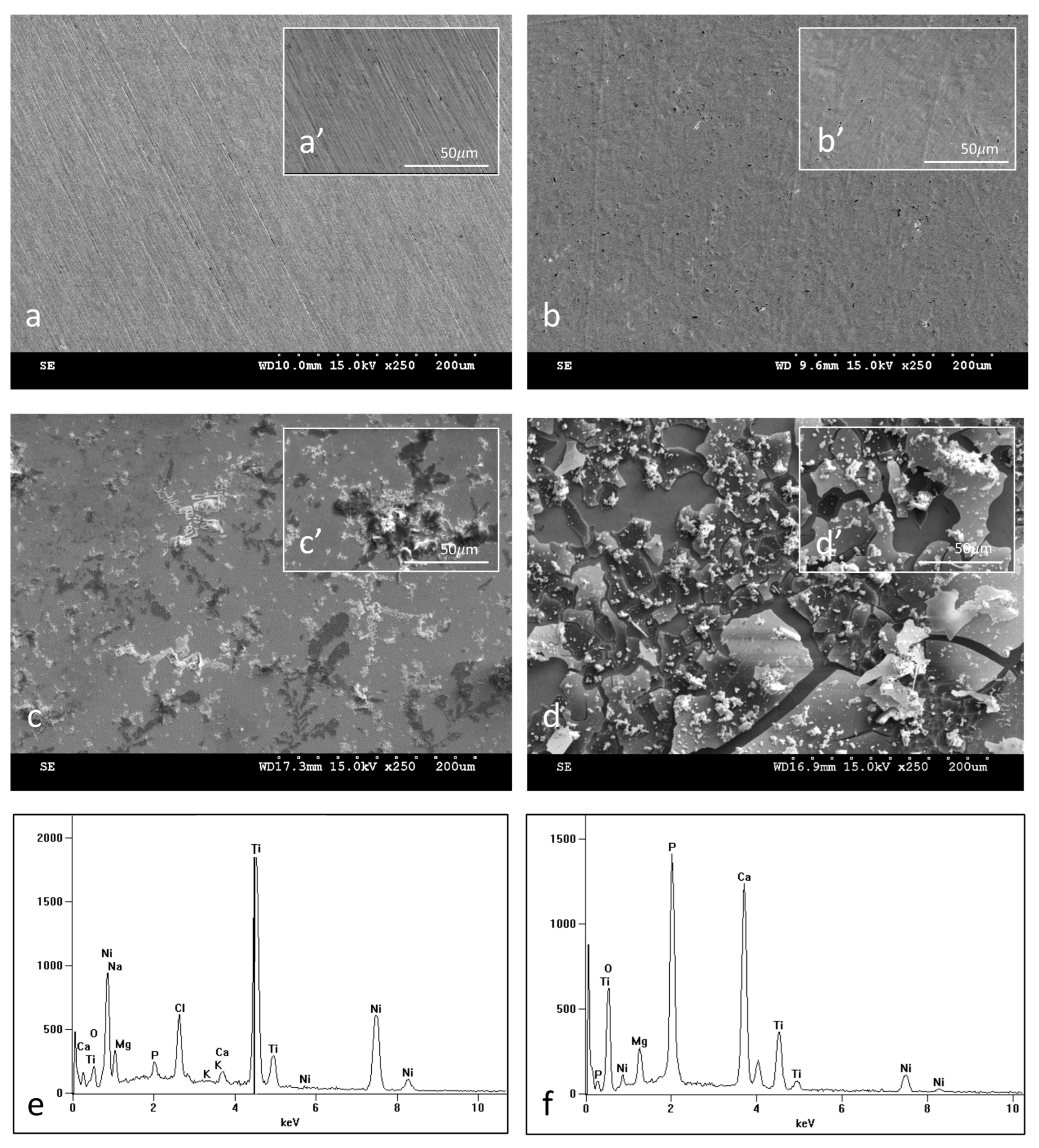
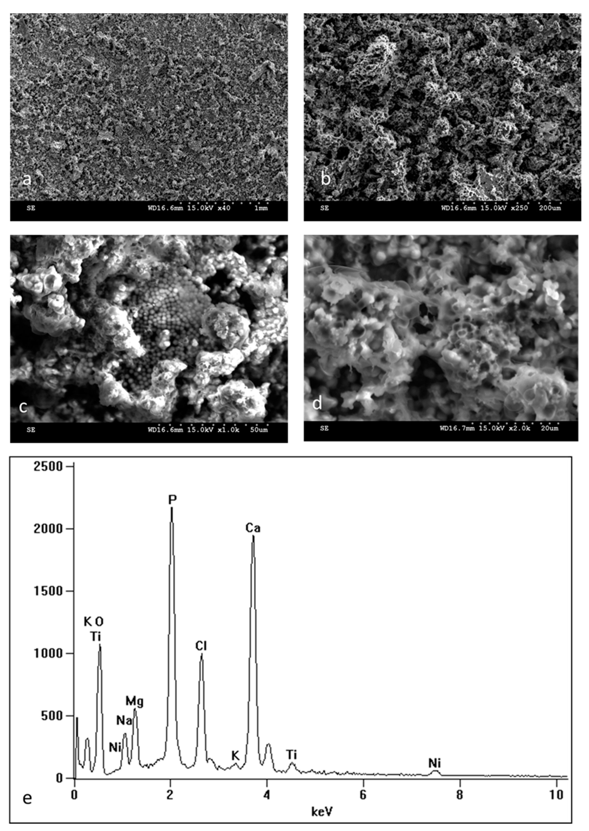
Scanning electron microscope (SEM) observations show the surface morphology of NiTi samples in the initial state (Figure 2a,a’,c,c’) and with an oxidized surface layer (Figure 2b,b’,d,d’) both before (Figure 2a,a’,b,b’) and after (Figure 2c,c’,d,d’) 14 days of incubation in SBF. The surface topography of the oxidized layer before incubation reflects the surface of the initial samples which include delicate scratches resulting from grinding the samples. Observations of the samples after 14 days of incubation (Figure 2c,c’,d,d’) show numerous deposits formed in the biomimetic process of deposition of apatite layers as a result of the interaction of the material surface with the ions present in the SBF solution. It is clearly visible from the SEM images that apatite deposits are formed more easily on the surface of the samples with the oxidized layer (Figure 2d,d’), and there are many more of them after the same incubation time than on the non-oxidized samples (Figure 2c,c’). The EDS analysis confirmed the predicted chemical composition of apatite deposits, which according to the spectra presenting the elemental analysis of the whole image (Figure 2e,f), consist of, among others, calcium, phosphorus, sodium, magnesium, potassium, oxygen etc. The other peaks that belong to titanium and nickel come from the substrate and the surface layer. The amount of calcium and phosphorus in the elemental composition of the tested surfaces is higher for samples with an oxidized layer than for non-oxidized samples (Table 2). Both the images of the formed deposits and their chemical composition testify to the increased bioactivity of the samples with the oxidized layer. To confirm the bioactivity of the prepared surface layer, the incubation process of the samples with the TiO2 layer was extended to 30 days. Surface images (Figure 3a–d) as well as the EDS spectra from the entire image area (Figure 3e) after 30 days of incubation clearly show a solid apatite layer of calcium phosphates that has been formed on the surface, which further confirms the excellent bioactivity of this layer. Also, a point analysis of apatite deposits for samples soaked for 14 and 30 days confirms the formation of calcium phosphates (Table 2). The ratio of Ca to P ranges from 1.26 to 1.53 and is the highest for samples with an oxidized layer soaked for 30 days. It is worth noting that this ratio is close to that found in ideal hydroxyapatite (1.67), which indicates the formation of calcium phosphates that can potentially promote better osseointegration of the material.
Usually, the individual components of different types of calcium phosphate compositions are mixed together and strongly integrated with each other, and therefore, the X-ray diffraction technique is very helpful in identifying phases [36]. XRD analysis of the samples after 30 days of incubation in the SBF solution showed that on the surface of NiTi with a TiO2 surface layer, a multiphasic composition of calcium phosphates consisting of calcium pyrophosphate dihydrate Ca2P2O7·2H2O (known as CPPD) and hydroxyapatite was formed (Figure 4). Datasheets indicate that it is a non-stoichiometric calcium-deficient hydroxyapatite Ca8.8(PO4)6(OH)1.92 which is often found with biomimetic compounds formed naturally when samples are soaked in solutions resembling human body fluids and is referred to as bone-like apatite. This would explain the observation that although the EDS analysis shows much more Ca and P after 14 days of incubation on samples with the oxide surface layer than on the non-oxidized samples, the Ca/P ratio is lower, indicating more CDHAP (calcium-deficient hydroxyapatite) produced, which is in line with the SEM observations.
The contact angle of NiTi samples increased after low-temperature oxidizing, which means decreased wettability for both polar (distilled water) and non-polar liquids (diiodomethane) (Table 3). Considering the classification based on 90 degrees as the critical point, it can be stated that the surface changed its character from slightly hydrophilic to slightly hydrophobic. The results were used to calculate the surface free energy, which decreased after the oxidation process (Table 3).
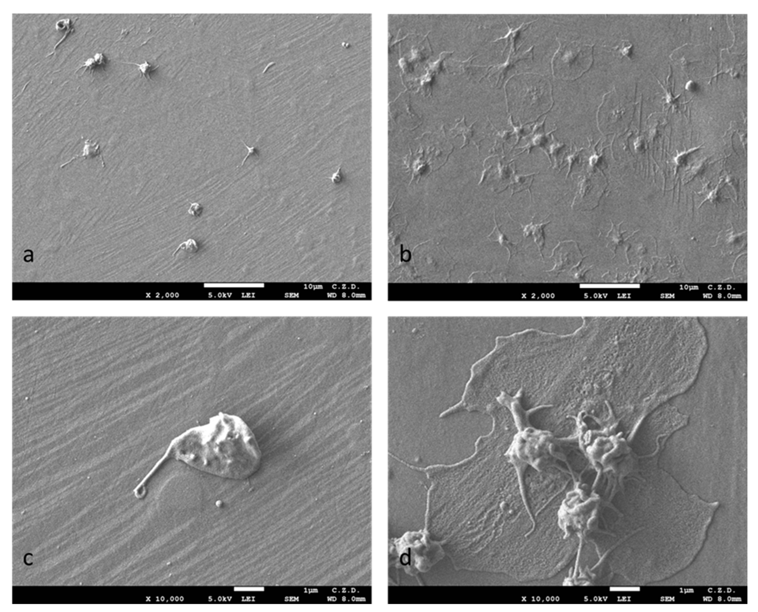
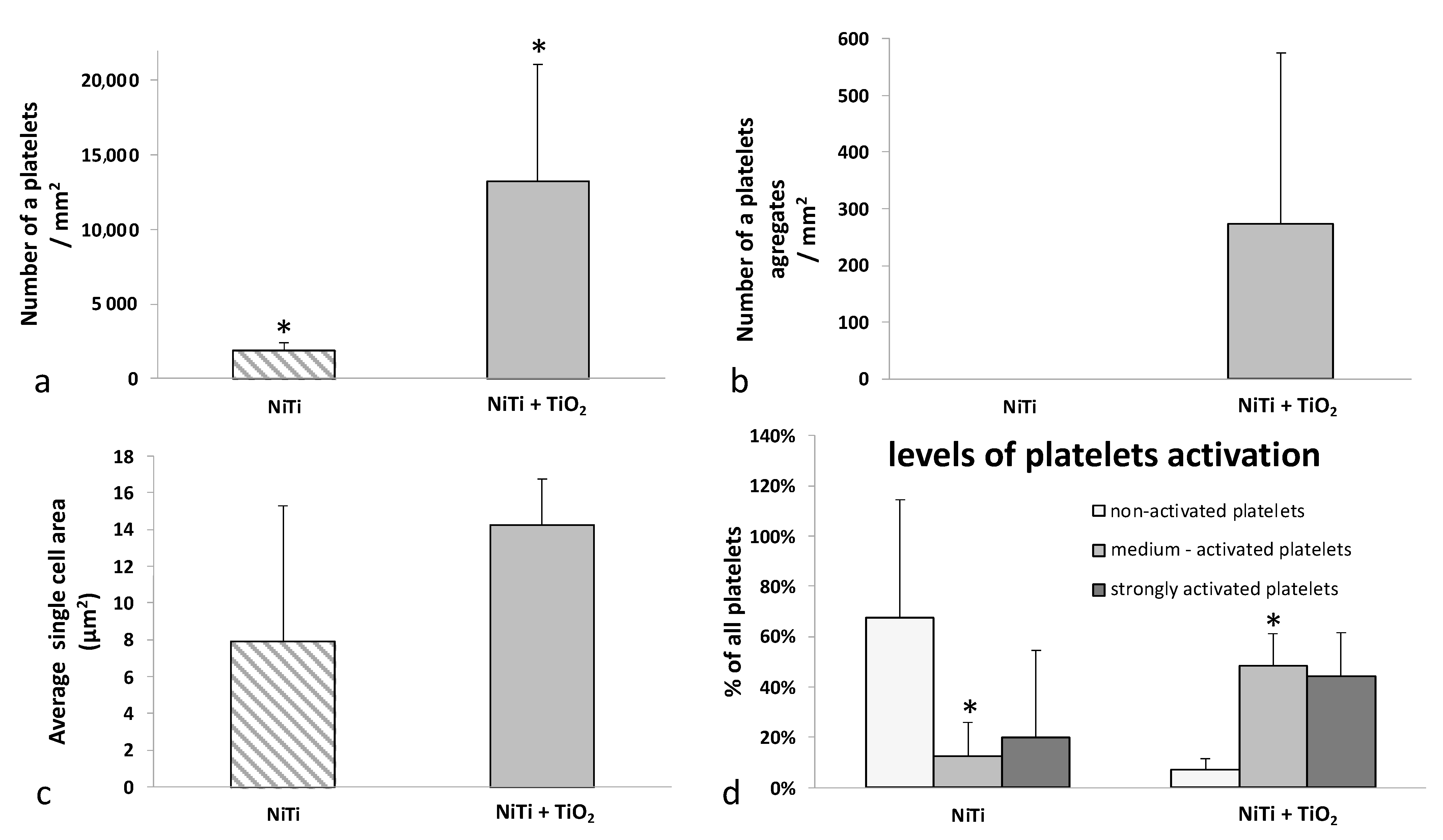
Platelet adhesion studies showed statistically significant differences in cell adhesion for samples with a TiO2 oxidized layer compared with the non-oxidized NiTi samples. The number of thrombocytes was much higher for samples with oxidized layers, and furthermore, they formed numerous aggregates, which was revealed both by microscopic observations of adhered cells using a scanning electron microscope (SEM) (Figure 5a–d) and by measurements made using morphometric software, as presented in graph form in Figure 6a–d. No platelet aggregation was observed for the samples without the surface layer, and the average size of a single cell was almost half that of the samples with the oxidized layer. Differences in aggregation and cell size are clearly noticeable but not statistically significant.
Platelet activation was assessed on the basis of their morphology according to the classification described in the literature [37]. The platelets were divided into “non-activated” with a spherical shape, “medium-activated”, i.e., those with numerous dendritic protrusions, and “strongly activated”—the most flattened. On both types of surface (with and without an oxidized surface layer) cells with different degrees of activation were found, with the majority of cells observed in the non-oxidized samples being non-activated cells, and strongly activated cells being moderately represented in the samples with the oxidized layer (Figure 6d).
Osteoblast proliferation was preserved on both sample types and within the first 48 h they proliferated at a similar rate. Between days 2 and 6, osteoblast growth was inhibited on the samples in the initial state, while on samples with an oxidized layer, they continued to proliferate at only a slightly reduced rate, as evidenced by the slope of the curve in the graph (Figure 7). As a result, after 6 days of incubation, the number of osteoblast cells was significantly higher on the surface of the samples with the oxidized layer compared with the initial samples.
The morphology of osteoblasts growing on samples with and without an oxidized surface layer was normal and slightly changed during the experiment (Figure 8). After 24 h (Figure 8a,a’,b,b’) and 48 h (Figure 8c,c’,d,d’) of incubation, the population of osteoblasts was heterogeneous, i.e., some of the cells had a flattened shape, and some were oblong or spindle-shaped (Figure 8a,a’,b,b’). At the end of the experiment (Figure 8e,e’,f,f’), most of the cells were oblong. After a 6-day incubation period, osteoblasts evenly covered the surface of both types of samples and overlapped each other.

4. Discussion

The NiTi shape memory alloy, being self-passivating, is considered a material with good biocompatibility [38,39,40]. However, surface modifications are necessary in the context of medical applications [39,41]. A large amount of literature on the use of various surface engineering methods to modify NiTi alloy [8,17,22,24,26,33,39,40,41,42,43,44,45,46,47,48,49,50,51,52,53,54,55,56,57,58] indicates that improving its biocompatibility and bioactivity is an important and current problem, especially in the case of long-term implants.
The produced surface layers were characterized by a nanocrystalline structure of titanium oxide TiO2 (rutile), which is the most thermodynamically stable polymorphic variety of titanium oxide [6]. The main motivations for studying the bioactivity of this layer type were the results of our previous studies, which indicated that the layers produced under glow discharge conditions increase corrosion resistance, thereby providing good protection against the release of nickel ions into the biological environment [22,24]. Moreover, in our preliminary studies [27], it was shown that the TiO2 layer of titanium oxide increases adhesion to the NiTi substrate. Those studies investigated amorphous carbon coating deposition. Our current study shows that the TiO2 layer also ensures faster surface deposition of an apatite layer composed of calcium phosphates.
The use of a glow discharge process temperature below 300 °C made it possible to produce surface layers that retain specific properties of the NiTi alloy, i.e., shape memory and superelasticity, which was previously demonstrated [33,59] on the samples of nitrided (TiN) or oxynitrided (TiO2 + TiN) layers with a thickness of several dozen nanometers and produced on NiTi alloy at different temperatures. It was found that during glow discharge processes carried out at temperatures above 300 °C, Ni4Ti3 phase precipitates appear in the structure of the alloy [33]. These processes can also form an Ti2N sublayer [60], as well change the kinetics of martensitic transformation [61] (appearance of an additional rhombendric R phase between the transition from austenitic to martensitic phase). It has been observed that processes carried out at temperatures below 300 °C do not adversely affect the shape memory phenomenon, and may even have a positive effect on the superelastic properties of NiTi alloy, expanding the hysteresis loop in the stress–strain relationship [61]. Moreover, nitrided and oxynitrided layers with a nanocrystalline structure that is several tens of nanometer thick did not show any cracks or damage after shape recovery tests [61], which is mainly due to their nanocrystalline structure and low thickness.
Bioactivity, defined as the ability to form a biomimetic bone-like apatite layer in the environment of solutions simulating human body fluids, is especially important in the context of the long-term use implants. There are numerous studies confirming that in vitro studies utilizing the biomimetic method also produce results in vivo, as well as a palpably better connection between the bioactively enhanced material and the bone [34]. This is based on the assumption that the biomimetic apatite layer, which is formed more easily on more bioactive surfaces, resembles biological bone crystals, and therefore, new bone formation can occur more readily on the surface [62]. Many types of calcium phosphates are used and synthetically produced as materials or layers for orthopedic or dental applications [36]. Of these, the most common and also the most stable is hydroxyapatite (HA). However, biologically occurring HA differs from synthetically produced HA, which has a Ca to P ratio of 1.67. In biological systems, calcium-deficient hydroxyapatite (CDHA, with the formula Ca10−x(HPO4)x(PO4)6−x (OH)2−x is found, most often also containing other ions (Na, K, Mg, Cl, etc.). In this respect, biomimetic CDHA is more like an inorganic composition of natural bone, which also shows a lower Ca/P ratio than synthetic bone. Therefore, CDHA, which has a higher specific surface area than HA or tricalcium phosphate (TCP) [63], exhibits considerable promise for industrial production of synthetic bone substitutes [64], with potential applications extending to drug delivery systems. Its presence in the biomimetically deposited layer ensures good biocompatibility and bioactivity of oxidized NiTi. According to the various in vitro experiments available in the literature, HA or CDHA is not precipitated directly from solutions containing calcium and orthophosphate ions, but some phases are present earlier as precursors [65,66,67,68], for example, dicalcium phosphate dihydrate (DCPD, brushite), amorphous calcium phosphate (ACP) or octacalcium phosphate (OCP) [36]. They are also discussed as precursors of apatite formation in vivo. Many researchers also use concentrated SBF solutions with higher ion concentrations (e.g., 1.5 SBF) to speed up in vitro deposition observations [69].
In addition to the observed deposition of CDHA, calcium pyrophosphate dihydrate Ca2P2O7·2H2O (CPPD) was also found in the apatite layers deposited on the samples, which although less popular, has recently been intensively studied as a material of biological interest [70]. The research included, for example, the production of ceramics based on calcium pyrophosphate for potential osteoplastic applications [71], as well as the use of calcium pyrophosphate for localized drug delivery in dental applications, resulting in a strong antibacterial effect towards E. coli (74% growth inhibition) and S. aureus (48% growth inhibition) [72]. It was also used for fabrication of a new composite material based on β-calcium pyrophosphate and Ti-13Nb-13Zr, which resulted in higher bioactivity compared with the pure Ti-13Nb-13Zr alloy [73].
Better proliferation of such cells as osteoblasts, mesenchymal cells, or platelet adhesion in vitro suggest higher in vivo bioactivity. It is known that platelets contain growth factors, which ensure better healing as well as greater growth of various types of cells. PRP is used as a recovery agent for musculoskeletal injuries. The results presented in this paper are consistent with these observations since the material with the oxidized layer was characterized by both increased proliferation of osteoblasts, and platelet adhesion and aggregation. In our study, osteoblast proliferation was similar for the first 48 h for both types of material. Only after 6 days of proliferation can a statistically significant difference be seen in favor of samples with a surface layer. This is due to the greater biocompatibility of the titanium oxide surface and probably the absence of the cytotoxic effect of nickel. This is consistent with studies from the literature [15,20] which indicate that sometimes the negative effect of nickel appears after 72 h, but is not observed after only 24 h. In addition, the layer of calcium phosphates indicates the formation of compounds beneficial for accelerated osseointegration. It can, therefore, be concluded that the presented modification of the shape memory alloy surface allows for a significant improvement in bioactivity. It is, therefore, advantageous in terms of increasing the biocompatibility of the material for potential applications as bone implants.
The response of cells, as well as the bioactivity of the surface, is certainly influenced by many factors, one of them undoubtedly being the wettability of the surface. The findings of this study align with previous literature findings suggesting that surfaces with lower surface energy and increased hydrophobicity promote platelet adhesion [74,75,76,77,78,79,80,81] or proliferation of other cell types. Many studies propose that this phenomenon may be attributed to the enhanced adsorption of proteins, which facilitate cell adhesion, on hydrophobic surfaces compared with hydrophilic surfaces where water molecules strongly adhere and hinder protein adsorption [82,83]. However, conflicting reports can also be found in the literature, indicating that hydrophobic surfaces may inhibit adhesion or cell growth on the surface [8,84,85,86,87], while other studies claim there is no significant effect caused by differences in contact angles on the biological response of cells (e.g., platelets [88]) on the surface of the tested biomaterials.
The chemical composition, phase composition, and surface wettability are important, yet are not the sole factors determining material surface interaction with the biological environment. Surface topography and surface charge are additional significant factors that can influence cell adhesion [89,90] but were not specifically investigated in this work. Future research should delve into these aspects and also examine the composition and distribution of the protein biofilm on the sample surfaces. It is well established that the types of proteins present and their relative proportions play a role in determining cell adhesion outcomes [88].

5. Conclusions

In this study, we have introduced a surface modification approach for the NiTi shape memory alloy. This modification involves oxidation conducted under glow-discharge conditions in a low-temperature plasma environment, yielding a continuous nanometer-thick layer of nanocrystalline titanium oxide TiO2 (rutile). The low temperature in this process ensures the preservation of the alloy’s specific properties inherent to shape memory. Our research has demonstrated that this newly formed oxide layer significantly heightens the bioactivity of the NiTi shape memory alloy. This was substantiated by the rapid and spontaneous biomimetic deposition of apatite from a simulated body fluid (SBF), a phenomenon that occurred more expeditiously on the modified surface compared with the unmodified material. Additionally, our investigations revealed enhanced osteoblast proliferation on the modified surface, as well as increased adhesion and activation of platelets obtained from platelet-rich plasma (PRP). Furthermore, the presence of hydroxyapatite among the calcium phosphates within the biomimetic layer formed in vitro holds promise for improved in vivo bioactivity. This optimism is derived from the fact that the SBF solution utilized closely mimics both the composition and pH of human body fluids. Consequently, our proposed surface modification presents a prospective avenue for enhancing the biocompatibility of NiTi alloy employed in the development of long-term bone implants.

Author Contributions

Conceptualization, J.W., J.S. and T.W.; methodology, J.W., T.B., A.S., E.C., D.M., J.M., J.S. and T.W.; software, J.W., T.B., A.S., E.C., D.M. and J.M.; validation, J.S. and T.W.; formal analysis, J.W., J.S. and T.W.; investigation, J.W., T.B., A.S., E.C., D.M. and J.M.; resources, J.W., J.S. and T.W.; data curation, J.W.; writing—original draft preparation, J.W.; writing—review and editing, J.W., T.B., A.S., E.C., D.M., J.M., J.S. and T.W.; visualization, J.W.; supervision, J.S. and T.W.; project administration, J.S.; funding acquisition, J.S. All authors have read and agreed to the published version of the manuscript.

Funding

This research was funded by the Warsaw University of Technology within the Excellence Initiative: Research University (IDUB) programme.

Institutional Review Board Statement

Not applicable.

Informed Consent Statement

Not applicable.

Data Availability Statement

Not applicable.

Conflicts of Interest

The authors declare no conflict of interest.

References

  1. Biesiekierski, A.; Wang, J.; Abdel-Hady Gepreel, M.; Wen, C. A new look at biomedical Ti-based shape memory alloys. Acta Biomater. 2012, 8, 1661–1669. [Google Scholar] [CrossRef]
  2. Yoneyama, T.; Miyazaki, S. Shape Memory Alloys for Biomedical Applications; Woodhead Publishing: Cambridge, UK, 2008. [Google Scholar] [CrossRef]
  3. Duerig, T.; Pelton, A.; Stöckel, D. An overview of nitinol medical applications. Mater. Sci. Eng. A 1999, 273–275, 149–160. [Google Scholar] [CrossRef]
  4. Morgan, N. Medical shape memory alloy applications—The market and its products. Mater. Sci. Eng. A 2004, 378, 16–23. [Google Scholar] [CrossRef]
  5. Elahinia, M.H.; Hashemi, M.; Tabesh, M.; Bhaduri, S.B. Manufacturing and processing of NiTi implants: A review. Prog. Mater. Sci. 2012, 57, 911–946. [Google Scholar] [CrossRef]
  6. Brunette, D.M.; Tengvall, P.; Textor, M.; Thomsen, P. Titanium in Medicine: Material Science, Surface Science, Engineering, Biological Responses and Medical Applications; Springer: Berlin/Heidelberg, Germany, 2001. [Google Scholar]
  7. Rondelli, G.; Torricelli, P.; Fini, M.; Rimondini, L.; Giardino, R. In vitro corrosion study by EIS of an equiatomic NiTi alloy and an implant quality AISI 316 stainless steel. J. Biomed. Mater. Res. Part B Appl. Biomater. 2006, 79, 320–324. [Google Scholar] [CrossRef] [PubMed]
  8. Zhao, T.; Li, Y.; Zhao, X.; Chen, H.; Zhang, T. Ni ion release, osteoblast-material interactions, and hemocompatibility of hafnium-implanted NiTi alloy. J. Biomed. Mater. Res. Part B Appl. Biomater. 2012, 100B, 646–659. [Google Scholar] [CrossRef]
  9. Shabalovskaya, S.A.; Rondelli, G.C.; Undisz, A.L.; Anderegg, J.W.; Burleigh, T.D.; Rettenmayr, M.E. The electrochemical characteristics of native Nitinol surfaces. Biomaterials 2009, 30, 3662–3671. [Google Scholar] [CrossRef]
  10. Hanawa, T. Metal ion release from metal implants. Mater. Sci. Eng. C 2004, 24, 745–752. [Google Scholar] [CrossRef]
  11. Toker, S.; Canadinc, D.; Maier, H.; Birer, O. Evaluation of passive oxide layer formation–biocompatibility relationship in NiTi shape memory alloys: Geometry and body location dependency. Mater. Sci. Eng. C 2014, 36, 118–129. [Google Scholar] [CrossRef]
  12. Ryhänen, J. Biocompatibility of nitinol. Minim. Invasive Ther. Allied Technol. 2000, 9, 99–105. [Google Scholar] [CrossRef]
  13. Arndt, M.; Brück, A.; Scully, T.; Jäger, A.; Bourauel, C. Nickel ion release from orthodontic NiTi wires under simulation of realistic in-situ conditions. J. Mater. Sci. 2005, 40, 3659–3667. [Google Scholar] [CrossRef]
  14. Denkhaus, E.; Salnikow, K. Nickel essentiality, toxicity, and carcinogenicity. Crit. Rev. Oncol. 2002, 42, 35–56. [Google Scholar] [CrossRef]
  15. Chrzanowski, W.; Szade, J.; Hart, A.D.; Knowles, J.C.; Dalby, M.J. Biocompatible, Smooth, Plasma-Treated Nickel–Titanium Surface—An Adequate Platform for Cell Growth. J. Biomater. Appl. 2012, 26, 707–731. [Google Scholar] [CrossRef] [PubMed]
  16. Plant, S.D.; Grant, D.M.; Leach, L. Behaviour of human endothelial cells on surface modified NiTi alloy. Biomaterials 2005, 26, 5359–5367. [Google Scholar] [CrossRef] [PubMed]
  17. Plant, S.D.; Grant, D.M.; Leach, L. Surface modification of NiTi alloy and human platelet activation under static and flow conditions. Mater. Lett. 2007, 61, 2864–2867. [Google Scholar] [CrossRef]
  18. Barrett, R.D.; Bishara, S.E.; Quinn, J.K. Biodegradation of orthodontic appliances. Part I. Biodegradation of nickel and chromium in vitro. Am. J. Orthod. Dentofac. Orthop. 1993, 103, 8–14. [Google Scholar] [CrossRef] [PubMed]
  19. Chrzanowski, W. Engineering Surface Properties of Nickel-Titanium Alloy for Improved Osteointegration; Wydawnictwo Politechniki Śląskiej: Gliwice, Poland, 2012. [Google Scholar]
  20. Wataha, J.C.; Ratanasathien, S.; Hanks, C.T.; Sun, Z. In vitro IL-1β and TNF-α release from THP-1 monocytes in response to metal ions. Dent. Mater. 1996, 12, 322–327. [Google Scholar] [CrossRef]
  21. JWataha, C.; Lockwood, P.E.; Marek, M.; Ghazi, M. Ability of Ni-containing biomedical alloys to activate monocytes and en-dothelial cells in vitro. J. Biomed. Mater. Res. 1999, 45, 251–257. [Google Scholar] [CrossRef]
  22. Czarnowska, E.; Borowski, T.; Sowińska, A.; Lelątko, J.; Oleksiak, J.; Kamiński, J.; Tarnowski, M.; Wierzchoń, T. Structure and properties of nitrided surface layer produced on NiTi shape memory alloy by low temperature plasma nitriding. Appl. Surf. Sci. 2015, 334, 24–31. [Google Scholar] [CrossRef]
  23. Witkowska, J.; Sowińska, A.; Czarnowska, E.; Płociński, T.; Borowski, T.; Wierzchoń, T. NiTi shape-memory alloy oxidized in low-temperature plasma with carbon coating: Characteristic and a potential for cardiovascular applications. Appl. Surf. Sci. 2017, 421, 89–96. [Google Scholar] [CrossRef]
  24. Witkowska, J.; Sowińska, A.; Czarnowska, E.; Płociński, T.; Kamiński, J.; Wierzchoń, T. Hybrid a-CNH+TiO2+TiN-type surface layers produced on NiTi shape memory alloy for cardiovascular applications. Nanomedicine 2017, 12, 2233–2244. [Google Scholar] [CrossRef] [PubMed]
  25. Witkowska, J.; Kamiński, J.; Płociński, T.; Tarnowski, M.; Wierzchoń, T. Corrosion resistance of NiTi shape memory alloy after hybrid surface treatment using low-temperature plasma. Vacuum 2017, 137, 92–96. [Google Scholar] [CrossRef]
  26. Witkowska, J.; Sowińska, A.; Czarnowska, E.; Płociński, T.; Rajchel, B.; Tarnowski, M.; Wierzchoń, T. Structure and properties of composite surface layers produced on NiTi shape memory alloy by a hybrid method. J. Mater. Sci. Mater. Med. 2018, 29, 110. [Google Scholar] [CrossRef] [PubMed]
  27. Chlanda, A.; Witkowska, J.; Morgiel, J.; Nowińska, K.; Choińska, E.; Swieszkowski, W.; Wierzchoń, T. Multi-scale characterization and biological evaluation of composite surface layers produced under glow discharge conditions on NiTi shape memory alloy for potential cardiological application. Micron 2018, 114, 14–22. [Google Scholar] [CrossRef] [PubMed]
  28. Forsgren, J.; Svahn, F.; Jarmar, T.; Engqvist, H. Formation and adhesion of biomimetic hydroxyapatite deposited on titanium substrates. Acta Biomater. 2007, 3, 980–984. [Google Scholar] [CrossRef] [PubMed]
  29. Jahan, A.; Ismail, M.; Sapuan, S.; Mustapha, F. Material screening and choosing methods—A review. Mater. Des. 2010, 31, 696–705. [Google Scholar] [CrossRef]
  30. Bahraminasab, M.; Jahan, A. Material selection for femoral component of total knee replacement using comprehensive VIKOR. Mater. Des. 2011, 32, 4471–4477. [Google Scholar] [CrossRef]
  31. Chen, M.; Yang, X.; Hu, R.; Cui, Z.; Man, H. Bioactive NiTi shape memory alloy used as bone bonding implants. Mater. Sci. Eng. C 2004, 24, 497–502. [Google Scholar] [CrossRef]
  32. Liu, X.; Chu, P.K.; Ding, C. Surface modification of titanium, titanium alloys, and related materials for biomedical applications. Mater. Sci. Eng. R Rep. 2005, 47, 49–121. [Google Scholar] [CrossRef]
  33. Lelątko, J.; Goryczka, T.; Wierzchoń, T.; Ossowski, M.; Łosiewicz, B.; Rówiński, E.; Morawiec, H. Structure of Low Temperature Nitrided/Oxidized Layer Formed on NiTi Shape Memory Alloy. Solid State Phenom. 2010, 163, 127–130. [Google Scholar] [CrossRef]
  34. Kokubo, T.; Takadama, H. How useful is SBF in predicting in vivo bone bioactivity? Biomaterials 2006, 27, 2907–2915. [Google Scholar] [CrossRef]
  35. Owens, D.K.; Wendt, R.C. Estimation of the surface free energy of polymers. J. Appl. Polym. Sci. 1969, 13, 1741–1747. [Google Scholar] [CrossRef]
  36. Dorozhkin, S.V. Calcium orthophosphates: Occurrence, properties, biomineralization, pathological calcification and biomimetic applications. Biomatter 2011, 1, 121–164. [Google Scholar] [CrossRef] [PubMed]
  37. Goodman, S.L.; Grasel, T.G.; Cooper, S.L.; Albrecht, R.M. Platelet shape change and cytoskeletal reorganization on polyurethaneureas. J. Biomed. Mater. Res. 1989, 23, 105–123. [Google Scholar] [CrossRef]
  38. Es-Souni, M.; Es-Souni, M.; Fischer-Brandies, H. Assessing the biocompatibility of NiTi shape memory alloys used for medical applications. Anal. Bioanal. Chem. 2005, 381, 557–567. [Google Scholar] [CrossRef]
  39. A Shabalovskaya, S. Surface, corrosion and biocompatibility aspects of Nitinol as an implant material. Bio-Med. Mater. Eng. 2002, 12, 69–109. [Google Scholar]
  40. Nagaraja, S.; Brown, R.; Saylor, D.; Undisz, A. Oxide Layer Formation, Corrosion, and Biocompatibility of Nitinol Cardiovascular Devices. Shape Mem. Superelasticity 2022, 8, 45–63. [Google Scholar] [CrossRef]
  41. Shabalovskaya, S.; Anderegg, J.; Van Humbeeck, J. Critical overview of Nitinol surfaces and their modifications for medical applications. Acta Biomater. 2008, 4, 447–467. [Google Scholar] [CrossRef]
  42. Tan, L.; Crone, W. Surface characterization of NiTi modified by plasma source ion implantation. Acta Mater. 2002, 50, 4449–4460. [Google Scholar] [CrossRef]
  43. Shevchenko, N.; Pham, M.-T.; Maitz, M. Studies of surface modified NiTi alloy. Appl. Surf. Sci. 2004, 235, 126–131. [Google Scholar] [CrossRef]
  44. Firstov, G.; Vitchev, R.; Kumar, H.; Blanpain, B.; Van Humbeeck, J. Surface oxidation of NiTi shape memory alloy. Biomaterials 2002, 23, 4863–4871. [Google Scholar] [CrossRef]
  45. Michiardi, A.; Aparicio, C.; Planell, J.; Gil, F. New oxidation treatment of NiTi shape memory alloys to obtain Ni-free surfaces and to improve biocompatibility. J. Biomed. Mater. Res. Part B Appl. Biomater. 2006, 77B, 249–256. [Google Scholar] [CrossRef] [PubMed]
  46. Chrzanowski, W.; Neel, E.A.A.; Armitage, D.A.; Zhao, X.; Knowles, J.C.; Salih, V. In vitrostudies on the influence of surface modification of Ni–Ti alloy on human bone cells. J. Biomed. Mater. Res. Part A 2010, 93A, 1596–1608. [Google Scholar] [CrossRef] [PubMed]
  47. Starosvetsky, D.; Gotman, I. TiN coating improves the corrosion behavior of superelastic NiTi surgical alloy. Surf. Coat. Technol. 2001, 148, 268–276. [Google Scholar] [CrossRef]
  48. Yokoyama, K.; Kanemura, T.; Sakai, J. Improvements in fracture properties of Ni–Ti superelastic alloy in physiological saline solution containing hydrogen peroxide by surface modification. Mater. Sci. Eng. A 2009, 513–514, 267–275. [Google Scholar] [CrossRef]
  49. Cheng, F.; Shi, P.; Man, H. Anatase coating on NiTi via a low-temperature sol–gel route for improving corrosion resistance. Scr. Mater. 2004, 51, 1041–1045. [Google Scholar] [CrossRef]
  50. Sui, J.; Cai, W. Formation of diamond-like carbon (DLC) film on the NiTi alloys via plasma immersion ion implantation and deposition (PIIID) for improving corrosion resistance. Appl. Surf. Sci. 2006, 253, 2050–2055. [Google Scholar] [CrossRef]
  51. Wong, M.; Cheng, F.; Man, H. Laser oxidation of NiTi for improving corrosion resistance in Hanks’ solution. Mater. Lett. 2007, 61, 3391–3394. [Google Scholar] [CrossRef]
  52. Man, H.C.; Cui, Z.D.; Yue, T.M. Surface characteristics and corrosion behavior of laser surface nitrided NiTi shape memory alloy for biomedical applications. J. Laser Appl. 2002, 14, 242–247. [Google Scholar] [CrossRef]
  53. Liu, J.-X.; Yang, D.-Z.; Shi, F.; Cai, Y.-J. Sol–gel deposited TiO2 film on NiTi surgical alloy for biocompatibility improvement. Thin Solid Films 2003, 429, 225–230. [Google Scholar] [CrossRef]
  54. Dudek, K.; Goryczka, T. Electrophoretic deposition and characterization of thin hydroxyapatite coatings formed on the surface of NiTi shape memory alloy. Ceram. Int. 2016, 42, 19124–19132. [Google Scholar] [CrossRef]
  55. Hu, T.; Chu, C.-L.; Yin, L.-H.; Pu, Y.-P.; Dong, Y.-S.; Guo, C.; Sheng, X.-B.; Chung, J.-C.; Chu, P.-K. In vitro biocompatibility of titanium-nickel alloy with titanium oxide film by H2O2 oxidation. Trans. Nonferrous Met. Soc. China 2007, 17, 553–557. [Google Scholar] [CrossRef]
  56. Poon, R.; Yeung, K.; Liu, X.; Chu, P.; Chung, C.; Lu, W.; Cheung, K.; Chan, D. Carbon plasma immersion ion implantation of nickel–titanium shape memory alloys. Biomaterials 2005, 26, 2265–2272. [Google Scholar] [CrossRef] [PubMed]
  57. Yeung, K.; Chan, R.; Lam, K.; Wu, S.; Liu, X.; Chung, C.; Chu, P.K.; Lu, W.; Chan, D.; Luk, K.; et al. In vitro and in vivo characterization of novel plasma treated nickel titanium shape memory alloy for orthopedic implantation. Surf. Coat. Technol. 2007, 202, 1247–1251. [Google Scholar] [CrossRef]
  58. Maleki-Ghaleh, H.; Khalil-Allafi, J.; Sadeghpour-Motlagh, M.; Shakeri, M.S.; Masoudfar, S.; Farrokhi, A.; Khosrowshahi, Y.B.; Nadernezhad, A.; Siadati, M.H.; Javidi, M.; et al. Effect of surface modification by nitrogen ion implantation on the electrochemical and cellular behaviors of super-elastic NiTi shape memory alloy. J. Mater. Sci. Mater. Med. 2014, 25, 2605–2617. [Google Scholar] [CrossRef] [PubMed]
  59. Lelątko, J.; Freitag, M.; Rak, J.; Wierzchoń, T.; Goryczka, T. Structure of Nitride and Nitride/Oxide Layers Formed on NiTi Alloy. Solid State Phenom. 2012, 186, 259–262. [Google Scholar] [CrossRef]
  60. Lelatko, J.; Paczkowski, P.; Wierzchoń, T.; Morawiec, H. TEM studies of the nitrided Ni-Ti surface layer. J. Microsc. 2006, 223, 234–236. [Google Scholar] [CrossRef]
  61. Lelątko, J.; Lekston, Z.; Freitag, M.; Wierzchoń, T.; Goryczka, T. Influence of low temperature glow discharge nitriding and ox-ynitriding process on microstructural and shape memory effect in NiTi alloy. Mater. Eng. 2012, 4, 256–261. [Google Scholar]
  62. Loty, C.; Sautier, J.M.; Boulekbache, H.; Kokubo, T.; Kim, H.M.; Forest, N. In vitro bone formation on a bone-like apatite layer prepared by a biomimetic process on a bioactive glass-ceramic. J. Biomed. Mater. Res. 2000, 49, 423–434. [Google Scholar] [CrossRef]
  63. Koppala, S.; Swamiappan, S.; Gangarajula, Y.; Xu, L.; Sadasivuni, K.K.; Ponnamma, D.; Rajagopalan, V. Calcium deficiency in hydroxyapatite and its drug delivery applications. Micro Nano Lett. 2018, 13, 562–564. [Google Scholar] [CrossRef]
  64. Bourgeois, B.; Laboux, O.; Obadia, L.; Gauthier, O.; Betti, E.; Aguado, E.; Daculsi, G.; Bouler, J.-M. Calcium-deficient apatite: A firstin vivo study concerning bone ingrowth. J. Biomed. Mater. Res. 2003, 65, 402–408. [Google Scholar] [CrossRef] [PubMed]
  65. O’Neill, W. The fallacy of the calcium-phosphorus product. Kidney Int. 2007, 72, 792–796. [Google Scholar] [CrossRef] [PubMed]
  66. LeGeros, R. Formation and transformation of calcium phosphates: Relevance to vascular calcification. Z. Kardiol. 2001, 90 (Suppl. 3), 116–124. [Google Scholar] [CrossRef]
  67. Gasga, J.R.; Carbajal-De-La-Torre, G.; Bres, E.; Gil-Chavarria, I.M.; Rodríguez-Hernández, A.G.; Garcia-Garcia, R. STEM-HAADF electron microscopy analysis of the central dark line defect of human tooth enamel crystallites. J. Mater. Sci. Mater. Med. 2008, 19, 877–882. [Google Scholar] [CrossRef]
  68. Nancollas, G.H.; Wu, W. Biomineralization mechanisms: A kinetics and interfacial energy approach. J. Cryst. Growth 2000, 211, 137–142. [Google Scholar] [CrossRef]
  69. Wan, B.; Ruan, Y.; Shen, C.; Xu, G.; Forouzanfar, T.; Lin, H.; Wu, G. Biomimetically precipitated nanocrystalline hydroxyapatite. Nano TransMed 2022, 1, e9130008. [Google Scholar] [CrossRef]
  70. Griesiute, D.; Garskaite, E.; Antuzevics, A.; Klimavicius, V.; Balevicius, V.; Zarkov, A.; Katelnikovas, A.; Sandberg, D.; Kareiva, A. Synthesis, structural and luminescent properties of Mn-doped calcium pyrophosphate (Ca2P2O7) polymorphs. Sci. Rep. 2022, 12, 7116. [Google Scholar] [CrossRef]
  71. Dorozhkin, S.V.; Epple, M. Biological and Medical Significance of Calcium Phosphates. Angew. Chem. Int. Ed. 2002, 41, 3130–3146. [Google Scholar] [CrossRef]
  72. Anastasiou, A.; Nerantzaki, M.; Brown, A.; Jha, A.; Bikiaris, D. Drug loading capacity of microporous β-pyrophosphate crystals. Mater. Des. 2019, 168, 107661. [Google Scholar] [CrossRef]
  73. Hu, H.; Zhang, L.; He, Z.; Jiang, Y.; Tan, J. Microstructure evolution, mechanical properties, and enhanced bioactivity of Ti-13Nb-13Zr based calcium pyrophosphate composites for biomedical applications. Mater. Sci. Eng. C 2019, 98, 279–287. [Google Scholar] [CrossRef]
  74. Lopez-Santos, C.; Colaux, J.L.; Laloy, J.; Fransolet, M.; Mullier, F.; Michiels, C.; Dogné, J.-M.; Lucas, S. Bioactivity and hemocompatibility study of amorphous hydrogenated carbon coatings produced by pulsed magnetron discharge. J. Biomed. Mater. Res. Part A 2013, 101A, 1800–1812. [Google Scholar] [CrossRef] [PubMed]
  75. Logothetidis, S.; Karagkiozaki, V.; Karagiannidis, P.G.; Kalfagiannis, N.; Patsalas, P.; Georgiou, D.; Kavatzikidou, P. Novel nanostructured biomaterials: Implications for coronary stent thrombosis. Int. J. Nanomed. 2012, 7, 6063–6076. [Google Scholar] [CrossRef] [PubMed]
  76. Wen, F.; Huang, N.; Leng, Y.; Zhao, A.; Jing, F. Study on wettabilities and platelet adhesion behavior of C:H and C:N:H films prepared by DC-MFCVA. Appl. Surf. Sci. 2008, 255, 469–472. [Google Scholar] [CrossRef]
  77. Chun, Y.; Levi, D.S.; Mohanchandra, K.; Carman, G.P. Superhydrophilic surface treatment for thin film NiTi vascular applications. Mater. Sci. Eng. C 2009, 29, 2436–2441. [Google Scholar] [CrossRef]
  78. Popov, C.; Vasilchina, H.; Kulisch, W.; Danneil, F.; Stüber, M.; Ulrich, S.; Welle, A.; Reithmaier, J. Wettability and protein adsorption on ultrananocrystalline diamond/amorphous carbon composite films. Diam. Relat. Mater. 2009, 18, 895–898. [Google Scholar] [CrossRef]
  79. Tavakalyan, N.B.; Karapetyan, A.G.; Pogosyan, A.S.; Abrahamyan, A.K.; Corti, A.; Pompella, A.; Mihranyan, A. Influence of unsaturated carbonic acids on hemocompatibility and cytotoxicity of poly-vinylacetate based co-polymers. J. Mater. Sci. Mater. Med. 2010, 21, 1693–1702. [Google Scholar] [CrossRef]
  80. Dey, R.K.; Ray, A.R. Synthesis, characterization, and blood compatibility of polyamidoamines copolymers. Biomaterials 2003, 24, 2985–2993. [Google Scholar] [CrossRef] [PubMed]
  81. Coelho, M.A.N.; Vieira, E.P.; Motschmann, H.; Möhwald, H.; Thünemann, A.F. Human Serum Albumin on Fluorinated Surfaces. Langmuir 2003, 19, 7544–7550. [Google Scholar] [CrossRef]
  82. Barberi, J.; Spriano, S. Titanium and Protein Adsorption: An Overview of Mechanisms and Effects of Surface Features. Materials 2021, 14, 1590. [Google Scholar] [CrossRef]
  83. Xu, L.-C.; Bauer, J.W.; Siedlecki, C.A. Proteins, platelets, and blood coagulation at biomaterial interfaces. Colloids Surf. B Biointerfaces 2014, 124, 49–68. [Google Scholar] [CrossRef]
  84. Tang, C.-M.; Fan, F.-Y.; Lin, W.-T.; Wang, L.; Lin, W.-C. Effect of Bioactivity of Surface Topography and Coating Forming by Infrared Light-Induced on Titanium for Bone Repair. Appl. Sci. 2020, 10, 8158. [Google Scholar] [CrossRef]
  85. Liu, Y.; Zhang, X.; Hao, P. The effect of topography and wettability of biomaterials on platelet adhesion. J. Adhes. Sci. Technol. 2016, 30, 878–893. [Google Scholar] [CrossRef]
  86. Nagashima, S.; Hasebe, T.; Kamijo, A.; Yoshimoto, Y.; Hotta, A.; Morita, H.; Terada, H.; Tanaka, M.; Takahashi, K.; Suzuki, T. Effect of oxygen plasma treatment on non-thrombogenicity of diamond-like carbon films. Diam. Relat. Mater. 2010, 19, 861–865. [Google Scholar] [CrossRef]
  87. Wen, X.-W.; Pei, S.-P.; Li, H.; Ai, F.; Chen, H.; Li, K.-Y.; Wang, Q.; Zhang, Y.-M. Study on an antifouling and blood compatible poly(ethylene–vinyl acetate) material with fluorinated surface structure. J. Mater. Sci. 2010, 45, 2788–2797. [Google Scholar] [CrossRef]
  88. Skoog, S.A.; Lu, Q.; Malinauskas, R.A.; Sumant, A.V.; Zheng, J.; Goering, P.L.; Narayan, R.J.; Casey, B.J. Effects of nanotopography on the in vitro hemocompatibility of nanocrystalline diamond coatings. J. Biomed. Mater. Res. Part A 2017, 105, 253–264. [Google Scholar] [CrossRef]
  89. Shibata, H.; Yokoi, T.; Goto, T.; Kim, I.Y.; Kawashita, M.; Kikuta, K.; Ohtsuki, C. Behavior of hydroxyapatite crystals in a simulated body fluid: Effects of crystal face. J. Ceram. Soc. Jpn. 2013, 121, 807–812. [Google Scholar] [CrossRef]
  90. Bodhak, S.; Bose, S.; Bandyopadhyay, A. Role of surface charge and wettability on early stage mineralization and bone cell–materials interactions of polarized hydroxyapatite. Acta Biomater. 2009, 5, 2178–2188. [Google Scholar] [CrossRef]
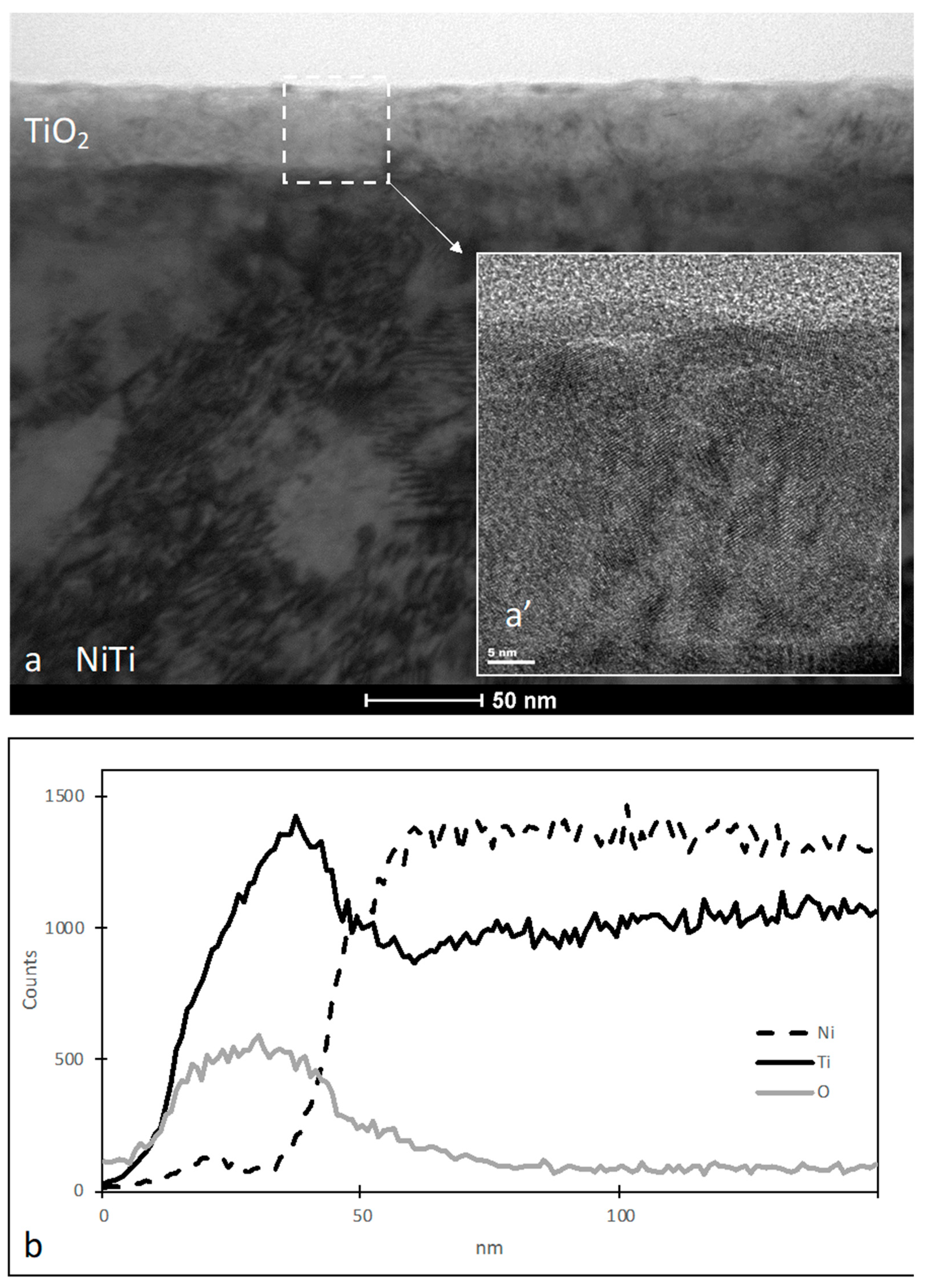
Figure 1. Microstructure of TiO2 layer produced on NiTi alloy (a); HRSTEM image of TiO2 layer (a’); and linear distribution of elements in the TiO2 layer (b).
Materials 16 06086 g001
Figure 2. Surface morphology of NiTi in initial state (a,a’,c,c’) and with a TiO2 surface layer (b,b’,d,d’) both before (a,a’,b,b’) and after (c,c’,d,d’) 14-day incubation in SBF solution: SEM images magnified ×250 (ad), ×1000 (a’d’). Chemical composition obtained by EDS from the entire image area for NiTi non-oxidized (e) and with a TiO2 surface layer (f) after 14-day incubation in SBF solution.
Materials 16 06086 g002
Figure 3. Surface morphology of calcium phosphates biomimetically deposited on NiTi alloy with a TiO2 surface layer after 30 days of incubation in SBF: SEM images magnified ×40 (a), ×250 (b), ×1000 (c), ×2000 (d). Chemical composition obtained by EDS from the entire image area for NiTi with a TiO2 surface layer after 30-day incubation in SBF solution (e).
Materials 16 06086 g003
Figure 4. XRD pattern for NiTi with a TiO2 surface layer after 30-day incubation in SBF solution: marked peaks for identified calcium phosphates.
Materials 16 06086 g004
Figure 5. Morphology of platelets adhering on the surfaces of NiTi alloy in the initial state (a,c) and with a TiO2 surface layer (b,d). SEM images magnified ×2000 (a,b) and ×10,000 (c,d).
Materials 16 06086 g005
Figure 6. Quantitative assessment of platelet adhesion (a), aggregation (b), their size (c) and level of activation according to morphological features (d) for NiTi in initial state and with a TiO2 surface layer; * p ≤ 0.05 NiTi versus NiTi + TiO2.
Materials 16 06086 g006
Figure 7. Proliferation of osteoblasts cultured on NiTi in the initial state and with a TiO2 surface layer represented as the number of cells per mm2 and measured after 24 h, 48 h and 6 days of growth; * p ≤ 0.05 NiTi versus NiTi + TiO2.
Materials 16 06086 g007
Figure 8. Morphology of osteoblasts cultured on the surfaces of NiTi alloy in the initial state (a,a’,c,c’,e,e’) and with a TiO2 surface layer (b,b’,d,d’,f,f’) for 24 h (a,a’,b,b’), 48 h (c,c’,d,d’) and 6 days (e,e’,f,f’). SEM images magnified ×60 (af), ×500 (a’f’).
Materials 16 06086 g008
Table 1. Composition of SBF according to Kokubo [34].
ComponentConcentration (g/L)
NaCl8.035
NaHCO30.355
KCl0.225
K2HPO4·3H2O0.231
MgCl2·6H2O0.311
CaCl20.292
Na2SO40.072
(HOCH₂)₃CNH₂(Tris)6.118
Table 2. The amount of calcium, phosphorus and oxygen expressed in atomic % determined by EDS measurements for non-oxidized NiTi after 14 days of SBF incubation and NiTi with a TiO2 oxidized surface layer after 14 and 30 days of SBF incubation. Results obtained for measurements from the image area and the average of 3 point measurements.
Ca (%at.)P (%at.)O (%at.)Ca/P
SampleImage AreaPointImage AreaPointImage AreaPointImage AreaPoint
NiTi (after 14 days in SBF)1.725.57 ± 0.131.323.78 ± 0.137.5511.16 ± 0.351.301.47
NiTi + TiO2 (after 14 days in SBF)19.7721.70 ± 0.2014.9217.22 ± 0.1148.0055.34 ± 0.551.321.26
NiTi + TiO2 (after 30 days in SBF)20.4924.83 ± 0.1814.7216.19 ± 0.1347.3933.82 ± 0.731.391.53
Table 3. Contact angles measured for NiTi in the initial state and with a TiO2 surface layer using distilled water and diiodomethane, and values of surface free energy calculated for NiTi in the initial state and with a TiO2 surface layer.
NiTiNiTi + TiO2
Contact Angledistilled water84.5 ± 4.3107.1 ± 0.7
diiodomethane45.5 ± 1.366.5 ± 0.9
Surface Free Energyγ (mN/m)36.3725.78
γd (mN/m)33.725.9
γp (mN/m)2.80.00
Abbreviations: γ: Total surface free energy; γd: Dispersive component of surface free energy; γp: Polar component of surface free energy.
Disclaimer/Publisher’s Note: The statements, opinions and data contained in all publications are solely those of the individual author(s) and contributor(s) and not of MDPI and/or the editor(s). MDPI and/or the editor(s) disclaim responsibility for any injury to people or property resulting from any ideas, methods, instructions or products referred to in the content.