Article

Optimization of Chitosan Extraction Process from Rapana venosa Egg Capsules Waste Using Experimental Design

by
Daniel Dinculescu
1,
Cristiana Luminița Gîjiu
1,
Manuela Rossemary Apetroaei
2,*,
Raluca Isopescu
1,
Ileana Rău
1,* and
Verginica Schröder
3
1
Faculty of Chemical Engineering and Biotechnologies, University POLITEHNICA of Bucharest, 011061 Bucharest, Romania
2
Faculty of Navigation and Naval Management, Mircea cel Batran Naval Academy, 900218 Constanta, Romania
3
Faculty of Pharmacy, Ovidius University of Constanta, 900527 Constanta, Romania
*
Authors to whom correspondence should be addressed.
Materials 2023, 16(2), 525; https://doi.org/10.3390/ma16020525
Submission received: 4 December 2022 / Revised: 23 December 2022 / Accepted: 1 January 2023 / Published: 5 January 2023

Abstract

:
New green and sustainable sources were chosen to obtain chitosan, an important material, with many applications in different fields. The present study is focused on egg capsules of Rapana venosa waste as raw material for chitosan oligomers. As previous studies revealed that chitosan extraction from this material takes place with a low yield, the present research aimed to optimize this step. A 22 experimental plan, with three replicates in the center, was proposed to investigate the influence of NaOH concentration and temperature on the yield extraction. After a primary analysis of the experimental data, a favorable temperature value was selected (90 °C) at which the total dissolution of the egg capsules was obtained. Then, at this temperature, the experimental plan was extended exploring the influence of the NaOH concentration on three levels (5, 6, and 7%) and the extraction duration on two levels (60 and 85 min). Based on all experimental data, a neural model was obtained and validated. The neural model was used to maximize the yield, applying Genetic Algorithm (GA) implemented in Matlab®. The resulting optimal solution is: NaOH concentration 6.47%, temperature 90 °C, duration 120 min, with a yield value of 7.05%.

Graphical Abstract

1. Introduction

Chitin and chitosan are biopolymers mainly of marine origin, considered materials support, which are met in organisms such as arthropods, mollusks, worms, coelenterate, and sponges.
Chitin is found particularly in the arthropod’s marine exoskeleton, in the mollusks’ shells, in the cephalopod’s structure (squids, octopuses, etc.) or in the cell walls of some fungi [1], while chitosan can be found in fungi, molds, yeasts, algae [2] or in other sources of rudimentary marine invertebrates.
The studies carried-out in the last fifty years have shown that the waste of crustaceans represents a rich source of products with added value, such as: chitosan, proteins, pigments, whose percentage in their composition, varies with the type of crustacean species and the harvest season [3,4,5].
Chitosan is the only alkaline polysaccharide in nature, is nontoxic, odorless, biocompatible and biodegradable [6]. Depending on their main characteristics (molar mass Mw and deacetylation degree DD), chitosan is made-up from different units of glucosamine and N-acetylglucosamine, or glucosamine only. In addition, the molar mass value is the criterion upon which the chitosan can be classified into high molar mass chitosan (HMw), medium molar mass (MMw), low molar mass (LMw) chitosan, and oligochitosan. Even if the borders between these chitosan types are fluidic, the “oligochitosan” term can be used only for chitosan molecules with fewer than 100 glucosamine units, or molar mass MW less than 16 kDa [4]. On the other side the literature data showed that the oligochitosan with degrees of polymerization (DPs) smaller than 20 and an average molecular weight less than 3.9 kDa are called chitosan oligomers (COs). COs are obtained by depolymerization of chitin or chitosan using acid hydrolysis, hydrolysis by physical methods and enzymatic degradation [5,6,7]. Due to the fact that COs are composed mainly of short chains they tend to change their conformation and form low-viscosity aqueous solutions, which may facilitate the interaction with other polyanions [8].
Oligochitosan is known to have several advantages over HMW and LMW chitosans in biomedical and industrial applications. In particular, due to its lower viscosity and higher compatibility with many additives, the applications of oligochitosan and COs have no undesirable impact on the physicochemical properties of consumer products. Additionally, oligochitosan has some advantages over HMW and LMW chitosan in its biological activity and biomedical applications [4].
Corroborating different literature data [9,10,11,12,13], the presence of particular chitosan or chitin types depends on the degree of evolution of the marine organism. Thus, primary forms, less evolved, without mineralization could be important sources of COs, while developed forms possess chitin, which in its hard-crystalline form, provides support and strength to marine organisms, resulting in the mineralized shell or shells forms. Taking into consideration this information, there is the possibility to find an eco-friendly marine source, susceptible to contain chitosan, a more economical, non-perishable, and clean source as an alternative to the crustacean wastes.
To the best of our knowledge there are few studies on chitosan obtained from such capsules. Our previous studies [11,14] showed the presence of COs in the egg capsules of Rapana venosa structure, together with other components, such as proteins and insoluble compounds. COs have a particular importance tacking into consideration their antimicrobial properties and, in this respect, the present study aimed to optimize the process of obtaining COs from Rapana venosa egg capsules, by using neural network modelling of the extraction process and GA to obtain the best solution in terms of chitosan yield. Process optimization attempts for chitosan extraction from various sources mention experimental design methods and statistical modelling coupled with nonlinear optimization techniques [15,16,17]. It is important to note that these capsules are either organic agglomerations found on the beach, after storms, or they are the remains obtained from harvesting and cleaning snails intended for consumption and their processing does not represent a threat to the marine environment.

2. Materials and Methods

2.1. Materials

Chitosan was extracted and identified from Rapana venosa egg capsules (Figure 1). The egg capsules free of egg mass, were collected (summer-autumn 2021), from the northern area of Navodari (44°19.3′ North–028°41.7′ East).
The reagents used in the extraction procedure were natrium hydroxide solutions (4%, 5%, 6%, 7%) prepared from NaOH pellets purchased from ChimReactiv S.R.L. (Bucharest, Romania), with purity higher than 99.3%. The acetic acid used for chitosan solubilization, as well as ethanol and acetone (p.a.) were purchased from Sigma-Aldrich (Merck KGaA, Darmstadt, Germany).

2.2. Chitosan Extraction Procedure

The experimental study used for chitosan extraction from capsule waste the procedure presented schematically in Figure 2. The chitosan extraction obtaining consisted of suspending the cleaned and crushed capsules in alkaline NaOH solutions, of different concentrations (4%, 5% or 6%), in a constant ratio capsule mass: NaOH solution volume = 1:40 (m/v), working time of 65, 85, and 120 min, at different deproteinization temperatures (80 °C, 85 °C or 90 °C), under constant stirring (500 rpm).
After the time chosen for extraction has elapsed, the alkaline suspension resulted was centrifuged at 3500 rpm rotations, in order to separate the precipitated chitosan from the alkaline supernatant loaded with the lipids and proteins that made up the capsular walls. The washing of the chitosan pellets was carried out by successive washings with distilled water until a pH = 7 of the last washing water was obtained.
In order to remove residues, the obtained chitosan was washed with a 1:1 mixture of ethyl alcohol and acetone, in the ratio of wet chitosan mass: solvent mixture volume = 1:10 (m/v). The organic solvents were removed by centrifugation and repeated washings with distilled water. At the end, the obtained wet chitosan cake was dried in an oven at a temperature of 105 °C.

2.3. Analytical Methods

2.3.1. Microscopic Analysis

The extracted chitosan from Rapana Venosa egg capsules was first analyzed and identified by fluorescence microscopy. Microscopic observations were carried-out by using an optical microscope OPTIKA B—353LD2 (blue filter: λex = 450–490 nm; λem = 515–520 nm), equipped with a camera OPTIKAM, with software Optika Vision Pro, Version 2.7, with (OPTIKA S.R.L., Ponteranica, BG, Italy) or acquisition and data processing.

2.3.2. Determination of Deacetylation Degree

Determination of deacetylation degree (DD) is one of the most important properties of the chitosan chemical characterization, which can influence its performance in many applications.
To determine the DD, the potentiometric titration [15] was taken into consideration, and the value of DD (%) was calculated according to [18] study (Equations (1) and (2)).
D D   ( % ) = 203 Q 1 + 42 Q
Q = c M Δ V m
where: cM—molar concentration of NaOH solution, used for titration (mol/L, in the present study: 0.1 mol/L);
ΔV—volume difference of NaOH added corresponding to the two inflection points (L);
m—mass of analyzed chitosan (g).

2.3.3. Molar Mass

The method used to determine the molar mass was based on intrinsic viscosity determination of the chitosan dissolved in dilute acidic solutions. The measurements were carried out using a capillary viscometer (Ostwald type), according to Pădurețu et al. [15].
This method is based on the relationship between the viscosimetric-average molar mass, Mv (g/mol) and the intrinsic viscosity, [η] (mL/g) of the chitosan solution given by the well-known Mark–Houwink–Sakurada Equation (3), where K and α are constants which depend on the nature of the solvent, temperature, and chemical structure of the polymer (K = 13.8 × 10⁻³ mL/g, α = 0.85).
[ η ] = K M v α

3. Results and Discussion

3.1. Optimization of the Extraction Process

The research started with an experimental plan 22 with three replicates in the center for the evaluation of the experimental error (variance 0.33). Two factors were considered in these steps: the concentration of NaOH and the temperature, while the duration was maintained constant at 120 min.
The obtained data are presented in Table 1.
The primary analysis of the results showed that not all the investigated conditions led to desired results, experimental point 1 corresponding to the concentration and temperature at minimum values even leading to the non-dissolution of 50% of the capsules. As a result, this point was excluded, and the experimental plan was completed with another 12 experiments carried out at a temperature of 90 °C, as a highest value of temperature, proved favorable for the total dissolution of the capsules (Table 1). A new experimental plan was proposed considering the factors the NaOH concentration and the process duration. A variable number of levels was chosen. For the process duration two levels were considered, while three levels were defined for the NaOH concentration in order to better evaluate its influence upon the extraction yield. These experiments were replicated twice at each point. The data are summarized in Table 2.

3.2. Optimizing the Extraction Yield Using Neural Networks

The data used for modeling are taken from Table 1 and Table 2, omitting the 50% undissolved samples, but keeping the partially dissolved and gelatinized samples, resulting in a total of 18 samples.
The evaluation of process yield as a function of NaOH solution concentration, temperature and duration was based on artificial neural networks (ANN) building. ANN belongs to the stochastic modelling and, even if they apparently are similar to regression, a totally different concept is applied to explain the influence of factors (inputs) upon the dependent variable (output). The process features are captured during a learning stage (training and testing) based of which the network will be capable to reproduce the process. During the learning stage, the sum of errors between the measured values of the output and the estimated ones is minimized according to a selected learning rule and optimization method. The resulting model is a “gray box” that reflects the relation between the inputs and outputs, even if a direct mathematical relation cannot be written. For the present study, a multilayer ANN was proposed (Figure 3) with three neurons in the input layer standing for the NaOH concentration, temperature, duration, four neurons in the hidden layer and one output neuron representing the extraction yield.
The network was trained and tested in Matlab (R2015a) using: Activation function: sigmoid; Learning Rule: Backpropagation of errors and Levenberg–Marquardt optimization algorithm implemented in the “trainlm” function, and population size 100.
In order to avoid overfitting, 70% of the data were used for training and 30% for testing and validation.
A first neural model obtained with the initial data achieved a correlation of the data with a coefficient of determination R2 = 0.92.
The optimization of the extraction process, in the sense of maximizing the yield, with the use of the neural model obtained at this stage, was carried out with the GA algorithm implemented in Matlab.
The resulting optimal solution is: NaOH concentration 6.7%, temperature 89 °C, duration 120 min, with a yield value of 5.75%.
To test the neural model, two experimental points were proposed in the «proximity» of this first estimate of the optimum (Table 3).
In order to obtain a better model, these two new points were added to the data set and a new improved neural model with the same structure, based on 20 experimental measurements, was obtained, for which R2 = 0.94 (Figure 4).
From the point of view of how the network captured the characteristics of the process, the convergence variation in the training, validation, and testing stages is analyzed, by identifying the minimum errors between the measured and estimated yield values, during the learning cycles (Figure 5).
Graphic representations of the yield variation in the investigated field showed that both the temperature and the working time must be at the maximum investigated values (90 °C and 120 min), while the optimal concentration of NaOH is close to the value of 6.5%, the increase in this concentration up to 7% or above this value not being favorable (Figure 6).
The optimum identified using the genetic algorithm suggested the operating conditions: NaOH concentration 6.47%, temperature 90 °C, duration 120 min at which the estimated yield is 7.05%.
Both the operating conditions and the yield value are very close to the values of experimental point two in the set of validation experiments; therefore, these conditions no longer require experimental validation, and the set of operating parameters [6.5 90 120] can be considered as optimal in the investigated field.

3.3. Chitosan Characterization

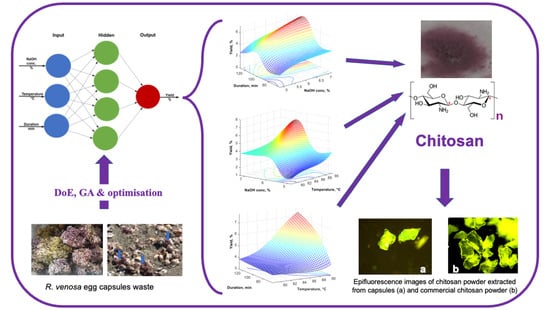
3.3.1. Fluorescence Microscopy

Cellulose, lignin, chitosan and collagen are important structural compounds existing in many organisms’ bodies. These biopolymers emit fluorescent radiation and these natural emissions are due to the presence, in various compartments of cell, of fluorescent compounds which emit radiations in the spectrum of visible light when they are excited with UV light [19]. The chitosan powder obtained by chemical extraction from the Rapana venosa egg capsules in alkaline dilute solutions was analyzed by the epifluorescence microscope, and its fluorescent emissions were compared with those emitted by the powder of a commercial chitosan (Figure 7a,b).
Comparing Figure 7a with Figure 7b, it can be observed that the two chitosan powders fluorescent emissions subjected to microscopic observations are similar, although the commercial chitosan is of a different origin.

3.3.2. Deacetylation Degree (DD) and Molar Mass (MW)

For the extracted chitosan, the DD values ranged between 74% and 76%. The obtained results were compared with the values of one low molar mass commercial chitosan (product number: 448869, CAS 9012-76-4, purchased from Sigma Aldrich, Darmstadt, Germany), with DD = 95%.
According to our experimental data, the obtained values for the molar mass of the chitosan extracted from egg capsules of Rapana venosa snail range between 1.5 and 3 kDa, which classifies this biopolymer into the category of chitosan oligomers (COs) [8,9]. The molar mass of extracted chitosan from egg capsules of Rapana venosa (1.5 kDa) was lower than the one obtained for the commercial chitosan (183 kDa—extracted from crab shells). This difference in the molar mass is due to the different sources of raw materials.

4. Conclusions

The present study proved that this new waste represented by Rapana venosa egg capsules represent an eco-friendly source for chitosan, particularly for chitosan oligomers with a deacetylation degree of 75%. This new source of COs-type chitosan presents, on one hand, the advantage to be obtained at low costs, by valorizing some marine waste, and on the other hand, it could be used as a biocide, in various fields of environmental engineering. By tailoring the extraction parameters, a reasonable yield (around 7%) can be achieved despite the fact that these capsules contain a low content of chitosan. The artificial neural network modelling and the genetic algorithms used in the optimization study revealed that the optimal operating parameters are a NaOH concentration of 6.5%, a temperature of 90 °C and an extraction time of 120 min.

Author Contributions

Conceptualization: R.I. and I.R.; methodology: M.R.A. and V.S.; software: C.L.G. and R.I.; validation: D.D. and C.L.G.; formal analysis: C.L.G., R.I. and D.D.; investigation: M.R.A. and V.S.; resources: I.R.; writing—original draft preparation: D.D., C.L.G., R.I. and M.R.A.; writing—review and editing: I.R., M.R.A. and D.D.; visualization: C.L.G.; supervision: I.R.; project administration: I.R.; funding acquisition: I.R. All authors have read and agreed to the published version of the manuscript.

Funding

This work was supported by a grant of a Romanian Ministry of Education and Research, CNCS—UEFISCDI, project number PN-III-P4-ID-PCE-2020-2243.

Institutional Review Board Statement

Not applicable.

Informed Consent Statement

Not applicable.

Data Availability Statement

Not applicable.

Conflicts of Interest

The authors declare no conflict of interest.

References

  1. Ioelovich, M. Crystallinity and Hydrophility of Chitin and Chitosan. Res. Rev. J. Chem. 2014, 3, 7–14. [Google Scholar]
  2. Knežević-Jugović, Z.D.; Bezbradica, D.I.; Mijin, D.Ž.; Antov, M.G. The Immobilization of Enzyme on Eupergit® Supports by Covalent Attachment. In Enzyme Stabilization and Immobilization. Methods in Molecular Biology; Minteer, S., Ed.; Humana Press: Totowa, NJ, USA, 2011; pp. 99–111. [Google Scholar]
  3. Bakshia, P.S.; Selvakumara, D.; Kadirvelub, K.; Kumara, N.S. Chitosan as an environment friendly biomaterial—A review on recent modifications and applications. Int. J. Biol. Macromol. 2020, 150, 1072–1083. [Google Scholar] [CrossRef]
  4. Blagodatskikh, I.V.; Kulikov, S.N.; Vyshivannaya, O.V.; Bezrodnykh, E.A.; Yamskov, I.A.; Tikhonov, V.E. Influence of glucosamine on oligochitosan solubility and antibacterial activity. Carbohydr. Res. 2013, 381, 28–32. [Google Scholar] [CrossRef]
  5. Lodhi, G.; Kim, Y.-S.; Hwang, J.-W.; Kim, S.-K.; Jeon, Y.-J.; Je, J.-Y.; Ahn, C.-B.; Moon, S.-H.; Jeon, B.-T.; Park, P.-J. Chitooligosaccharide and Its Derivatives: Preparation and Biological Applications. BioMed Res. Int. 2014, 2014, 654913. [Google Scholar] [CrossRef] [Green Version]
  6. Mourya, V.K.; Inamdar, N.N.; Choudhari, Y.M. Chitooligosaccharides: Synthesis, characterization and applications. Polym. Sci. Ser. A 2011, 53, 583–612. [Google Scholar] [CrossRef]
  7. Aam, B.B.; Heggset, E.B.; Norberg, A.L.; Sørlie, M.; Varum, K.M.; Eijsink, V.G.H. Production of chitooligosaccharides and their potential applications in medicine. Mar. Drugs 2010, 8, 1482–1517. [Google Scholar] [CrossRef] [PubMed] [Green Version]
  8. Jiang, G.-L.; Zhu, M.-J. Preparation of astaxanthin-encapsulated complex with zein and oligochitosan and its application in food processing. LWT 2019, 106, 179–185. [Google Scholar] [CrossRef]
  9. Green, J.H.; Mattick, J.F. Fishery waste management. In Food Processing Waste Management; Green, J.H., Kramer, A., Eds.; AVI Publishing: Westport, UK, 1979; pp. 202–307. [Google Scholar]
  10. Subhapradha, N.; Ramasamy, P.; Shanmugam, V.; Madeswaran, P.; Srinivasan, A.; Shanmugam, A. Physicochemical characterisation of β-chitosan from Sepioteuthis lessoniana gladius. Food Chem. 2013, 141, 907–913. [Google Scholar] [CrossRef] [PubMed]
  11. Apetroaei, M.R.; Zgârian, R.G.; Manea, A.M.; Rău, I.; Tihan, G.T.; Schröder, V. New source of chitosan from Black Sea marine organisms identification. Mol. Cryst. Liq. Cryst. 2016, 628, 102–109. [Google Scholar] [CrossRef]
  12. Lee, J.W.; Yeomans, W.G.; Allen, A.L.; Deng, F.; Gross, R.A.; Kaplan, D.L. Production of Chitosan-and Chitin-Like Exopolymers, Patent No. US6534294B1, 18 March 2003. [Google Scholar]
  13. Chatterjee, S.; Adhya, M.; Guha, A.K.; Chatterjee, B.P. Chitosan from Mucor rouxii: Production and physico-chemical characterization. Process Biochem. 2005, 40, 395–400. [Google Scholar] [CrossRef]
  14. Schröder, V.; Rău, I.; Dobrin, N.; Stefanov, C.; Mihali, C.-V.; Pădureţu, C.-C.; Apetroaei, M.R. Micromorphological details and identification of chitinous wall structures in Rapana venosa (Gastropoda, Mollusca) egg capsules. Sci. Rep. 2020, 10, 14550. [Google Scholar] [CrossRef] [PubMed]
  15. Pădurețu, C.C.; Isopescu, R.; Rău, I.; Apetroaei, M.R.; Schröder, V. Influence of the parameters of chitin deacetylation process on the chitosan obtained from crab shell waste. Korean J. Chem. Eng. 2019, 36, 1890–1899. [Google Scholar] [CrossRef]
  16. Bello, V.E.; Olafadeha, O.A. Comparative investigation of RSM and ANN for multi-response modeling and optimization studies of derived chitosan from Archachatina marginate shell. Alex. Eng. J. 2021, 60, 3869–3899. [Google Scholar] [CrossRef]
  17. Abdel-Gawad, K.M.; Hifney, A.F.; Fawzy, M.A.; Gomaa, M. Technology optimization of chitosan production from Aspergillus niger biomass and its functional activities. Food Hydrocoll. 2017, 63, 593–601. [Google Scholar] [CrossRef]
  18. Dima, J.B.; Sequeiros, C.; Zaritzky, N. Chitosan from Marine Crustaceans: Production, Characterization and Applications. In Biological Activities and Application of Marine Polysaccharides, 1st ed.; Shalaby, E.A., Ed.; InTech Open: London, UK, 2017; pp. 39–56. [Google Scholar]
  19. Roshchina, V.V. Vital Autofluorescence: Application to the study of plant living cells. Int. J. Spectrosc. 2012, 2012, 124672. [Google Scholar] [CrossRef]
Figure 1. R. venosa egg capsules waste.
Materials 16 00525 g001
Figure 2. Chitosan obtaining process from Rapana venosa egg capsules.
Materials 16 00525 g002
Figure 3. ANN architecture.
Materials 16 00525 g003
Figure 4. Parity plot for the experimental and computed data.
Materials 16 00525 g004
Figure 5. Evolution of the error in approximation of experimental data, during the learning process.
Materials 16 00525 g005
Figure 6. Yield variation for 90 °C temperature (a), 120 min working time (b) and 6.5% NaOH concentration (c).
Materials 16 00525 g006
Figure 7. Fluorescent emissions (epifluorescence microscopy (×100)) of: (a) chitosan powder extracted from capsules; (b) commercial chitosan powder.
Materials 16 00525 g007
Table 1. Yields values for the initial 22 experimental plan, considering an extraction time of 120 min.
Exp.
No.
NaOH Conc.
(%)
Temperature
(°C)
Yield
(%)
Observations
Deproteinization Time = 120 min
megg:vol NaOH = 1/40
14801.72At the end, about 50% egg capsules did not dissolve
26804.16At the end, part of the capsules remained undissolved and were found in the centrifuged filtrate
34904.76At the end, all the capsules were dissolved
46905.20At the end, all the capsules were dissolved
55853.00At the end, when filtering the obtained suspension, a small number of capsules remained in the form of gelatine (partially dissolved)
65852.00
75852.00
Table 2. Yields values for the supplementary 22 experimental plan for the deproteinization temperature of 90 °C.
Duration (min.)6085
NaOH Conc., (%)
5 1.963.04
1.843.20
6 3.324.92
3.283.92
7 2.884.00
3.124.92
Table 3. New experimental points, and the yield values.
Exp. No.NaOH Conc.
(%)
Temperature
(°C)
Duration
(min)
Yield (%)
Estimated by the Preliminary ModelExperimental
17851204.154.13
26.5901206.187.00
Disclaimer/Publisher’s Note: The statements, opinions and data contained in all publications are solely those of the individual author(s) and contributor(s) and not of MDPI and/or the editor(s). MDPI and/or the editor(s) disclaim responsibility for any injury to people or property resulting from any ideas, methods, instructions or products referred to in the content.

