Article

Scanning with Laser Beam over the TiO2 Nanotubes Covered with Thin Chromium Layers towards the Activation of the Material under the Visible Light

1
Centre for Plasma and Laser Engineering, Institute of Fluid-Flow Machinery, Polish Academy of Sciences, Fiszera 14 Street, 80-231 Gdańsk, Poland
2
Faculty of Applied Physics and Mathematics, Institute of Nanotechnology and Materials Engineering, Gdańsk University of Technology, Narutowicza 11/12 Street, 80-233 Gdańsk, Poland
3
Department of Biomedical Engineering, Faculty of Electronics, Telecommunications and Informatics, Gdańsk University of Technology, Narutowicza 11/12 Street, 80-233 Gdańsk, Poland
*
Author to whom correspondence should be addressed.
Materials 2023, 16(7), 2572; https://doi.org/10.3390/ma16072572
Submission received: 22 February 2023 / Revised: 17 March 2023 / Accepted: 21 March 2023 / Published: 23 March 2023
(This article belongs to the Special Issue Advances in Thin Film Technology and Laser Processing)

Abstract

:
This work presents pulsed UV laser treatment (355 nm, 2 Hz) of TiO2 nanotubes decorated with chromium oxides. The modification was performed in a system equipped with a beam homogenizer, and during the irradiation, the samples were mounted onto the moving motorized table. In such a system, both precisely selected areas and any large area of the sample can be modified. Photoelectrochemical tests revealed photoresponse of laser-treated samples up to 1.37- and 18-fold under the illumination with ultraviolet-visible and visible light, respectively, in comparison to bare titania. Optimal beam energy fluence regarding sample photoresponse has been established. Scanning electron microscopy images, X-ray diffraction patterns, along with Raman and X-ray photoelectron spectra, suggest that the enhanced photoresponse results from changes solely induced in the layer of chromium oxides. It is believed that the results of the present work will contribute to a wider interest in laser modification of semiconductors exhibiting improved photoelectrochemical activity.

1. Introduction

The shortage of natural resources due to the burning of fossil fuels has become a global concern over the past decades [1,2,3]. Moreover, rapid industrial development as well as population increment caused the raising of organic and inorganic contaminants that can influence not only the environment but also human health [4,5]. Taking the above into account, as well as the infinite energy demand in current society, new and green technologies are required to overcome these problems. The common sources of renewable energy are, e.g., solar, wind, geothermal, and ocean energies. Among those, solar energy is not only the most abundant one but is also constantly being replenished. Moreover, the usage of photoelectrochemical devices employing semiconductor electrodes is already widely recognized as an efficient and eco-friendly alternative to explore sustainable energy sources [6,7,8,9].
The reason why semiconductors are used as electrodes for electrochemical processes lies in the energy specificity of semiconductor reactions [10]. It is known that the change in potential of such an electrode may lead to changes in the number of charge carriers [11]. Therefore, various kinds of semiconductors such as Fe2O3 [12], WO3 [13], BiVO4 [14], or TiO2 [15] are being investigated. Nonetheless, due to the great chemical stability, low toxicity, and cheap price, titanium dioxide is the most favored candidate. In particular, titanium dioxide in the form of nanotubes has been intensively studied due to the excellent electron transfer, easily controllable architecture, and high specific surface area [16]. However, one cannot forget about the drawbacks that hamper the application of pure titania. Most of all, titania exhibits wide band gap of 3.2 eV and 3.0 eV for anatase and rutile phases, respectively [17], meaning that it is not matching to the visible light region, and UV light is mainly absorbed by such material [18]. Another limitation is the fast recombination of electron–hole pairs formed by the titania [19]. According to the literature survey, the modification of titania with non-metals, metals, and metal oxides causes not only the reduction of the energy band gap contributing to the visible light absorption, but also improves the separation of charge carriers leading to the inhibition of electron–hole recombination [15,20].
As transition metals are earth abundant, they stand as a great alternative for noble metals such as gold, platinum, or palladium. Especially, taking into account the deficient resources and high prices of noble metals that make their usage on the large scale pointless. Going further, the main advantages of metal oxides are stability, low cost, and relatively easy preparation routes such as, e.g., electrochemical deposition, photoreduction, wet impregnation, or magnetron deposition combined with annealing [15,21]. In our previous work, we studied titanium dioxide nanotubes decorated with chromium, molybdenum, and tungsten oxides [22]. These oxides are in general regarded as wide band gap semiconductors [23,24,25]. Moreover, chromium oxide exhibits p-type semiconducting properties [26], whereas molybdenum and tungsten oxides exhibit n-type [27,28]. We demonstrated that the deposition of chromium oxides onto the top of the TiO2 nanotubes (TNT) increased their photoresponse. Such behavior can be attributed to the intraband states provided by chromium oxides and the change in electronic properties; namely, the additional direct (allowed) pathway for photon absorption. Meanwhile, the deposition of molybdenum and tungsten oxides resulted in decreased photoresponse of the studied material. It was concluded that optical transition occurring in these electrodes has a forbidden nature.
In this work, we extended our studies of titania nanotubes decorated with chromium oxides [22] by exploring the possibility of UV laser modification. It should be noted in here that previously we also examined laser-treated TiO2 nanotubes with deposited thin films of chromium [29]. The 532 nm wavelength of Nd: YAG laser was used, though, the photoresponse of prepared material under UV-vis illumination was decreased after modification in comparison to pure titania, while for visible light it increased. Meanwhile surface modification of bare TiO2 nanotubes via pulsed UV laser treatment can improve their photoresponse if processing parameters are satisfied [30,31]. Surface melting (the top of the nanotubes) [30,31], degradation of crystal structure [30,31], optical band gap decrement [30], and change in electronic properties, including the increase in donor density [30,31] and positive shift of flat band potential [30], may accompany the enhanced photoresponse. Nonetheless, the general concept of laser treatment is to manipulate crystal structure, which contributes to the emergence of new additional mid-gap states. These states provide additional carriers which narrow optical band gap and enhance sample photoresponse. Therefore, one can expect the synergistic effect of chromium oxide decoration and UV laser processing of titania material.
Indeed, in here we report on optimized pulsed UV laser processing of TiO2 nanotubes decorated with aggregates of chromium oxides, resulting in the superior photoresponse. The effect of laser modification on morphology, crystallinity, surface chemical composition, and optical properties was studied by scanning electron microscopy (SEM), X-ray diffraction (XRD), X-ray photoelectron spectroscopy (XPS), and ultraviolet-visible (UV-vis) spectroscopy, respectively. Electrochemical and photoelectrochemical properties of materials were tested by means of cyclic and linear voltammetry; the latter being investigated under UV-vis and vis illumination. Electronic properties were evaluated by analysis of Mott–Schottky plots. Thanks to the use of a beam homogenizer ensuring a square laser spot and motorized table on which the samples were mounted, both precisely selected areas as well as any large area of the sample can be modified. Therefore, such an approach can be further scaled up from the laboratory to a commercial level.

2. Methods

TiO2 nanotubes were prepared via an anodization process of Ti foil (99.7%, Strem, Newburyport, MA, USA) in a two-electrode system. First of all, titanium foils of 2 × 3 cm2 pieces were cleaned in an ultrasonic bath in acetone (Chempur, Piekary Śląskie, Poland, p.a. grade), ethanol (Chempur, p.a. grade), and deionized water (HYDROLAB, Straszyn, Poland, 0.05 µS) for 10 min in each solvent, followed by drying in a stream of cold air (Air Liquide, Paris, France, ALPHAGAZ™ 1 Air). Afterwards, Ti foil served as an anode while Pt mesh as a cathode. The electrolyte consisted of 0.5 g NH4F (Chempur, p.a. grade), 7.5 mL deionized H2O, and 42.5 mL ethylene glycol (Chempur, p.a. grade). 50 mL of electrolyte was used for the anodization process and the distance between the electrodes was 2 cm. The temperature was controlled by a thermostat (Julabo F-12, Seelbach, Germany) and kept at 23 °C. The voltage of 40 V was maintained for 15 min while the voltage ramp-up and ramp-down rates were set to 6 V min−1. As-anodized samples were rinsed with ethanol and were dried in the air freely. Finally, amorphous TiO2 nanotubes were annealed in a tubular furnace (Nabertherm, Lilienthal, Germany, 120/1000/12-P330) at 450 °C in the air for 2 h (ramp-up rate: 2 °C min−1) to ensure the crystalline phase. The samples were then allowed to cool down freely [22].
In the next step, TiO2 nanotubes were covered with 15 nm of chromium film by means of a magnetron sputtering system (Quorum, Q150T S, target: TK8845 Cr, Quorum Technologies, Lewes, UK) equipped with quartz crystal microbalance. Prior the deposition, the target was cleaned by applying 150 mA with the shutter closed. For deposition, the pressure and current were established to 3 × 10⁻³ mBar and 120 mA, respectively. Afterwards, the samples were thermally treated in the tubular furnace to both crystallize and oxidize the deposited material. The processing parameters were the same as in the case of crystallization of titania nanotubes.
Finally, laser modifications were performed using Nd: YAG pulsed laser (Quantel, Lannion, France, Q-850) operated at 355 nm with 2 Hz repetition rate and 6 ns pulse duration. Beam homogenizer was utilized to ensure the uniform intensity spot of square shape. Moreover, the samples were mounted on the precise programmable motorized table (SmarAct, Oldenburg, Germany). The constant speed of the table of 4 mm min−1 was maintained during processing. The energy fluences were set to 10, 20, 30, 40, and 50 mJ cm−2, while the pressure to 5 × 10⁻⁵ mbar [30]. The electrode surface was scanned twice (forward and backwards) with a laser beam. The laser spot that reaches the sample is of square shape with 2.3 mm side. It means that to process the 2 × 3 cm2 sample the position of laser beam after each treated row of 2.3 mm width should be changed. Every row of the sample is treated with the same energy fluence, and the number of rows is limited only with the size of the sample, enabling this technique to be easily adapted for large-area sample modifications.
The samples’ morphology was investigated using scanning electron microscope technique (FEI, Waltham, MA, USA, Quanta FEG 250) with field emission and ET secondary electron detector. The voltage was kept at 10 kV.
High-resolution X-ray photoelectron spectroscopy analyses were carried out in the core-level binding energy of Ti 2p, O 1s, and Cr 2p to evaluate the surface chemical states of the studied samples. Escalab 250 Xi was used, operating with Al Kα X-ray source, 250 μm spot diameter. The pass energy was 20 eV. Low-energy electron and Ar+ ions flow was used for charge compensation, with final x-axis calibration at C 1s (284.8 eV) adventitious carbon.
The structural analysis was performed by means of X-ray diffractometer (Bruker D2 Phaser 2nd generation, Billerica, MA, USA) using CuKa radiation and a LynxEye XE-T detector at room temperature and Raman spectrometer (InVia, Renishaw, Wotton-under-Edge, Gloucestershire, UK) with laser excitation at 514 nm and power of 2.5 mW.
Optical properties of prepared materials were tested using Lambda 35 UV-Vis Spectrometer in the range of 200–900 nm. The scanning speed was set to 120 nm min−1. Band gap energy values were determined on the basis of the Kubelka–Munk function [32].
Bare and the set of modified titania were characterized as electrode materials using different electrochemical techniques. Autolab PGSTAT302N potentiostat–galvanostat system (Methrom, Herisau, Switzerland) was used to control the electrode potential and record the current or impedance data depending on the selected technique. The measurements were carried out in three electrode arrangements, where Pt mesh acted as the counter electrode, Ag/AgCl/0.1 M KCl served as the reference electrode, and the obtained material was treated as the working electrode. The electrolyte was 0.5 M Na2SO4 with pH = 6.8 deaerated with argon (5.0, Air Liquide, ALPHAGAZ™) for 40 min before measurements. During electrochemical tests, the argon flow was maintained above the solution. Cyclic voltammetry (CV) was carried out in the range from +1.0 V to −1.0 V, with the scanning speed of 50 mV s−1. The cyclic voltammetry curves were recorded several times to verify the stability of the electrode material in contact with an electrolyte within the investigated potential range.
Linear sweep voltammetry (LV) measurements were performed in dark and under UV-vis and visible light illumination at 10 mV s−1 in the potential range of −0.2–+1.2 V. As a light source, an xenon lamp (Osram XBO 150, Munich, Germany) equipped with an AM 1.5 filter was used. In order to illuminate electrode material with the visible light, an optical filter (420GG Schott) was mounted in the photoelectrochemical setup enabling the cutting off of the wavelengths below 420 nm.
The Mott–Schottky plot was constructed on the basis of the impedance measurements carried out at a constant frequency of 1 kHz, in the potential range from +0.6 V to −0.4 V vs. Ag/AgCl/0.1 M KCl. Before recording data for each potential, the electrode was polarized for 60 s. The donor densities were calculated according to the Mott–Schottky space charge capacitance from the following equation:
N d = 2 ε 0 ε e [ d 1 C SC 2 dE ] 1
where ε is the dielectric constant of TiO2, ε0 is the vacuum permittivity, e stays for the electron charge, and E is the applied potential; in calculations ε0 = 8.85 × 10⁻¹² F/m, ε = 38 for anatase-TiO2 and e = 1.602 × 10⁻¹⁹ C were used [33].

3. Results and Discussion

SEM images of the pristine TiO2 nanotubes can be found in our previous works [22]. Nonetheless, the length of the nanotubes is of 1.4 ± 0.1 µm, while their internal diameter and wall thickness of 90 ± 8 nm and 13 ± 3 nm, respectively. After deposition, the chromium species are located at the top part of nanotubes in the form of aggregates—see Figure 1a, which is in agreement with our previous reports [22]. SEM images of laser-processed samples are show in Figure 1b–f. It can be easily observed that the lowest laser fluence, namely 10 mJ cm−2, did not influence the morphology of the sample in a significant way. However, increasing the laser fluence results in the partial melting of the deposited layer on the top of the nanotubes, and the area of melted area increases with the fluence up to 50 mJ cm−2. Similar behavior was previously reported for the bare titania nanotubes [30]; the melted layer can partially or fully cover the nanotube mouths leading to the decrease in surface area. Nonetheless, underneath the melted layer, the architecture of nanotubes remained intact (see Figure S1 in the Supplementary Materials) and the length of TNT was preserved regardless of the parameters of laser processing.
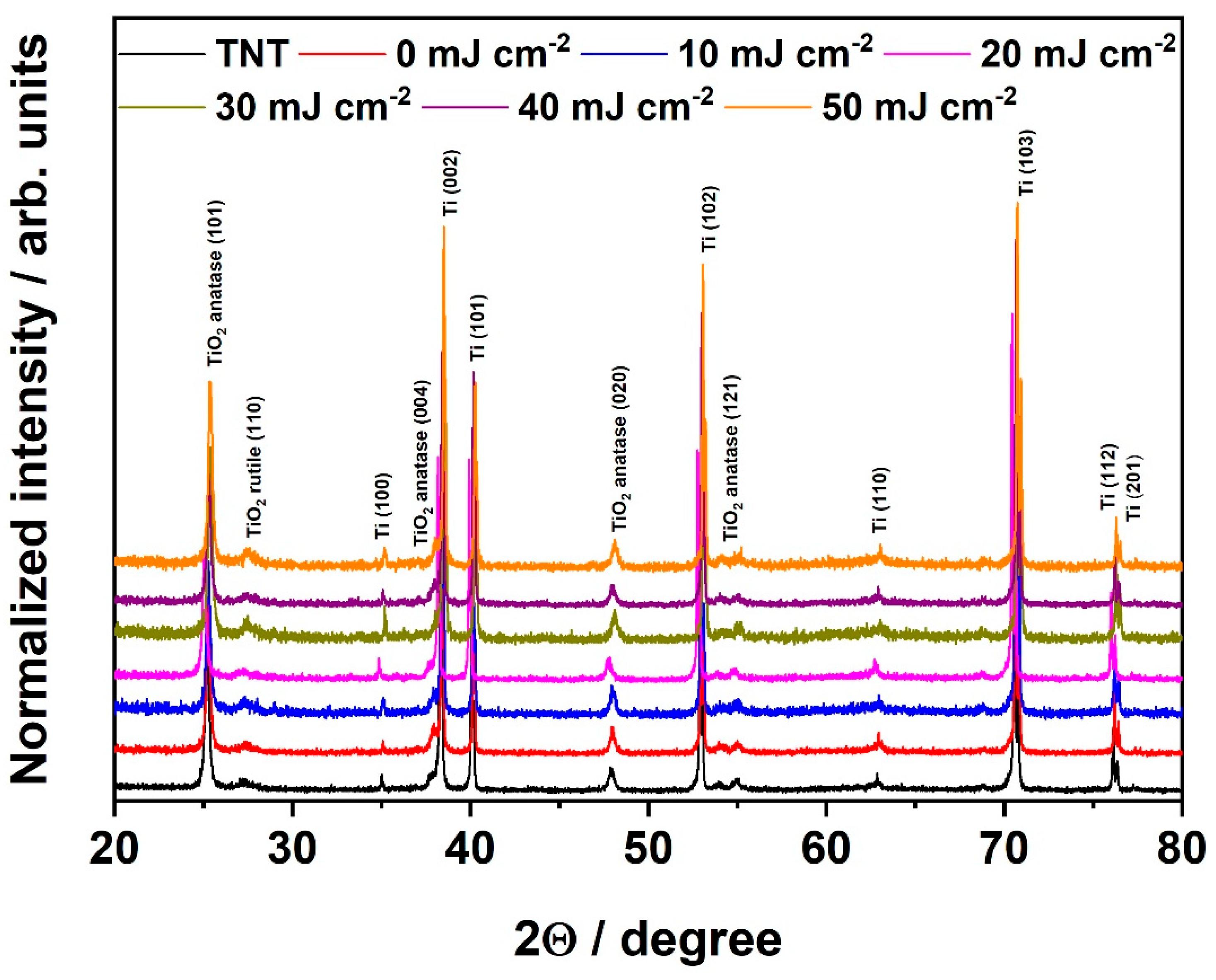
The structural properties of obtained materials were investigated by means of the XRD technique, and collected XRD patterns confirm the crystallinity of all prepared electrodes. Particular peak positions were identified according to the [ICDD PDF-4+] and are indicated in Figure 2. The anatase is a dominant phase; however, trace rutile peaks can be easily observed. The material phase composition was calculated using the given formulas [34]:
W A = k A A A k A A A + A R
W R = A R k A A A + A R
where WA and WR are weight fractions of anatase and rutile, AA and AR are integrated intensities of anatase and rutile, and kA is equal to 0.886. Anatase (101) (2Θ = 25.21°) and rutile (110) (2Θ = 27.22°) peaks were analyzed to assess the anatase–rutile fraction.
The reference sample, namely pure TiO2 nanotubes, is composed of ca. 88% of anatase and ca. 11% of rutile. Further chromium deposition, thermal annealing in tubular furnace, and laser processing do not influence on the materials composition as well as on the intensity of peaks in a significant way. It can therefore be concluded that the laser beam fluence was not high enough to degrade the crystal structure of the samples [35,36]. It should be also mentioned in here that the diffraction signals of titanium arise from the titanium foil on which nanotubes were grown.
As it comes to material decorated with chromium species, no additional peaks originating from chromium oxide presence are visible in XRD patterns. This can be justified by the low amount of the deposited species which is below the detection limit of XRD instruments. This is in agreement with our previous work [37], in which the XRD patterns of the TiO2 nanotubes, decorated with spherical nickel nanoparticles with an average size of 34 ± 10 nm, revealed no peaks corresponding to nickel species.
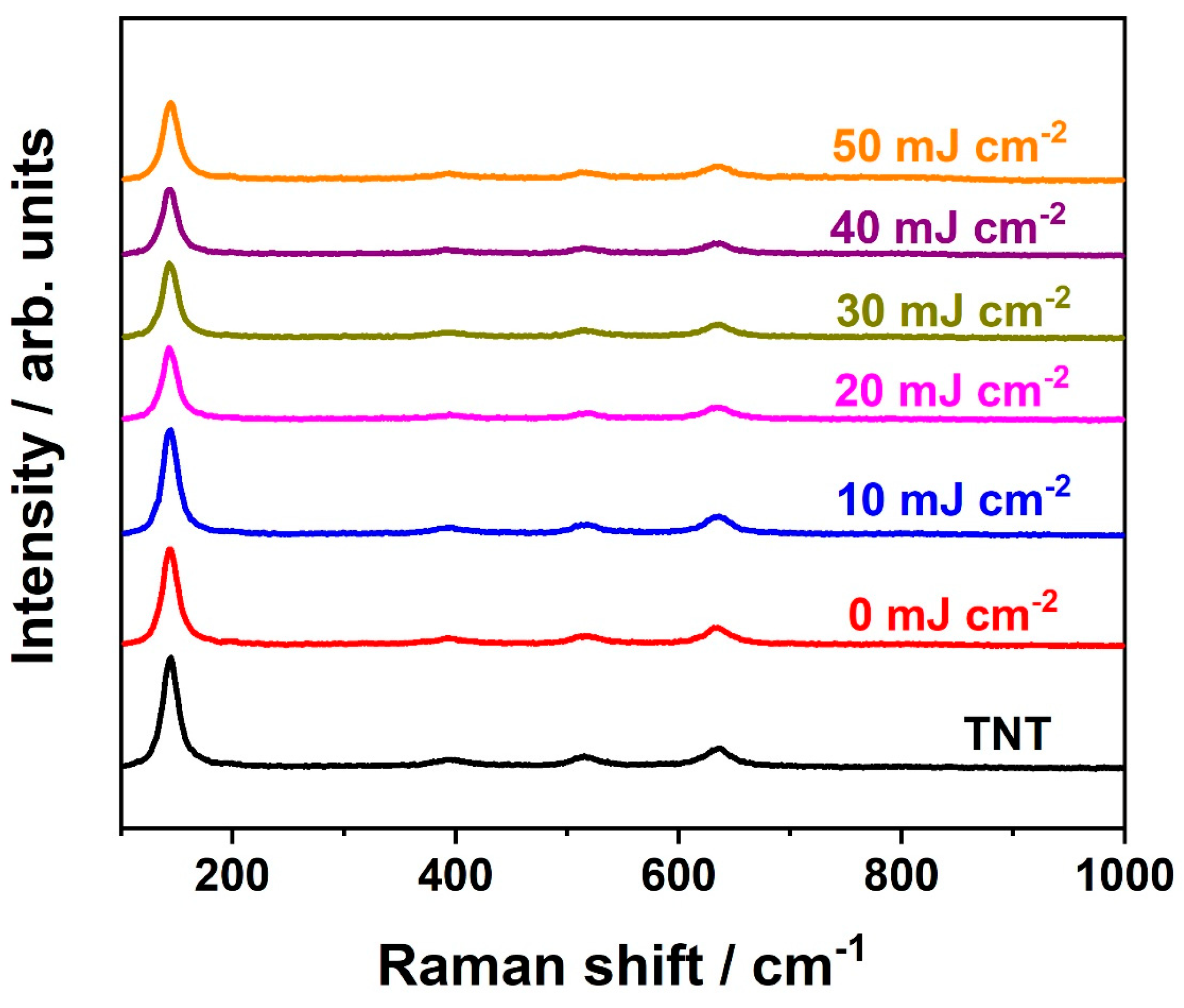
Further analysis of structural properties was conducted via Raman spectroscopy, and the spectra collected for all the samples are given in Figure 3. Signals registered at 144, 197, 395, 515, and 636 cm−1 correspond to Eg(1), Eg(2), B1g, A1g, and Eg(3) active anatase modes, respectively [38]. On contrary to the X-ray diffractograms, no bands assigned to rutile are observed. This may be explained by the fact that rutile phase is formed underneath the nanotubes, i.e., at the metal/oxide interface [39] and the small amount of this phase. Moreover, the position of the main anatase peak at 144 cm−1 remained unchanged regardless of the applied energy fluence used to modify electrode material (Figure S2). It should be mentioned that the change in position was previously observed for pristine laser-treated titania nanotubes [30,40]. Nonetheless, similar behavior, namely no peak shift due to the laser treatment, was also reported for thin gold layers deposited onto TNT [40]. Typically, shift of the 144 cm−1 Raman mode as well as its broadening is associated with creation of oxygen vacancies [41,42]. Therefore, obtained results indicate that the additional chromium layer may protect the electrode material from the oxygen vacancies formation which was also assumed in the case of gold film [40]. Nonetheless, it can be also observed that the intensity of the main anatase band varies with different values of energy fluence (Figure S2). It may be caused by the lower degree of crystallinity of laser-treated samples due to the non-homogenous laser irradiation in terms of pulse energy [36] or changes in sample morphology [40]. However, the XRD results do not confirm the former explanation. Therefore, we anticipate that it is attributed to the geometry change—as can be observed in Figure 1.
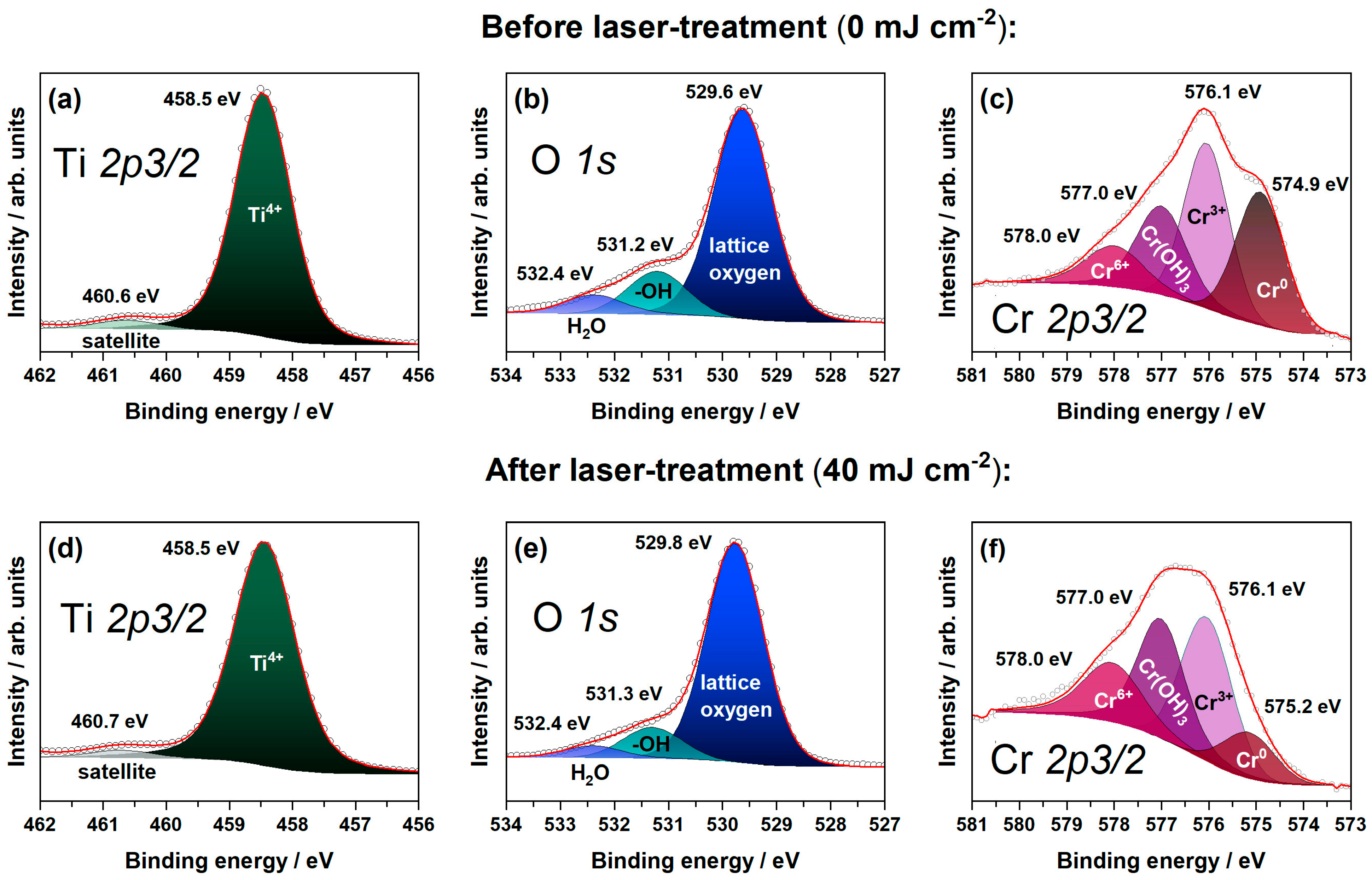
The surface chemical compositions of the TiO2 nanotubes decorated with chromium layer before (Figure 4a–c) and after laser treatment (Figure 4d–f) were investigated by means of the XPS technique. A sample treated with 40 mJ cm−2 was selected as a representative one on the basis of electrochemical measurements described later on. The XPS results corresponding to the binding energies for Ti 2p3/2, O 1s and Cr 2p3/2 were analyzed. For laser-untreated electrode material, the high resolution scan of Ti 2p3/2 indicates the presence of only Ti4+ in the TiO2 crystal lattice (peak at 458.5 eV) [43,44]. The signal at 460.6 eV is recognized as a satellite based on its intensity and position from the Ti4+ peak [22]. Additionally, no peaks corresponding to the Ti3+ are observed. The high-resolution scan of O 1s can be deconvoluted into three peaks assigned to lattice oxygen, OH, and H2O bonds at 529.6 eV, 531.2 eV, and 532.4 eV, respectively [45,46]. For Cr 2p3/2, the spectrum deconvolution exhibits that peaks at 574.9 eV, 576.1 eV, 577.0 eV, and 578.0 eV correspond to metallic chromium, Cr3+, chromium-hydroxide, and Cr6+, respectively [47,48,49,50]. The presence of metallic chromium indicates that the sputtered chromium layer did not undergo full oxidation after thermal annealing in the tubular furnace. Additionally, 30% of the total chromium content in the sample can be ascribed to a metallic state; 34%—to Cr3+, 23%—to chromium in chromium hydroxide, and 13%—to Cr6+.
After laser treatment, no changes in chemical nature of titania are observed as the position of the peak corresponding to Ti4+ in the Ti 2p3/2 region remains intact. Also, no Ti3+ has been identified. Therefore, XPS measurements verify the preliminary conclusions derived from XRD and Raman spectra regarding the preservation of crystal structure after laser processing. Furthermore, in O 1s and Cr 2p3/2 regions, insignificant peak shifts can be noticed—see Figure 4e,f. However, taking into account the area under the peaks, it can be concluded that the amount of lattice oxygen increases, while the decrease of OH species occurs with practically no changes for H2O bonds. Meanwhile, the overall content of chromium in the sample is decreased over 2 times in the case of the laser-treated sample in comparison to the untreated one. This may be attributed to the evaporation of the deposited layer [51]. Moreover, the metallic chromium share decreased to 12%, while the Cr3+, chromium in chromium-hydroxide, and Cr6+ increased to 35%, 32%, and 21%, respectively. It should be mentioned in here that lasers have been applied for oxidation of thin films, including chromium layers [52], and the oxidation is still possible under the vacuum conditions [53]. Additionally, the chromium–oxygen bonds were already present due to the initial thermal treatment in air after chromium deposition onto titania nanotubes; therefore, the structural reordering of the oxide species layer due to the subsequent laser heating cannot be excluded [54].
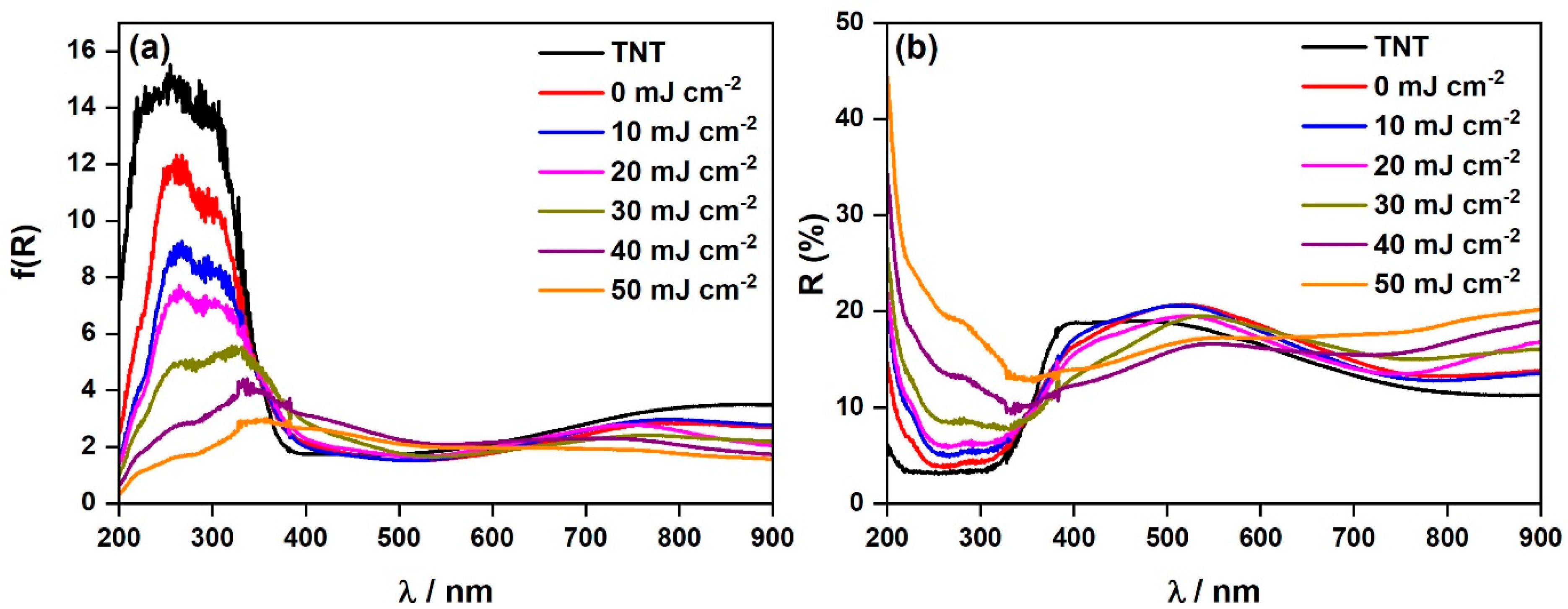
The results of the reflectance measurements and respective Kubelka–Munk functions are presented in Figure 5. For all the prepared samples, the absorption is primarily limited to the UV part of the spectrum that is typical for titania-based materials [55]. As it comes to pristine titania, it is well-known that the energy band gap values of rutile and anatase are 3.0 eV and 3.2 eV, respectively. For bulk material [56] therefore, the absorption of this material is consistent with the reported wide band gap. The TiO2 nanotubes decorated with chromium oxides (0 mJ cm−2) exhibit an Urbach tail [57], indicating that the chromium oxides provide intraband gap states. Subsequent laser treatment (10–50 mJ cm−2) contributes, however, to a decrease of absorbance (i.e., f(R)) in the UV range. Nonetheless, the absorption edge is redshifted with the increased laser fluence. To access the energy band gap values, the Tauc plots were constructed (Figure S3) and derived values are given in Table S1. For pristine titania nanotubes, indirect (allowed) optical transition was assumed according to the literature [32,58]. The estimated value of 3.01 eV is close to the reported data, nonetheless one cannot forget that it is morphology dependent [59]. In the case of chromium species, for chromium oxide, two different transitions are reported; namely, direct [60] and indirect [23]. Nonetheless, in our previous work we experimentally determined the Tauc exponent that could be assigned to direct (allowed) transition [32]. From Table S1 it is clear that with the increase of laser fluence, the band gap value shifts from 3.14 eV to even 1.68 eV. As no oxygen vacancies were detected by XPS measurements, the changes both in the absorbance and in band gap values are most likely correlated with the differences in sample morphology [61]. This is verified with the reflectance data (Figure 5b) and SEM analysis (Figure 1) as the increase of reflectance with the lowering of the surface area is observed.
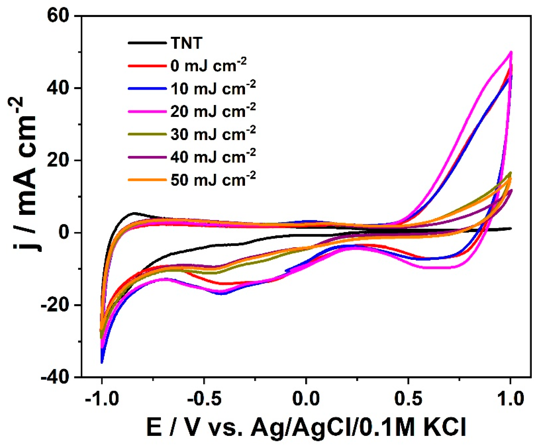
The electrochemical characterization was started with the recording of voltammetry curves in the range from +1.0 to −1.0 V vs. Ag/AgCl/0.1 M KCl that are presented in Figure 6. The working electrode was cycled several times and any current diminishing was not observed, indicating the material stability in neutral electrolyte in the investigated potential range (see Figure S4).
Pure titanium dioxide exhibits the lowest capacitive current densities and no redox reaction especially in the range from −0.7 to +1.0 V [62,63]. The increase of cathodic current observed in the negative range is attributed to the reduction/oxidation of titanium on the surface bonded to the hydroxyl groups [64]. Such activity is many times attributed to doping/de-doping with H+ with simultaneous reduction/oxidation of Ti4+/Ti3+ for TiO2 in the form of both nanotubes and single crystal [65,66]. For the electrode modified with chromium oxide, one may observe additional electrochemical activity and the increased current density compared to bare titania in the anodic regime. This feature is well-seen for titania decorated with chromium oxide but without laser treatment and treated with laser beam of low fluence (10 and 20 mJ cm−2), and can be ascribed to the oxidation of Cr3+ (present in the form of Cr2O3 as indicated by XPS data) to Cr6+. The activity towards water splitting is rather debatable because no arising bubbles on the electrode surface were observed.
Regarding the cathodic branch, we can distinguish two wide reduction bands. The first one with the maximum at +0.65 V can be identified as reduction of Cr6+ to Cr5+. The other one at −0.42 V could be attributed to the reduction of Cr5+ to Cr3+ [67]. Unfortunately, literature studies on the titania modified with chromium oxide are poor in terms of detailed characterization using cyclic voltammetry, and the electrochemical investigations are mostly carried out with linear voltammetry in different light conditions or by performing impedance studies. Thus, taking into account data of standard redox potentials [68], for this signal, we also consider another reaction; namely, reduction of Cr3+ to Cr2+. Once again, we would like to underline that even though the sample was cycled numerous times, each CV curve overlaps, indicating that those reactions are reversible.
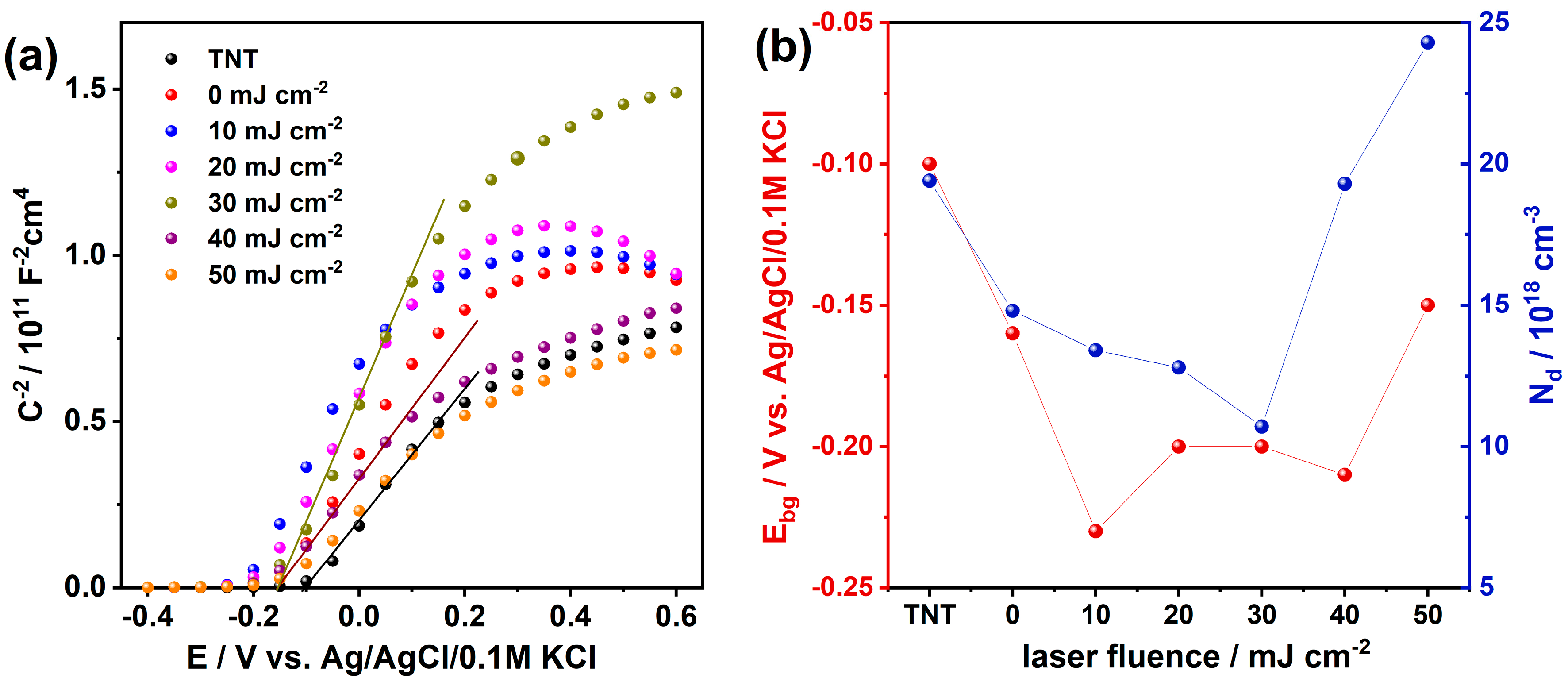
Then, the impedance measurements were carried out in the range from +0.6 towards −0.4 V vs. Ag/AgCl/0.1M KCl to prepare the Mott–Schottky plot (Figure 7a). The positive slope indicates the characteristic of an n-type semiconductor. The intersection of the tangent of the linear region of the Mott–Schottky plot enables the determining of the position of flat band potential for the surface of fabricated materials.
The flat band potential for pure titania is localized at −0.1 V vs. Ag/AgCl/0.1M KCl, whereas for the laser modified TNT with chromium species the position is negatively shifted, see Figure 7b. When the laser fluence fits the range of 10–40 mJ cm−2 the flat band is located at −0.2 V vs. Ag/AgCl/0.1M KCl. In the case of previous work concerning titania modification with chromium oxide, we did not observe any shift [29], but here a cathodic change of 0.1 V is noticed. A similar change was observed by Radecka et al. [69] for Cr-doped titania magnetron sputtered onto the amorphous silica and reported by Li et al. [70] for Zr-doped titania nanotubes modified with urea as well as for iodine-doped titania [71]. In the case of n-type semiconductor, negative shift of flat band potential can indicate that the position of the conduction band is going up and among other factors, can play a role in the charge transfer at the electrode/electrolyte interface.
According to He et al. [72], this negative shift is ascribed to introducing defects levels, thus changing the surface state distribution. However, we should be aware that this change will not only result from the presence of chromium oxide surface species, but can be the effect of laser annealing inducing changes in the morphology and hydroxide groups’ distribution that finally affect the contact area at the electrode/electrolyte interface.
Afterwards, the free-charge carrier concentration was determined on the basis of the slope of the Mott–Schottky plots and using equation (Equation (1)). The change in donor density is depicted in Figure 7b. First, the number of donors decreases reaching the minimum value for fluence equaled to 30 mJ cm−2. Then, the donor density increases reaching the maximum for the material annealed with the highest laser fluence and exhibiting the most distorted surface; namely, initially open nanotubes are fully or partially closed. Those changes result both from the changes in the contribution of the oxidation state of Cr in its different species present on TNT surface as well as the decrease of OH species according to XPS investigation.
Finally, the photoactivity of the fabricated materials was tested. Figure 8a displays linear voltammograms registered under chopped UV-vis illumination of the investigated samples. Note that photocurrent is defined as a difference between current density registered under sample illumination and in dark. In general, all the samples exhibit positive photocurrent along with anodic polarization, indicating n-type semiconducting properties [73]. Current spikes observed at lower potentials, particularly in a range from −0.2 to +0.2 V are related to electron–hole surface recombination during light-off action where these holes are temporarily stored at the photoelectrode/electrolyte interface [74]. Moreover, this can be the result of decrease in band bending [75] due to the cathodic shift of flad band potential, as was confirmed by Mott–Schottky analysis. Bare titania can generate photocurrent of 43 µA cm−2 at +0.65 V. Higher photoresponse of about 51 µA cm−2 was found for the TiO2 nanotubes decorated with Cr oxides but without laser annealing. Subsequent laser treatment with 10–40 mJ cm−2 enhanced the photoresponse, and the current density reaches 59 µA cm−2. A decrease in photoresponse was found for 50 mJ cm−2, indicating that laser fluence higher than 40 mJ cm−2 does not ensure greater enhancement of the photoactivity. Additionally, the stability of the material under long-term irradiation was verified and during the whole irradiation period the current was stable and any significant decrease was not observed (Figure S5).
Figure 8b displays linear voltammograms registered under chopped illumination by visible light (λ > 420 nm). Similar character of current spikes was noted as in the case of measurements carried out under UV-vis illumination, suggesting that the same surface recombination processes occur also when the electrode material is exposed to the visible light. As one can expect, this time bare TNT exhibits much lower photocurrent of only ca. 1.5 µA cm−2 at +0.65 V due to the wide band gap of titania. When the TiO2 nanotubes decorated with Cr oxides (0 mJ cm−2) were tested, a huge improvement was recorded, and the sample reaches 23 µA cm−2 at +0.65 V vs. Ag/AgCl/0.1 M KCl. It should be mentioned in here that although this sample has wider energy band gap (see Table S1), it also exhibits the Urbach tail (Figure 5) which indicates that the chromium oxides provide intraband gap states. The additional intraband gap states may provide the pathway for electron transfer between semiconductor and the acceptor species, and the excess of electron originating from localized states can affect the surface chemistry of electrode. Therefore, it can be assumed that the intraband gap states can play a major role in visible light response [22]. Subsequent laser modification ensures enhancement of current up to 27 µA cm−2 for a beam energy fluence of 30 mJ cm−2. However, it should be noted that the current density obtained for the material modified with 40 mJ cm−2 is not of much lower value. Thus, it can be stated that the optimum laser fluence fits the range of 30–40 mJ cm−2. Such an increase can result from several factors; namely, the decrease of band gap energy, change of the surface morphology that plays the crucial role in contact between the electrode and electrolyte and the charge transfer processes occurring at this interface, and change in the oxidation state of surface chromium oxide species as well as hydroxide groups. Finally, we should also take into account that laser treatment leads to manipulation of band bending that affects the charge transfer, and in this process intraband gap states can also be included.
Combined modification covering the chromium oxides decoration and subsequent laser treatment give a contribution to enhanced photoresponse up to 1.37-fold under radiation, covering the full solar spectrum compared to the bare TiO2 nanotubes. Whereas, the laser treatment itself, compared to the unmodified TiO2 nanotubes decorated with chromium oxides, provides ca. 20% current density improvement. In the case of visible light, all applied modification routes provide significant enhancement compared to bare titania. Without laser annealing, current density increased over 15 times; whereas, for the laser fluence of 30 mJ cm−2, the enhancement factor equals 18. Observed behavior upon both UV-vis and vis irradiation differs from our previous achievements [29], where we worked with the spaced titania nanotubes and no thermal treatment was applied after Cr sputtering. This differences in the fabrication route indicates how initial architecture of nanotubes and the sequence of thermal processing impact the overall photoresponse of the fabricated material decorated with chromium oxide. Moreover, recorded photocurrent improvement upon radiation from the visible range is much higher than obtained for Cr2O3-loaded titania nanotubes as reported by Ding et al. [76], when potassium dichromate was used as a Cr precursor, and by Radecka et al. [69], who showed that doping with Cr leads to photocurrent lowering due to the acceleration of the recombination process on the point defects. On the other hand, for the titania modified with chromium oxide using chromium nitride as a precursor, Shaislamov et al. [77] reported lower photoactivity under UV-vis light comparing to unmodified titania, while in the visible light conditions, photocurrent of modified electrode was much higher comparing to the bare substrate. A similar case was described by Song et al. [50], who found how annealing atmosphere affects the photoactivity since lack of access to oxygen leads to the formation of oxygen vacancies, affecting in consequence the oxidation state of Cr. In our case, manipulation with laser parameters enables us to change the content of the Cr species while the oxidation state of Ti is preserved. Thus, taking into account former experience [29] and works focused onto the modification with chromium oxide but based on different synthesis routes, the reported increase here of photocurrent for laser-modified titania decorated initially with Cr is a result of the manipulation within the chemical nature of Cr species and surface architecture.

4. Conclusions

In this work, a laser-based method of modification of chromium species coated titanium dioxide nanotubes, which leads to enhancement of the photoactivity of the studied materials is presented. Titania nanotubes were prepared via an optimized anodization process followed by thermal annealing to ensure crystallization. After the deposition of Cr layer, additional thermal treatment was applied for both the crystallization and oxidation. Finally, the material underwent annealing by means of UV laser. SEM inspection revealed the changes in the nanotubes’ morphology in terms of partial or full closing of the nanotubes’ tops. Nonetheless, underneath the melted area, the initial architecture of nanotubes remained intact. XRD and Raman measurements confirmed the mainly anatase phase of nanotubes and indicated on no oxygen vacancies present, respectively. The latter was verified with XPS analysis. Moreover, it was proven that the laser treatment influences on the chemical nature of the Cr species, as well as the narrowing of the energy band gap, derived from reflectance spectra. Both Mott–Schottky analysis and LV measurements confirmed the n-type character of prepared electrode materials. For the optimized laser working parameters, the current density of 59 µA cm−2 and 27 µA cm−2 was registered for samples under UV-vis and vis illumination, respectively. It should be underlined that it gives 1.37-fold and 18 times higher photoresponse, respectively, in comparison to bare titania. Moreover, the usage of beam homogenizer and motorized table ensures the homogenous modification of material of any shape and size, indicating that the proposed route can be easily scaled up from the laboratory to the commercial level.

Supplementary Materials

The following supporting information can be downloaded at: https://www.mdpi.com/article/10.3390/ma16072572/s1, Figure S1. SEM images (cross-section) of the TiO2 nanotubes decorated with chromium oxides (a) before and (b–f) after laser treatment with (b) 10 mJ cm−2, (c) 20 mJ cm−2, (d) 30 mJ cm−2, (e) 40 mJ cm−2, (f) 50 mJ cm−2 fluence. Figure S2. Raman spectra of the TiO2 nanotubes decorated with chromium oxides before (0 mJ cm−2) and after laser-treatment (10–50 mJ cm−2); the enlarged portion of Figure 3. Figure S3. Tauc plots of (a) the bare TiO2 nanotubes and (b) the laser-treated TiO2 nanotubes decorated with chromium oxides (0–50 mJ cm−2). Figure S4. 20 subsequent CV cycles recorded for the material modified with the laser fluence of 40 mJ cm−2. Scan rate was set to 50 mV/s. Figure S5. Chronoamperometry curve recorded under prolonged irradiation of the electrode material modified with 40 mJ cm−2 laser fluence. The electrode was polarized at +0.5 V vs. Ag/AgCl/0.1M KCl. Table S1. Optical bandgap values of the bare TiO2 nanotubes and the laser-treated TiO2 nanotubes decorated with chromium oxides (0–50 mJ cm−2).

Author Contributions

Formal analysis, K.G., Ł.H. and K.S.; Investigation, Ł.H., J.K., K.J. and K.S.; Writing—original draft, K.G., Ł.H. and K.S.; Writing—review & editing, K.G. and K.S.; Supervision, K.S.; Funding acquisition, K.S. All authors have read and agreed to the published version of the manuscript.

Funding

This research was financed by the National Science Centre (Poland): grant no 2017/26/E/ST5/00416.

Data Availability Statement

The data presented in this study are available on request from the corresponding author. The data are not publicly available due to technical or time limitations.

Conflicts of Interest

The authors declare no conflict of interest.

References

  1. Wang, R.; Bai, J.; Li, Y.; Zeng, Q.; Li, J.; Zhou, B. BiVO4/TiO2(N2) Nanotubes Heterojunction Photoanode for Highly Efficient Photoelectrocatalytic Applications. Nano-Micro Lett. 2017, 9, 14. [Google Scholar] [CrossRef] [PubMed] [Green Version]
  2. Pinchuk, V.A.; Kuzmin, A.V. The Effect of the Addition of TiO2 Nanoparticles to Coal-Water Fuel on Its Thermophysical Properties and Combustion Parameters. Fuel 2020, 267, 117220. [Google Scholar] [CrossRef]
  3. Nguyen, T.P.; Nguyen, D.L.T.; Nguyen, V.-H.; Le, T.-H.; Vo, D.-V.N.; Trinh, Q.T.; Bae, S.-R.; Chae, S.Y.; Kim, S.Y.; Le, Q.V. Recent Advances in TiO2-Based Photocatalysts for Reduction of CO2 to Fuels. Nanomaterials 2020, 10, 337. [Google Scholar] [CrossRef] [PubMed] [Green Version]
  4. Del Prado-Audelo, M.L.; García Kerdan, I.; Escutia-Guadarrama, L.; Reyna-González, J.M.; Magaña, J.J.; Leyva-Gómez, G. Nanoremediation: Nanomaterials and Nanotechnologies for Environmental Cleanup. Front. Environ. Sci. 2021, 9, 793765. [Google Scholar] [CrossRef]
  5. Al-Madanat, O.; AlSalka, Y.; Ramadan, W.; Bahnemann, D.W. TiO2 Photocatalysis for the Transformation of Aromatic Water Pollutants into Fuels. Catalysts 2021, 11, 317. [Google Scholar] [CrossRef]
  6. Zhang, L.; Cui, P.; Yang, H.; Chen, J.; Xiao, F.; Guo, Y.; Liu, Y.; Zhang, W.; Huo, F.; Liu, B. Metal–Organic Frameworks as Promising Photosensitizers for Photoelectrochemical Water Splitting. Adv. Sci. 2016, 3, 1500243. [Google Scholar] [CrossRef]
  7. Kusior, A.; Wnuk, A.; Trenczek-Zajac, A.; Zakrzewska, K.; Radecka, M. TiO2 Nanostructures for Photoelectrochemical Cells (PECs). Int. J. Hydrogen Energy 2015, 40, 4936–4944. [Google Scholar] [CrossRef]
  8. Wang, D.-Y.; Li, C.-H.; Li, S.-S.; Kuo, T.-R.; Tsai, C.-M.; Chen, T.-R.; Wang, Y.-C.; Chen, C.-W.; Chen, C.-C. Iron Pyrite/Titanium Dioxide Photoanode for Extended Near Infrared Light Harvesting in a Photoelectrochemical Cell. Sci. Rep. 2016, 6, 20397. [Google Scholar] [CrossRef] [Green Version]
  9. Liu, C.; Zhang, C.; Yin, G.; Zhang, T.; Wang, W.; Ou, G.; Jin, H.; Chen, Z. A Three-Dimensional Branched TiO2 Photoanode with an Ultrathin Al 2O3 Passivation Layer and a NiOOH Cocatalyst toward Photoelectrochemical Water Oxidation. ACS Appl. Mater. Interfaces 2021, 13, 13301–13310. [Google Scholar] [CrossRef]
  10. Williams, R. Electrochemical Reactions of Semiconductors. J. Vac. Sci. Technol. 1976, 13, 12–18. [Google Scholar] [CrossRef]
  11. Frank, S.N.; Bard, A.J. Semiconductor Electrodes. II. Electrochemistry at n-Type Titanium Dioxide Electrodes in Acetonitrile Solutions. J. Am. Chem. Soc. 1975, 97, 7427–7433. [Google Scholar] [CrossRef]
  12. Makimizu, Y.; Nguyen, N.T.; Tucek, J.; Ahn, H.; Yoo, J.; Poornajar, M.; Hwang, I.; Kment, S.; Schmuki, P. Activation of A-Fe2O3 for Photoelectrochemical Water Splitting Strongly Enhanced by Low Temperature Annealing in Low Oxygen Containing Ambient. Chem. Eur. J. 2020, 26, 2685–2692. [Google Scholar] [CrossRef] [Green Version]
  13. Kalanur, S.S.; Duy, L.T.; Seo, H. Recent Progress in Photoelectrochemical Water Splitting Activity of WO3 Photoanodes. Top. Catal. 2018, 61, 1043–1076. [Google Scholar] [CrossRef]
  14. Barawi, M.; Gomez-Mendoza, M.; Oropeza, F.E.; Gorni, G.; Villar-Garcia, I.J.; Giménez, S.; de la Peña O’Shea, V.A.; García-Tecedor, M. Laser-Reduced BiVO4 for Enhanced Photoelectrochemical Water Splitting. ACS Appl. Mater. Interfaces 2022, 14, 33200–33210. [Google Scholar] [CrossRef]
  15. Arifin, K.; Yunus, R.M.; Minggu, L.J.; Kassim, M.B. Improvement of TiO2 Nanotubes for Photoelectrochemical Water Splitting: Review. Int. J. Hydrogen Energy 2021, 46, 4998–5024. [Google Scholar] [CrossRef]
  16. Abdullah, M.; Kamarudin, S.K. Titanium Dioxide Nanotubes (TNT) in Energy and Environmental Applications: An Overview. Renew. Sustain. Energy Rev. 2017, 76, 212–225. [Google Scholar] [CrossRef]
  17. Luttrell, T.; Halpegamage, S.; Tao, J.; Kramer, A.; Sutter, E.; Batzill, M. Why Is Anatase a Better Photocatalyst than Rutile?—Model Studies on Epitaxial TiO2 Films. Sci. Rep. 2014, 4, 4043. [Google Scholar] [CrossRef] [Green Version]
  18. Mahajan, V.K.; Mohapatra, S.K.; Misra, M. Stability of TiO₂ Nanotubes Arrays in Photoelectrochemical Studies. Int. J. Hydrogen Energy 2008, 33, 5369–5374. [Google Scholar] [CrossRef]
  19. Momeni, M.M.; Motalebian, M. Chromium-Doped Titanium Oxide Nanotubes Grown via One-Step Anodization for Efficient Photocathodic Protection of Stainless Steel. Surf. Coat. Technol. 2021, 420, 127304. [Google Scholar] [CrossRef]
  20. Momeni, M.M.; Mahvari, M.; Ghayeb, Y. Photoelectrochemical Properties of Iron-Cobalt WTiO2 Nanotube Photoanodes for Water Splitting and Photocathodic Protection of Stainless Steel. J. Electroanal. Chem. 2019, 832, 7–23. [Google Scholar] [CrossRef]
  21. Mohammadi, T.; Sharifi, S.; Ghayeb, Y.; Sharifi, T.; Momeni, M.M. Photoelectrochemical Water Splitting and H2 Generation Enhancement Using an Effective Surface Modification of W-Doped TiO2 Nanotubes (WT) with Co-Deposition of Transition Metal Ions. Sustainability 2022, 14, 13251. [Google Scholar] [CrossRef]
  22. Haryński, Ł.; Olejnik, A.; Karczewski, J.; Ryl, J.; Grochowska, K.; Siuzdak, K. Linking Optical and Electronic Properties to Photoresponse of Heterojunctions Based on Titania Nanotubes and Chromium, Molybdenum, and Tungsten Oxides. Opt. Mater. 2022, 134, 113183. [Google Scholar] [CrossRef]
  23. Cheng, C.-S.; Gomi, H.; Sakata, H. Electrical and Optical Properties of Cr₂O₃ Films Prepared by Chemical Vapour Deposition. Phys. Status Solidi A 1996, 155, 417–425. [Google Scholar] [CrossRef]
  24. Shafeeq, K.M.; Rizwana, E.; Kishor, C.H.R.; Aneesh, P.M. Growth and Characterization of α-MoO₃ Thin Films Grown by Spray Pyrolysis Technique. AIP Conf. Proc. 2019, 2082, 050010. [Google Scholar] [CrossRef]
  25. Gullapalli, S.K.; Vemuri, R.S.; Ramana, C.V. Structural Transformation Induced Changes in the Optical Properties of Nanocrystalline Tungsten Oxide Thin Films. Appl. Phys. Lett. 2010, 96, 171903. [Google Scholar] [CrossRef]
  26. Metikoš-Huković, M.; Ceraj-Cerić, M. P-Type and N-Type Behavior of Chromium Oxide as a Function of the Applied Potential. J. Electrochem. Soc. 1987, 134, 2193–2197. [Google Scholar] [CrossRef]
  27. Peelaers, H.; Chabinyc, M.L.; Van de Walle, C.G. Controlling N-Type Doping in MoO3. Chem. Mater. 2017, 29, 2563–2567. [Google Scholar] [CrossRef]
  28. Gillet, M.; Aguir, K.; Gillet, E.; Schirbaum, K. The Structure and Electrical Conductivity of Vacuum-Annealed WO₃ Thin Films. Thin Solid Film. 2004, 467, 239–246. [Google Scholar] [CrossRef]
  29. Haryński, Ł.; Karczewski, J.; Ryl, J.; Grochowska, K.; Siuzdak, K. Rapid Development of the Photoresponse and Oxygen Evolution of TiO2 Nanotubes Sputtered with Cr Thin Films Realized via Laser Annealing. J. Alloys Compd. 2021, 877, 160316. [Google Scholar] [CrossRef]
  30. Haryński, Ł.; Grochowska, K.; Karczewski, J.; Ryl, J.; Siuzdak, K. Scalable Route towards Superior Photoresponse of UV-Laser-Treated TiO₂ Nanotubes. ACS Appl. Mater. Interfaces 2020, 12, 3225–3235. [Google Scholar] [CrossRef]
  31. Xu, Y.; Melia, M.A.; Tsui, L.; Fitz-Gerald, J.M.; Zangari, G. Laser-Induced Surface Modification at Anatase TiO₂ Nanotube Array Photoanodes for Photoelectrochemical Water Oxidation. J. Phys. Chem. C 2017, 121, 17121–17128. [Google Scholar] [CrossRef]
  32. Haryński, Ł.; Olejnik, A.; Grochowska, K.; Siuzdak, K. A Facile Method for Tauc Exponent and Corresponding Electronic Transitions Determination in Semiconductors Directly from UV–Vis Spectroscopy Data. Opt. Mater. 2022, 127, 112205. [Google Scholar] [CrossRef]
  33. Park, B.H.; Li, L.S.; Gibbons, B.J.; Huang, J.Y.; Jia, Q.X. Photovoltaic Response and Dielectric Properties of Epitaxial Anatase-TiO2 Films Grown on Conductive La0.5Sr0.5CoO3 Electrodes. Appl. Phys. Lett. 2001, 79, 2797–2799. [Google Scholar] [CrossRef]
  34. Slimen, H.; Houas, A.; Nogier, J.P. Elaboration of Stable Anatase TiO2 through Activated Carbon Addition with High Photocatalytic Activity under Visible Light. J. Photochem. Photobiol. A Chem. 2011, 221, 13–21. [Google Scholar] [CrossRef]
  35. Haryński, Ł.; Grochowska, K.; Kupracz, P.; Karczewski, J.; Coy, E.; Siuzdak, K. The In-Depth Studies of Pulsed UV Laser-Modified TiO2 Nanotubes: The Influence of Geometry, Crystallinity, and Processing Parameters. Nanomaterials 2020, 10, 430. [Google Scholar] [CrossRef] [Green Version]
  36. Sopha, H.; Mirza, I.; Turčičova, H.; Pavlinak, D.; Michalicka, J.; Krbal, M.; Rodriguez-Pereira, J.; Hromadko, L.; Novák, O.; Mužík, J.; et al. Laser-Induced Crystallization of Anodic TiO2 Nanotube Layers. RSC Adv. 2020, 10, 22137–22145. [Google Scholar] [CrossRef]
  37. Haryński, Ł.; Grochowska, K.; Karczewski, J.; Ryl, J.; Rysz, J.; Siuzdak, K. Free-Standing TiO₂ Nanotubes Decorated with Spherical Nickel Nanoparticles as a Cost-Efficient Electrocatalyst for Oxygen Evolution Reaction. RSC Adv. 2021, 11, 219–228. [Google Scholar] [CrossRef]
  38. Chen, X.; Mao, S.S. Titanium Dioxide Nanomaterials: Synthesis, Properties, Modifications, and Applications. Chem. Rev. 2007, 107, 2891–2959. [Google Scholar] [CrossRef]
  39. Albu, S.P.; Tsuchiya, H.; Fujimoto, S.; Schmuki, P. TiO2 Nanotubes—Annealing Effects on Detailed Morphology and Structure. Eur. J. Inorg. Chem. 2010, 2010, 4351–4356. [Google Scholar] [CrossRef]
  40. Grochowska, K.; Nedyalkov, N.; Karczewski, J.; Haryński, Ł.; Śliwiński, G.; Siuzdak, K. Anodic Titania Nanotubes Decorated with Gold Nanoparticles Produced by Laser-Induced Dewetting of Thin Metallic Films. Sci. Rep. 2020, 10, 20506. [Google Scholar] [CrossRef]
  41. Wang, B.; Shen, S.; Mao, S.S. Black TiO2 for Solar Hydrogen Conversion. J. Mater. 2017, 3, 96–111. [Google Scholar] [CrossRef]
  42. Sarkar, A.; Khan, G.G. The Formation and Detection Techniques of Oxygen Vacancies in Titanium Oxide-Based Nanostructures. Nanoscale 2019, 11, 3414–3444. [Google Scholar] [CrossRef] [PubMed]
  43. Mahdi, N.; Kumar, P.; Goswami, A.; Perdicakis, B.; Shankar, K.; Sadrzadeh, M. Robust Polymer Nanocomposite Membranes Incorporating Discrete TiO2 Nanotubes for Water Treatment. Nanomaterials 2019, 9, 1186. [Google Scholar] [CrossRef] [PubMed] [Green Version]
  44. Farsinezhad, S.; Sharma, H.; Shankar, K. Interfacial Band Alignment for Photocatalytic Charge Separation in TiO2 Nanotube Arrays Coated with CuPt Nanoparticles. Phys. Chem. Chem. Phys. 2015, 17, 29723–29733. [Google Scholar] [CrossRef] [Green Version]
  45. Zhang, W.-X.; Liang, Y.; Luo, J.-W.; Jia, A.-P.; Wang, Y.-J.; Lu, J.-Q.; Luo, M.-F. Morphological Effects of Ordered Cr2O3 Nanorods and Cr2O3 Nanoparticles on Fluorination of 2-Chloro-1,1,1-Trifluoroethane. J. Mater. Sci. 2016, 51, 6488–6496. [Google Scholar] [CrossRef]
  46. Akshay, V.R.; Arun, B.; Mandal, G.; Vasundhara, M. Visible Range Optical Absorption, Urbach Energy Estimation and Paramagnetic Response in Cr-Doped TiO2 Nanocrystals Derived by a Sol–Gel Method. Phys. Chem. Chem. Phys. 2019, 21, 12991–13004. [Google Scholar] [CrossRef]
  47. Liu, X.; Wu, J. Coupling Interface Constructions of NiO–Cr2O3 Heterostructures for Efficient Electrocatalytic Oxygen Evolution. Electrochim. Acta 2019, 320, 134577. [Google Scholar] [CrossRef]
  48. Nemanič, V.; Kovač, J.; Žumer, M.; Zavašnik, J. Impact of Surface Oxide on Hydrogen Permeability of Chromium Membranes. Int. J. Hydrogen Energy 2022, 48, 9723–9733. [Google Scholar] [CrossRef]
  49. Biesinger, M.C.; Brown, C.; Mycroft, J.R.; Davidson, R.D.; McIntyre, N.S. X-Ray Photoelectron Spectroscopy Studies of Chromium Compounds. Surf. Interface Anal. 2004, 36, 1550–1563. [Google Scholar] [CrossRef]
  50. Song, X.; Li, W.; Liu, X.; Wu, Y.; He, D.; Ke, Z.; Cheng, L.; Jiang, C.; Wang, G.; Xiao, X.; et al. Oxygen Vacancies Enable the Visible Light Photoactivity of Chromium-Implanted TiO2 Nanowires. J. Energy Chem. 2021, 55, 154–161. [Google Scholar] [CrossRef]
  51. Shirato, N.; Strader, J.; Kumar, A.; Vincent, A.; Zhang, P.; Karakoti, A.; Nacchimuthu, P.; Cho, H.-J.; Seal, S.; Kalyanaraman, R. Thickness Dependent Self Limiting 1-D Tin Oxidenanowire Arrays by Nanosecond Pulsed Laser Irradiation. Nanoscale 2011, 3, 1090–1101. [Google Scholar] [CrossRef] [Green Version]
  52. Kotsedi, L.; Furlan, V.; Bharadwaj, V.; Kaviyarasu, K.; Sotillo, B.; Mtshali, C.B.; Matinise, N.; Demir, A.G.; Previtali, B.; Ramponi, R.; et al. Chromium Oxide Formation on Nanosecond and Femtosecond Laser Irradiated Thin Chromium Films. Opt. Mater. 2019, 95, 109206. [Google Scholar] [CrossRef] [Green Version]
  53. Xie, B.; Sun, M.; Xu, B.; Wang, C.; Jiang, H.; Li, D.; Li, Y. Oxidation of Stainless Steel in Vacuum and Evolution of Surface Oxide Scales during Hot-Compression Bonding. Corros. Sci. 2019, 147, 41–52. [Google Scholar] [CrossRef]
  54. Veiko, V.P.; Poleshchuk, A.G. Laser-Induced Local Oxidation of Thin Metal Films: Physical Fundamentals and Applications. In Fundamentals of Laser-Assisted Micro- and Nanotechnologies; Veiko, V.P., Konov, V.I., Eds.; Springer Series in Materials Science; Springer International Publishing: Cham, Switzerland, 2014; Volume 195, pp. 149–171. [Google Scholar] [CrossRef]
  55. Abouhaswa, A.S. Physical Properties of Anatase TiO2 Nanocrystallites: Based Photoanodes Doped with Cr2O3. Opt. Quantum Electron. 2020, 52, 144. [Google Scholar] [CrossRef]
  56. Roy, P.; Kim, D.; Lee, K.; Spiecker, E.; Schmuki, P. TiO2 Nanotubes and Their Application in Dye-Sensitized Solar Cells. Nanoscale 2010, 2, 45–59. [Google Scholar] [CrossRef]
  57. Yaghoubi, H.; Li, Z.; Chen, Y.; Ngo, T.H.; Bhethanabotla, V.R.; Joseph, B.; Ma, S.; Schlaf, R.; Takshi, A. Toward a Visible Light-Driven Photocatalyst: The Effect of Midgap-States-Induced Energy Gap of Undoped TiO₂ Nanoparticles. ACS Catal. 2015, 5, 327–335. [Google Scholar] [CrossRef] [Green Version]
  58. Peighambardoust, N.S.; Khameneh Asl, S.; Mohammadpour, R.; Asl, S.K. Band-Gap Narrowing and Electrochemical Properties in N-Doped and Reduced Anodic TiO2 Nanotube Arrays. Electrochim. Acta 2018, 270, 245–255. [Google Scholar] [CrossRef]
  59. Freitas, R.G.; Santanna, M.A.; Pereira, E.C. Dependence of TiO2 Nanotube Microstructural and Electronic Properties on Water Splitting. J. Power Sources 2014, 251, 178–186. [Google Scholar] [CrossRef] [Green Version]
  60. Hu, Z.; Xu, M.; Shen, Z.; Yu, J.C. A Nanostructured Chromium(III) Oxide/Tungsten(vi) Oxide p–n Junction Photoanode toward Enhanced Efficiency for Water Oxidation. J. Mater. Chem. A 2015, 3, 14046–14053. [Google Scholar] [CrossRef]
  61. Pishkar, N.; Jedi-soltanabadi, Z.; Ghoranneviss, M. Reduction in the Band Gap of Anodic TiO2 Nanotube Arrays by H2 Plasma Treatment. Results Phys. 2018, 10, 466–468. [Google Scholar] [CrossRef]
  62. Zhou, M.; Glushenkov, A.M.; Kartachova, O.; Li, Y.; Chen, Y. Titanium Dioxide Nanotube Films for Electrochemical Supercapacitors: Biocompatibility and Operation in an Electrolyte Based on a Physiological Fluid. J. Electrochem. Soc. 2015, 162, A5065–A5069. [Google Scholar] [CrossRef]
  63. Fabregat-Santiago, F.; Mora-Seró, I.; Garcia-Belmonte, G.; Bisquert, J. Cyclic Voltammetry Studies of Nanoporous Semiconductors. Capacitive and Reactive Properties of Nanocrystalline TiO2 Electrodes in Aqueous Electrolyte. J. Phys. Chem. B 2003, 107, 758–768. [Google Scholar] [CrossRef]
  64. Oliva, F.Y.; Avalle, L.B.; Cámara, O.R. Electrochemical Behaviour of Human Serum Albumin–TiO2 Nanocrystalline Electrodes Studied as a Function of PH. J. Electroanal. Chem. 2002, 534, 19–29. [Google Scholar] [CrossRef]
  65. Macak, J.M.; Gong, B.G.; Hueppe, M.; Schmuki, P. Filling of TiO2 Nanotubes by Self-Doping and Electrodeposition. Adv. Mater. 2007, 19, 3027–3031. [Google Scholar] [CrossRef]
  66. Pelouchova, H.; Janda, P.; Weber, J.; Kavan, L. Charge Transfer Reductive Doping of Single Crystal TiO2 Anatase. J. Electroanal. Chem. 2004, 566, 73–83. [Google Scholar] [CrossRef]
  67. Sab, N.; Claes, P.; Glibert, J. Chemical and Electrochemical Behaviour in Molten Hydroxides. VII. Chromium Species in Sodium Hydroxide. Electrochim. Acta 1998, 43, 2089–2100. [Google Scholar] [CrossRef]
  68. Cramer, S.D.; Covino, B.S. (Eds.) Electrochemical Series. In Corrosion: Materials; ASM International: Singapore, 2005; pp. 665–671. [Google Scholar] [CrossRef] [Green Version]
  69. Radecka, M.; Rekas, M.; Trenczek-Zajac, A.; Zakrzewska, K. Importance of the Band Gap Energy and Flat Band Potential for Application of Modified TiO2 Photoanodes in Water Photolysis. J. Power Sources 2008, 181, 46–55. [Google Scholar] [CrossRef]
  70. Li, Y.; Xiang, Y.; Peng, S.; Wang, X.; Zhou, L. Modification of Zr-Doped Titania Nanotube Arrays by Urea Pyrolysis for Enhanced Visible-Light Photoelectrochemical H2 Generation. Electrochim. Acta 2013, 87, 794–800. [Google Scholar] [CrossRef]
  71. Siuzdak, K.; Szkoda, M.; Sawczak, M.; Lisowska-Oleksiak, A.; Karczewski, J.; Ryl, J. Enhanced Photoelectrochemical and Photocatalytic Performance of Iodine-Doped Titania Nanotube Arrays. RSC Adv. 2015, 5, 50379–50391. [Google Scholar] [CrossRef]
  72. He, Z.-K.; Lu, Y.; Zhao, J.; Zhao, J.; Gao, Z.; Song, Y.-Y. Engineering Carrier Density at TiO2 Nanotube Metasurface with Hole Reservoir for Enhanced Photo-Electrocatalysis. Appl. Surf. Sci. 2023, 613, 155974. [Google Scholar] [CrossRef]
  73. Hardee, K.L.; Bard, A.J. Semiconductor Electrodes: X. Photoelectrochemical Behavior of Several Polycrystalline Metal Oxide Electrodes in Aqueous Solutions. J. Electrochem. Soc. 1977, 124, 215–224. [Google Scholar] [CrossRef] [Green Version]
  74. Wang, P.; Li, D.; Chi, H.; Zhao, Y.; Wang, J.; Li, D.; Pang, S.; Fu, P.; Shi, J.; Li, C. Unveiling the Hydration Structure of Ferrihydrite for Hole Storage in Photoelectrochemical Water Oxidation. Angew. Chem. Int. Ed. 2021, 60, 6691–6698. [Google Scholar] [CrossRef]
  75. Tafalla, D.; Salvador, P. Kinetic Approach to the Photocurrent Transients in Water Photoelectrolysis at N-TiO₂ Electrodes. J. Electrochem. Soc. 1990, 137, 1810–1815. [Google Scholar] [CrossRef]
  76. Ding, J.; Ming, J.; Lu, D.; Wu, W.; Liu, M.; Zhao, X.; Li, C.; Yang, M.; Fang, P. Study of the Enhanced Visible-Light-Sensitive Photocatalytic Activity of Cr2O3-Loaded Titanate Nanosheets for Cr(VI) Degradation and H2 Generation. Catal. Sci. Technol. 2017, 7, 2283–2297. [Google Scholar] [CrossRef]
  77. Shaislamov, U.; Yang, B.; Park, K. Enhanced Photocatalytic Properties of TiO2 Nanotube Arrays with Cr2O3 Nanoparticles under Visible Light. J. Korean Phys. Soc. 2012, 61, 759–763. [Google Scholar] [CrossRef]
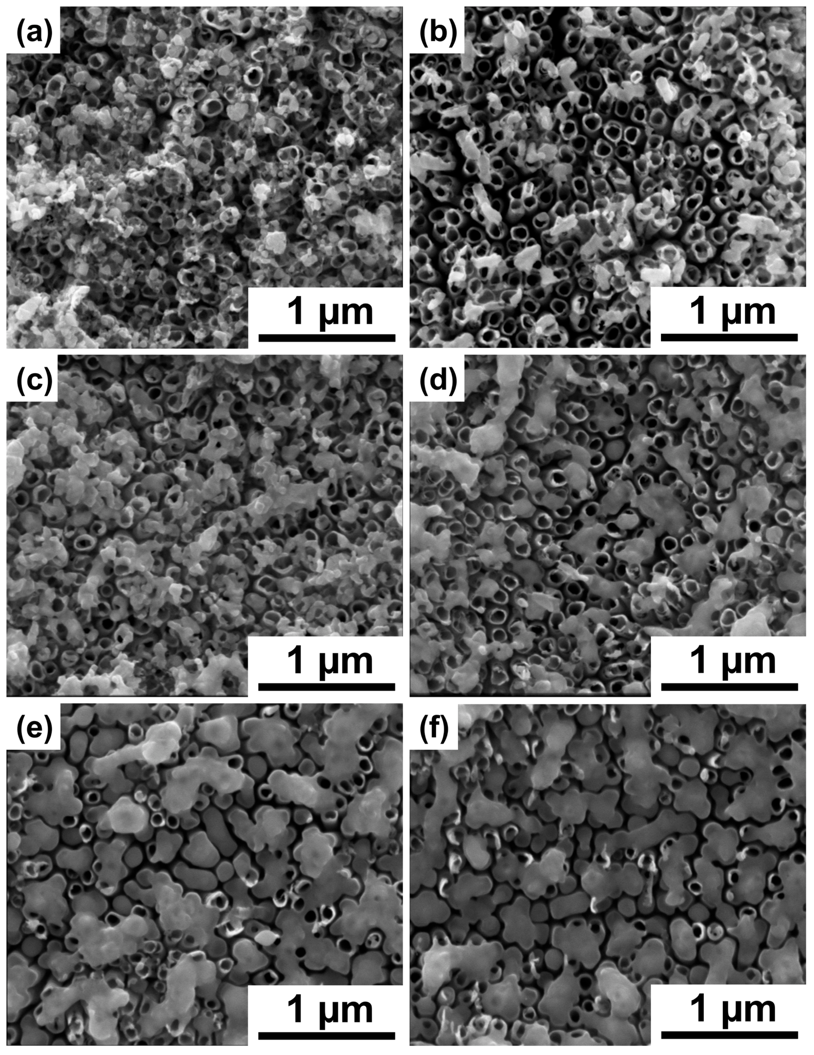
Figure 1. SEM images of the TiO2 nanotubes decorated with chromium oxides (a) before and (bf) after laser treatment with (b) 10 mJ cm−2, (c) 20 mJ cm−2, (d) 30 mJ cm−2, (e) 40 mJ cm−2, and (f) 50 mJ cm−2 fluence.
Materials 16 02572 g001
Figure 2. XRD patterns of the TiO2 nanotubes decorated with chromium oxides before (0 mJ cm−2) and after laser treatment (10–50 mJ cm−2).
Materials 16 02572 g002
Figure 3. Raman spectra of the TiO2 nanotubes decorated with chromium oxides before (0 mJ cm−2) and after laser treatment (10–50 mJ cm−2).
Materials 16 02572 g003
Figure 4. XPS spectra of the (a,d) Ti 2p, (b,e) O 1s, and (c,f) Cr 2p regions registered for the TiO2 nanotubes decorated with chromium oxides (ac) before and (df) after laser-treatment with 40 mJ cm−2.
Materials 16 02572 g004
Figure 5. Absorption data of the bare TiO2 nanotubes and the laser-treated TiO2 nanotubes decorated with chromium oxides (0–50 mJ cm−2) displayed as (a) Kubelka–Munk function and (b) reflectance vs. wavelength.
Materials 16 02572 g005
Figure 6. Cyclic voltammetry curves recorded for bare and modified titania recorded with 50 mV s−1 in 0.5 M Na2SO4.
Materials 16 02572 g006
Figure 7. (a) Mott–Schottky plot of bare and modified titania in 0.5 M Na2SO4 solution. (b) The change in band gap energy (Ebg) position and donor density depending on the applied laser fluence. The values of those parameters were determined on the basis of the Mott–Schottky plot given in (a).
Materials 16 02572 g007
Figure 8. Linear voltammograms registered for the bare and modified titania nanotubes. The LV curves were recorded under chopped (a) UV-vis and (b) vis irradiation.
Materials 16 02572 g008
Disclaimer/Publisher’s Note: The statements, opinions and data contained in all publications are solely those of the individual author(s) and contributor(s) and not of MDPI and/or the editor(s). MDPI and/or the editor(s) disclaim responsibility for any injury to people or property resulting from any ideas, methods, instructions or products referred to in the content.