Review

The Recent Applications of Magnetic Nanoparticles in Biomedical Fields

by
Jiaqi Hong
,
Linhao Wang
,
Qikai Zheng
,
Changyu Cai
,
Xiaohua Yang
and
Zhenlin Liao
*
College of Food Science, South China Agricultural University, Guangzhou 510642, China
*
Author to whom correspondence should be addressed.
These authors contributed equally to this work.
Materials 2024, 17(12), 2870; https://doi.org/10.3390/ma17122870
Submission received: 19 March 2024 / Revised: 5 June 2024 / Accepted: 5 June 2024 / Published: 12 June 2024

Abstract

:
Magnetic nanoparticles (MNPs) have found extensive application in the biomedical domain due to their enhanced biocompatibility, minimal toxicity, and strong magnetic responsiveness. MNPs exhibit great potential as nanomaterials in various biomedical applications, including disease detection and cancer therapy. Typically, MNPs consist of a magnetic core surrounded by surface modification coatings, such as inorganic materials, organic molecules, and polymers, forming a nucleoshell structure that mitigates nanoparticle agglomeration and enhances targeting capabilities. Consequently, MNPs exhibit magnetic responsiveness in vivo for transportation and therapeutic effects, such as enhancing medical imaging resolution and localized heating at the site of injury. MNPs are utilized for specimen purification through targeted binding and magnetic separation in vitro, thereby optimizing efficiency and expediting the process. This review delves into the distinctive functional characteristics of MNPs as well as the diverse bioactive molecules employed in their surface coatings and their corresponding functionalities. Additionally, the advancement of MNPs in various applications is outlined. Additionally, we discuss the advancements of magnetic nanoparticles in medical imaging, disease treatment, and in vitro assays, and we anticipate the future development prospects and obstacles in this field. The objective is to furnish readers with a thorough comprehension of the recent practical utilization of MNPs in biomedical disciplines.

1. Introduction

When compared to other nanostructures, magnetic nanoparticles (MNPs) are one of the most significant and often employed classes of nanomaterials because of their specific characteristics. MNPs possess the capacity to significantly impact diagnostic and therapeutic practices in the medical field due to their distinctive characteristics, such as super-paramagnetic moment, magnetic resonance, and efficient interactions at the molecular and cellular levels [1,2]. Additionally, the application of diverse surface coatings can enhance the medicinal properties of MNPs, leading to desirable pharmacokinetic effects and reduced toxicity. Apart from properties, attractive and durable medicinal properties can be produced for these particles by using various surface coatings, and the pharmacokinetic effects and toxicity of magnetic nanoparticles caused by interactions with cells or biological proteins can be avoided, resulting in the increased biocompatibility and practicality of magnetic nanoparticles [3,4]. MNPs are used in a wide variety of fields, especially in biomedical applications [5] (Figure 1). MNPs have demonstrated utility as a medical imaging contrast agent [6,7,8], as a carrier for pharmaceutical agents [9,10], and in cancer therapy through magnetically induced hyperthermia (MHT) [11,12,13], photodynamic therapy (PDT) [6,14,15], and photothermal therapy (PTT) [16,17,18].
On the other hand, MNPs have garnered significant interest as valuable tools for in vitro diagnosis with a variety of appealing applications such as trace analysis, magnetic separation, quantitative detection, and rapid testing, making them highly suitable for a range of biomedical uses [19]. Furthermore, MNPs serve as safe and efficient vectors for gene delivery, facilitating various biotechnological techniques including magnetofection [20], molecular recognition systems [21], single-nucleotide polymorphism (SNP) genotyping [22,23], and gene-specific PCR assays [24,25].
Therefore, MNPs are extensively researched due to their diverse applications. This paper aims to delve into the characteristics of MNPs, including their magnetic properties, size, and the bioactive molecules incorporated in the shell layer. Additionally, it discusses the advancements in utilizing MNPs for medical imaging, disease treatment, and in vitro assays as well as the potential future directions and obstacles in the development of MNPs. The core–shell structure of MNPs and their functions are comprehensively introduced in this study. We present a comprehensive view of MNPs from the inside out, from the core–shell structure of MNPs, the roles of cores and coatings, to the applications of mature products, which include various landed MNP products, and MNP clinical applications approved for use in various countries, with the aim of providing readers with a comprehensive understanding of the practical applications of magnetic nanoparticles in biomedical fields. The objective is to furnish readers with a thorough comprehension of the practical utilization of MNPs in biomedical disciplines.

2. Characteristics of MNPs

2.1. Functional Characteristics of MNPs

Distinguished from conventional nanomaterials, MNPs may exhibit distinctive properties: namely, superparamagnetism, magnetic guidance, high transverse relaxivity, magnetothermal effects and other magnetic properties [26]. When smaller than the critical size, for temperatures below the Curie temperature but above the blocking temperature, MNPs will be in a superparamagnetic state. Superparamagnetic MNPs are highly magnetizable under an applied external magnetic field, typically presenting high magnetic susceptibility and saturation magnetizations but with no magnetic coercivity and hence no remanent magnetization. This is particularly relevant since after removing the applied magnetic field, the nanoparticles will exhibit close to null magnetic-related agglomeration, hence enabling favorable dispersions. This superparamagnetic behavior facilitates targeting and magnetic separation in various product applications. Magnetic fluid hyperthermia (MFH) relies on MNPs to convert energy of alternating magnetic fields (AMF) into thermal energy. The MNPs located in tumors and other diseased areas generate localized heating under the application of AMF, inducing selective apoptosis or necrosis of the diseased cells [27]. The transverse relaxivity of MNPs induces the spin relaxation of water protons when imaging tissues in vivo, increasing the T1 (spin–lattice) and T2 (spin–spin) relaxivity of water protons, thereby further enhancing magnetic resonance imaging (MRI) contrast [28]. Notably, MNPs exhibit antimicrobial properties to a certain extent [29]. Some authors believe that electrostatic forces can cause NPs to bind to the bacterial cell membrane and disorganize their metabolic functionality, while others propose that the production of reactive oxygen species (ROS) is the primary bactericidal mechanism utilized by numerous antibacterial NP agents [30].

2.2. Biocompatibility of MNPs

MNPs utilized in various application scenarios have distinct requirements for desirable product characteristics. In vivo applications necessitate MNPs with optimal biocompatibility and particle size to prevent aggregation resulting from magnetic attraction, thereby ensuring stability. Additionally, it is crucial for MNPs to possess the ability to evade recognition and capture by the reticuloendothelial system, traverse the capillary network without inducing thrombosis, and enhance the likelihood of reaching target tissues [31]. As such, the size and surface coating of MNPs are pivotal factors influencing their non-toxicity and chemical properties. The utilization of biocompatible materials as coatings for magnetic nanoparticles can serve as a protective barrier, improving the overall biocompatibility of the particles by facilitating the continuous adaptation of more biocompatible surface coatings. This process can help prevent rejection, cytotoxicity, and carcinogenicity in vivo [32].
Within the tissue body, nanoparticles larger than 200 nm in diameter are readily cleared by the reticuloendothelial system [30]. However, nanoparticles with diameters smaller than 8 nm are easily excreted from the body through existent pores of the kidney’s basal lamina (renal clearance) [33], reducing the blood-circulating time of these nanostructures. The diameter range of 10–40 nm (including ultrasmall MNPs) is fundamental for prolonged blood circulation, allowing the nanoparticles to cross capillary walls and often be phagocytized by macrophages trafficking to the lymph nodes and bone marrow. MNPs sized between 50 and 100 nm have the ability to evade the reticuloendothelial system in vivo, resulting in extended circulation periods and circumventing clearance by alveolar macrophages [34]. Therefore, optimizing the size of MNPs so that they avoid pharmacokinetic effects and toxicity resulting from interactions with cellular or biological proteins, as well as overcoming in vivo barriers, can enhance the effective surface area (primarily applicable to particles with sizes below 100 nm) and improve tissue diffusion [35,36].
From the above, it can be seen that MNPs, as a unique presence in nanomaterials, possess irreplaceable magnetic properties and are widely favored as magnetic targeting carriers. As a result, MNPs are synthesized with different strategies focusing on different internal magnetic cores, sizes, and surface-modified layers are usually used to constitute MNPs to meet specific requirements. Furthermore, MNPs for medical imaging require high transverse relaxation and resolution, MNPs for hyperthermia are critical for heat efficiency, and targeted delivery requires high targeting precision and specificity. Magnetic nanoparticle products for in vitro applications are capable of high throughput, high sensitivity, and automated operation.

3. Functional Surface Coatings on MNPs

The agglomeration of metal oxides occurs, which seriously affects the stability and dispersion of the particles, thereby restricting the functionality of MNPs [37]. Furthermore, MNPs utilized in biological settings typically necessitate a protective coating to mitigate the detrimental effects of dissolved oxygen, ROS, and bioactive compounds. As a result, a compact and chemically stable shell grows on the core around the metal oxide. Common coating materials are categorized into inorganic materials, organic molecules, and polymers [38].
Functional surface coatings offer a range of possibilities for MNPs. Firstly, these coatings provide limited zwitterionic properties to enhance hydrophobicity and dispersion while safeguarding the magnetic core from damage and erosion [30]. Secondly, coatings act as a barrier, effectively shielding the magnetic core against the attack of chemical species in the aqueous solution and preventing rejection by the body. which completely improves the biocompatibility and optimizes the pharmacokinetics of the body via escaping the rapid elimination “stealth effect” [39]. Certain surface coatings impart specific recognition functions to MNPs in order to regulate the timing of drug release or improve the accuracy of binding to targeting units [40], which makes the delivery of MNPs in vivo more maneuverable and accurate [41].

3.1. Inorganic Material

Inorganic materials, specifically carbon, noble metals, and inert oxides like gold [42], silver [43], silica [44], and alumina [45], have been utilized in the coating of MNPs to improve magnetism stability. The decoration of MNPs with noble metals (gold or silver) enables new properties such as optical properties and enhanced bio-affinity, biocompatibility, and chemical and physical properties without affecting the magnetic features of the core. Thus, noble metal MNPs are extensively employed for surface modification of the core in various applications, such as electrochemical (bio)sensing, biological structure separation, targeted drug delivery, and bioimaging applications. In oncological diseases treatment, the combination of magnetic nanoparticles with Au has opened novel perspectives to attain more efficient and diverse therapies for different kinds of tumors. Au MNPs have high absorption and efficiency in generating secondary electrons under g-ray or X-ray irradiation, so much interest has been raised in applying AuMNPs as radiosensitizers in RT for cancer [46]. It has been shown that Au MNPs with different sizes and shapes can significantly improve the effectiveness of cancer therapy mediated by RT and HT. Based on the efficiency of the reported results, the composition of Au–iron oxide nanocomposites is a crucial factor in regulating their physicochemical properties, conditioning their performance concerning multimodal imaging and therapy [47]. The utilization of silica (SiO2) as a protective coating for the magnetic core is supported by its unique characteristics, including chemical and magnetic stability, as well as its compatibility for surface functionalization. Additionally, in addition to preserving the integrity of magnetic nanoparticles, silanol groups (-SiOH) can serve as attachment sites for additional modifications [2].

3.2. Multifunctional Organic Material

The organic material utilized in the production of MNPs serves two primary functions: first, by incorporating organic small molecules or surfactants (e.g., sodium citrate, oleic acid, etc.) as dispersants or stabilizers during the preparation process, and second, by grafting specific binding components of the target onto the MNPs to enable precise and effective delivery akin to a key fitting into a lock. Examples of such binding components include antibodies, aptamers, lectins, and phages, among others. Detailed information on the organic molecular materials presently employed in MNPs and their respective applications can be found in Table 1.

3.3. Polymer Material

Polymers, such as polyethylene glycol, polyvinyl pyrrolidone, polyvinyl alcohol, poly-(lactic-co-glycolic acid), chitosan, hyaluronic acid, and polyethyleneimine, are synthetic polymer materials that possess numerous functional groups on their surface, exhibiting excellent biocompatibility. Polymer-modified MNPs can be synthesized using various methods such as physical adsorption [63], chemical coupling [64], and surface self-assembly procedures [65]. These polymers on MNPs not only facilitate effective interaction with the target substances but also prevent nanoparticles agglomeration [19]. This approach enhances synthesis stability, pharmacokinetics, and biocompatibility. In MFH, the most clinically developed MNPs are typically stabilized by biocompatible hydrophilic surface coatings such as polyethylene glycol, aminosilane, or dextran [66]. Veloso et al. made the composites through the interplay of (di)phenylalanine-coated magnetic nanoparticles, PEGylated liposomes and doxorubicin co-assembly in dehydropeptide-based gels, enabling an enhancement of the gelation kinetics in a concentration-dependent manner, mainly through the use of PEGylated liposomes. The composites can not only control the behavior of the system through the externally applied magnetic field but also achieve the biocompatibility of the particles [67]. Research has demonstrated that the surface of bacteria contains lipopolysaccharides, teichoic acids, and other components, indicating a negative charge characteristic. Therefore, MNPs modified with positively charged polymers can efficiently and non-selectively adhere to bacterial surfaces through electrostatic interactions [68].

4. Applications of MNPs in Biomedical Fields

4.1. Applications of MNPs in Medical Imaging Technology

MRI is one of the main in vivo imaging modalities, being able to provide both anatomical and functional information with excellent image quality. Superparamagnetic iron oxide nanoparticles (SPIONs) have emerged as a promising alternative to conventional contrast agents for MRI [69]. SPIONs shorten T2 relaxation time, decrease the signal intensity of protons, and form dark areas in MRI images to enhance contrast. SPIONs have been used for applications in the diagnostic imaging of major cancer types [70]. Recently, an ultrasmall manganese ferrite nanoparticle modified with polyethylene glycol-ethoxy-benzyl ligand on the surface (MnFe2O4-EOB-PEG) developed by Fan Haiming’s team has achieved high hepatocyte specificity that is ultrasensitive, and the detection rate of ultrasmall liver tumors by MnFe2O4-EOB-PEG was as high as 92%, which is much higher than that of clinical hepatocyte specific contrast agent Gd-EOB-DTPA (48%). It is suitable for high-resolution hepatobiliary MRI in large animals, which can quickly detect ultrasmall liver tumors below 5 mm [71]. SPIONs serve as versatile contrast agents that are capable of being used individually or in combination as bimodal imaging agents for techniques like positron emission tomography (PET) [72]. Furthermore, SPIONs can be integrated into diagnostic and therapeutic materials for applications such as magnetic hyperthermia [73]. SPIONs have been approved by the U.S. Food and Drug Administration (FDA) for clinical use: Combidex® (U.S.) and Sinerem® (Europe) as a magnetic resonance imaging (MRI) agent. In addition to in vivo focal tissue imaging, Ferumoxytol is currently the only MRI angiography agent used to characterize and map metastatic lymph nodes [74].
In addition to MRI, the emerging medical imaging technology currently favored by researchers is magnetic particle imaging (MPI), which has higher sensitivity and contrast. MPI generates high-resolution three-dimensional images of the concentration and location of iron oxide nanoparticle (IONP) tracers after they have been injected into the bloodstream intravenously, which relies on the change in the direct magnetization of the IONPs. Unlike MRI, which is based on changes in the nuclear magnetization of surrounding water molecules, MPI is capable of utilizing IONP tracers for real-time site and concentration detection for true quantification [30]. Early applications of MPI have focused on various applications such as MPI cell tracking, multiplex MPI, perfusion and tumor MPI, lung MPI, and functional MPI [75,76,77].

4.2. Applications of MNPs in the Treatment of Diseases

4.2.1. Applications of MNPs in Magnetic Fluid Hyperthermia

Magnetothermal therapy is a form of physical therapy utilized in the clinical management of cancer either as a standalone treatment or in conjunction with other therapeutic approaches such as ionizing radiation therapy and chemotherapy. This therapy exploits the enhanced permeability and retention effects of tumor tissue vasculature to treatment. MFH with magnetic iron oxide nanoparticles was clinically approved in 2010 for treating recurrent glioblastoma with radiation following demonstrations of improved overall survival in clinical trials [74,78]. Issels et al. found that a combination of regional hyperthermia and neoadjuvant chemotherapy resulted in increased local progression-free survival as well as improved overall survival for patients with localized high-risk soft tissue sarcoma in a phase III randomized clinical trial (EORTC 62961-ESHO 95) [79]. Van Landeghem et al. present a study detailing the autopsy results of two glioblastoma patients who underwent treatment with MFH following the instillation of MNPs [80]. The majority of the nanoparticles were aggregated and preferentially localized in areas of geographic necrosis within the tumor, which was restricted in distribution to the sites of instillation. At the borders of the aggregates, the particles were phagocytosed mainly by macrophages. No bystander effect of the instillated nanoparticles could be observed regarding, e.g., sarcomatous tumor formation, the formation of a sterile abscess, or foreign body giant cell reaction.
Moreover, moderate hyperthermia (39–45 °C) demonstrates a promising therapeutic effect and activates local immune vitality. This phenomenon induces a cellular stress cascade, referred to as the unfolded protein response, within the tumor microenvironment, subsequently eliciting an immune response. In the study, 43 °C was able to trigger evidence of an anti-tumor CD8+ T cell response [68], whereas 45 °C did not, suggesting that a narrower target window may be required for effective immune modulation. Evidence that temperatures of 43 °C were able to elicit an anti-tumor CD8+ T cell response, whilst temperatures of 45 °C were not, in the same experimental system [81], indicate that there may be a narrow target window required for effective immunomodulation.

4.2.2. Drug Delivery and Targeting

With the application of nanotechnology in biomedicine, MNP drug-carrying systems have been applied to targeted drug therapy for inflammation, infection [82], autoimmune diseases [83], and cancer [84], which involves the use of biologic drugs, the target identification of markers, and metabolic property studies [74,85]. In the process of preparing MNPs, existing drugs are predominantly loaded onto nanoparticles through various methods. The first method involves the combination of nanoparticles and drugs, facilitated by weak intermolecular interaction forces such as hydrogen bonding, hydrophobic effects, electrostatic interactions, van der Waals’ forces, and spatial interactions. The second method entails the direct bonding of particles onto drugs through surface functional groups, including hydroxyl, amino, and carboxyl groups. Drug-loaded MNPs can successfully evade in vivo barrier interception and achieve in vivo shuttle freedom by maintaining a controlled particle size of 10–100 nm and subsequently delivering the drug precisely under the influence of an applied magnetic field [86].
The ideal delivery of drug-loaded MNPs can achieve precise localization, strong targeting, and lower toxicity, making it particularly advantageous for advanced or intolerant patients who cannot undergo conventional radiotherapy or chemotherapy. Therefore, drug-carrying MNP targeted therapy represents a contemporary approach to cancer treatment that significantly prolongs the survival period and improves the quality of patients’ survival [87]. Furthermore, drug-loaded MNPs play a crucial role in identifying disease targets and investigating drug metabolism properties. Japanese scientists Takumi Ito et al. used thalidomide beads to identify the target protein of thalidomide, demonstrating that cereblon (CRBN), a protein encoded by a candidate gene associated with mild mental retardation, serves as a primary target for thalidomide teratogenicity [88]. Zeid et al. found that IONPs formed a more stable complex with the spike protein receptor binding domain (S1-RBD) of SARS-CoV-2, leading to alterations in the conformation of the envelope and spiny protein subunits of SARS-CoV-2, ultimately resulting in viral inactivation. This finding suggests a potential role for IONPs in combating novel coronavirus pneumonia [89].

4.3. Applications of MNPs in Biomedical Assays

MNPs can achieve the purification of various biomolecules through techniques such as magnetic separation and magnetic solid-phase extraction. This approach facilitates the separation and purification of target substances without the need for intricate chromatographic equipment. Therefore, MNPs are widely used in biomedical fields, such as nucleic acid preparation, immunoassay, cell sorting, protein purification, early diagnosis of tumor cells and biomarkers, and the isolation of cells, viruses, and exosomes [90].
The surface modification of MNPs utilized various functional groups, which can immobilize different biological ligands in the analyte [59]. Commonly used MNPs include histidine-tagged MNPs [91], diethylaminoethylcellulose MNPs [92], heparin MNPs [93], flag-labeled MNPs, antibody MNPs (Protein A/Protein G/Protein L) [94,95], Strep-tag II MNPs [96], adapter MNPs [97], and solid-phase peptide MNPs [98,99], primarily for the purpose of separating and purifying the analyte.

4.3.1. Detection of Nucleic Acid

The use of MNPs in nucleic acid testing is mainly for the extraction and purification of nucleic acid molecules as well as the determination of specific sequences of nucleic acids (Figure 2), such as the measuring of single nucleotide polymorphisms (genetic variations) [100], real-time PCR quantification [101], multiplex PCR assays [102], forensic identification [103], and clinical diagnostics [104].
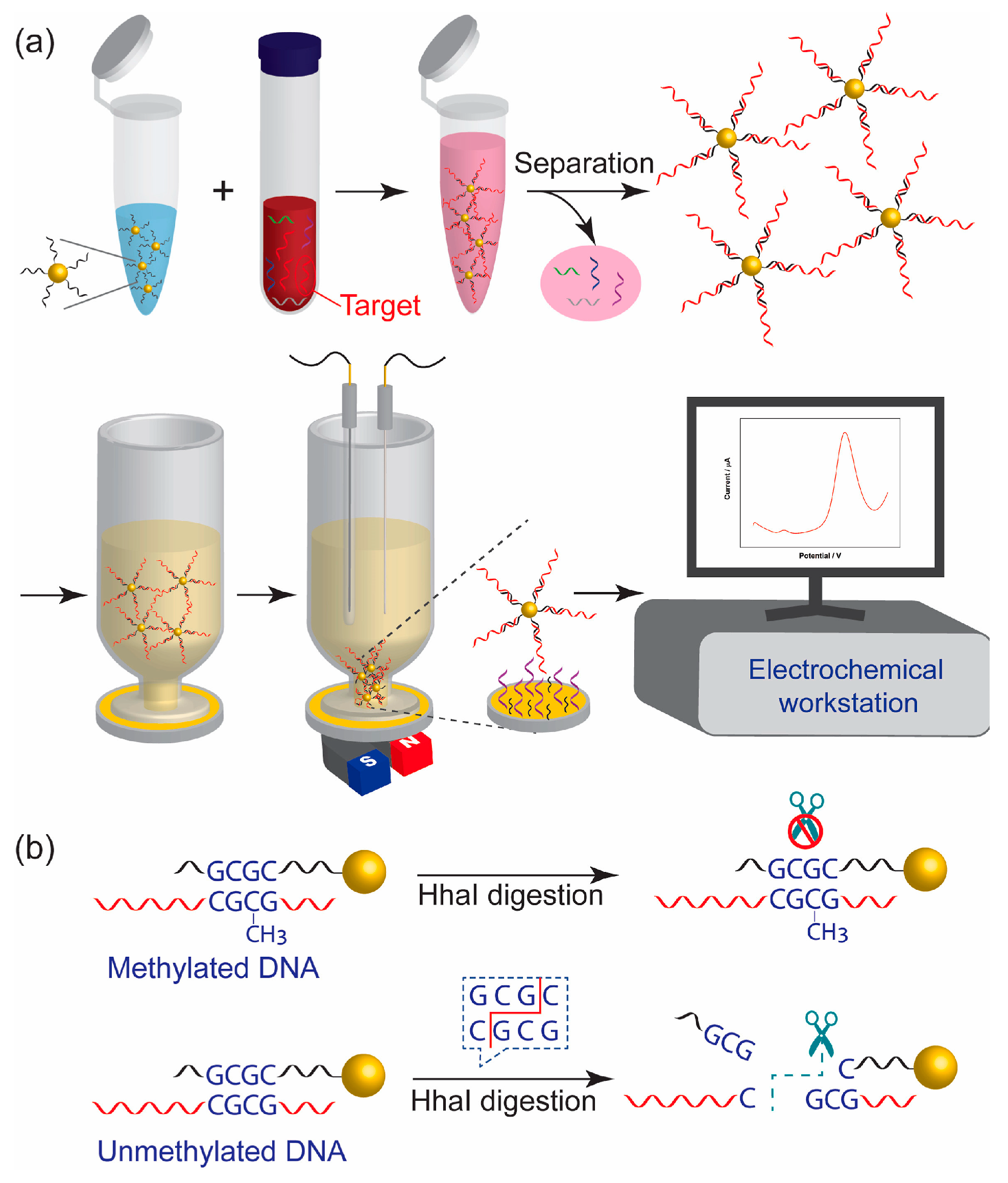
The automatic and rapid nucleic acid extractor (NAE) designed by Chen et al. can create 16 samples simultaneously, and the extraction process can be finished within 30 min [105]. Li et al. employed a pair of universal dual-color probes to four different SNP loci (C667T, A1298C, M235T, and G93A), thus greatly reducing typing costs [106]. Wang et al. designed a novel competitive chemiluminescent DNA method based on Fe3O4@SiO2@Au-functionalized magnetic nanoparticles (Au-MNPs) for the detection of the p53 tumor suppressor gene with a detection limit of 0.001 ng/mL (0.16 pM) and a wide linear response range (0.001 ng/mL~6.6 ng/mL) [107]. The DNA-Au@MNPs-based sensor developed by Chen et al. demonstrates potential for the swift identification of DNA methylation in blood, offering a response time of 35 min and enabling a minimally invasive diagnosis of ovarian cancer. The biosensor presents a dynamic range from 2 aM to 20 nM for 110 nucleotide DNA sequences containing single-site methylation with the lowest detected concentration of 2 aM [108] (Figure 3).
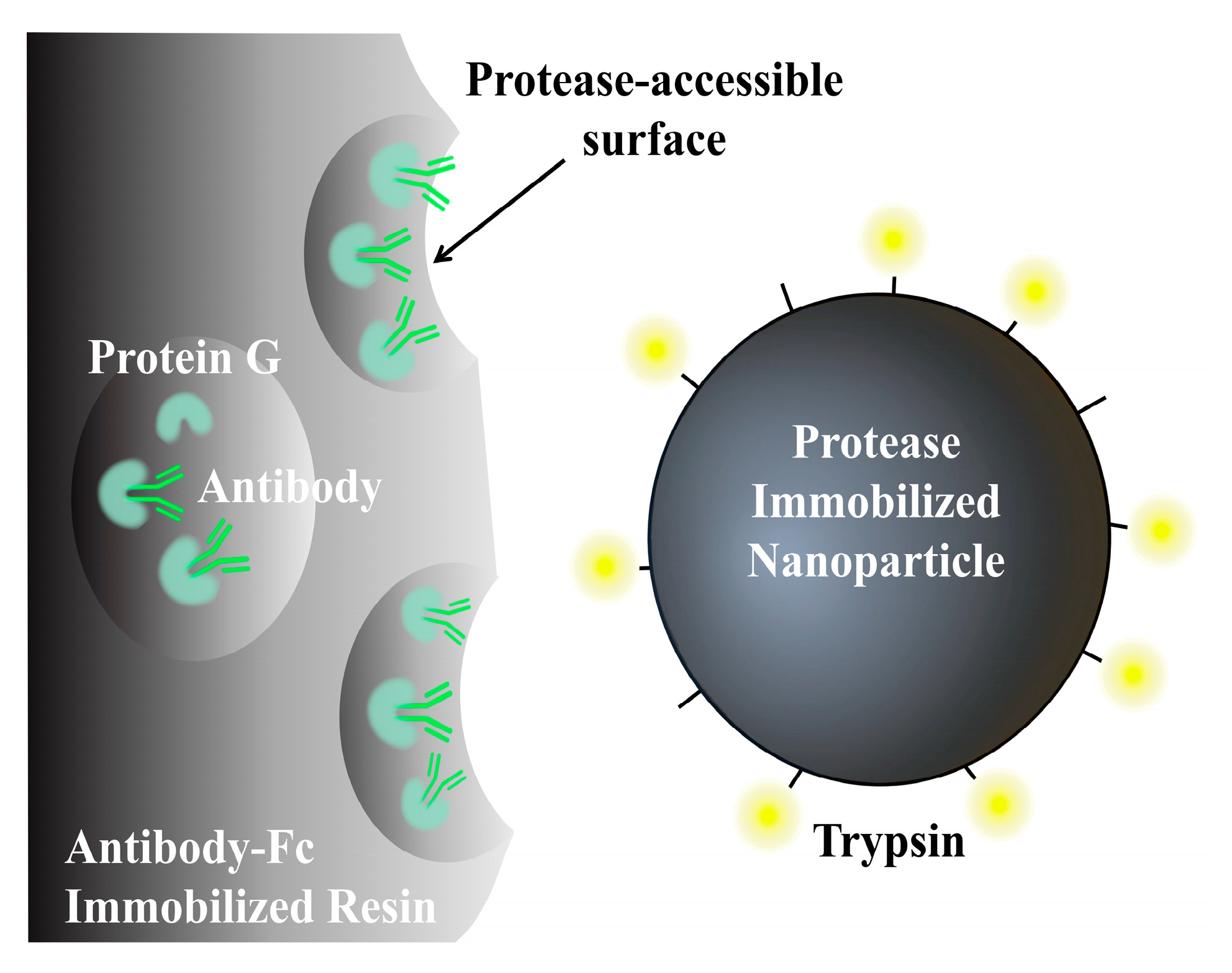
At present, there is a wide array of high-throughput, high-sensitivity, and automated MNP products available in the commercial market. Commonly utilized commercial products include Primer Design from Eastleigh, UK, Magtivio from Thermiekstraat, the Netherlands, Omega Bio-Tek from Norcross, GA, USA, as well as BIOBASE (Jinan, China), MAGIC-BIO (Lishui, China), and BIO-LAB (Xi’an, Chin) from China. Additionally, a collaborative effort between a Japanese research team and Shimadzu Corporation (Kyoto, Japan) has resulted in the development of 200-nanometer-sized magnetic beads (FG beads) capable of identifying specific sequences of monoclonal antibodies for clinical monitoring of antibody–drug metabolism [109], which have been widely used in the contract research organization (CRO) of biopharmaceuticals (Figure 4).

4.3.2. Cell Separation

During cell separation, MNPs are bound to specific biorecognition molecules such as antigens, glycoproteins, and specific structural components on the cell surface by Magnetic Activated Cell Sorting (MACS) [110,111,112], nucleic acid probes, antibiotics, phages [113], and lectins [114,115]. Subsequently, the MNP-labeled cells are separated from the mixtures through the application of a magnetic field, leading to the enrichment and purification of the target cells. Current MNP cell sorting techniques primarily consist of positive and negative sorting methods. Positive sorting involves MNPs binding to target cells for isolation and purification, making it suitable for flow analysis and cell-based analysis. Positive sorting can be further categorized into direct and indirect positive sorting. Indirect positive sorting involves the labeling target cells with a non-coupled, biotinylated, or fluorescein-coupled primary antibody and then attaching it to the MNPs using lgG, anti-biotin–streptavidin, or anti-fluorescein secondary antibodies to separate virtually any cell type based on the specific binding, affinity, and adsorption. In contrast, negative sorting involves MNPs binding to unwanted cells, leaving behind the target cells (Figure 5). When applying nanoscale MNPs for cell sorting, MNPs with sizes less than 250 nm are stable and biocompatible and do not affect the physiological function of cells, which has a wide range of applications in tumor research, cellular immunotherapy, and single-cell analysis [116].
At present, the FDA and the European Union have approved the operation of two cell separation systems based on immunomagnetic separation technology. The first is the CELLSEARCH system for clinical diagnoses and enumeration of circulating tumor cells (CTCs) in patients bearing metastatic breast cancer and metastatic colorectal or prostate cancer [117]. The CELLSEARCH system is a semi-automated in vitro diagnostic device that identifies, isolates, and counts CTCs from a simple blood test using a ferrofluid of IONPs modified with the anti-epithelial cell adhesion molecule antibody EpCAM. The second was Miltenyi Biote’s introduction of the CliniMACS CD34 Reagent System as the first semi-automated immunomagnetic cell separation system for clinical allogeneic stem cell transplantation and enrichment of hematopoietic stem cells expressing CD34+ in patients with acute myeloid leukemia [118].

4.3.3. Determination of Other Biomolecules

Various techniques, such as magnetic solid-phase extraction [119], immobilization [120], and paramagnetic probes [121], are commonly employed for the purification and characterization of biomolecules, including proteins, pathogenic bacteria, viruses, and biomarkers. The SARS-CoV-2 aerosol detection platform developed by Chen H et al. used a wet-wall cyclone in combination with immunomagnetic nanoparticle adsorption sampling and ddPCR for the detection of airborne SARS-CoV-2 aerosols [122] with a minimum detection limit of 250 copies per unit volume of aerosol (102 copies/mL, concentration factor 2.5).
MNPs also play an indelible role in the detection of much-anticipated cancer biomarkers. Magnetoimmunoassay-based electrochemical sensors, photoelectrochemical sensors, and magnetofluidic systems are capable of rapidly and efficiently extracting and determining biomarkers in complex biological samples for in vitro detection [123,124]. Chikkaveeraiah et al. used a microfluidic system equipped with heavy enzyme-labeled MNPs to detect the cancer biomarker proteins prostate-specific antigen (PSA) and interleukin-6 (IL-6) in serum [125], and the detection limit of PSA was 0.23 pg/mL and 0.30 pg/mL for IL-6 in diluted serum mixtures.

4.4. Other Applications of MNPs in the Treatment of Diseases

In addition to the aforementioned biomedical applications, MNPs are extensively utilized in magnetic transfection technology, cell therapy, deep brain stimulation (DBS), and the repair and regeneration of damaged tissues and organs. Among the physical methods to modulate neuronal activity without hardware implants, only magnetic fields can penetrate the brain without absorption or scattering [126], so various methods of neuronal modulation by MNPs have been swiftly developed to improve existing therapeutic strategies and provide new therapeutic ideas [72]. The predominant magnetic DBS methods are mainly magnetothermal and magneto-mechanical stimulation. Magnetic nanostructures (MNSs) used in the fields of tissue engineering and regenerative medicine have the capability to provide magnetic targeting to stem cells, thereby facilitating cell differentiation through the mechanical stimulation induced by MNSs. Furthermore, the scaffolds created by MNSs enhance cell proliferation and adhesion, consequently enhancing the effectiveness of the scaffolds in vivo and promoting tissue repair [127]. In addition, mechanical stimulation generated by applying a magnetic field may activate some receptors on the cell surface and thus upregulate growth-related genes [128].
From the above discussion, the diverse applications of MNP product development encompass both in vivo and in vitro applications such as medical imaging, magnetic hyperthermia, targeted drug delivery, cellular therapy, DBS, nucleic acid detection, cell separation, magnetic transfection, and magnetic solid-phase extraction technologies. Among these applications, in vitro MNP products have been slowly replacing previous technologies as the mainstream choice due to their characteristic advantages and are widely used. In vivo MNP products, due to the longer clinical trial period, can be applied to a limited number of products at present, although the results are still objective.

5. Conclusions and Future Perspectives

Magnetic nanomaterials, as a distinctive component within the field of nanobiomaterials, have garnered significant attention from researchers and are increasingly being acknowledged for their diverse applications. The emergence of magnetic nanotechnology presents novel opportunities in the medical realm, particularly in addressing complex human diseases such as tumors and neurodegenerative disorders. The consumer preference for various magnetic nanomaterial products in the market is attributed to their convenience and eco-friendly nature, rendering them well-suited for contemporary in vitro biomedical applications. Consequently, MNPs are anticipated to play a pivotal role as the “material of the future” and will have a significant impact on all areas of nanobiomedicine.
Despite the promising results of previous studies on MNPs, there remain numerous obstacles to be addressed during the clinical trial phase. These challenges include investigating the internalization effects of MNPs, their clearance in vivo, and potential barriers such as long-term toxicity (including cytotoxicity, hematotoxicity, teratogenicity, and mutagenicity). Strategies to mitigate these issues have involved adjusting the size and functional modifications of MNPs to minimize their toxic effects and promote targeted accumulation at specific pathological sites. In vitro assay studies, the targeted adsorption of specific genera and specific microorganisms (e.g., Mycoplasma, Chlamydia, and parasites) is rarely studied [89], and the cost associated with targeting these microorganisms to address diseases and hazards remains high in terms of financial and resource allocation. Secondly, MNP detection products based on antibodies, lectins, and antibiotics account for a major share of the market. Except for antibody MNPs and nucleic acid detection MNP products, it is difficult to dissociate the target and reuse other MNP products, so many of them are disposable products, which greatly increases the cost of use and resource waste. By employing ion substitution and reversible competitive action, the substances attached to MNPs can be dissociated, enabling the reuse of MNP products. Consequently, the development of multifunctional applications utilizing MNPs is anticipated to become a common practice in industrial and biomedical markets, offering promising opportunities for future utilization.

Author Contributions

Conceptualization, L.W.; methodology, Q.Z.; investigation, X.Y.; writing—original draft preparation, J.H.; project administration and writing—review as well as editing, Z.L.; visualization, C.C. All authors have read and agreed to the published version of the manuscript.

Funding

This research was funded by the Key-Area Research and Development Program of Guangdong Province, grant number 2020B020226008.

Institutional Review Board Statement

Not applicable.

Informed Consent Statement

Not applicable.

Data Availability Statement

Not applicable.

Acknowledgments

This work was supported by South China Agricultural University.

Conflicts of Interest

The authors declare no conflicts of interest.

References

  1. Kianfar, E. Magnetic nanoparticles in targeted drug delivery: A review. J. Supercond. Nov. Magn. 2021, 34, 1709–1735. [Google Scholar] [CrossRef]
  2. Hosu, O.; Tertis, M.; Cristea, C. Implication of Magnetic Nanoparticles in Cancer Detection, Screening and Treatment. Magnetochemistry 2019, 5, 55. [Google Scholar] [CrossRef]
  3. Gessner, I.; Park, J.H.; Lin, H.Y. Magnetic gold nanoparticles with idealized coating for enhanced point-of-care sensing. Adv. Healthc. Mater. 2022, 11, 2102035. [Google Scholar] [CrossRef]
  4. Daya, R.; Xu, C.; Nguyen, N.Y.T. Angiogenic hyaluronic acid hydrogels with curcumin-coated magnetic nanoparticles for tissue repair. ACS Appl. Mater. Interfaces 2022, 14, 11051–11067. [Google Scholar] [CrossRef] [PubMed]
  5. Katz, E. Magnetic nanoparticles. Magnetochemistry 2020, 6, 6. [Google Scholar] [CrossRef]
  6. Zhang, Y.; Zhang, Q.; Zhang, A. Multifunctional co-loaded magnetic nanocapsules for enhancing targeted MR imaging and in vivo photodynamic therapy. Nanomed. Nanotechnol. Biol. Med. 2019, 21, 102047. [Google Scholar] [CrossRef] [PubMed]
  7. Yang, Y.; Li, T.; Jing, W. Dual-modality and Noninvasive Diagnostic of MNP–PEG–Mn Nanoprobe for Renal Fibrosis Based on Photoacoustic and Magnetic Resonance Imaging. ACS Appl. Mater. Interfaces 2023, 15, 12797–12808. [Google Scholar] [CrossRef]
  8. Ma, R.; Hou, Y.; Wang, S. Imaging-Guided Cancer Therapy Based on Multifunctional Magnetic Nanoparticles. Chin. J. Chem. 2023, 41, 2557–2573. [Google Scholar] [CrossRef]
  9. Wong, J.; Prout, J.; Seifalian, A. Magnetic nanoparticles: New perspectives in drug delivery. Curr. Pharm. Des. 2017, 23, 2908–2917. [Google Scholar] [CrossRef]
  10. Wang, N.; Guan, Y.; Yang, L. Magnetic nanoparticles (MNPs) covalently coated by PEO–PPO–PEO block copolymer for drug delivery. J. Colloid Interface Sci. 2013, 395, 50–57. [Google Scholar] [CrossRef]
  11. Demessie, A.A.; Park, Y.; Singh, P. An Advanced Thermal Decomposition Method to Produce Magnetic Nanoparticles with Ultrahigh Heating Efficiency for Systemic Magnetic Hyperthermia. Small Methods 2022, 6, 2200916. [Google Scholar] [CrossRef]
  12. Kaushik, S.; Thomas, J.; Panwar, V.A. drug-free strategy to combat bacterial infections with magnetic nanoparticles biosynthesized in bacterial pathogens. Nanoscale 2022, 14, 1713–1722. [Google Scholar] [CrossRef]
  13. Willis, A.J.; Pernal, S.P.; Gaertner, Z.A. Rotating magnetic nanoparticle clusters as microdevices for drug delivery. Int. J. Nanomed. 2020, 15, 4105–4123. [Google Scholar] [CrossRef]
  14. Ni, D.; Ferreira, C.A.; Barnhart, T.E. Magnetic targeting of nanotheranostics enhances cerenkov radiation-induced photodynamic therapy. J. Am. Chem. Soc. 2018, 140, 14971–14979. [Google Scholar] [CrossRef] [PubMed]
  15. Ostroverkhov, P.; Semkina, A.; Naumenko, V. HSA—Coated magnetic nanoparticles for MRI-guided photodynamic cancer therapy. Pharmaceutics 2018, 10, 284. [Google Scholar] [CrossRef]
  16. Chen, C.; Wang, S.; Li, L. Bacterial magnetic nanoparticles for photothermal therapy of cancer under the guidance of MRI. Biomaterials 2016, 104, 352–360. [Google Scholar] [CrossRef] [PubMed]
  17. Yeboah, I.B.; Hatekah, S.W.; Yaya, A. Photothermally-heated superparamagnetic polymeric nanocomposite implants for interstitial thermotherapy. Nanomaterials 2022, 12, 955. [Google Scholar] [CrossRef]
  18. Kang, S.; Baskaran, R.; Ozlu, B. T1-positive Mn2+-doped multi-stimuli responsive poly (L-DOPA) nanoparticles for photothermal and photodynamic combination cancer therapy. Biomedicines 2020, 8, 417. [Google Scholar] [CrossRef] [PubMed]
  19. Xiao, F.; Li, W.; Xu, H. Advances in magnetic nanoparticles for the separation of foodborne pathogens: Recognition, separation strategy, and application. Compr. Rev. Food Sci. Food Saf. 2022, 21, 4478–4504. [Google Scholar] [CrossRef]
  20. Azadpour, B.; Aharipour, N.; Paryab, A. Magnetically-assisted viral transduction (magnetofection) medical applications: An update. Biomater. Adv. 2023, 154, 213657. [Google Scholar] [CrossRef]
  21. Guo, Y.; Li, S.; Wang, Y. Diagnosis–Therapy integrative systems based on magnetic RNA nanoflowers for Co-drug delivery and targeted therapy. Anal. Chem. 2017, 89, 2267–2274. [Google Scholar] [CrossRef]
  22. Farooq, A.; Tosheva, L.; Azzawi, M. Real-time observation of aortic vessel dilation through delivery of sodium nitroprusside via slow release mesoporous nanoparticles. J. Colloid Interface Sci. 2016, 478, 127–135. [Google Scholar] [CrossRef]
  23. Liu, H.; Li, S.; Wang, Z. Automated detection system of single nucleotide polymorphisms using two kinds of functional magnetic nanoparticles. Appl. Surf. Sci. 2008, 255, 600–603. [Google Scholar] [CrossRef]
  24. Zhang, J.; Zhang, T.; Gao, J. Biocompatible iron oxide nanoparticles for targeted cancer gene therapy: A review. Nanomaterials 2022, 12, 3323. [Google Scholar] [CrossRef] [PubMed]
  25. Brestovac, B.; Harnett, G.B.; Smith, D.W. Multiplex nested PCR (MNP) assay for the detection of 15 high risk genotypes of human papillomavirus. J. Clin. Virol. 2005, 33, 116–122. [Google Scholar] [CrossRef]
  26. Jun, Y.; Seo, J.; Cheon, J. Nanoscaling laws of magnetic nanoparticles and their applicabilities in biomedical sciences. Acc. Chem. Res. 2008, 41, 179–189. [Google Scholar] [CrossRef]
  27. Bai, S.; Hou, S.; Chen, T. Magnetic nanoparticle-mediated hyperthermia: From heating mechanisms to cancer theranostics. Innov. Mater. 2024, 2, 100051-1–100051-16. [Google Scholar] [CrossRef]
  28. Fallows, T.W.; McGrath, A.J.; Silva, J. High-throughput chemical and chemoenzymatic approaches to saccharide-coated magnetic nanoparticles for MRI. Nanoscale Adv. 2019, 1, 3597–3606. [Google Scholar] [CrossRef]
  29. Quan, K.; Zhang, Z.; Ren, Y. Possibilities and impossibilities of magnetic nanoparticle use in the control of infectious biofilms. J. Mater. Sci. Technol. 2021, 69, 69–78. [Google Scholar] [CrossRef]
  30. Bruschi, M.L.; de Toledo, L.A.S. Pharmaceutical applications of iron-oxide magnetic nanoparticles. Magnetochemistry 2019, 5, 50. [Google Scholar] [CrossRef]
  31. Obaidat, I.M.; Narayanaswamy, V.; Alaabed, S. Principles of magnetic hyperthermia: A focus on using multifunctional hybrid magnetic nanoparticles. Magnetochemistry 2019, 5, 67. [Google Scholar] [CrossRef]
  32. Rezaei, B.; Yari, P.; Sanders, S.M. Magnetic nanynchronous disintegration of ferroptosis defense axis via engineered exosome-conjugated magnetic nanopaapplications. Small 2024, 20, 2304848. [Google Scholar] [CrossRef] [PubMed]
  33. Arias, L.S.; Pessan, J.P.; Vieira, A.P.M. Iron Oxide Nanoparticles for Biomedical Applications: A Perspective on Synthesis, Drugs, Antimicrobial Activity, and Toxicity. Antibiotics 2018, 7, 46. [Google Scholar] [CrossRef] [PubMed]
  34. Li, B.; Chen, X.; Qiu, W. Synchronous disintegration of ferroptosis defense axis via engineered exosome-conjugated magnetic nanoparticles for glioblastoma therapy. Adv. Sci. 2022, 9, 2105451. [Google Scholar] [CrossRef] [PubMed]
  35. Bilal, M.; Mehmood, S.; Rasheed, T. Bio-catalysis and biomedical perspectives of magnetic nanoparticles as versatile carriers. Magnetochemistry 2019, 5, 42. [Google Scholar] [CrossRef]
  36. Slimani, Y.; Hannachi, E.; Tombuloglu, H. Magnetic nanoparticles based nanocontainers for biomedical application. In Smart Nanocontainers; Elsevier: Amsterdam, The Netherlands, 2020; pp. 229–250. [Google Scholar]
  37. Bao, S.; Yang, W.; Wang, Y. One-pot synthesis of magnetic graphene oxide composites as an efficient and recoverable adsorbent for Cd (II) and Pb (II) removal from aqueous solution. J. Hazard. Mater. 2020, 381, 120914. [Google Scholar] [CrossRef] [PubMed]
  38. Tran, H.V.; Ngo, N.M.; Medhi, R. Multifunctional Iron Oxide Magnetic Nanoparticles for Biomedical Applications: A Review. Materials 2022, 15, 503. [Google Scholar] [CrossRef] [PubMed]
  39. Abd Elrahman, A.A.; Mansour, F.R. Targeted magnetic iron oxide nanoparticles: Preparation, functionalization and biomedical application. J. Drug Deliv. Sci. Technol. 2019, 52, 702–712. [Google Scholar] [CrossRef]
  40. Dulińska-Litewka, J.; Łazarczyk, A.; Hałubiec, P. Superparamagnetic Iron Oxide Nanoparticles—Current and Prospective Medical Applications. Materials 2019, 12, 617. [Google Scholar] [CrossRef]
  41. Hepel, M. Magnetic Nanoparticles for Nanomedicine. Magnetochemistry 2020, 6, 3. [Google Scholar] [CrossRef]
  42. Sabale, S.; Kandesar, P.; Jadhav, V. Recent developments in the syn-thesis, properties, and biomedical applications of core/shell superparamagnetic iron oxide nanoparticles with gold. Biomater. Sci. 2017, 5, 2212–2225. [Google Scholar] [CrossRef] [PubMed]
  43. Zhou, X.; Li, P.; Wu, X. Multifunctional biosensor constructed by Ag-coating magnetic-assisted unique urchin core porous shell structure for dual SERS enhancement, enrichment, and quantitative detection of multi-components inflammatory markers. Biosens. Bioelectron. 2022, 210, 114257. [Google Scholar] [CrossRef] [PubMed]
  44. Alyassin, Y.; Sayed, E.G.; Mehta, P. Application of mesoporous silica nanoparticles as drug delivery carriers for chemotherapeutic agents. Drug Discov. Today 2020, 25, 1513–1520. [Google Scholar] [CrossRef] [PubMed]
  45. Bai, Y.L.; Shahed-Al-Mahmud, M.; Selvaprakash, K. Tail fiber protein-immobilized magnetic nanoparticle-based affinity approaches for detection of Acinetobacter baumannii. Anal. Chem. 2019, 91, 10335–10342. [Google Scholar] [CrossRef] [PubMed]
  46. Montiel Schneider, M.G.; Martín, M.J.; Otarola, J. Bio-medical Applications of Iron Oxide Nanoparticles: Current Insights Progress and Perspectives. Pharmaceutics 2022, 14, 204. [Google Scholar] [CrossRef]
  47. Yang, D.; Yang, G.; Yang, P. Assembly of Au Plasmonic Photothermal Agent and Iron Oxide Nanoparticles on Ultrathin Black Phosphorus for Targeted Photothermal and Photodynamic Cancer Therapy. Adv. Funct. Mater. 2017, 27, 1700371. [Google Scholar] [CrossRef]
  48. Bai, Y.; Roncancio, D.; Suo, Y. A method based on amino-modified magnetic nanoparticles to extract DNA for PCR-based analysis. Colloids Surf. B Biointerfaces 2019, 7, 87–93. [Google Scholar] [CrossRef] [PubMed]
  49. Li, P.; Li, M.; Zhang, F. High-efficient nucleic acid separation from animal tissue samples via surface modified magnetic nanoparticles. Sep. Purif. Technol. 2021, 262, 118348. [Google Scholar] [CrossRef]
  50. Zhou, Z.; Kadam, U.S.; Irudayaraj, J. One-stop genomic DNA extraction by salicylic acid-coated magnetic nanoparticles. Anal. Biochem. 2013, 442, 249–252. [Google Scholar] [CrossRef]
  51. Sahoo, S.L.; Liu, C.H. Adsorption behaviors of DNA by modified magnetic nanoparticles: Effect of spacer and salt. Colloids Surf. A Physicochem. Eng. Asp. 2015, 482, 184–194. [Google Scholar] [CrossRef]
  52. Maeda, Y.; Toyoda, T.; Mogi, T. DNA recovery from a single bacterial nanoparticles. Colloids Surf. B Biointerfaces 2016, 139, 117–122. [Google Scholar] [CrossRef] [PubMed]
  53. Hoyt, K.; Barr, J.R.; Kalb, S.R. Detection of ricin activity and structure by using novel galactose-terminated magnetic bead extraction coupled with mass spectrometric detection. Anal. Biochem. 2021, 631, 114364. [Google Scholar] [CrossRef]
  54. Abdelaziz, M.M.; Hefnawy, A.; Anter, A. Respirable spray dried vancomycin coated magnetic nanoparticles for localized lung delivery. Int. J. Pharm. 2022, 611, 121318. [Google Scholar] [CrossRef] [PubMed]
  55. Zhan, S.; Fang, H.; Chen, Q. M13 bacteriophage as biometric component for orderly assembly of dynamic light scattering immunosensor. Biosens. Bioelectron. 2022, 217, 114693. [Google Scholar] [CrossRef] [PubMed]
  56. Antal, I.; Strbak, O.; Khmara, I. MRI Relaxivity Changes of the Magnetic Nanoparticles Induced by Different Amino Acid Coatings. Nanomaterials 2020, 10, 394. [Google Scholar] [CrossRef] [PubMed]
  57. Kuan, W.C.; Lai, J.W.; Lee, W.C. Covalent binding of glutathione on magnetic nanoparticles: Application for immobilizing small fragment ubiquitin-like-specific protease 1. Enzym. Microb. Technol. 2021, 143, 109697. [Google Scholar] [CrossRef]
  58. Matveeva, V.G.; Bronstein, L.M. Magnetic Nanoparticle-Containing Supports as Carriers of Immobilized Enzymes: Key Factors Influencing the Biocatalyst Performance. Nanomaterials 2021, 11, 2257. [Google Scholar] [CrossRef] [PubMed]
  59. Sosa-Acosta, J.R.; Iriarte-Mesa, C.; Ortega, G.A. DNA–Iron Oxide Nanoparticles Conjugates: Functional Magnetic Nanoplatforms in Biomedical Applications. Top. Curr. Chem. 2020, 378, 13. [Google Scholar] [CrossRef] [PubMed]
  60. Millart, E.; Lesieur, S.; Faivre, V. Superparamagnetic lipid-based hybrid nanosystems for drug delivery. Expert Opin. Drug Deliv. 2018, 15, 523–540. [Google Scholar] [CrossRef]
  61. Liu, S.; Chen, X.; Bao, L. Treatment of infarcted heart tissue via the capture and local delivery of circulating exosomes through antibody-conjugated magnetic nanoparticles. Nat. Biomed. Eng. 2020, 4, 1063–1075. [Google Scholar] [CrossRef]
  62. Sizikov, A.A.; Kharlamova, M.V.; Nikitin, M.P. Nonviral locally injected magnetic vectors for in vivo gene delivery: A review of studies on magnetofection. Nanomaterials 2021, 11, 1078. [Google Scholar] [CrossRef] [PubMed]
  63. You, S.M.; Jeong, K.B.; Luo, K. based colorimetric detection of pathogenic bacteria in food through magnetic separation and enzyme-mediated signal amplification on paper disc. Anal. Chim. Acta 2021, 1151, 338252. [Google Scholar] [CrossRef] [PubMed]
  64. Deng, M.; Wang, Y.; Chen, G. Poly-l-lysine-functionalized magnetic beads combined with polymerase chain reaction for the detection of Staphylococcus aureus and Escherichia coli O157: H7 in milk. Dairy Sci 2021, 104, 12342–12352. [Google Scholar] [CrossRef] [PubMed]
  65. Gao, X.; Yao, X.; Zhong, Z. Rapid and sensitive detection of Staphylococcus aureus assisted by polydopamine modified magnetic nanoparticles. Talanta 2018, 186, 147–153. [Google Scholar] [CrossRef] [PubMed]
  66. Carter, T.J.; Agliardi, G.; Lin, F.Y. Potential of magnetic hyperthermia to stimulate localized immune activation. Small 2021, 17, 2005241. [Google Scholar] [CrossRef]
  67. Veloso, S.R.; Tiryaki, E.; Spuch, C. Tuning the drug multimodal release through a co-assembly strategy based on magnetic gels. Nanoscale 2022, 14, 5488–5500. [Google Scholar] [CrossRef]
  68. Cremin, K.; Jones, B.A.; Teahan, J. Scanning ion conductance microscopy reveals differences in the ionic environments of gram-positive and negative bacteria. Anal. Chem. 2020, 92, 16024–16032. [Google Scholar] [CrossRef]
  69. Etemadi, H.; Buchanan, J.K.; Kandile, N.G. Iron oxide nanoparticles: Physicochemical characteristics and historical developments to commercialization for potential technological applications. ACS Biomater. Sci. Eng. 2021, 7, 5432–5450. [Google Scholar] [CrossRef]
  70. Bakhtiary, Z.; Saei, A.A.; Hajipour, M.J. Targeted superparamagnetic iron oxide nanoparticles for early detection of cancer: Possibilities and challenges. Nanomed. Nanotechnol. Biol. Med. 2016, 12, 287–307. [Google Scholar] [CrossRef]
  71. Zhang, H.; Guo, Y.; Jiao, J. A hepatocyte-targeting nanoparticle for enhanced hepatobiliary magnetic resonance imaging. Nat. Biomed. Eng. 2023, 7, 221–235. [Google Scholar] [CrossRef]
  72. Thomas, G.; Boudon, J.; Maurizi, L. Innovative magnetic nanoparticles for PET/MRI bimodal imaging. ACS Omega 2019, 4, 2637–2648. [Google Scholar] [CrossRef] [PubMed]
  73. Myrovali, E.; Maniotis, N.; Samaras, T. Spatial focusing of magnetic particle hyperthermia. Nanoscale Adv. 2020, 2, 408–416. [Google Scholar] [CrossRef] [PubMed]
  74. Neuwelt, A.; Sidhu, N.; Hu, C.A.A. Iron-based superparamagnetic nanoparticle contrast agents for MRI of infection and inflammation. Am. J. Roentgenol. 2015, 204, W302–W313. [Google Scholar] [CrossRef] [PubMed]
  75. Bulte, J.W. Superparamagnetic iron oxides as MPI tracers: A primer and review of early applications. Adv. Drug Deliv. Rev. 2019, 138, 293–301. [Google Scholar] [CrossRef] [PubMed]
  76. Ling, W.; Wang, M.; Xiong, C. Synthesis, surface modification, and applications of magnetic iron oxide nanoparticles. J. Mater. Res. 2019, 34, 1828–1844. [Google Scholar] [CrossRef]
  77. Healy, S.; Bakuzis, A.F.; Goodwill, P.W. Clinical magnetic hyperthermia requires integrated magnetic particle imaging. Wiley Interdiscip. Rev. Nanomed Nanobiotechnol. 2022, 14, e1779. [Google Scholar] [CrossRef] [PubMed]
  78. Marchal, S.; Hor, A.E.; Millard, M. Anticancer drug delivery: An update on clinically applied nanotherapeutics. Drugs 2015, 75, 1601–1611. [Google Scholar] [CrossRef] [PubMed]
  79. Issels, R.D.; Lindner, L.H.; Verweij, J. Effect of neoadjuvant chemotherapy plus regional hyperthermia on long-term outcomes among patients with localized high-risk soft tissue sarcoma: The EORTC 62961-ESHO 95 randomized clinical trial. JAMA Oncol. 2018, 4, 483–492. [Google Scholar] [CrossRef]
  80. van Landeghem, F.K.; Maier-Hauff, K.; Jordan, A. Post-mortem studies in glioblastoma patients treated with thermotherapy using magnetic nanoparticles. Biomaterials 2009, 30, 52–57. [Google Scholar] [CrossRef]
  81. Toraya-Brown, S.; Sheen, M.R.; Zhang, P. Local hyperthermia treatment of tumors induces CD8+ T cell-mediated resistance against distal and secondary tumors. Nanomed. Nanotechnol. Biol. Med. 2014, 10, 1273–1285. [Google Scholar] [CrossRef]
  82. Wang, J.; Wang, L.; Pan, J. Magneto-based synergetic therapy for im-plant-associated infections via biofilm disruption and innate immunity regulation. Adv. Sci. 2021, 8, 2004010. [Google Scholar] [CrossRef]
  83. Atluri, V.S.R.; Jayant, R.D.; Pilakka-Kanthikeel, S. Development of TIMP1 magnetic nanoformulation for regulation of synaptic plasticity in HIV-1 infection. Int. J. Nanomed. 2016, 11, 4287–4298. [Google Scholar] [CrossRef] [PubMed]
  84. Bui, M.P.; Le, T.A.; Yoon, J. A magnetic particle imaging-based navigation platform for magnetic nano-particles using interactive manipulation of a virtual field free point to ensure targeted drug delivery. IEEE Trans. Ind. Electron. 2020, 68, 12493–12503. [Google Scholar] [CrossRef]
  85. Gul, S.; Khan, S.B.; Rehman, I.U. A comprehensive review of magnetic nanomaterials modern day theranostics. Front. Mater. 2019, 6, 179. [Google Scholar] [CrossRef]
  86. Liu, Y.; Cao, F.; Sun, B. Magnetic nanoparticles: A new diagnostic and treatment platform for rheumatoid arthritis. J. Leucoc. Biol. 2021, 109, 415–424. [Google Scholar] [CrossRef]
  87. Liu, X.; Zhang, Y.; Wang, Y. Comprehensive understanding of magnetic hyperthermia for improving antitumor therapeutic efficacy. Theranostics 2020, 10, 3793. [Google Scholar] [CrossRef]
  88. Ito, T.; Ando, H.; Suzuki, T. Identification of a primary target of thalidomide teratogenicity. Science 2010, 327, 1345–1350. [Google Scholar] [CrossRef]
  89. Abo-Zeid, Y.; Ismail, N.S.M.; McLean, G.R. A molecular docking study repurposes FDA approved iron oxide nanoparticles to treat and control COVID-19 infection. Eur. J. Pharm. Sci. 2020, 153, 105465. [Google Scholar] [CrossRef] [PubMed]
  90. Ali, A.; Shah, T.; Ullah, R. Review on recent progress in magnetic nanoparticles: Synthesis, characterization, and diverse applications. Front. Chem. 2021, 9, 629054. [Google Scholar] [CrossRef]
  91. Han, X.; Cao, M.; Zhou, B.; Yu, C. Specifically immobilizing His-tagged allergens to magnetic nanoparticles for fast and quantitative detection of allergen-specific IgE in serum samples. Talanta 2022, 19, 121301. [Google Scholar] [CrossRef]
  92. Ajibade, P.A.; Paca, A.M. Tris (dithiocarbamato) iron (III) complexes as precursors for iron sulfide nano-crystals and iron sulfide-hydroxyethyl cellulose composites. J. Sulfur Chem. 2019, 40, 52–64. [Google Scholar] [CrossRef]
  93. Pires, F.; Silva, J.C.; Ferreira, F.C. Heparinized Acellular Hydrogels for Magnetically In-duced Wound Healing Applications. ACS Appl. Mater. Interfaces 2024, 16, 9908–9924. [Google Scholar] [CrossRef] [PubMed]
  94. Vu, T.T.; Zhang, A.; Wang, R. Using single antigen specificity magnetic beads for the isolation of specific antibodies against HLA antigens. HLA 2024, 103, e15490. [Google Scholar] [CrossRef] [PubMed]
  95. Källsten, M.; Ghorasaini, M.; Hartmann, R. Magnetic beads for desalting of monoclonal antibodies and antibody–drug Conjugates. Anal. Chem. 2020, 92, 9001–9007. [Google Scholar] [CrossRef] [PubMed]
  96. Guo, X.; Hu, F.; Zhao, S. Immunomagnetic separation method integrated with the Strep-Tag II system for rapid enrichment and mild release of exosomes. Anal. Chem. 2023, 95, 3569–3576. [Google Scholar] [CrossRef] [PubMed]
  97. Zhang, M.; Wang, Y.; Wu, P. Development of a highly sensitive detection method for TTX based on a magnetic bead-aptamer competition system under triple cycle amplification. Anal. Chim. Acta 2020, 1119, 18–24. [Google Scholar] [CrossRef] [PubMed]
  98. Khadsai, S.; Seeja, N.; Rutnakornpituk, M. Selective enrichment of zein gene of maize from cereal products using magnetic support having pyrrolidinyl peptide nucleic acid probe. Food Chem. 2021, 338, 127812. [Google Scholar] [CrossRef]
  99. Nan, K.; He, M.; Chen, B. Histidine tag modified magnetic beads for analysis of arsenic binding proteins. Anal. Chim. Acta 2024, 1304, 342554. [Google Scholar] [CrossRef] [PubMed]
  100. Lee, M.H.; Leu, C.C.; Lin, C.C. Gold-decorated magnetic nanoparticles modified with hairpin-shaped DNA for fluorometric discrimination of single-base mismatch DNA. Microchim. Acta 2019, 186, 80. [Google Scholar] [CrossRef]
  101. Chen, Y.; Liu, Y.; Shi, Y. Magnetic particles for integrated nucleic acid purification, amplification and detection without pipetting. TrAC Trends Anal. Chem. 2020, 127, 115912. [Google Scholar] [CrossRef]
  102. Xuhong, Y.; Sinong, Z.; Jianping, L. A PCR-lateral flow assay system based on gold magnetic nanoparticles for CYP2C19 genotyping and its clinical applications. Artif. Cells Nanomed. Biotechnol. 2019, 47, 636–643. [Google Scholar] [CrossRef] [PubMed]
  103. Nadar, S.S.; Kelkar, R.K.; Pise, P.V. The un-tapped potential of magnetic nanoparticles for forensic investigations: A comprehensive review. Talanta 2021, 230, 122297. [Google Scholar] [CrossRef] [PubMed]
  104. Cheng, H.; Liu, J.; Ma, W. Low background cascade signal amplification electrochemical sensing platform for tumor-related mRNA quantification by target-activated hybridization chain reaction and electroactive cargo release. Anal. Chem. 2018, 90, 12544–12552. [Google Scholar] [CrossRef] [PubMed]
  105. Chen, Z.; Wu, Y.; Chen, H. Design and application of automatic and rapid nucleic acid extractor using magnetic nanoparticles. J. Nanosci. Nanotechnol. 2016, 16, 6998–7004. [Google Scholar] [CrossRef]
  106. Li, S.; Liu, H.; Jia, Y. An automatic high-throughput single nucleotide polymorphism genotyping approach based on universal tagged arrays and magnetic nanoparticles. J. Biomed. Nanotechnol. 2013, 9, 689–698. [Google Scholar] [CrossRef]
  107. Wang, L.; Yao, M.; Fang, X. Novel competitive chemiluminescence DNA assay based on Fe3O4@ SiO2@Au-functionalized magnetic nanoparticles for sensitive detection of p53 tumor suppressor gene. Appl. Biochem. Biotechnol. 2019, 187, 152–162. [Google Scholar] [CrossRef] [PubMed]
  108. Chen, D.; Wu, Y.; Tilley, R.D. Rapid and ultrasensitive electrochemical detection of DNA methylation for ovarian cancer diagnosis. Biosens. Bioelectron. 2022, 206, 114126. [Google Scholar] [CrossRef]
  109. Iwamoto, N.; Shimada, T.; Umino, Y. Selective detection of complementarity-determining regions of monoclonal antibody by limiting protease access to the substrate: Nano-surface and molecular-orientation limited proteolysis. Analyst 2014, 139, 576–580. [Google Scholar] [CrossRef] [PubMed]
  110. Oh, S.; Jung, S.H.; Seo, H. Magnetic activated cell sorting (MACS) pipette tip for immunomagnetic bacteria separation. Sens. Actuators B Chem. 2018, 272, 324–330. [Google Scholar] [CrossRef]
  111. González-Ravina, C.; Castro, A.P.; Palomino, M.C. P–081 Microfluidic sorting does not improve clinical outcomes compared to magnetic activated cell sorting (MACS) in Assisted Re-production. Hum. Reprod. 2021, 36, deab130-080. [Google Scholar] [CrossRef]
  112. Adams, J.D.; Kim, U.; Soh, H.T. Multitarget magnetic activated cell sorter. Proc. Natl. Acad. Sci. USA 2008, 105, 18165–18170. [Google Scholar] [CrossRef]
  113. Wang, L.; Lin, H.; Zhang, J. Phage long tail fiber protein-immobilized magnetic nanoparticles for rapid and ultrasensitive detection of Salmonella. Talanta 2022, 248, 123627. [Google Scholar] [CrossRef] [PubMed]
  114. Monje, P.V. Human Schwann Cells in vitro II. Passaging, Purification, Banking, and Labeling of Established Cultures. Bio-Protocol 2023, 13, e4882. [Google Scholar] [CrossRef]
  115. Yang, G.; Huang, M.; Wang, Y. Streptavidin-exposed magnetic nanoparticles for lectin magnetic separation (LMS) of Staphylococcus aureus prior to three quantification strategies. Microchim. Acta 2019, 186, 813. [Google Scholar] [CrossRef]
  116. Lin, Z.; Lin, S.Y.; Xie, P. Rapid Assessment of Surface Markers on Cancer Cells Using Immuno-Magnetic Separation and Multi-frequency Impedance Cytometry for Targeted Therapy. Sci. Rep. 2020, 10, 3015. [Google Scholar] [CrossRef]
  117. Wang, L.; Balasubramanian, P.; Chen, A.P. Promise and limits of the CellSearch platform for evaluating pharmacodynamics in circulating tumor cells. Semin. Oncol. 2016, 43, 464–475. [Google Scholar] [CrossRef]
  118. FDA Approves CliniMACS CD34 Reagent System for the Prevention of Graft-vs-Host Disease in AML. Available online: https://www.ascopost.com/issues/february-15-2014/fda-approves-clinimacs-cd34-reagent-system-for-the-prevention-of-graft-vs-host-disease-in-aml/ (accessed on 14 November 2020).
  119. Manousi, N.; Rosenberg, E.; Deliyanni, E. Magnetic solid-phase extraction of organic compounds based on graphene oxide nanocomposites. Molecules 2020, 25, 1148. [Google Scholar] [CrossRef] [PubMed]
  120. Mylkie, K.; Nowak, P.; Rybczynski, P. Polymer-Coated Magnetite Nanoparticles for Protein Immobilization. Materials 2021, 14, 248. [Google Scholar] [CrossRef] [PubMed]
  121. Miao, Q.; Nitsche, C.; Orton, H. Paramagnetic Chemical Probes for Studying Biological Macromolecules. Chem. Rev. 2022, 122, 9571–9642. [Google Scholar] [CrossRef]
  122. Chen, H.; Ma, X.; Zhang, X. Novel aerosol detection platform for SARS-CoV-2: Based on specific magnetic nanoparticles adsorption sampling and digital droplet PCR detection. Chin. Chem. Lett. 2023, 34, 107701. [Google Scholar] [CrossRef]
  123. Kavetskyy, T.; Alipour, M.; Smutok, O. Magneto-immunoassay of cancer biomarkers: Recent progress and challenges in biomedical analysis. Microchem. J. 2021, 167, 106320. [Google Scholar] [CrossRef]
  124. Gheorghiu, E. Detection of pathogenic bacteria by magneto-immunoassays: A review. J. Biomed. Res. 2021, 35, 277. [Google Scholar] [CrossRef] [PubMed]
  125. Chikkaveeraiah, B.V.; Mani, V.; Patel, V. Microfluidic electrochemical immunoarray for ultrasensitive detection of two cancer biomarker proteins in serum. Biosens. Bioelectron. 2011, 26, 4477–4483. [Google Scholar] [CrossRef] [PubMed]
  126. Chen, R.; Canales, A.; Anikeeva, P. Neural recording and modulation technologies. Nat. Rev. Mater. 2017, 2, 16093. [Google Scholar] [CrossRef] [PubMed]
  127. Ovejero, J.G.; Spizzo, F.; Morales, M.P. Nanoparticles for Magnetic Heating: When Two (or More) Is Better Than One. Materials 2021, 14, 6416. [Google Scholar] [CrossRef]
  128. Van de Walle, A.; Perez, J.E.; Abou-Hassan, A. Magnetic nanoparticles in regenerative medicine: What of their fate and impact in stem cells? Mater. Today Nano 2020, 11, 100084. [Google Scholar] [CrossRef]
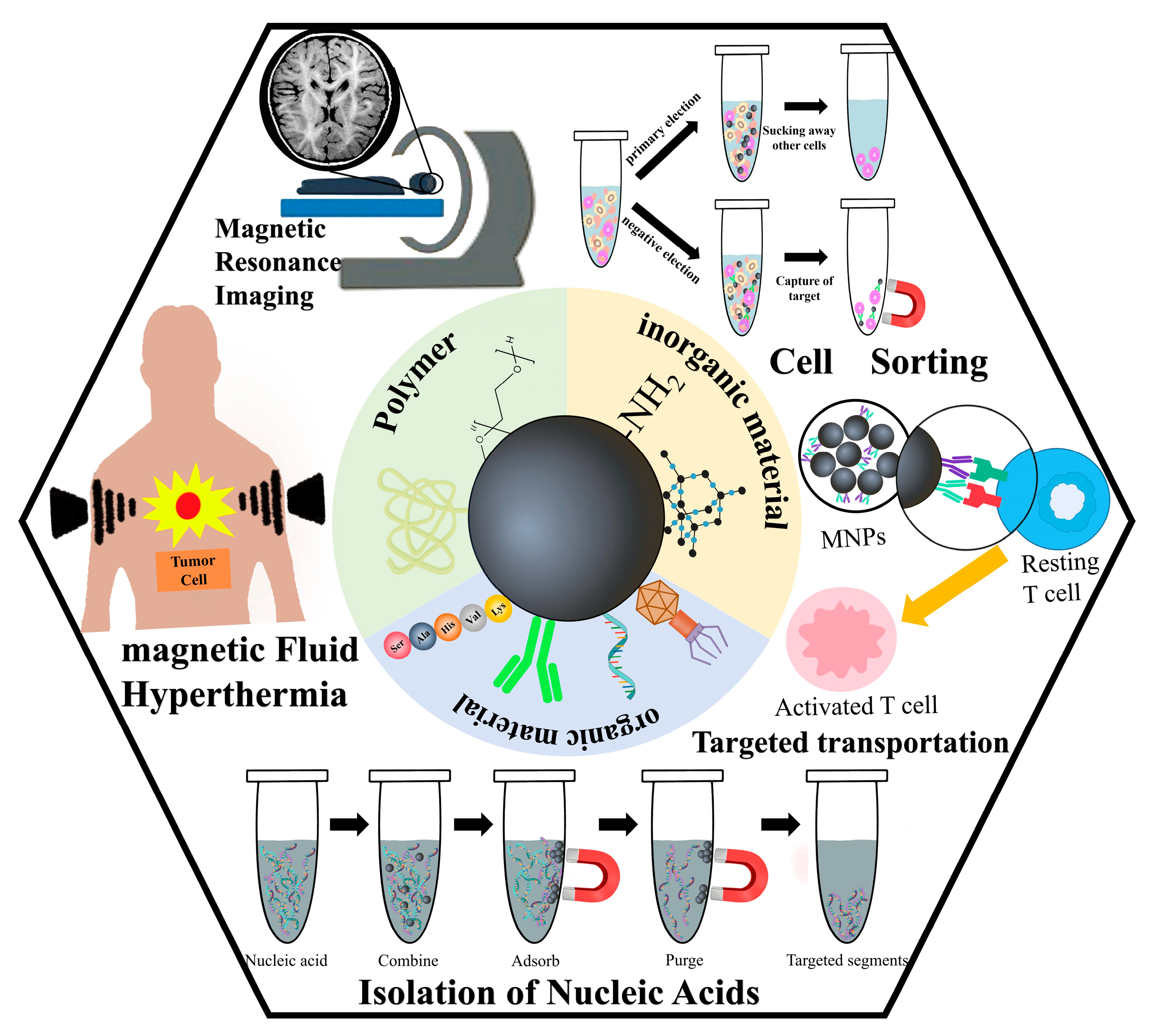
Figure 1. Schematic illustration of the main applications of magnetic nanoparticles (MNPs) with functional surface coatings.
Materials 17 02870 g001
Figure 2. A schematic diagram depicting the process of nucleic acid molecule extraction and purification utilizing magnetic nanoparticles (MNPs). The MNPs selectively bind to the target nucleic acid fragment, facilitating enrichment and separation under the influence of a magnetic field. Subsequently, the MNPs disassociate from the nucleic acid fragment, allowing for the collection of the desired target fragment.
Materials 17 02870 g002
Figure 3. Schematic illustration of the detection of DNA methylation based on nucleic acid hybridization on a network of gold-coated magnetic nanoparticles. (a) Workflow for the measurement of methylated DNA. (b) Principle of the specific detection of DNA methylation. Methylated DNA survived HhaI digestion, whereas unmethylated DNA was digested by the HhaI restriction enzyme. The use of Hhal restriction endonuclease facilitates the selective and sensitive detection of 110 nucleotide DNA targets with a single-site 5-methylcytosine. Image used with kind permission from Biosensors and Bioelectronics (ref. [76]). Copyright 2024, Biosensors and Bioelectronics.
Materials 17 02870 g003
Figure 4. Conceptual representation of nano-surface and molecular-orientation limited (nSMOL) proteolysis. Protease is immobilized on spherical nanoparticles and antibodies on fine-pore Protein G resin. The arrowed area represents the protease-accessible surface.
Materials 17 02870 g004
Figure 5. Schematic illustration of two cell separation methods for magnetic nanoparticles (MNPs). Positive selection involves enriching and purifying target cells by binding MNPs to them, while negative selection separates target cells by binding MNPs to unwanted cells.
Materials 17 02870 g005
Table 1. Organic molecules modified on MNPs and their mechanism of action.
Organic
Molecules
Mechanism of ActionApplianceReference
amino groupThe positively charged surfaces of MNPs facilitate
electrostatic interactions and hydrogen bonding
Attachment of groups, binding of DNA, or capture of bacteriaBai et al. [48]
carboxyl group
(-COOH)
The carboxyl group facilitates the formation of ionic
bridges between sodium ions in solution and the
phosphate groups of nucleic acid molecules
Linkage groups, specific adsorption of nucleic acidsLi et al. [49]
Salicylic acid (SA)Introducing carboxylic acid and phenolic functional
groups onto MNPs
Make MNPs have good adsorption propertiesZhou et al. [50]
Acridine Orange,
(ACO)
ACO is a cell-permeable fluorescent and water-soluble stain,
while MNPs@ACO exhibits the ability to interact with DNA
and RNA through embedding or electrostatic attraction
binding nucleic
acid
Sahoo et al. [51]
Imidazole
(IMI)
The charge of MNPs@IMI can reach neutrality and exhibits
reversible charge behavior upon pH modification
adsorbing DNA
by electrostatic
action
Maeda et al. [52]
agglutininThis sugar-binding protein possesses one or more glycosyl
binding sites within its three-dimensional structure,
enabling it to interact with peptidoglycan and
lipopolysaccharide present on the surface of diverse
cell types, thereby inducing agglutination or glycoconjugate
precipitation in a wide range of cellular contexts
Binding bacteria
in a broad
spectrum
Kaitlin et al. [53]
antibioticsAntibiotic-modified MNPs exhibit antibacterial activity
through specific binding interactions with bacterial
surface structures
Specific recognition
of ligands, target
drugs
Abdelaziz et al. [54]
bacteriophageThe phage tail fibers exhibit a recognition and binding
capability toward bacteria
Biometric ligands,
specific isolation of
target pathogens
Zhan et al. [55]
amino acidsNumerous side-chain amino acids possess plentiful
carboxyl, hydroxyl, and sulfhydryl groups, thereby
offering a significant quantity of binding sites for
nanoparticles
Functionalized
modification, capture of bacteria
Antal et al. [56]
polypeptidesSelective and potent signaling molecules that bind to
specific cell surface receptors (e.g., G protein-coupled
receptors or ion channels) to trigger intracellular effects
Specific recognition of ligands and target drugsKuan et al. [57]
enzymesMNPs@enzyme serves to safeguard enzyme activity
while concurrently functioning as a magnetic separation
and recovery tool
Enzyme-carrier
complexes with high stability and selectivity
Matveeva et al. [58]
streptomycin (antibiotic)Streptavidin demonstrates a high degree of specificity
and a robust affinity for tetrameric biotin binding
Commonly used as
affinity-adsorbed MNPs for biological use
Sosa-Acosta et al. [59]
liposomeMagnetic-fluid-loaded liposomes (MFLs) possess a positively charged surface that enables them to interact with phosphorylates in DNA. MFLs have the ability to adsorb to cell membranes, which are negatively charged, and subsequently enter the cell through membrane depressions, thereby leveraging the benefits of both magnetic materials and liposomesCarriers of
targeted drugs
Millart et al. [60]
antibodiesAntibody-modified MNPs, commonly referred to
as immunomagnetic beads, exhibit specific binding
capabilities to antigens
Specific binding
ligands and targeting drugs
Liu et al. [61]
aptamers1. The recognition of ligands occurs through the mutual
alignment of spatial conformations, resulting in high selectivity and affinity for their respective targets.
2. The termini of aptamer sequences may be adorned with a
variety of functional groups or molecules to facilitate chemical
modification and sensing, including but not limited to amino,
carboxyl, biotin, and fluorescein
Acting as affinity
adsorption and specific binding, applying in
the fields of magnetic transfection and gene therapy
Sizikov et al. [62]
Disclaimer/Publisher’s Note: The statements, opinions and data contained in all publications are solely those of the individual author(s) and contributor(s) and not of MDPI and/or the editor(s). MDPI and/or the editor(s) disclaim responsibility for any injury to people or property resulting from any ideas, methods, instructions or products referred to in the content.