

Dental Metal Matrix Composites: The Effects of the Addition of Titanium Nanoparticle Particles on Dental Amalgam


Abstract
1. Introduction
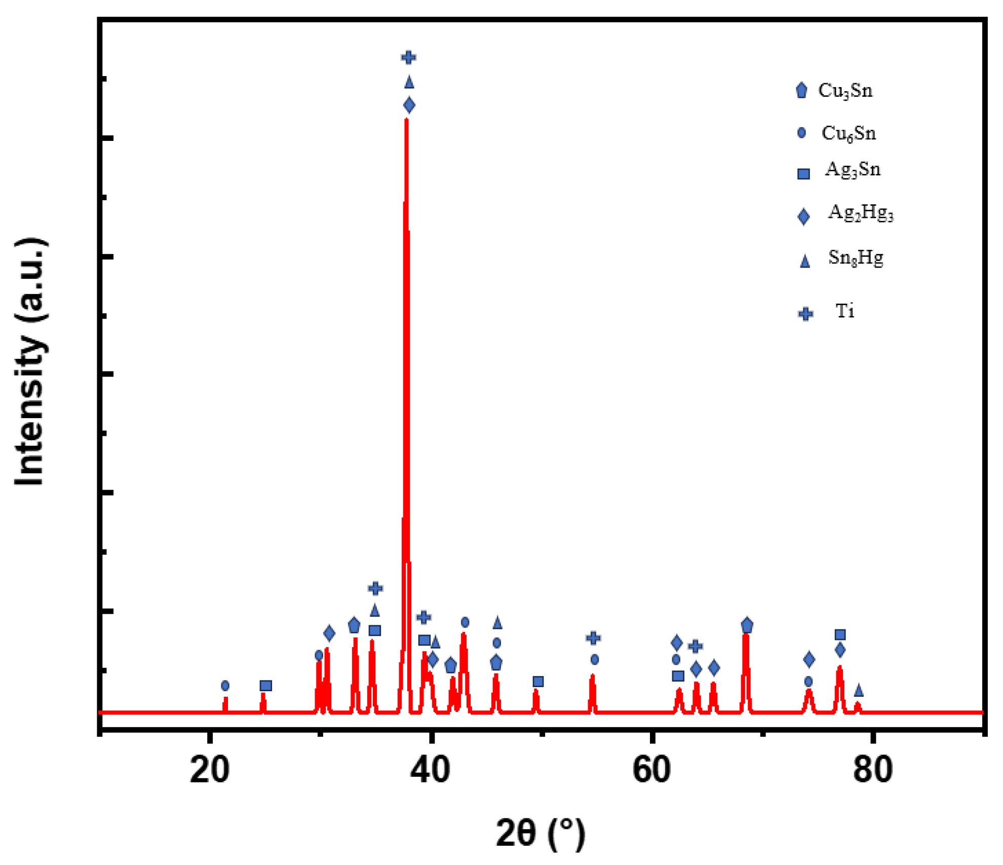
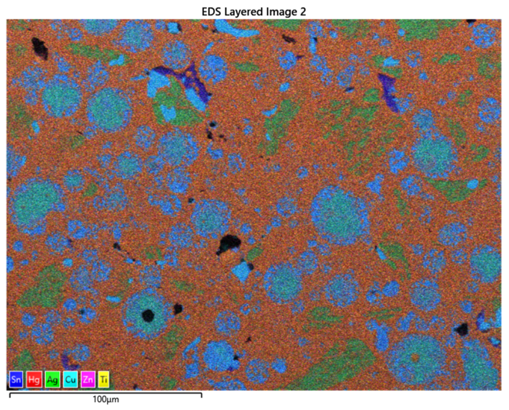
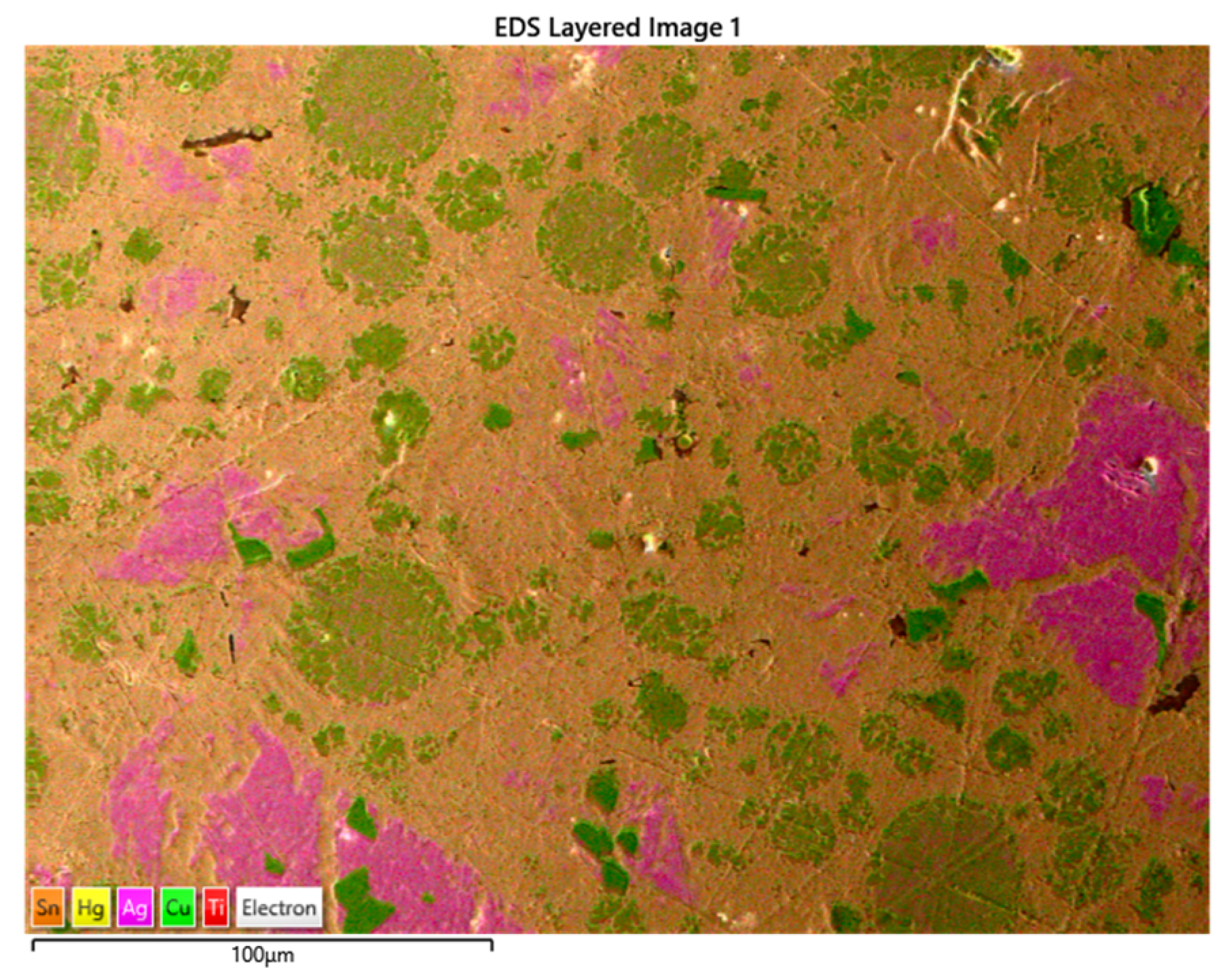
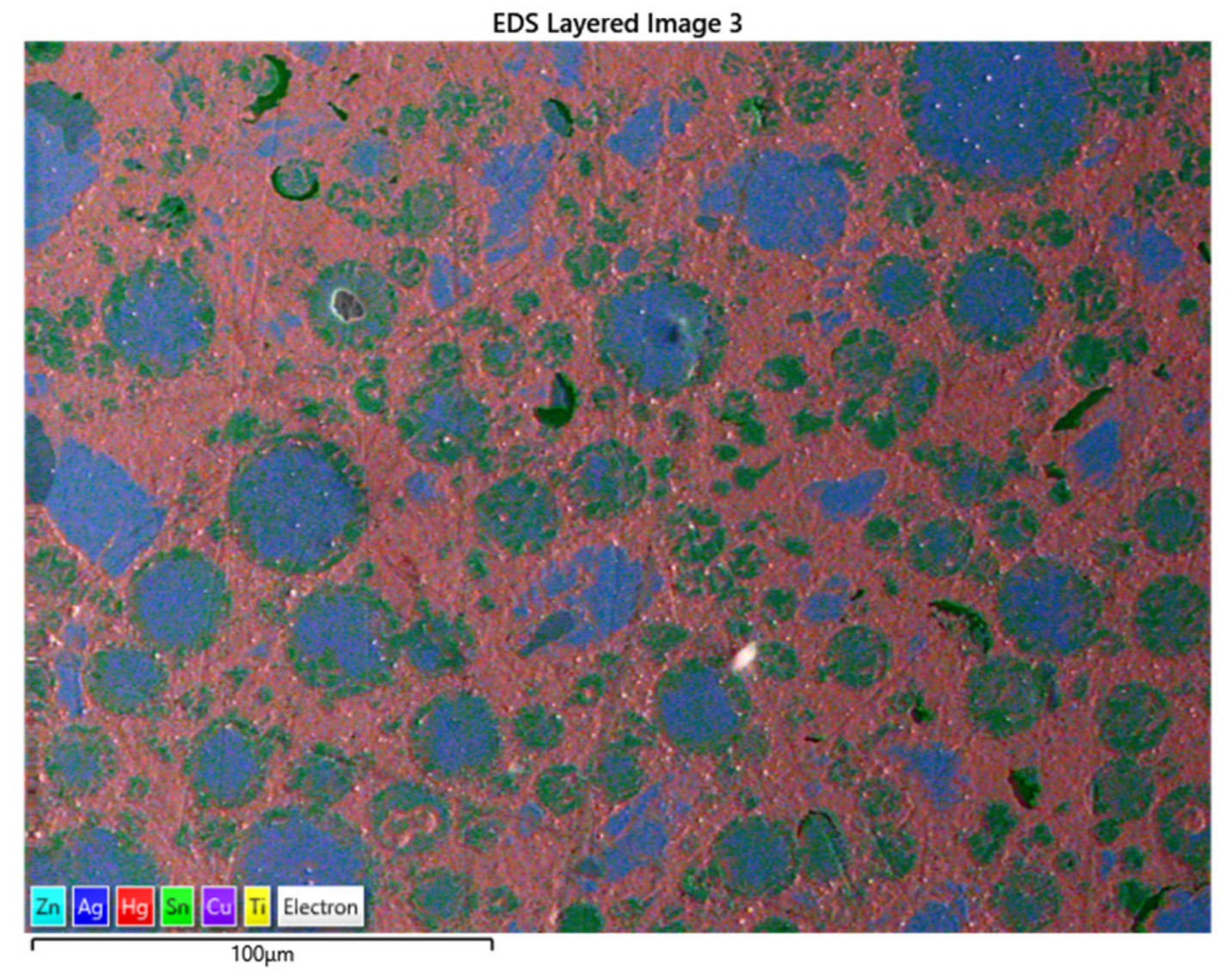
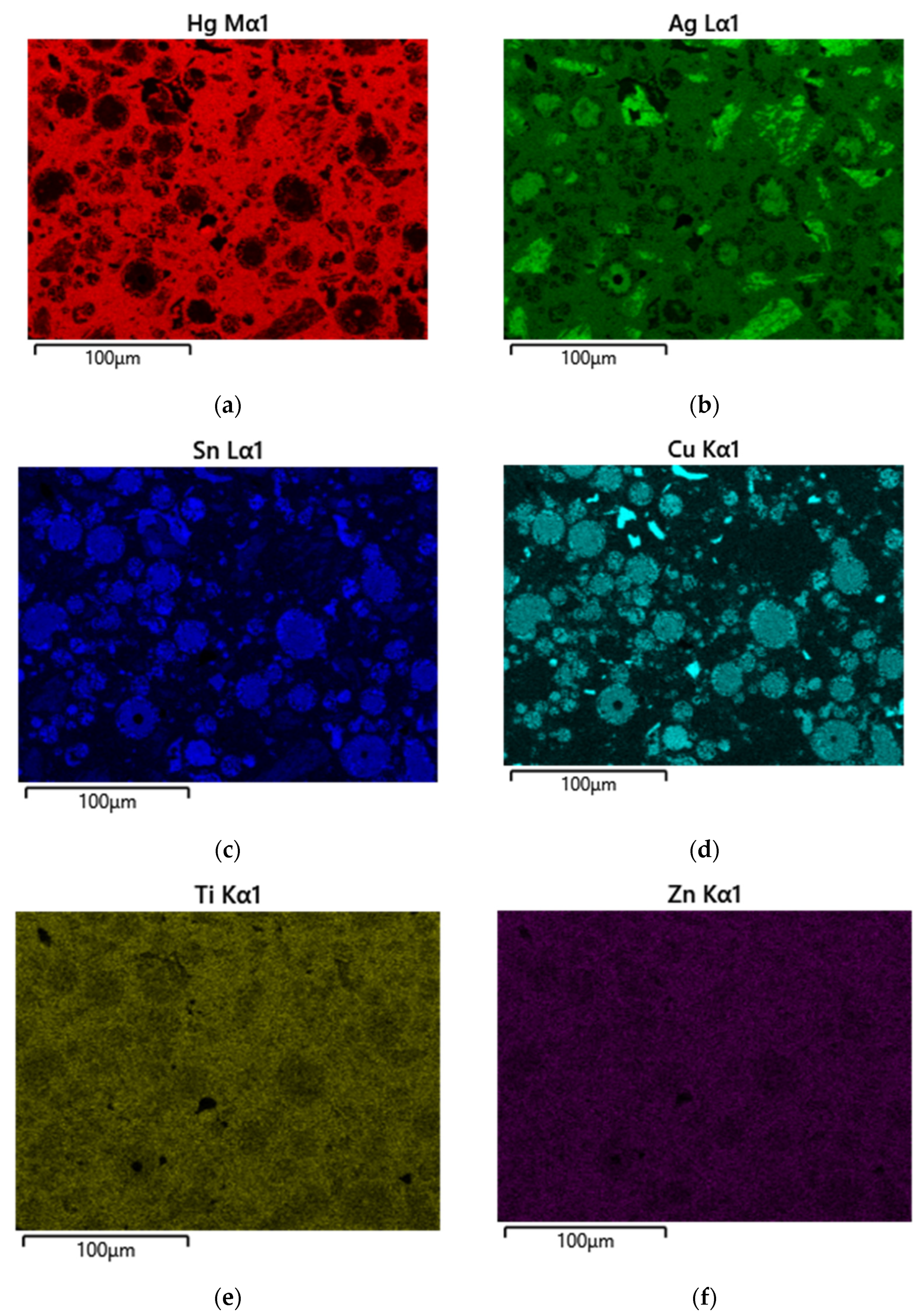
- The γ-phase (Ag3Sn), γ (the strongest phase). It is a closed-pack hexagonal structure, as determined by Nial, Almin, and Westgren (1931). Moreover, Murphy (1926) studied the equilibrium conditions of the silver–tin binary system and confirmed that the γ-phase is of the hexagonal close-pack structure [28,29,30].
- The γ-1 phase (Ag2Hg3), γ1-phase (dominant phase in the set amalgam). The unit cell of this γ-1 phase is of the cubic crystal structure, as determined by G. V. Black in 1895, and appears to exist in the (112) plane [31].
2. Materials and Methods
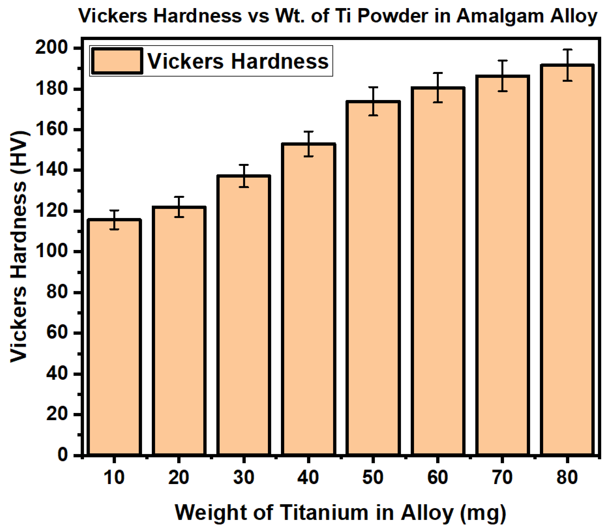
3. Results and Discussion
4. Conclusions
Author Contributions
Funding
Institutional Review Board Statement
Informed Consent Statement
Data Availability Statement
Conflicts of Interest
References
- Moxon, R.; Xu, Z.; Chris-Okoro, I.; Cherono, S.; Kumar, D. Determination and Calculations of Mercury Vapor Concentration and Energy Released from Freshly Condensed Dental Amalgams Having Various Copper Percentages within the Alloy. Materials 2023, 16, 3452. [Google Scholar] [CrossRef]
- Okabe, T.; Yamashita, T.; Nakajima, H.; Berglund, A.; Zhao, L.; Guo, I.; Ferracane, J.L. Reduced mercury vapor release from dental amalgams prepared with binary Hg-In liquid alloys. J. Dent. Res. 1994, 73, 1711–1716. [Google Scholar] [CrossRef]
- Demaree, N.C.; Taylor, D.F. Properties of dental amalgams made from spherical alloy particles. J. Dent. Res. 1962, 41, 890–906. [Google Scholar] [CrossRef]
- Shaini, F.J.; Fleming GJ, P.; Shortall, A.C.C.; Marquis, P.M. A comparison of the mechanical properties of a gallium-based alloy with a spherical high-copper amalgam. Dent. Mater. 2001, 17, 142–148. [Google Scholar] [CrossRef] [PubMed]
- Sun, Y.; Yu, H.; Kesim, M.T.; Alpay, S.P.; Aindow, M. Microstructural stability, defect structures and deformation mechanisms in a Ag3Sn/Cu3Sn alloy. J. Mater. Sci. 2017, 52, 2944–2956. [Google Scholar] [CrossRef]
- Marshall, G.W.; Finkelstein, G.F.; Marshall, S.J.; Greener, E.H. Microstructural changes of dental amalgam by copper additions. J. Oral Rehabil. 1976, 3, 359–370. [Google Scholar] [CrossRef]
- Uçar, Y.; Brantley, W. Biocompatibility of dental amalgams. Biocompat. Dent. Biomater. 2017, 95–111. [Google Scholar] [CrossRef]
- Pihl, C.F.; Beasley, W.M. Compounds Formed in Silver Dental Amalgam. J. Dent. Res. 2016, 47, 418–426. [Google Scholar] [CrossRef] [PubMed]
- Donly, K.J.; Sasa, I.S. Dental Materials Pediatric Dentistry, 6th ed.; Elsevier: Amsterdam, The Netherlands, 2019; pp. 293–303. [Google Scholar] [CrossRef]
- Aerts, A.; Danaci, S.; Prieto, B.G.; Van den Bosch, J.; Neuhausen, J. Evaporation of mercury impurity from liquid lead–bismuth eutectic. J. Nucl. Mater. 2014, 448, 276–281. [Google Scholar] [CrossRef]
- Shi-Duk, L.; Yukuo, T.K.-H.; Osamu, O. Ions Released from Dental Amalgam in contact with titanium. Dent. Mater. J. 2003, 22, 96–110. [Google Scholar]
- Hoque, M.E.; Showva, N.N.; Ahmed, M.; Rashed, A.B.; Sadique, S.E.; El-Bialy, T.; Xu, H. Titanium and Titanium Alloys in Dentistry current trend. Heliyon 2022, 8, 1–6. [Google Scholar] [CrossRef] [PubMed]
- Prajna, P.N.; Sudarshan, K.; Kishore, G.; Deepika, P. Effect of shape of titanium dioxide nanofillers on the properties of dental composites. Odontology 2023, 111, 697–707. [Google Scholar]
- Fairhurst, C.W.; Cohen, J.B. The crystal structures of two compounds found in dental amalgam: Ag2Hg3 and Ag3 Sn. Acta Crystallogr. Sect. B Struct. Crystallogr. Cryst. Chem. 1972, 28, 371–378. [Google Scholar] [CrossRef]
- Yahya, N.; Puspitasari, P.; Latiff, N.R.A. Hardness improvement of dental amalgam using zinc oxide and aluminum oxide nanoparticles. Charact. Dev. Biosyst. Biomater. Advanced Structured Materials. 2013, 29, 9–32. [Google Scholar]
- Patsurakos, A.; Moberg, L.E. Corrosion behavior and microhardness of three amalgams. Eur. J. Oral Sci. 1988, 96, 376–383. [Google Scholar] [CrossRef]
- Javid, M.A.; Rafique, M.; Hussain, A.; Nadeem, K.; Nabi, G.; Mehmood, K.; Ajaz-un-Nabi, M. Synthesis and mechanical properties of dental amalgam. Mater. Today Proc. 2021, 47, S33–S37. [Google Scholar]
- Zhang, B.B.; Zheng, Y.F.; Liu, Y. Effect of Ag on the corrosion behavior of Ti–Ag alloys in artificial saliva solutions. Dent. Mater. 2009, 25, 672–677. [Google Scholar] [CrossRef]
- Slokar, L.; Pranjić, J.; Carek, A. Metallic materials for use in dentistry. The holistic approach to environment. Croat. Sci. Prof. J. 2017, 7, 39–58. [Google Scholar]
- Bauer, S.; Schmuki, P.; Von Der Mark, K.; Park, J. Engineering biocompatible implant surfaces: Part I: Materials and surfaces. Prog. Mater. Sci. 2013, 58, 261–326. [Google Scholar] [CrossRef]
- Shin, D.H. An Experimental Study on The Microhardness of Dental Amalgams. Restor. Dent. Endod. 1982, 8, 89–96. [Google Scholar]
- Chung, K.H.; Hsiao, L.Y.; Lin, Y.S.; Duh, J.G. Morphology and electrochemical behavior of Ag–Cu nanoparticle-doped amalgams. Acta Biomater. 2008, 4, 717–724. [Google Scholar] [CrossRef] [PubMed]
- Ryge, G.; Telford, R.F.; Fairhurst, C.W. Strength and phase formation of dental amalgam. J. Dent. Res. 1957, 36, 986–991. [Google Scholar] [CrossRef] [PubMed]
- Um, C.M.; Kim, Y.H. X-ray diffraction analysis of dental amalgams. J. Korean Dent. Assoc. 1985, 23, 229–235. [Google Scholar]
- Mateer, R.S.; Reitz, C.D. Corrosion of amalgam restorations. J. Dent. Res. 1970, 49, 399–407. [Google Scholar] [CrossRef] [PubMed]
- Koike, M.; Ferracane, J.L.; Adey, J.D.; Fujii, H.; Okabe, T. Initial mercury evaporation from experimental Ag–Sn–Cu amalgams containing Pd. Biomaterials 2004, 25, 3147–3153. [Google Scholar] [CrossRef] [PubMed]
- Vrijhoef, M.M.A.; Driessens, F.C.M. The primary factor of the creep resistance of dispersant amalgams. J. Oral Rehabil. 1975, 2, 165–167. [Google Scholar] [CrossRef] [PubMed]
- Darvell, B.W. Effect of corrosion on the strength of dental silver amalgam. Dent. Mater. 2012, 28, e160–e167. [Google Scholar] [CrossRef] [PubMed]
- Eames, W.B. Preparation and condensation of amalgam with a low mercury-alloy ratio. J. Am. Dent. Assoc. 1959, 58, 78–83. [Google Scholar] [CrossRef] [PubMed]
- Bahari, M.; Oskoee, P.A.; Oskoee, S.S.; Pouralibaba, F.; Ahari, A.M. Mercury release of amalgams with various silver contents after exposure to bleaching agent. J. Dent. Res. Dent. Clin. Dent. Prospect. 2016, 10, 118. [Google Scholar] [CrossRef]
- Johnson, L.B., Jr. X-ray diffraction evidence for the presence of β (Ag Hg) in dental amalgam. J. Biomed. Mater. Res. 1967, 1, 285–297. [Google Scholar] [CrossRef]
- Snapp, K.R.; Boyer, D.B.; Peterson, L.C.; Svare, C.W. The contribution of dental amalgam to mercury in blood. J. Dent. Res. 1989, 68, 780–785. [Google Scholar] [CrossRef] [PubMed]
- Ghatee, M.H.; Karimi, H.; Shekoohi, K. Structural, mechanical and thermodynamical properties of silver amalgam filler: A Monte Carlo simulation study. J. Mol. Liq. 2015, 211, 96–104. [Google Scholar] [CrossRef]
- Espevik, S. Dental amalgam. Annu. Rev. Mater. Sci. 1977, 7, 55–72. [Google Scholar] [CrossRef]
- Davies, R.A.; Ardalan, S.; Mu, W.H.; Tian, K.; Farsaikiya, F.; Darvell, B.W.; Chasse, G.A. Geometric, electronic and elastic properties of dental silver amalgam γ-(Ag3Sn), γ1-(Ag2Hg3), γ2-(Sn8Hg) phases, comparison of experiment and theory. Intermetallics 2010, 18, 756–760. [Google Scholar] [CrossRef]
- Bracho-Troconis, C.; Colon, P.; Bartout, J.D.; Bienvenu, Y. Influence of thermal treatments on Ag Sn Cu powders in order to reduce mercury contents in dental amalgam. J. Mater. Sci. Mater. Med. 2000, 11, 1–9. [Google Scholar] [CrossRef] [PubMed]
- Neme, A.L.; Wagner, W.C.; O’Brien, W.J. Effects of palladium addition on emission of mercury vapor from dental amalgam. Dent. Mater. 1999, 15, 382–389. [Google Scholar] [CrossRef]
- Okabe, T.; Ohmoto, K.; Nakajima, H.; Woldu, M.; Ferracane, J.L. Effect of Pd and in on mercury evaporation. Dent. Mater. J. 1987, 16, 191–199. [Google Scholar] [CrossRef] [PubMed]
- Aaseth, J.; Hilt, B.; Bjørklund, G. Mercury exposure, and health impacts in dental personnel. Environ. Res. 2018, 164, 65–69. [Google Scholar] [CrossRef] [PubMed]
- De Oliveira, M.T.; Pereira, J.R.; Ghizoni, J.S.; Bittencourt, S.T.; Molina, G.O. Effects from exposure to dental amalgam on systemic mercury levels in patients and dental school students. Photomed. Laser Surg. 2010, 28, S111–S114. [Google Scholar] [CrossRef]
- Khwaja, M.A.; Abbasi, M.S. Mercury poisoning dentistry: High-level indoor air mercury contamination at selected dental sites. Rev. Environ. Health. 2015, 29, 29–31. [Google Scholar] [CrossRef]
- Bernhoft, R.A. Mercury toxicity and treatment: A review of the literature. J. Environ. Public Health 2012, 2012, 460508. [Google Scholar] [CrossRef] [PubMed]
- Jamil, N.; Baqar, M.; Ilyas, S.; Qadir, A.; Arslan, M.; Salman, M.; Ahsan, N.; Zahid, H. Use of Mercury in dental silver amalgam: An occupational and environmental assessment. BioMed Res. Int. 2016, 6126385. [Google Scholar] [CrossRef] [PubMed]
- Dhar, V.; Hsu, K.L.; Coll, J.A.; Ginsberg, E.; Ball, B.M.; Chhibber, S.; Johnson, M.; Kim, M.; Modaresi, N.; Tinanoff, N. Evidence-base update of pediatric dental restoration procedures; dental materials. J. Clin. Pediatr. Dent. 2015, 39, 303–310. [Google Scholar] [CrossRef] [PubMed]
- Kopperud, S.E.; Tveit, A.B.; Gaarden, T.; Sandvik, L.; Espelid, I. Longevity of posterior dental restoration and restorations and reasons for failure. Eur. J. Oral. Sci. 2012, 120, 539–548. [Google Scholar] [CrossRef]
- Kall, J.; Just, A.; Aschner, M. What is the risk? Dental amalgam, mercury exposure, and human health risks throughout the life span. In Epigenetics, the Environment, and Children’s Health Across Lifespans; Hollar, D., Ed.; Springer: Cham, Switzerland, 2016; pp. 159–206. [Google Scholar]
- Powell, L.V.; Johnson, G.H.; Bales, D.J. Effect of Admixed Indium on Mercury Vapor Release from Dental Amalgam. J. Dent. Res. 2016, 68, 1231–1233. [Google Scholar] [CrossRef]
- Arizona Instrument Inc, Jerome J505 Mercury Vapor Analyzer, Operation Manual, Chandler, Arizona, USA. 2015, pp. 1–28. Available online: https://www.raecorents.com/amfile/file/download/file/238/product/633/ (accessed on 21 October 2023).


| Amalgam | Silver | Tin | Copper | Zinc | Mercury |
|---|---|---|---|---|---|
| Dispersalloy (Before) | 69.5% | 17.70% | 11.80% | 1.00% | - |
| (After) | 34.75% | 8.85% | 5.90% | 0.50% | 50.00% |
| Sybralloy (Before) | 40.00% | 26.80% | 33.00% | 0.20% | - |
| (After) | 18.4% | 12.33% | 15.18% | 0.09% | 46.00% |
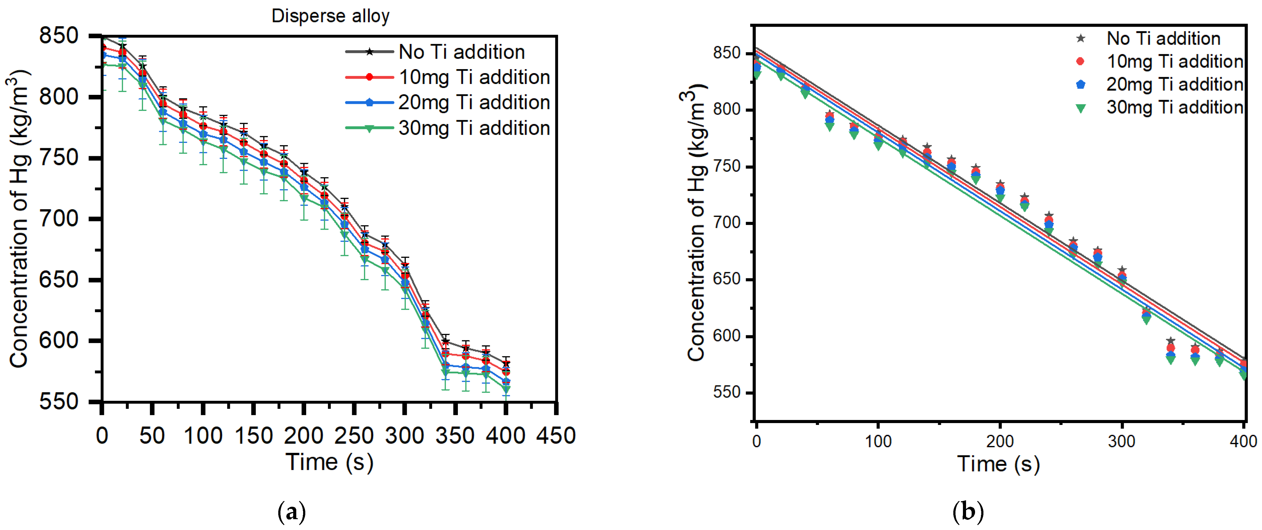
| Time (s) | Disperse Alloy without Titanium (kg/m3) | 10 mg Ti (kg/m3) | 20 mg Ti (kg/m3) | 30 mg Ti (kg/m3) |
|---|---|---|---|---|
| 0 | 846.085 | 840.912 | 837.684 | 831.746 |
| 20 | 838.5543 | 836.753 | 834.506 | 830.610 |
| 40 | 821.8564 | 819.484 | 817.615 | 814.582 |
| 60 | 796.6544 | 794.553 | 790.832 | 786.064 |
| 80 | 787.0785 | 785.748 | 781.475 | 778.509 |
| 100 | 780.6533 | 776.378 | 772.671 | 768.917 |
| 120 | 773.851 | 771.637 | 768.083 | 762.455 |
| 140 | 767.3113 | 762.788 | 758.149 | 752.788 |
| 160 | 756.4582 | 753.443 | 749.743 | 744.506 |
| 180 | 748.9329 | 745.518 | 741.826 | 738.884 |
| 200 | 734.831 | 731.667 | 728.925 | 722.617 |
| 220 | 723.0086 | 719.471 | 716.337 | 714.841 |
| 240 | 706.5386 | 702.661 | 698.586 | 692.789 |
| 260 | 684.1459 | 680.305 | 677.990 | 672.447 |
| 280 | 675.8653 | 673.548 | 669.812 | 663.599 |
| 300 | 658.6462 | 653.709 | 650.716 | 647.443 |
| 320 | 623.0678 | 620.806 | 617.448 | 614.909 |
| 340 | 595.9563 | 589.642 | 583.067 | 579.712 |
| 360 | 590.5342 | 587.812 | 581.549 | 578.801 |
| 380 | 586.6308 | 583.904 | 580.172 | 577.759 |
| 400 | 577.8899 | 574.817 | 569.553 | 565.762 |
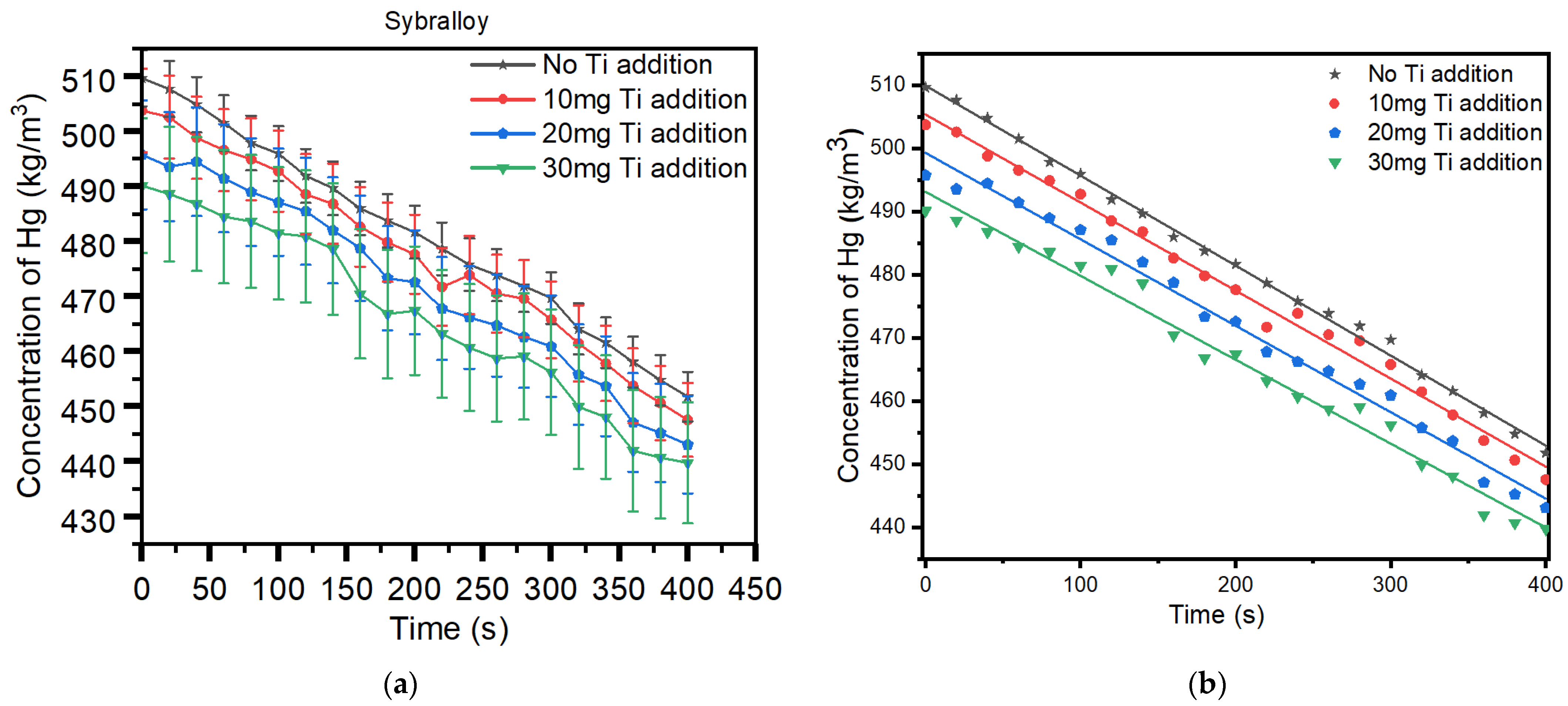
| Time (s) | Sybralloy without Titanium (kg/m3) | 10 mg Ti (kg/m3) | 20 mg Ti (kg/m3) | 30 mg Ti (kg/m3) |
|---|---|---|---|---|
| 0 | 509.658 | 503.772 | 495.746 | 490.128 |
| 20 | 507.684 | 502.607 | 493.581 | 488.607 |
| 40 | 504.855 | 498.818 | 494.473 | 486.790 |
| 60 | 501.534 | 496.573 | 491.455 | 484.482 |
| 80 | 497.867 | 494.914 | 488.964 | 483.617 |
| 100 | 495.953 | 492.763 | 487.109 | 481.448 |
| 120 | 491.894 | 488.548 | 485.476 | 480.891 |
| 140 | 489.657 | 486.817 | 481.990 | 478.609 |
| 160 | 485.967 | 482.636 | 478.754 | 470.445 |
| 180 | 483.746 | 479.837 | 473.305 | 466.781 |
| 200 | 481.676 | 477.649 | 472.568 | 467.408 |
| 220 | 478.656 | 471.715 | 467.783 | 463.172 |
| 240 | 475.767 | 473.883 | 466.191 | 460.688 |
| 260 | 473.866 | 470.499 | 464.736 | 458.711 |
| 280 | 471.860 | 469.565 | 462.643 | 459.073 |
| 300 | 469.699 | 465.772 | 460.908 | 456.189 |
| 320 | 464.076 | 461.439 | 455.757 | 449.909 |
| 340 | 461.568 | 457.808 | 453.664 | 448.055 |
| 360 | 458.063 | 453.733 | 447.067 | 441.927 |
| 380 | 454.768 | 450.614 | 445.196 | 440.675 |
| 400 | 451.755 | 447.547 | 443.063 | 439.742 |
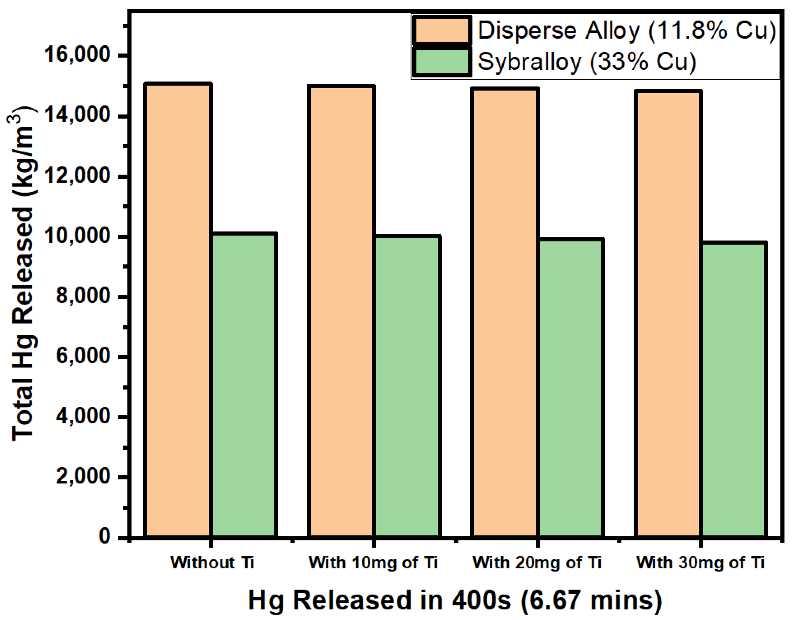
| Disperse Alloy—11.8% Cu (kg/m3) | Sybralloy—33% Cu (kg/m3) | |
|---|---|---|
| Total Concentration of Hg Without Titanium | 15,074.5499 | 10,110.569 |
| Total Concentration of Hg with 10 mg of Titanium | 15,005.556 | 10,027.509 |
| Total Concentration of Hg with 20 mg of Titanium | 14,926.739 | 9910.429 |
| Total Concentration of Hg with 30 mg of Titanium | 14,839.74 | 9797.347 |
| Slope | DF | Sum of Squares | Mean Square | F Value | Prob > F | ||
|---|---|---|---|---|---|---|---|
| Disperse Alloy Without Titanium (R2 = 0.97621) | −0.686 | Model | 1 | 144,988.2712 | 144,988.2712 | 779.48714 | 1.11 × 10⁻¹⁶ |
| Error | 19 | 3534.08927 | 186.0047 | ||||
| Total | 20 | 148,522.3605 | |||||
| 10 mg Ti (R2 = 0.97626) | −0.688 | Model | 1 | 145,777.7609 | 145,777.7609 | 781.45524 | 1.11 × 10⁻¹⁶ |
| Error | 19 | 3544.38401 | 186.54653 | ||||
| Total | 20 | 149,322.1449 | |||||
| 20 mg Ti (R2 = 0.97459) | −0.693 | Model | 1 | 147,814.1458 | 147,814.1458 | 728.70775 | 1.11 × 10⁻¹⁶ |
| Error | 19 | 3854.03994 | 202.84421 | ||||
| Total | 20 | 151,668.1857 | |||||
| 30 mg Ti (R2 = 0.97563) | −0.689 | Model | 1 | 146,448.5597 | 146,448.5597 | 760.55004 | 1.11 × 10⁻¹⁶ |
| Error | 19 | 3658.56614 | 192.55611 | ||||
| Total | 20 | 150,107.1259 |
| Slope | DF | Sum of Squares | Mean Square | F Value | Prob > F | ||
|---|---|---|---|---|---|---|---|
| Sybralloy Without Titanium (R2 = 0.99721) | −0.143 | Model | 1 | 6265.08552 | 6265.08552 | 6802.26144 | 0 |
| Error | 19 | 17.49957 | 0.92103 | ||||
| Total | 20 | 6282.58509 | |||||
| 10 mg Ti (R2 = 0.99216) | −0.14 | Model | 1 | 5995.10186 | 5995.10186 | 2405.08619 | 0 |
| Error | 19 | 47.36085 | 2.49268 | ||||
| Total | 20 | 6042.46271 | |||||
| 20 mg Ti (R2 = 0.98835) | −0.137 | Model | 1 | 5782.32621 | 5782.32621 | 1611.21658 | 0 |
| Error | 19 | 68.18711 | 3.5888 | ||||
| Total | 20 | 5850.51332 | |||||
| 30 mg Ti (R2 = 0.9834) | −0.133 | Model | 1 | 5445.9406 | 5445.9406 | 1125.60529 | 0 |
| Error | 19 | 91.92643 | 4.83823 | ||||
| Total | 20 | 5537.86702 |
Disclaimer/Publisher’s Note: The statements, opinions and data contained in all publications are solely those of the individual author(s) and contributor(s) and not of MDPI and/or the editor(s). MDPI and/or the editor(s) disclaim responsibility for any injury to people or property resulting from any ideas, methods, instructions or products referred to in the content. |
© 2024 by the authors. Licensee MDPI, Basel, Switzerland. This article is an open access article distributed under the terms and conditions of the Creative Commons Attribution (CC BY) license (https://creativecommons.org/licenses/by/4.0/).
Share and Cite
Moxon, R.; Xu, Z.; Tettey, F.; Chris-Okoro, I.; Kumar, D. Dental Metal Matrix Composites: The Effects of the Addition of Titanium Nanoparticle Particles on Dental Amalgam. Materials 2024, 17, 1662. https://doi.org/10.3390/ma17071662
Moxon R, Xu Z, Tettey F, Chris-Okoro I, Kumar D. Dental Metal Matrix Composites: The Effects of the Addition of Titanium Nanoparticle Particles on Dental Amalgam. Materials. 2024; 17(7):1662. https://doi.org/10.3390/ma17071662
Chicago/Turabian StyleMoxon, Ryan, Zhigang Xu, Felix Tettey, Ikenna Chris-Okoro, and Dhananjay Kumar. 2024. "Dental Metal Matrix Composites: The Effects of the Addition of Titanium Nanoparticle Particles on Dental Amalgam" Materials 17, no. 7: 1662. https://doi.org/10.3390/ma17071662
APA StyleMoxon, R., Xu, Z., Tettey, F., Chris-Okoro, I., & Kumar, D. (2024). Dental Metal Matrix Composites: The Effects of the Addition of Titanium Nanoparticle Particles on Dental Amalgam. Materials, 17(7), 1662. https://doi.org/10.3390/ma17071662