Article

Unraveling Broadband Near-Infrared Luminescence in Cr3+-Doped Ca3Y2Ge3O12 Garnets: Insights from First-Principles Analysis

1
School of Science and Optoelectronic Engineering, Chongqing University of Posts and Telecommunications, Chongqing 400065, China
2
Center of Innovative Development of Science and New Technologies, National Academy of Sciences of Tajikistan, Dushanbe 734025, Tajikistan
3
Institute of Plasma Physics of the Czech Academy of Sciences, U Slovanky 2525/1a, 18200 Prague, Czech Republic
4
Current Lighting Solutions LLC., 1099 Ivanhoe Road, Cleveland, OH 44110, USA
5
Centre of Excellence for Photoconversion, Vinča Institute of Nuclear Sciences—National Institute of the Republic of Serbia, University of Belgrade, 11351 Belgrade, Serbia
6
Institute of Physics, University of Tartu, W. Ostwald Str. 1, 50411 Tartu, Estonia
7
Faculty of Science and Technology, Jan Długosz University, Armii Krajowej 13/15, PL-42200 Częstochowa, Poland
8
Academy of Romanian Scientists, 3 Ilfov, 050044 Bucharest, Romania
9
Ministry of Education Key Laboratory of Bioinorganic and Synthetic Chemistry, State Key Laboratory of Optoelectronic Materials and Technologies, School of Chemistry, Sun Yat-Sen University, Guangzhou 510275, China
*
Authors to whom correspondence should be addressed.
These authors contributed equally to this work.
Materials 2024, 17(7), 1709; https://doi.org/10.3390/ma17071709
Submission received: 6 March 2024 / Revised: 1 April 2024 / Accepted: 4 April 2024 / Published: 8 April 2024

Abstract

:
In this study, we conducted an extensive investigation into broadband near-infrared luminescence of Cr3+-doped Ca3Y2Ge3O12 garnet, employing first-principles calculations within the density functional theory framework. Our initial focus involved determining the site occupancy of Cr3+ activator ions, which revealed a pronounced preference for the Y3+ sites over the Ca2+ and Ge4+ sites, as evidenced by the formation energy calculations. Subsequently, the geometric structures of the excited states 2E and 4T2, along with their optical transition energies relative to the ground state 4A2 in Ca3Y2Ge3O12:Cr3+, were successfully modeled using the ΔSCF method. Calculation convergence challenges were effectively addressed through the proposed fractional particle occupancy schemes. The constructed host-referred binding energy diagram provided a clear description of the luminescence kinetics process in the garnet, which explained the high quantum efficiency of emission. Furthermore, the accurate prediction of thermal excitation energy yielded insights into the thermal stability of the compound, as illustrated in the calculated configuration coordinate diagram. More importantly, all calculated data were consistently aligned with the experimental results. This research not only advances our understanding of the intricate interplay between geometric and electronic structures, optical properties, and thermal behavior in Cr3+-doped garnets but also lays the groundwork for future breakthroughs in the high-throughput design and optimization of luminescent performance and thermal stability in Cr3+-doped phosphors.

1. Introduction

Among various crystalline solids that are used for optical applications, the compounds with the cubic garnet structure are of special importance and significance. This is a very large family of compounds, whose structure offers numerous opportunities for chemical composition alterations, such as creating solid solutions and/or introducing optically active impurity ions. The garnets can easily accommodate transition metal and rare earth ions, rendering them potential candidates for various optical applications. There has been a recent surge in interest in Cr3+-doped garnets due to their broadband near-infrared (NIR) luminescence, with potential applications in medical diagnostics, food analysis, horticultural lighting, night vision, etc. [1,2,3,4,5,6,7]. For instance, Ca3Y2Ge3O12: Cr3+, synthesized via the solid-state reaction method, exhibits a broadband NIR emission spanning from 700 to 1100 nm, with a peak centered at 800 nm [8]. This emission spectrum aligns perfectly with the absorption frequencies of hydrogen-containing groups X-H (where X=C, N and O), making it an ideal non-destructive testing tool for food safety applications [9]. Theoretical investigations that provide a comprehensive understanding of the luminescence mechanisms are necessary for enabling the next generation of highly efficient Cr3+-activated garnet phosphors.
Extensive systematic spectroscopic analyses have been conducted on garnet crystals doped with Cr3+ ions, utilizing the well-established Tanabe–Sugano energy level diagram for 3d3 ions in solids [10,11]. Additionally, the exchange charge model within the framework of semi-empirical crystal-field (CF) theory can provide valuable insights into the relationship between the spectroscopic properties of Cr3+ ions and their local coordination environments [12]. However, the number of first-principles studies focusing on Cr3+-doped garnets within the density functional theory (DFT) framework remains relatively small compared to experimental and semi-empirical theoretical papers on the same topic. This is primarily due to the rather complicated structure of garnets, characterized by a large number of atoms in a unit cell, thus incurring high computational costs. Moreover, most reported DFT calculations on Cr3+-doped compounds have predominantly concentrated on ground-state properties, as exemplified by the case of Ca4ZrGe3O12: Cr3+ discussed in reference [13]. However, such studies are limiting since they fail to provide the knowledge of Cr3+ ions’ 2E and 4T2 excited states, which are important in the design of new useful phosphors. Fortunately, Duan et al. [14,15,16] have successfully applied the ΔSCF-DFT method with non-Aufbau occupations on Kohn–Sham (KS) orbitals to model the excited states 2E and 4T2 of Cr3+ ions doped in some oxides. However, such calculations probing Cr3+-doped garnets are lacking.
The main goal of the present work is to provide a deeper fundamental understanding of the excited states and the associated luminescence phenomena in Cr3+-doped garnets by integrating the first-principles ΔSCF-DFT technique. This integration is essential for addressing the aforementioned gaps in our knowledge. Specifically, we chose the garnet Ca3Y2Ge3O12: Cr3+ as a case study. In this paper, we conducted an extensive theoretical analysis of its structural, electronic, and optical properties. Special attention was devoted to factors such as the site occupancy, luminescence mechanism, and thermal stability of Cr3+ dopants within this garnet matrix. Furthermore, we delved into the challenge of achieving calculation convergence in modeling the excited 4T2 state of Cr3+ ions, employing the approximation of the single-electron configuration t2 2ge1 g, utilizing either the DFT+U or hybrid DFT method. Notably, our previous investigation encountered a computational breakdown when describing the excited 4T2 state of Mn4+ ions in K2SiF6 due to the significant mixing between the constrainedly occupied and unoccupied 3d KS orbitals in the hybrid DFT calculations [17]. Consequently, these predictive calculations and technique development can be readily applied to other systems doped with Cr3+ ions, thus offering potential for the high-throughput design of Cr3+-doped NIR materials.
This paper is organized as follows: Section 2 contains a description of the calculation method. Section 3 contains all obtained results and their analysis. Finally, the paper is concluded with a summary of our findings.

2. Method of Calculations

Our study employed first-principles calculations within the DFT framework, utilizing the Vienna ab initio simulation package (VASP, version 5.4.4.) [18]. Geometric structure relaxations and defect formation calculations were conducted using the Perdew–Burke–Ernzerhof (PBE) functional [19], incorporating an empirical U value (Ueff = 4.0 eV) specifically tailored for the Cr3+-3d orbitals [14,20]. The electronic structure and optical transitions of both neat and Cr3+-doped Ca3Y2Ge3O12 were calculated using the hybrid functional of PBE0 with an additional 25% Hartree–Fock exchange [21]. The treatment of semicore electrons for Ca (3s23p64s2), Y (4s24p65s24d1), Ge (4s23d104p2), O (2s22p4), and Cr (3p63d54s1) was explicitly addressed using the projector augmented-wave pseudopotentials [22,23]. Modeling Cr3+ defects in Ca3Y2Ge3O12 required a supercell containing 160 atoms, with one Y3+/Ca2+/Ge4+ ion substituted by a Cr3+ ion. Sampling the Brillouin zone involved a single k-point Γ for the total energy and relaxation calculations of the constructed supercell, while a 3 × 3 × 3 k-points mesh, based on the Monkhorst–Pack scheme [24], was employed for the host’s unit cell. For both the neat and doped systems, the closed-shell and spin-polarized DFT calculation forms were applied, respectively. A plane-wave basis cutoff energy of 520 eV was employed, with convergence criteria set at 10−6 eV for electronic energy minimization and 0.01 eV/Å for Hellman–Feynman forces on each atom.
The formation energy of a defect X in the charge state of q can be determined as follows [25]: E f X q = E t o t X q E t o t b u l k i n i μ i + q E F , where Etot[Xq] and Etot[bulk] represent the calculated total energies of the defective and perfect supercells, respectively. The variables ni, μI, and EF correspond to the change in the atom number of element i (added if ni > 0 or removed if ni < 0 with respect to the perfect supercell), the chemical potential of species i, and the Fermi energy level, respectively. To account for image charge interaction at periodic boundary conditions and changes in electrostatic potential caused by the defect, the total energies of charged defects were corrected using the method proposed by Durrant et al. [26]. The charge transition level of a defect X from its charged states q to q′ (where q > q′) can be assessed as ε(q/q′) = (Etot[Xq′] − Etot[Xq])/(qq′) − EVBM, where EVBM represents the energy of the host’s valence band maximum (VBM).
The standard ΔSCF-DFT procedure [27,28] was employed to model the excited states 2E and 4T2 of Cr3+ ions in Ca3Y2Ge3O12. These states correspond to a spin flip of one t2g electron and a transition of the KS orbital from t2g to eg, respectively. Modeling the excited state 2E presented no challenges, although the 4A2-2E optical transition energy required adjustment by a scaling factor of 1.5 compared to the DFT-generated value due to the spin contamination effect between the ground 4A2 and excited 2E states [14]. However, it proved to be challenging to represent the excited state 4T2 in Ca3Y2Ge3O12:Cr3+. The constrained separation of a pair of electrons and holes to the lowest eg and the highest t2g KS orbitals led to a significant calculation convergence issue in the DFT+U and hybrid DFT calculations. This is not surprising, given that the narrow t2g-eg energy gap of Cr3+ ions in Ca3Y2Ge3O12 intensifies the mixing between the lowest occupied eg and the highest unoccupied t2g KS orbitals, which pushes the calculations towards collapse. This problem is exacerbated by the fact that many Cr3+-doped garnets with broadband emission are associated with the weak CF case [29]. Considering that the structural disparity between the ground 4A2 and excited 4T2 states of Cr3+ ions primarily stems from the distinction in the electronic density profiles of the 3d-t2g and eg single-electron states, it is imperative to maintain the single-electron configuration t2g2eg1 for modeling the excited state 4T2 of the Cr3+ dopants. However, the two t2g electrons can partially infiltrate into the highest empty t2g KS orbitals to counteract the approach of the lowest occupied eg KS orbital. Therefore, in this study, we proposed two sets of fractional particle occupancy schemes to characterize the geometric structure of the Cr3+ 4T2 excited state in Ca3Y2Ge3O12, as illustrated in Figure 1. In Scheme 1, one of the two t2g electrons is uniformly distributed among the highest two t2g KS orbitals, while the other occupies the lowest t2g KS orbital entirely. In Scheme 2, the allocation of the two t2g electrons is straightforward, with equal distribution among the three t2g KS orbitals.
Slater’s transition-state method [30,31] was employed to estimate the 4A2-4T2 excitation and emission energies, respectively, at the equilibrium geometric structures of the ground 4A2 and excited 4T2 states. This process involves examining the disparities in energy between the lowest eg and highest t2g KS orbitals in the density of states (DOS) diagrams obtained from such calculations based on the single-electron configuration t2g2.5eg0.5, as depicted in Figure 1. The associated zero-phonon line (ZPL) energy can be readily determined by applying the Franck–Condon principle. Additionally, the Stokes shift energy can be calculated by evaluating the difference between the excitation and emission energies of the corresponding optical transitions.

3. Results and Discussion

3.1. Ground States of Both Neat and Cr3+-Doped Ca3Y2Ge3O12

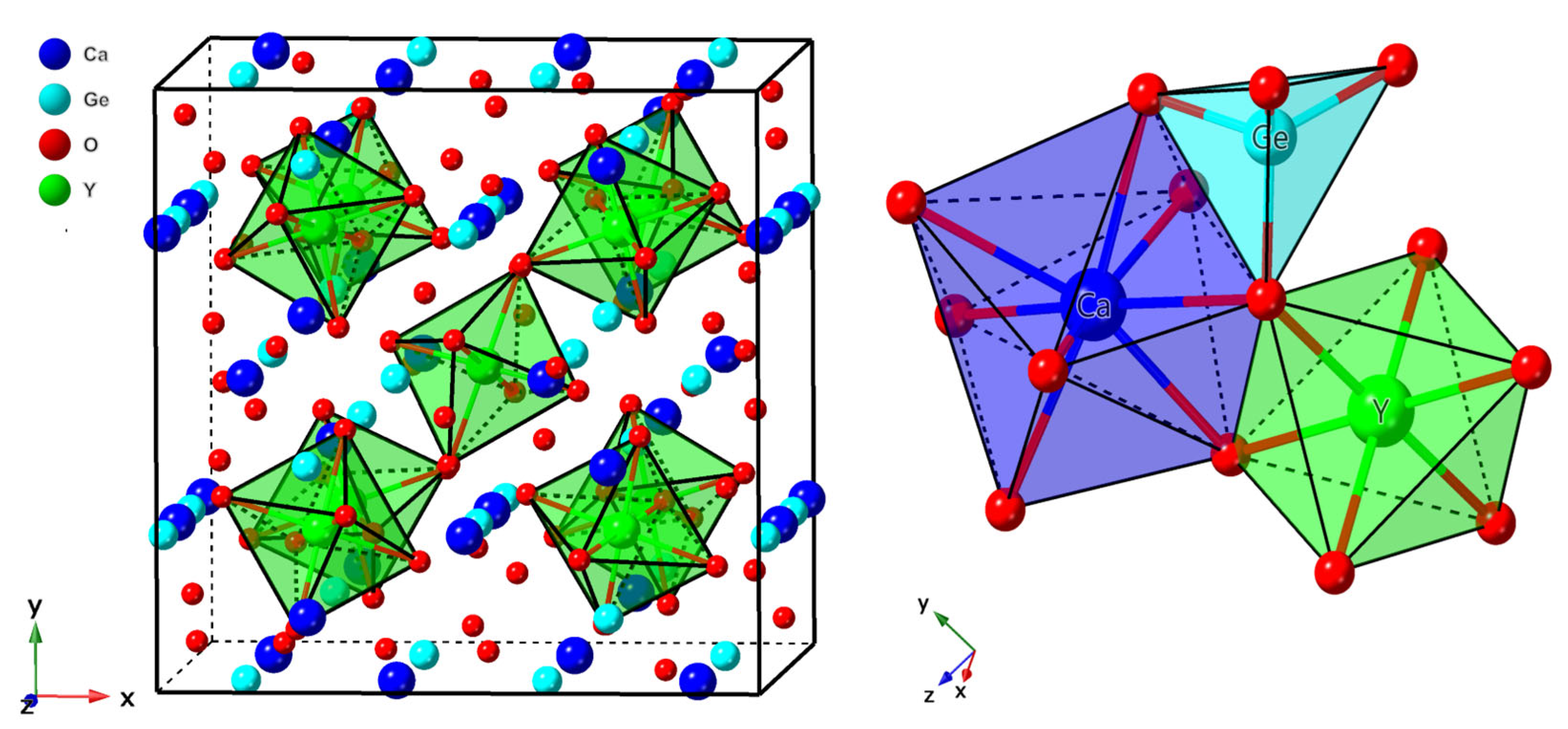
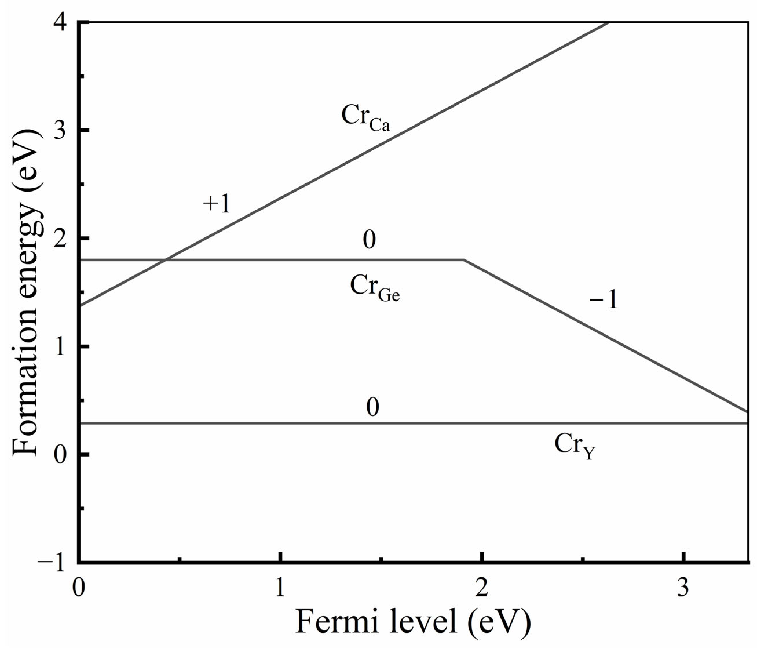
3.1.1. Structural Properties and Defect Site Occupancy

Ca3Y2Ge3O12 crystallizes in the conventional cubic garnet structure, with the I a 3 ¯ d space group and an experimental lattice constant of 12.8059 Å [32]. Within this crystalline framework, the coordination environments of the constituent cations manifest intriguing symmetries and spatial arrangements, as shown in Figure 2. Specifically, the Y3+ ions occupy octahedral coordination sites, characterized by point group symmetry S6, wherein each Y3+ ion is coordinated with six O2− ions at an equidistant Y-O distance of 2.234 Å. In contrast, the Ge4+ ions reside in tetrahedral sites, displaying point group symmetry S4, with a coordinated arrangement of four O2− ions at an identical distance of 1.766 Å. Meanwhile, the Ca2+ ions are found within dodecahedral coordination environments, distinguished by point group symmetry D2, each surrounded by eight neighboring O2− ions. Notably, such a coordination environment results in two distinct Ca-O distances (2.469 and 2.560 Å).
The calculated structural data for the Ca3Y2Ge3O12 host, encompassing lattice constants, internal anion position, unit cell volume, and bond lengths of Y3+-O2−, Ge4+-O2− and Ca2+-O2−, demonstrate substantial agreement with the earlier-discussed experimental descriptions, as outlined in Table 1. The observed slight overestimation is ascribed to the inherent characteristics of the generalized gradient approximation employed in the PBE functional. Considering the ionic radius difference between Cr3+ dopants and the three substitutional sites available [33], it is anticipated that the introduction of Cr3+ at the Y3+ and Ca2+ sites will induce a contraction in their local coordination environments, while the opposite effect is expected at the Ge4+ sites. The Cr3+-O2− bond lengths and the unit cell volume changes upon Cr3+ doping at the three cationic sites, calculated using the PBE+U method, strongly corroborate this empirical conclusion, as evidenced by the data comparisons presented in Table 1. The experimentally refined unit cell volume after Cr3+ doping tends to decrease compared to the host case (refer to Figure 2c in the reference [8]). This, combined with the calculated findings, suggests a preference for Cr3+ dopants to substitute at Y3+ and Ca2+ sites over the Ge4+ sites. A further inference can be drawn, indicating that the Y3+ sites are more accommodating to Cr3+ ions than the Ca2+ sites, owing to the closer alignment of ionic radii between Y3+ and Cr3+ ions within a six-coordinated-ligand environment.
To conclusively determine the preferential site occupancy of Cr3+ ions in Ca3Y2Ge3O12, we calculated the formation energies of Cr dopants at the three cationic sites within the PBE+U framework. In this study, the chemical potential for oxygen atoms was established by considering a gas of O2 molecules, expressed as μ O = 1 2 E O 2 g a s + Δ μ O . Here, E O 2 g a s represents the calculated total energy per formula unit for O2 gas, and ΔμO is related to the contribution arising from gas partial pressure (P) and sintering temperature (T). Under the specified experimental conditions (T = 1450 °C and P = 1 atm [8]), ΔμO was determined as −2.051 eV, following the formula expression provided in the reference [34]. The chemical potentials of other atoms (Ca, Y, Ge, and Cr) were straightforwardly derived from the calculated total energies per formula unit of their respective bulk binary oxides, based on the obtained oxygen chemical potential. These are determined by the following equations: μ C a = E C a O b u l k μ O , μ Y = 1 / 2   ( E Y 2 O 3 b u l k 3 μ O ) , μ G e = E G e O 2 [ b u l k ] 2 μ O , and μ C r = 1 / 2   ( E C r 2 O 3 b u l k 3 μ O ) . Figure 3 illustrates the formation energies of Cr ions substituting at the three cationic sites as a function of Fermi energy. Inspection of Figure 3 reveals that the charge state of Cr ions located at the Ge4+ sites undergoes a transition from “+4” to “+3” as the Fermi energy increases. This aligns with the common understanding in coordination chemistry, where transition metal ions with a 3d3 electronic configuration tend to be oxidized at a tetrahedral site in the absence of additional constraints from physics or chemistry. In contrast, those occupying the Ca2+ and Y3+ sites consistently maintain a “+3” charge state. Simultaneously, the formation energy of Cr3+ ions substituting the Y3+ sites consistently remains lower than those in the Ca2+ and Ge4+ sites. Consequently, defects involving Cr ions substituting at the Y3+ sites dominate, and the charge state of Cr ions is predominantly “+3” in Ca3Y2Ge3O12. This theoretical fact is fully confirmed by the experimental XRD analysis reported previously [8] and aligns with the calculated structural properties of Ca3Y2Ge3O12: Cr3+ discussed above. Hereafter, if not specifically emphasized, we exclusively focus on the case wherein Cr3+ ions occupy the Y3+ sites in Ca3Y2Ge3O12 for the description of the structural, electronic, and optical properties of Ca3Y2Ge3O12: Cr3+.
Despite the results obtained by Cui et al. [9], only the results for a single Cr3+ location in the host (at the Y3+ site) are shown here, which is based on the Cr3+ preference to occupy the octahedral sites in crystalline solids (see Figure 3).

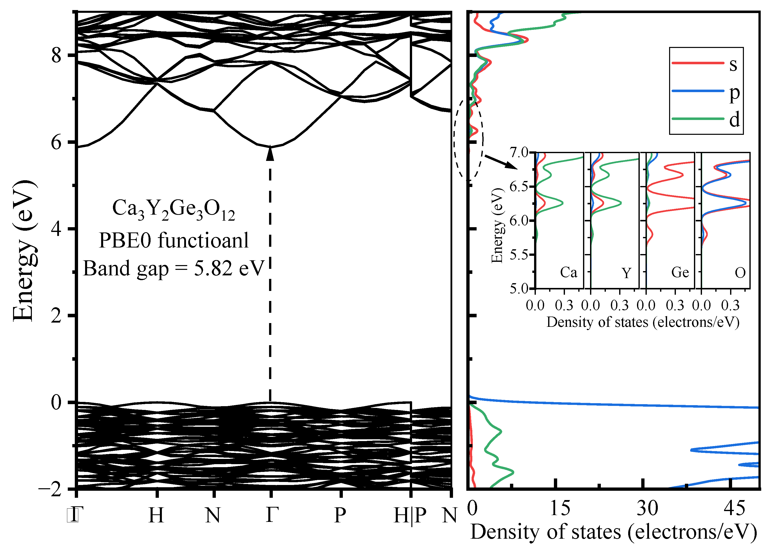
3.1.2. Electronic Properties

The band structure, along with the DOSs, were computed for pristine Ca3Y2Ge3O12 utilizing the PBE0 functional, taking into account the optimized geometric structure of the host, as illustrated in Figure 4. The calculated band gap displays a direct character and measures 5.82 eV, marking a significant improvement compared to the result of 3.32 eV obtained with the PBE functional. This closely aligns with the experimentally determined optical band gap of the host (5.71 eV), determined through the Kubelka–Munk function and the Tauc relation applied to the measured diffuse reflection spectra [8]. The top of the valence bands (VBs) appears relatively flat, similar to other oxygen-based garnets [35], while the bottom of the conduction bands (CBs) exhibits notable dispersion, with a single CB dipping down at the Γ point. This observation strongly suggests high electron mobility in the CBs and the localization behavior of holes in the VBs. The calculated DOS diagrams provide further insight into the composition of the band edges. The VBs’ top is predominantly influenced by the O-2p orbitals, whereas the CBs’ bottom is primarily composed of the Ca-3d, Y-4d, Ge-4s, and O-2sp orbitals.
Figure 5 depicts the DOS diagrams of Cr3+-doped Ca3Y2Ge3O12, obtained from the PBE0 calculations using the optimized geometric structure when Cr3+ ions occupy the Y3+ sites. As anticipated, new states associated with the Cr-3d orbitals emerge within the band gap. The lower Cr-3d-t2g KS orbitals with spin up are observed to subtly split into two bands, positioned slightly above the top of the VBs. Meanwhile, the higher Cr-3d-eg KS orbitals with spin up, localized in the CBs, remain as one band without any observable splitting. The calculated results fully align with the fundament knowledge in group theory: the triply degenerate t2g transforms into a single-fold A and a doubly degenerate E, whereas the doubly degenerate eg is maintained as E when Cr3+ ions occupy the Y3+ octahedral sites with the point group symmetry S6 [36]. All the Cr-3d KS orbitals with spin down are deeply buried into the CBs.

3.2. Excited States 2E and 4T2 of Cr3+-Doped Ca3Y2Ge3O12

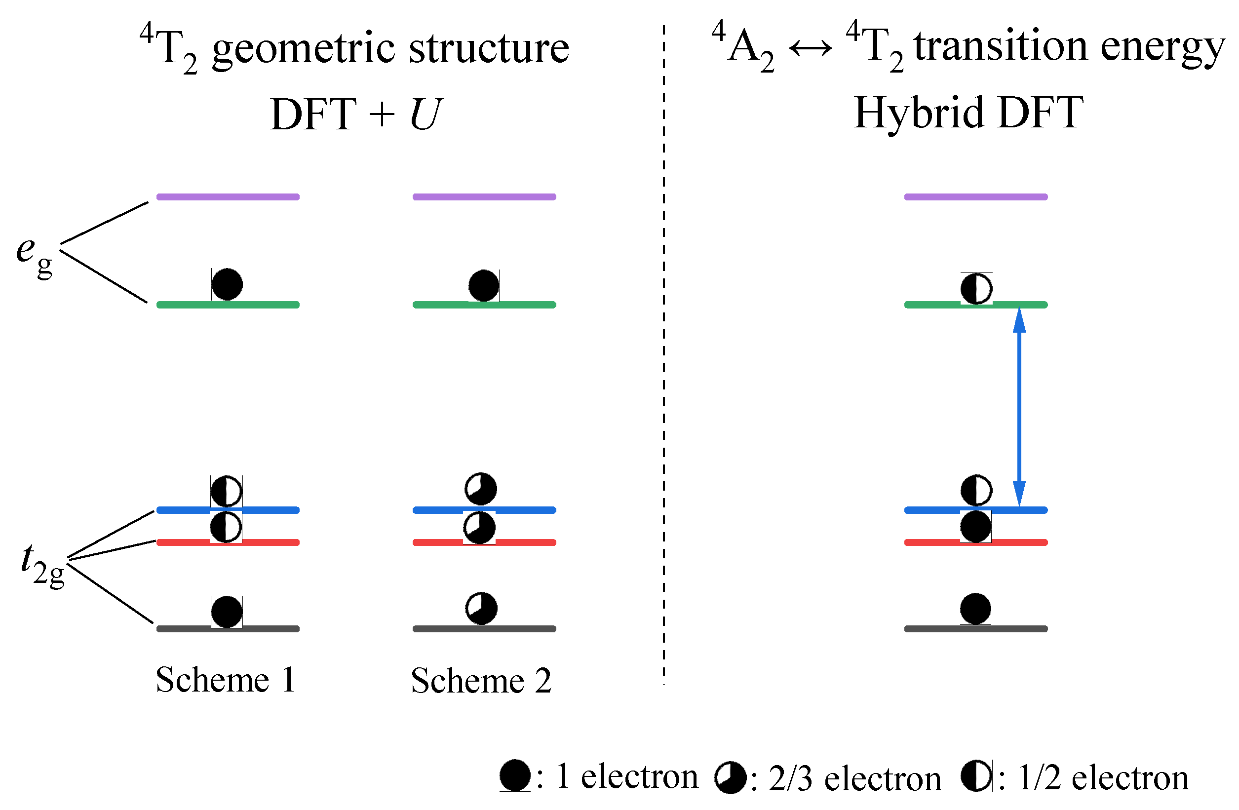
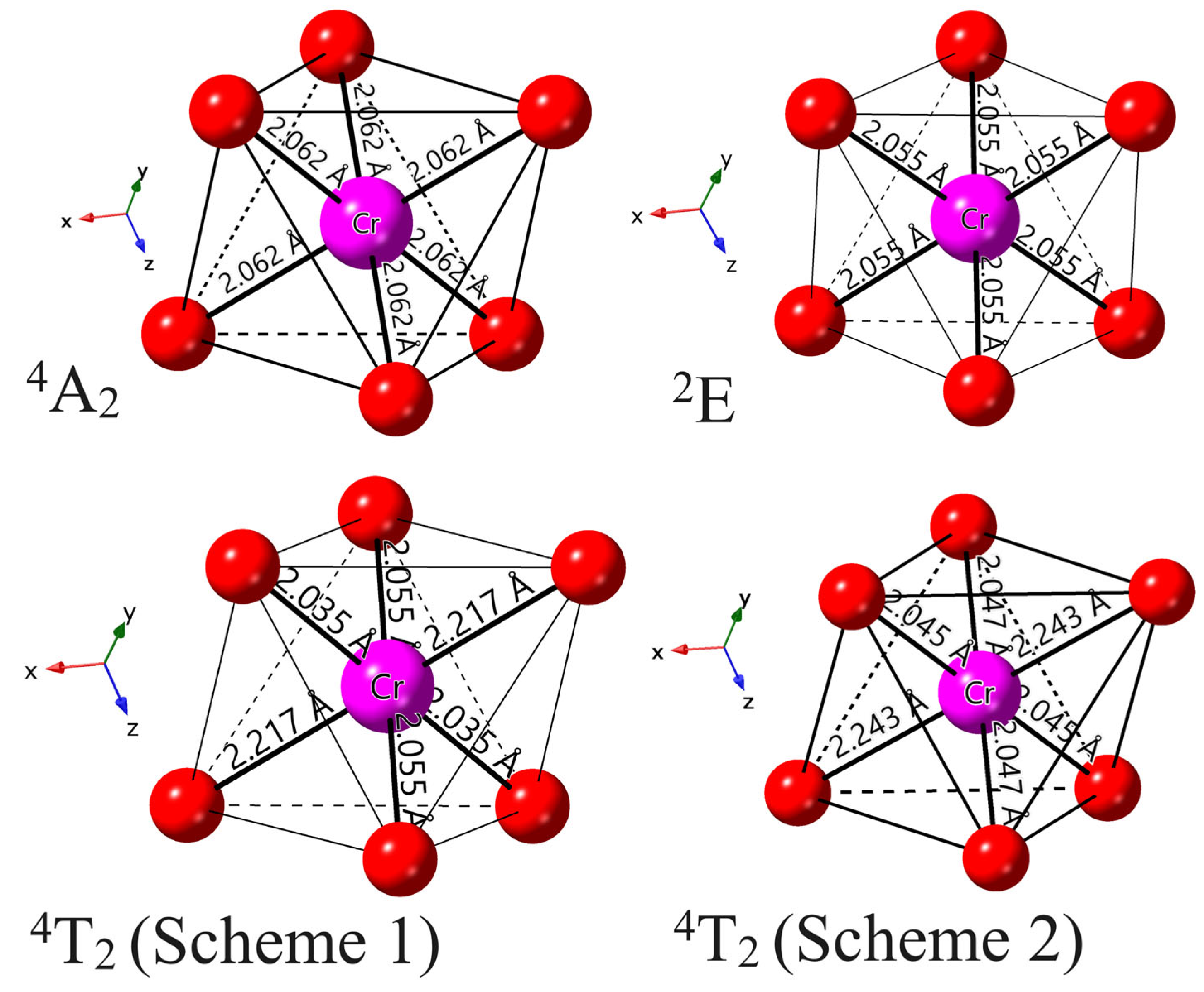
3.2.1. Structural Properties

The equilibrium geometric structures of the excited states 2E and 4T2 of Cr3+ ions in Ca3Y2Ge3O12 were determined using the ΔSCF technique with the PBE+U method, as illustrated in Figure 6. In the case of the excited state 2E, the calculated local coordination environment of Cr3+ ions maintains the initial point group symmetry of S6, with a slight bond length contraction of 0.007 Å in comparison to its ground state 4A2. This minor change is anticipated since the 2E state is not associated with an orbital change but rather a spin flip when compared to 4A2. In contrast, the optimized equilibrium geometric structures of the excited state 4T2, utilizing two sets of fractional particle occupancy schemes tailored for the calculation convergence issues mentioned in the computational methodology section, undergo a significant Jahn–Teller distortion, resulting in a symmetry descent from S6 to its subgroup Ci. This distortion involves a notable axial expansion (at least an increase of 0.15 Å) and a slight equatorial compression in the [CrO6]9− complex. And the Cr3+-O2− bond lengths, initially identical, split into three groups. These calculation results align with semi-empirical CF analyses on the Jahn–Teller effect for 3d ions in solids [37]. Additionally, we considered the volume of the [CrO6]9− complex as an index to characterize the distortion level of the excited state 4T2 with respect to the ground state 4A2. The calculated results of 11.6742 (4A2), 12.3496 (Scheme 1 for 4T2), and 12.5107 (Scheme 2 for 4T2) Å3 on this parameter indicate a potential over-relaxation risk in the equilibrium geometric structure obtained from the second fractional particle occupancy scheme, or an under-relaxation case for Scheme 1 (which will be discussed later).

3.2.2. Optical Properties and Luminescence Mechanism

Upon analysis of the obtained geometric structures for the ground state 4A2 and excited states 2E and 4T2 of Cr3+ in Ca3Y2Ge3O12, we conducted the PBE0 calculations to determine the excitation, emission, ZPL, and Stokes shift energies for the optical transitions 4A2-2E and 4A2-4T2, employing the ΔSCF technique within Slater’s transition-state method. The resulting values, along with the available experimental data, are tabulated in Table 2. The observed comparison between the ZPL energies of the excited states 2E and 4T2 and the ground state 4A2 indicates a weak CF case in Ca3Y2Ge3O12: Cr3+. Consequently, the experimentally observed broad NIR emission should be attributed to the 4A2-4T2 optical transition, not solely due to its spin-allowed transition nature. Remarkably, the calculated excitation, emission, and Stokes shift energies for the 4A2-4T2 optical transition demonstrate excellent agreement with the experimental values, particularly when predicated on the 4T2 geometric structure optimized with the first fractional particle occupancy scheme, as opposed to Scheme 2. An over-relaxation phenomenon is discerned in the 4T2 geometric structure optimized by Scheme 2, manifesting through a markedly larger calculated Stokes shift energy. The superiority of Scheme 1 becomes apparent, as the chosen particle occupations on the three t2g KS orbitals align aptly with their energy distribution corresponding to the combination of a single-fold A and a doubly degenerate E. Furthermore, the 4A2-2E optical transition displays a negligible configuration coordinate change, evidenced by the calculated Stokes shift energy of 0.02 eV. This observation, in conjunction with the significantly larger Stokes shift energy of 0.27 eV observed in the optical transition of 4A2-4T2, receives robust support from the structural data presented in the previous section.
To understand the luminescence mechanism of the materials under investigation, we constructed a host-referred binding energy (HRBE) diagram for Cr3+-doped Ca3Y2Ge3O12 following Dorenbos’s standardization [38], as illustrated in Figure 7. The energy level position of the ground state 4A2 of Cr dopants within the band gap was determined by employing the charge transition level ε(+1/0) (denoted as ε(Cr4+/Cr3+)), derived from the PBE0 total energy calculations. Simultaneously, the positions of the excited states 2E, 4T2, and 4T1 were ascertained by considering their calculated ZPL energies relative to the ground state 4A2. It is worth noting that the estimation of the 4A2-4T1 ZPL energy involves the energy difference between the two experimentally observed 4A2-4T2 and 4A2-4T1 excitation energies (i.e., ~0.72 eV taken from reference [8]). Inspection of Figure 7 reveals three distinct excitation pathways that induce the luminescence: host absorption from VB to CB and the 4A24T1 and 4A24T2 transitions of Cr3+ dopants. Evidently, the excitation efficiency of the host absorption is the lowest, given the considerable separation of the ground 4A2 and luminescent 4T2 energy levels from the top of the VBs and the bottom of the CBs, respectively. The isolated nature of the luminescence, free from the interference of the host’s electronic structure, also ensures potentially excellent quantum efficiency for applications in NIR light sources. Both anticipated observations are substantiated by the experiments (refer to Figure 4 and see Section 3.4 in reference [8]).

3.2.3. Thermal Stability

The luminescent energy level observed in the investigated compound has been attributed to the excited state 4T2 of Cr3+ dopants. Consequently, the thermal quenching effect in Cr3+-doped Ca3Y2Ge3O12 arises primarily from the thermally activated crossover between the potential surfaces of the ground 4A2 and excited 4T2 states [39]. It is crucial to note that the crossover between the potential surfaces of the ground 4A2 and excited 2E states appears challenging due to a nearly negligible change in configuration coordinates relative to the ground state 4A2. The thermal excitation energy (Ea) of Ca3Y2Ge3O12: Cr3+, a pivotal parameter for describing the thermal stability of materials, can be defined as the energy difference between the crossover point of the potential surfaces of the ground 4A2 and excited 4T2 states and the equilibrium structure point of the excited state 4T2. To evaluate Ea, we constructed a configuration coordinate diagram of Cr3+ ions in Ca3Y2Ge3O12. This involved considering the equilibrium structure points of the ground 4A2 and excited 4T2 states (denoted as Qg and Qe, respectively), utilizing the calculated excitation, emission, and ZPL energies of the optical transition 4A2-4T2 (denoted as Ex., Em. and EZPL, respectively), and applying a one-dimensional harmonic approximation for the potential surfaces of the ground 4A2 and excited 4T2 states, as illustrated in Figure 8.
By utilizing the calculated average Cr3+-O2− bond lengths of the [CrO6]9− complex in the ground 4A2 and excited 4T2 states as the horizon coordinate values of the Qg and Qe points, respectively, the crossover point (denoted as QT) between the potential surfaces of the ground 4A2 and excited 4T2 states can be ascertained. Consequently, the thermal excitation energy Ea was determined to be 0.266 eV. This value closely aligns with the reported thermal excitation energy of 0.25 eV, derived by fitting a modified Arrhenius equation to the measured temperature dependence of the emission intensity of Ca3Y2Ge3O12: Cr3+ [8]. This good agreement between the calculated and experimentally estimated thermal barrier values serves as solid proof of the validity of the performed analysis and allows for a further investigation of the role of non-radiative processes in the deactivation of the excited electronic states. Such a prediction concerning thermal excitation energy holds significant value for the smart search for novel NIR Cr3+-doped phosphors with high thermal stability. Additionally, the energy differences between the two Qg and Qe points on the potential surfaces of the ground 4A2 and excited 4T2 states were assessed, yielding values of 0.21 and 0.06 eV, respectively (the sum of these values corresponds to the Stokes shift energy). This indicates that the primary energy loss during the luminescence kinetics process takes place in the ground-state relaxation following emission.
The reliable outcomes of the present paper validate the robustness of the fractional particle occupancy scheme developed in this study, effectively overcoming the calculation convergence challenges present in the DFT+U and hybrid DFT modeling of the excited 4T2 state of 3d3 ions in solids. Additionally, this scheme can serve as a complement to an alternative approach quite recently proposed by Duan et al., which involves deactivating the 3d subspace diagonalization to address the same encountered problem [16]. Beyond enriching our fundamental understanding, this study establishes a foundation for future endeavors in the high-throughput design of novel Cr3+-doped phosphors, placing emphasis on both high thermal stability and the luminescent properties required for NIR applications.

4. Conclusions

In conclusion, our thorough investigation, employing first-principles calculations within the DFT framework, has successfully unraveled broadband NIR luminescence in Cr3+-doped Ca3Y2Ge3O12 garnets. This comprehensive exploration has provided valuable insights into the intricate interplay among geometric and electronic structures, optical properties, and thermal behavior. The findings are summarized below:
  • The results from both the structural analysis and the defect formation energy calculations indicate a tendency for Cr3+ dopants to preferentially occupy Y3+ sites rather than Ca2+ and Ge4+ sites. Comparing the optimized geometric structure of the ground state 4A2 of Cr3+ ions, the excited state 4T2 exhibits a significant Jahn–Teller distortion, characterized by a notable axial expansion and a slight equatorial compression in the [CrO6]9− complex. In contrast, the excited state 2E primarily retains the initial ground-state structure, undergoing a negligible change.
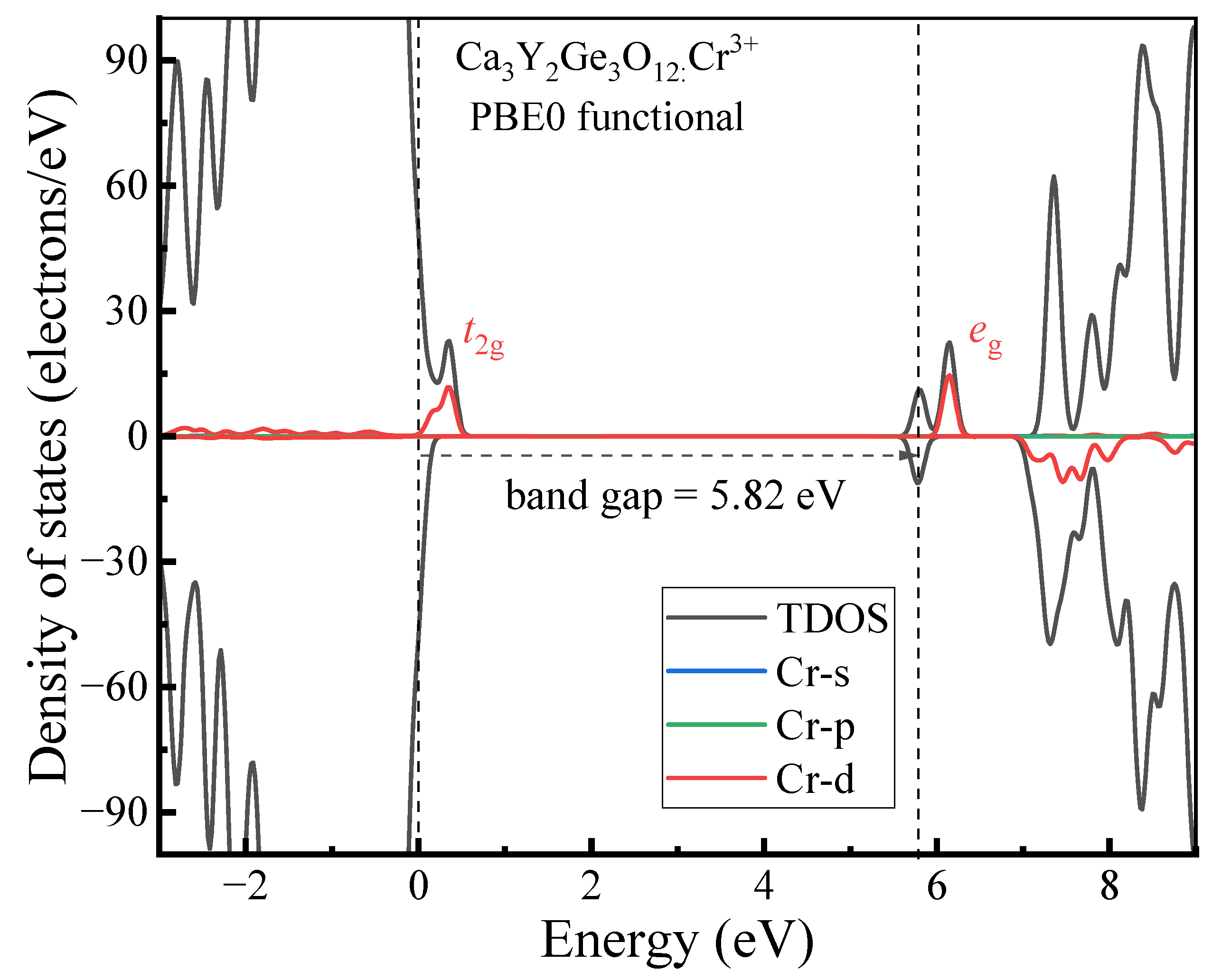
  • The host material Ca3Y2Ge3O12 features a direct band gap of 5.82 eV, allowing sufficient space to accommodate the multiple energy levels of Cr3+ dopants. The calculated positions of the ground 4A2 and excited 4T2 energy levels within the band gap underscore the isolated nature of Cr3+ optical centers from the host’s electronic structure. This discovery further supports the observed higher quantum efficiency.
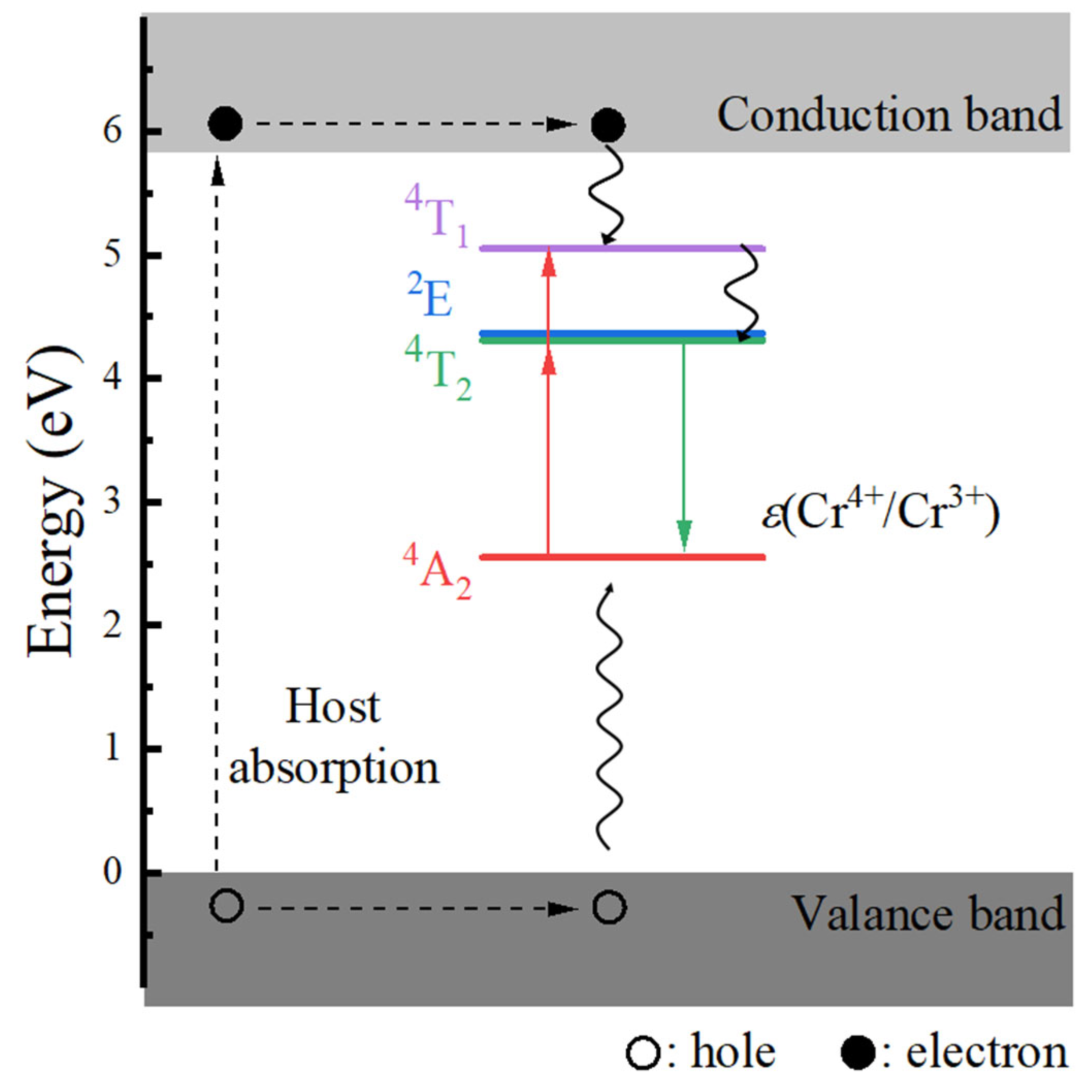
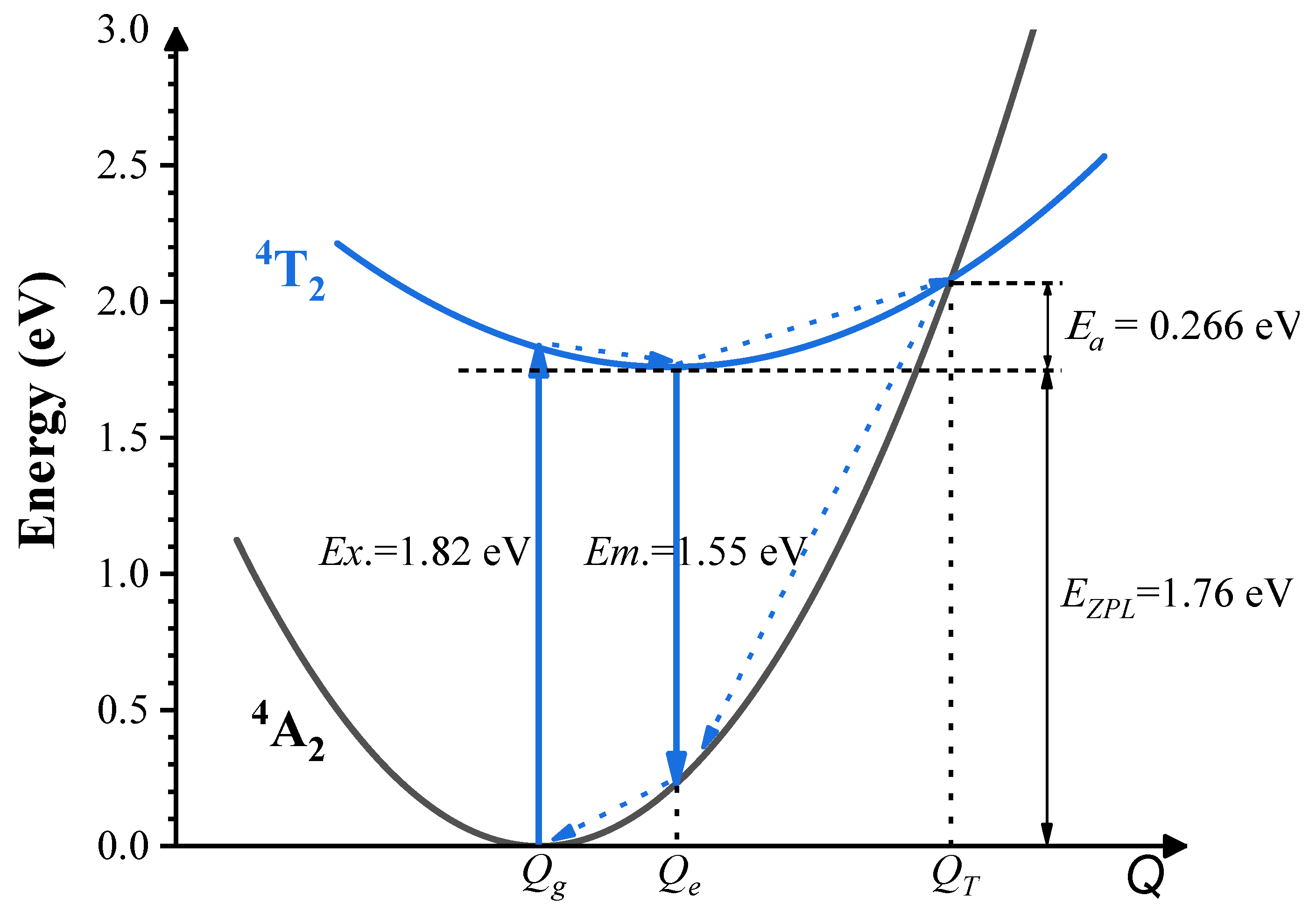
  • The calculated energies for the excitation, emission, and Stokes shift associated with the optical transitions 4A2-2E and 4A2-4T2 show a much better agreement with the experimental values. The energy comparison of the optical transitions 4A2-2E and 4A2-4T2 indicates that Cr3+ ions are located in a weak CF. The identification of three distinct excitation pathways that induce the 4T24A2 luminescence suggests that the excitations of Cr3+ ions to the 4T1 and 4T2 states are more efficient.
  • Our accurate prediction of thermal excitation energy has paved a direct path to providing fundamental analysis of the thermal quenching process in phosphors doped with 3d3 ions, using the configuration coordinate diagram.

Author Contributions

W.Z.: Investigation, Formal Analysis, Visualization, Data Curation, Writing—Original Draft Preparation. B.L.: Investigation, Formal Analysis, Validation, Writing—Original Draft Preparation. M.S.K.: Investigation, Formal analysis, Validation. M.B.: Investigation, Formal analysis, Validation. F.R.: Writing—Review and Editing. A.M.S.: Writing—Review and Editing. M.G.B.: Supervision, Writing—Review and Editing. J.W.: Project Administration, Writing—Review and Editing. C.M.: Methodology, Conceptualization, Writing—Review and Editing. All authors have read and agreed to the published version of the manuscript.

Funding

This work was financially supported by the National Natural Science Foundation of China (Grant Nos. 52161135110, 12274048 and 12304439). B.L. acknowledges the support from the China Postdoctoral Science Foundation (Grant No. 2023MD744135) and the Scientific and Technological Research Program of Chongqing Municipal Education Commission (Grant No. KJQN202200629). MSK appreciates the support from the National Young Foreign Talents Plan (Grant No. QN2023035001L) and the 2021 Chongqing Postdoctoral International Exchange Program of China Postdoctoral Science Foundation (Grant No. YJ20210346). MGB thanks the support from the Overseas Talents Plan of Chongqing Association for Science and Technology (Grant No. 2022[60]), the Polish NCN projects 2021/40/Q/ST5/00336, the Estonian Research Council grant (PRG 2031), and the Ministry of Science, Technological Development, and Innovation of the Republic of Serbia under contract 451-03-47/2023–01/200017.

Institutional Review Board Statement

Not applicable.

Data Availability Statement

Data are contained within the article and can be obtained from the authors upon request.

Conflicts of Interest

Author Alok M. Srivastava was employed by the company Current Lighting Solutions LLC. The remaining authors declare that the research was conducted in the absence of any commercial or financial relationships that could be construed as a potential conflict of interest.

References

  1. Zhang, L.L.; Wang, D.D.; Hao, Z.D.; Zhang, X.; Pan, G.H.; Wu, H.J.; Zhang, J.H. Cr3+-doped broadband NIR garnet phosphor with enhanced luminescence and its application in NIR spectroscopy. Adv. Opt. Mater. 2019, 7, 1900185. [Google Scholar] [CrossRef]
  2. He, S.; Zhang, L.L.; Wu, H.; Wu, H.J.; Pan, G.H.; Hao, Z.D.; Zhang, X.; Zhang, L.G.; Zhang, H.; Zhang, J.H. Efficient super broadband NIR Ca2LuZr2Al3O12: Cr3+, Yb3+ garnet phosphor for pc-LED light source toward NIR spectroscopy applications. Adv. Opt. Mater. 2020, 8, 1901684. [Google Scholar] [CrossRef]
  3. Basore, E.T.; Xiao, W.G.; Liu, X.F.; Wu, J.H.; Qiu, J.R. Broadband near-infrared garnet phosphors with near-unity internal quantum efficiency. Adv. Opt. Mater. 2020, 8, 2000296. [Google Scholar] [CrossRef]
  4. Mao, M.Q.; Zhao, T.L.; Zeng, H.T.; Wang, L.; Huang, F.; Tang, X.Y.; Xie, R.J. Broadband near-infrared (NIR) emission realized by the crystal-field engineering of Y3-xCaxAl5-xSixO12: Cr3+ (x = 0–2.0) garnet phosphors. J. Mater. Chem. C 2020, 8, 1981–1988. [Google Scholar] [CrossRef]
  5. Zou, X.K.; Wang, X.J.; Zhang, H.R.; Kang, Y.Y.; Yang, X.; Zhang, X.J.; Molokeev, M.S.; Lei, B.F. A highly efficient and suitable spectral profile Cr3+-doped garnet near-infrared emitting phosphor for regulating photomorphogenesis of plants. Chem. Eng. J. 2021, 428, 132003. [Google Scholar] [CrossRef]
  6. Zhang, Q.Q.; Li, G.G.; Dang, P.P.; Liu, D.J.; Huang, D.Y.; Lian, H.Z.; Lin, J. Enhancing and tuning broadband near-infrared (NIR) photoluminescence properties in Cr3+-doped Ca2YHf2Al3O12 garnet phosphors via Ce3+/Yb3+-codoping for LED applications. J. Mater. Chem. C 2021, 9, 4815–4824. [Google Scholar] [CrossRef]
  7. Xiao, H.; Zhang, J.N.; Zhang, L.L.; Wu, H.; Wu, H.J.; Pan, G.H.; Liu, F.; Zhang, J.H. Cr3+ activated garnet phosphor with efficient blue to far-red conversion for pc-LED. Adv. Opt. Mater. 2021, 9, 2101134. [Google Scholar] [CrossRef]
  8. Mao, N.; Liu, S.Q.; Song, Z.; Liu, Q.L. A broadband near-infrared phosphor Ca3Y2Ge3O12: Cr3+ with garnet structure. J. Alloys Compd. 2021, 863, 158699. [Google Scholar] [CrossRef]
  9. Cui, J.; Li, P.L.; Cao, L.W.; Wang, X.J.; Yao, Y.; Zhang, M.Y.; Zheng, M.J.; Yang, Z.B.; Suo, H.; Wang, Z.J. Achievement of broadband near-infrared phosphor Ca3Y2Ge3O12: Cr3+, Ce3+ via energy transfer for food analysis. J. Lumin. 2021, 237, 118170. [Google Scholar] [CrossRef]
  10. Adachi, S. Photoluminescence spectroscopy and crystal-field parameters of Cr3+ ion in red and deep red-emitting phosphors. ECS J. Solid State Sci. Technol. 2019, 8, R164–R168. [Google Scholar] [CrossRef]
  11. Adachi, S. Review-Photoluminescence properties of Cr3+-activated oxide phosphors. ECS J. Solid State Sci. Technol. 2021, 10, 026001. [Google Scholar] [CrossRef]
  12. Brik, M.G.; Srivastava, A.M. Review-A Review of the electronic structure and optical properties of ions with d3 electron configuration (V2+, Cr3+, Mn4+, Fe5+) and main related misconceptions. ECS J. Solid State Sci. Technol. 2018, 7, R3079–R3085. [Google Scholar] [CrossRef]
  13. Xiang, J.M.; Zheng, J.M.; Zhao, X.Q.; Zhou, X.; Chen, C.H.; Jin, M.K.; Guo, C.F. Synthesis of broadband NIR garnet phosphor Ca4ZrGe3O12: Cr3+, Yb3+ for NIR pc-LED applications. Mater. Chem. Front. 2022, 6, 440–449. [Google Scholar] [CrossRef]
  14. Shang, L.B.; Chen, Q.L.; Jing, W.G.; Ma, C.-G.; Duan, C.K.; Du, J.F. First-principles study of transition metal dopants as spin qubits. Phys. Rev. Mater. 2022, 6, 086201. [Google Scholar] [CrossRef]
  15. Shang, L.B.; Liu, M.Z.; Duan, C.K. Does Cr3+ occupy tetrahedral sites and luminesce in oxides? A first-principles exploration. J. Phys. Chem. Lett. 2022, 13, 10635–10641. [Google Scholar] [CrossRef] [PubMed]
  16. Shang, L.B.; Quan, Q.S.; Wei, X.T.; Duan, C.K. Understanding near-infrared luminescence in Cr-doped La-gallogermanates through first-principles calculations and ligand-field theory. Phys. Rev. B 2023, 106, 155136. [Google Scholar] [CrossRef]
  17. Kurboniyon, M.S.; Lou, B.B.; Zafari, U.; Rahimi, F.; Srivastava, A.M.; Yamamoto, T.; Brik, M.G.; Ma, C.G. First-principles study of geometric and electronic structures, and optical transition energies of Mn4+ impurity ions: K2SiF6 as a prototype. J. Lumin. 2023, 263, 120103. [Google Scholar] [CrossRef]
  18. Kresse, G.; Furthmüller, J. Efficient iterative schemes for ab initio total-energy calculations using a plane-wave basis set. Phys. Rev. B 1996, 54, 11169–11186. [Google Scholar] [CrossRef] [PubMed]
  19. Perdew, J.P.; Burke, K.; Ernzerhof, M. Generalized gradient approximation made simple. Phys. Rev. Lett. 1996, 77, 3865–3868. [Google Scholar] [CrossRef]
  20. Dudarev, S.L.; Botton, G.A.; Savrasov, S.Y.; Humphreys, C.J.; Sutton, A.P. Electron-energy-loss spectra and the structural stability of nickel oxide: An LSDA+U study. Phys. Rev. B 1998, 57, 1505–1509. [Google Scholar] [CrossRef]
  21. Adamo, C.; Barone, V. Toward reliable density functional methods without adjustable parameters: The PBE0 model. J. Chem. Phys. 1999, 110, 6158–6170. [Google Scholar] [CrossRef]
  22. Kresse, G.; Joubert, D. From ultrasoft pseudopotentials to the projector augmented-wave method. Phys. Rev. B 1999, 59, 1758–1775. [Google Scholar] [CrossRef]
  23. Blochl, P.E. Projector augmented-wave method. Phys. Rev. B 1994, 50, 17953–17979. [Google Scholar] [CrossRef]
  24. Monkhorst, H.J.; Pack, J.D. Special points for Brillouin-zone integrations. Phys. Rev. B 1976, 13, 5188–5192. [Google Scholar] [CrossRef]
  25. Freysoldt, C.; Grabowski, B.; Hickel, T.; Neugebauer, J.; Kresse, G.; Janotti, A.; Van de Walle, C.G. First-principles calculations for point defects in solids. Rev. Mod. Phys. 2014, 86, 253–305. [Google Scholar] [CrossRef]
  26. Durrant, T.; Murphy, S.; Watkins, M.; Shluger, A. Relation between image charge and potential alignment corrections for charged defects in periodic boundary conditions. J. Chem. Phys. 2018, 149, 024103. [Google Scholar] [CrossRef] [PubMed]
  27. Hellman, A.; Razaznejad, B.; Lundqvist, B.I. Potential-energy surfaces for excited states in extended systems. J. Chem. Phys. 2004, 120, 4593–4602. [Google Scholar] [CrossRef] [PubMed]
  28. Canning, A.; Chaudhry, A.; Boutchko, R.; Gronbech-Jensen, N. First-principles study of luminescence in Ce-doped inorganic scintillators. Phys. Rev. B 2011, 83, 125115. [Google Scholar] [CrossRef]
  29. Adachi, S. Luminescence spectroscopy of Cr3+ in an oxide: A strong or weak crystal-field phosphor? J. Lumin. 2021, 234, 117965. [Google Scholar] [CrossRef]
  30. Liberman, D.A. Slater transition-state band-structure calculations. Phys. Rev. B 2000, 62, 6851–6853. [Google Scholar] [CrossRef]
  31. Chen, Q.L.; Shang, L.B.; Ma, C.-G.; Duan, C.K. Angular Jahn-Teller effect and photoluminescence of the tetrahedral coordinated Mn2+ activators in solids—A first-principles study. Inorg. Chem. 2022, 61, 13471–13480. [Google Scholar] [CrossRef]
  32. Levy, D.; Barbier, J. Normal and inverse garnets: Ca3Fe2Ge3O12, Ca3Y2Ge3O12 and Mg3Y2Ge3O12. Acta Crystallogr. C 1999, 55, 1611–1614. [Google Scholar] [CrossRef]
  33. Shannon, R.D. Revised effective ionic radii and systematic studies of interatomic distances in halides and chalcogenides. Acta Crystallogr. A 1976, 32, 751–767. [Google Scholar] [CrossRef]
  34. Van de Walle, C.G.; Neugebauer, J. First-principles calculations for defects and impurities: Applications to III-nitrides. J. Appl. Phys. 2004, 95, 3851–3879. [Google Scholar] [CrossRef]
  35. Xu, Y.N.; Ching, W.Y.; Brickeen, B.K. Electronic structure and bonding in garnet crystals Gd3Sc2Ga3O12, Gd3Sc2Al3O12, and Gd3Ga5O12 compared to Y3Al5O12. Phys. Rev. B 2000, 61, 1817–1824. [Google Scholar] [CrossRef]
  36. Butler, P.H. Point Group Symmetry Applications, Method and Tables; Plenum Press: New York, NY, USA, 1981. [Google Scholar]
  37. Brik, M.G.; Avram, N.M. (Eds.) Optical Properties of 3D-Ions in Crystals; Tsinghua University Press: Beijing, China; Springer: Berlin/Heidelberg, Germany, 2013. [Google Scholar]
  38. Dorenbos, P. Charge transfer bands in optical materials and related defect level. Opt. Mater. 2017, 69, 8–22. [Google Scholar] [CrossRef]
  39. Senden, T.; van Dijk-Moes, R.J.A.; Meijerink, A. Quenching of the red Mn4+ luminescence in Mn4+-doped fluoride LED phosphors. Light-Sci. Appl. 2018, 7, 8. [Google Scholar] [CrossRef]
Figure 1. Schematic diagrams depicting the fractional particle occupancy schemes employed to determine the geometric structure of the 4T2 excited state and the 4A2-4T2 optical transition energies of Cr3+ ions located in an octahedral environment. The left and the right parts are referred to in the text as Scheme 1 and Scheme 2, respectively.
Materials 17 01709 g001
Figure 2. Schematic representations of the crystal structure of Ca3Y2Ge3O12, illustrating the spatial arrangement of the constituent cations and their corresponding local coordination environments.
Materials 17 01709 g002
Figure 3. Calculated formation energies of Cr substitutions (CrY, CrCa, and CrGe) in Ca3Y2Ge3O12 plotted against Fermi energy. The VBM energy is referenced to zero, and the integer values on the line segments represent the total charges of the analyzed defective systems.
Materials 17 01709 g003
Figure 4. Calculated band structure and DOS diagrams for pristine Ca3Y2Ge3O12. The insets display the projected DOS diagrams focused on the CBs’ bottom. The VBM energy is referenced to zero. The symbols Γ, H, N, and P denote the high-symmetry k-points (0 0 0), (1/2 − 1/2 1/2), (0 0 1/2), and (1/4 1/4 1/4), respectively.
Materials 17 01709 g004
Figure 5. Calculated density of states diagrams of Cr3+-doped Ca3Y2Ge3O12 in the ground state 4A2. The valence band maximum energy is referenced to zero.
Materials 17 01709 g005
Figure 6. Schematic representations of the local coordination environments of Cr3+ dopants in the ground state 4A2 and the excited states 2E and 4T2, including the results obtained using two sets of fractional particle occupancy schemes.
Materials 17 01709 g006
Figure 7. Calculated host-referred binding energy diagram of Cr3+-doped Ca3Y2Ge3O12. The notation ε(Cr4+/Cr3+) represents the calculated charge transition level ε(+1/0). The details to determine the energy level positions of the ground state 4A2 and the excited states 2E, 4T2, and 4T1 can be found in the text.
Materials 17 01709 g007
Figure 8. Schematic depiction of the calculated configuration coordinate diagram of Cr3+ ions in Ca3Y2Ge3O12. The ground state 4A2 energy is used as the reference point (zero). Ex., Em., EZPL and Ea represent the calculated excitation, emission, and zero-phonon line energies of the optical transition 4A2-4T2, and the thermal excitation energy, respectively. Qg, Qe, and QT denote the equilibrium structure points of the ground 4A2 and excited 4T2 states, along with the potential surface crossover point between the two states, respectively. The blue dotted arrows indicate the non-radiative transitions. Excitation, emission, and zero-phonon line energies (1.82 eV, 1.55 eV, 1.76 eV) correspond to the wavelengths of 681 nm, 800 nm, and 704 nm.
Materials 17 01709 g008
Table 1. Comparison of the calculated and experimental structural properties of both neat and Cr3+-doped Ca3Y2Ge3O12 in their ground states: lattice constants (a = b = c, in Å), non-dimensional coordinates of internal anion position (x, y, z), unit cell volume V before and after Cr3+ doping (in Å3), and bond lengths of Y3+-O2−, Ge4+-O2− and Ca2+-O2− in the pure host, along with Cr3+-O2− bond lengths upon Cr3+ doping at the three cationic sites (in Å).
SystemParameterCalc.Expt. a
Ca3Y2Ge3O12a = b = c12.938112.8059
O (x, y, z)0.9644, 0.0557, 0.16040.9637, 0.0567, 0.1609
V(host)2165.75082100.0533
Y3+-6O2−2.2452.234
Ge4+-4O2−1.7891.766
Ca2+-4O(1)2−2.4862.469
Ca2+-4O(2)2−2.5962.560
Ca3Y2Ge3O12:Cr3+V(Cr3+/Y3+)2155.4599-
Cr3+/Y3+-6O2−2.062-
V(Cr3+/Ge4+)2187.9207-
Cr3+/Ge4+-4O2−1.932-
V(Cr3+/Ca2+)2148.4651-
Cr3+/Ca2+-4O(1)2−2.142-
Cr3+/Ca2+-4O(2)2−2.576-
Note: a Ref. [32].
Table 2. Comparison of the calculated and experimental excitation, emission, ZPL, and Stokes shift energies of the optical transitions between the 4A2 ground state and the excited states 2E and 4T2 of Cr3+ in Ca3Y2Ge3O12 (all in eV).
ExcitationEmissionZPLStokes Shift
2E1.801.781.790.02
4T2
Scheme 11.821.551.760.27
Scheme 21.821.391.710.43
Expt. a1.831.55-0.28
Note: a Ref. [8].
Disclaimer/Publisher’s Note: The statements, opinions and data contained in all publications are solely those of the individual author(s) and contributor(s) and not of MDPI and/or the editor(s). MDPI and/or the editor(s) disclaim responsibility for any injury to people or property resulting from any ideas, methods, instructions or products referred to in the content.

