Article

A Hydrothermal and Combustion-Reduction Process with Polyvinyl Pyrrolidone as a Restricted Growth Agent and Galactose as a Reducing Agent for the Fabrication of Rod-like α-Fe2O3/Fe3O4 Magnetic Nanocomposites

1
School of Medicine, Jiangsu University, Zhenjiang 212013, China
2
College of Vanadium and Titanium, Panzhihua University, Panzhihua 617000, China
*
Authors to whom correspondence should be addressed.
Materials 2025, 18(5), 1014; https://doi.org/10.3390/ma18051014
Submission received: 7 February 2025 / Revised: 23 February 2025 / Accepted: 24 February 2025 / Published: 25 February 2025

Abstract

:
A hydrothermal and combustion-reduction process with polyvinyl pyrrolidone (PVP) as a restricted growth agent and galactose as a reducing agent was developed for the fabrication of rod-like α-Fe2O3/Fe3O4 magnetic nanocomposites (MNCs). Firstly, β-FeOOH nanorods (NRs) were fabricated by the hydrothermal method, with PVP as a restricted growth agent. To obtain a smaller size for better applications in the biomedical field, the concentrations of FeCl3 and PVP, the hydrothermal temperature, and the hydrothermal time were optimized as 0.171 M, 0.163 mM, 100 °C, and 8 h, and the fabricated β-FeOOH NRs were 193.1 nm in average length and 43.2 nm in average diameter. Then, with β-FeOOH NRs as precursors, α-Fe2O3/Fe3O4 MNCs were prepared via the combustion-reduction process with galactose as a reducing agent; the factors of the calcination temperature and time and the mass ratio of β-FeOOH and galactose were assessed as 300 °C, 0.5 h, and 1:2, respectively. The prepared α-Fe2O3/Fe3O4 MNCs under the optimized conditions were 81.6 nm in average length and 23.9 nm in average diameter, while their saturation magnetization reached 69.8 emu/g.

Graphical Abstract

1. Introduction

Many novel magnetic nanomaterials have experienced wider interest in the last decades owing to many excellent properties that differ from traditional materials, such as stable physical and chemical properties, satisfying surfaces, interface effects, a large specific surface area, and so on [1,2], and magnetic nanomaterials also obtain wide applications in various fields. For example, in the biomedical field, magnetic nanomaterials are particularly promising for their unique physicochemical properties that can support multiple functions, including X-ray computed tomography, cancer diagnosis by magnetic resonance imaging [3], Raman and photoacoustic imaging [4], drug delivery [5,6], the target therapy of cancers, and plasmonic photothermal and photodynamic therapies [7,8]. In the electrochemical detection area, magnetic nanomaterials can be applied as electrode modifiers and sorbents to enhance electrochemical signals for sensitive detection [9,10].
Among nanomaterials, hematite (α-Fe2O3) and magnetite (Fe3O4) are chemically stable, non-toxic, environmentally friendly, and have other unique characteristics, such as a high surface-to-volume ratio and so on [11]; they are often applied in biomedicine research. However, these magnetic materials have excessive or excessively low magnetic properties in practical applications [12]. To solve the above problem, α-Fe2O3/Fe3O4 magnetic nanocomposites (MNCs) have attracted our attention due to their appropriate magnetic properties for bio-applications [13,14]; among them, rod-like α-Fe2O3/Fe3O4 MNCs have especially become a research focus in biomedicine owing to their being devoured by cells and achieving easy entry into cells [2]; so, rod-like α-Fe2O3/Fe3O4 MNCs have caught researchers’ fancy.
α-Fe2O3/Fe3O4 MNCs can be fabricated by the following methods: the sonication/sol–gel method [15,16], the solvothermal method [17,18], incubation–calcination method [19], chemical vapor deposition and calcination process [20], solution combustion and calcination process [21], etc. These methods have drawbacks of complexity, a high cost, and harm to the environment. The proposed hydrothermal process with PVP as the restricted growth agent and combustion-reduction process with galactose as the reducing agent have the advantages of simple operation, practicability, high stability, and low cost [22,23].
In this work, a hydrothermal and combustion-reduction process was introduced for the preparation of α-Fe2O3/Fe3O4 MNCs with FeCl3 and polyvinyl pyrrolidone (PVP) as materials and galactose as a reducing agent, and the conditions of the process for the fabrication of α-Fe2O3/Fe3O4 MNCs were optimized in detail, and the fabrication mechanism was revealed.

2. Experimental

2.1. Materials

Polyvinyl Pyrrolidone (AR), anhydrous ferric chloride (AR), and galactose (AR) were purchased from Sinopharm Chemical Reagent Co., Ltd. (Shanghai, China), the absolute ethanol (AR) was purchased from Xilong Scientific Co., Ltd. (Shantou, China), LO2 cells and RPMI 1640 were purchased from Hefei Wanwu Biotechnology Co., Ltd. (Hefei, China).

2.2. Fabrication and Characterization of β-FeOOH Nanorods

A novel hydrothermal process with PVP as the restricted growth agent was developed to fabricate β-FeOOH nanorods (NRs). Typically, PVP and anhydrous ferric chloride were dissolved into 70 mL distilled water to form homogeneous solutions containing various concentrations of anhydrous ferric chloride (0.171 M, 0.342 M, 0.513 M, 0.684 M, and 0.855 M) and PVP (0 mM, 0.054 mM, 0.109 mM, 0.163 mM, 0.217 mM, and 0.271 mM), which were transferred to hydrothermal reactors, followed by being heated to different hydrothermal temperatures (40 °C, 60 °C, 80 °C, and 100 °C) for various hydrothermal times (2 h, 4 h, 6 h, 8 h, 10 h, and 12 h) in a furnace; after the hydrothermal reactions finished, the hydrothermal reactors were cooled to room temperature. The mixtures were centrifugally separated under 10,000 rpm/min in a centrifuge, and the obtained solids were alternately washed with ethanol and deionized water at least three times and dried for 12 h, and they were ground to obtain β-FeOOH NRs. The composition and microstructure of the β-FeOOH NRs were investigated by X-ray diffraction (XRD), under the conditions of a Cu-Kα ray with a scan rate of 5°/min for 2θ of 20–80°, and by scanning electron microscope (SEM).

2.3. Preparation and Characterization of α-Fe2O3/Fe3O4 MNCs

α-Fe2O3/Fe3O4 MNCs were prepared via the combustion-reduction process with galactose as a reducing agent. Firstly, β-FeOOH NRs and galactose were uniformly mixed according to their diverse mass ratios (1:1, 1:2, 1:4, 1:6, and 1:8), and placed into crucibles. Then, the mixtures together with the crucibles were calcined at various calcination temperatures (200–700 °C, with intervals of 100 °C) for different calcination times (0.5–2.0 h, with intervals of 0.5 h) in a programmed temperature furnace; the products were ground after being naturally cooled to room temperature, and rod-like α-Fe2O3/Fe3O4 MNCs were obtained. The composition, morphology, and microstructure of the α-Fe2O3/Fe3O4 MNCs were verified by XRD and transmission electron microscope (TEM), and their hysteresis loops were measured on a vibrating sample magnetometer (VSM).

2.4. MTT Assay

After the LO2 cells were resuscitated, the LO2 cells were cultured in RPMI 1640. When the fusion degree of the LO2 cells reached 80%, the LO2 cells were planted in 96-well plates. After 24 h, the α-Fe2O3/Fe3O4 MNCs were sterilized by ultraviolet lamp and dispersed in RPMI 1640 to form suspensions with various concentrations (0–1000 μg/mL), and then the suspensions were added into the plates, and 20 μL MTT solutions (5%) were added to the plates and cultured for another 24 h, and then the absorbances of the plates were measured. The viabilities of the LO2 cells were calculated.

3. Results and Discussion

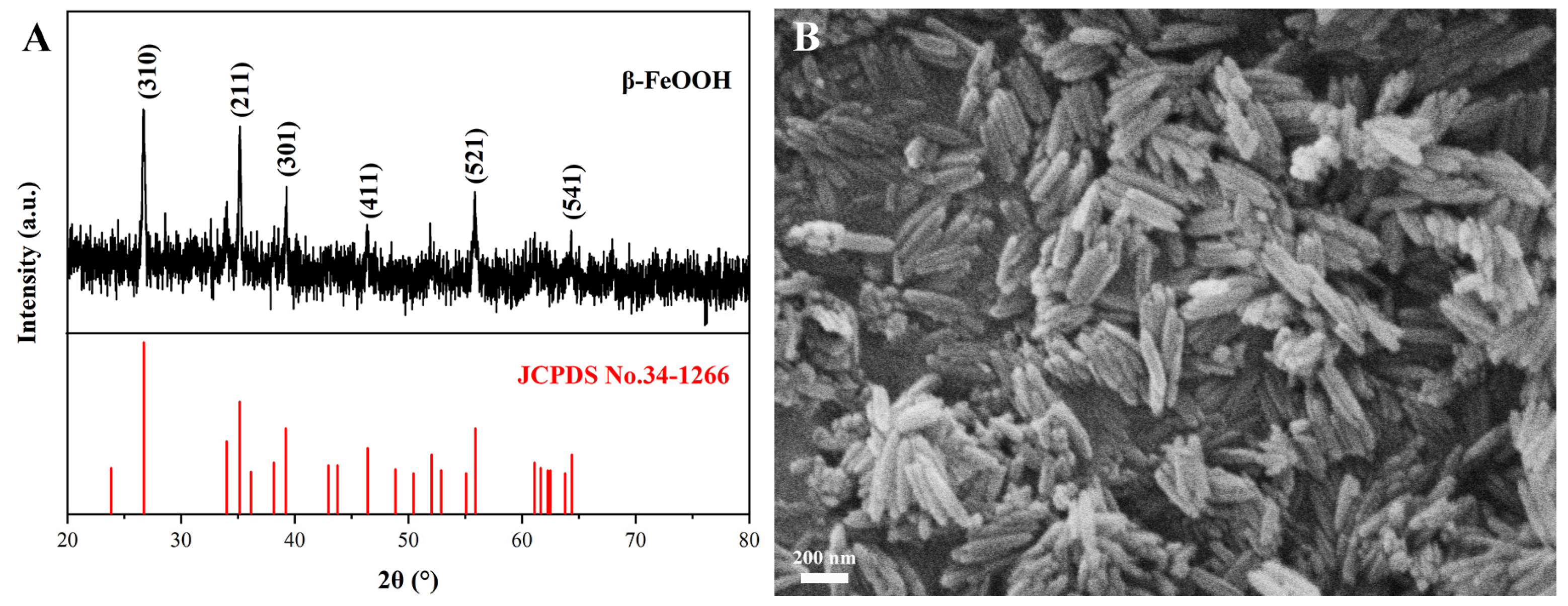
3.1. Characteristics of β-FeOOH NRs

The XRD pattern and SEM morphology of the β-FeOOH NRs fabricated at 100 °C for 8 h in a 70 mL solution containing FeCl3 of 0.171 M and PVP of 0.163 mM are revealed in Figure 1. It was found from the XRD pattern (Figure 1A) that the X-ray diffraction peaks at 26.7°, 35.1°, 39.2°, 46.4°, 55.9°, and 64.4° were perfectly fitted to the β-FeOOH standard PDF card (JCPDS No. 34-1266), which affirmed that the product belonged to the phase of β-FeOOH. Figure 1B shows their SEM morphology; obviously, the structures of the products were nanorods, and their average diameter and length reached 43.2 nm and 193.1 nm, respectively. All the above results demonstrated that β-FeOOH NRs were successfully fabricated.

3.2. Optimization of Fabrication Conditions for β-FeOOH NRs

During the hydrolysis process, FeCl3 was thermally decomposed to HCl and Fe(OH)3, and then the as-generated Fe(OH)3 continued to decompose and formed β-FeOOH. PVP acted as a dispersant and adsorbed on the β-FeOOH crystal nucleus through the coordination bond formed between the lone pair electrons of nitrogen or oxygen atoms in its structure and the atoms on the β-FeOOH surface, which enabled the C-H chain to fully expand in water and spread out in all directions. Therefore, the C-H chain prevented aggregation between the nanorod and made the β-FeOOH crystal nucleus grow in one direction and form β-FeOOH NRs, and it made them uniformly dispersed in the solution. Therefore, the effects of the FeCl3 concentration, PVP concentration, hydrothermal temperature, and hydrothermal time were the key factors, so all the preparation conditions were optimized.

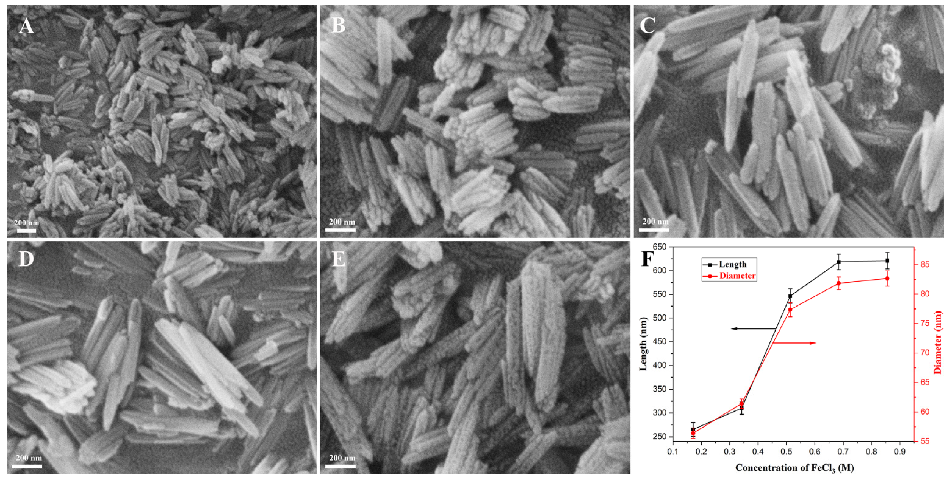
3.2.1. Optimization of FeCl3 Concentration

The solutions containing 0.054 mM PVP and FeCl3 of various concentrations were hydrothermally treated at 100 °C for 10 h; when the concentration of FeCl3 was below 0.088 M, the product was almost unobtained; the other yields are listed in Table 1, and the microstructures of the as-prepared β-FeOOH NRs fabricated with various concentrations of FeCl3 (0.171–0.855 M) and their size change rules are displayed in Figure 2. Obviously, the products prepared with various FeCl3 concentrations were rod-like in structure; their average lengths and diameters are shown in Figure 2F; with FeCl3 concentration increasing from 0.171 M to 0.855 M, their average length increased from 265.5 nm to 621.1 nm, while their average diameter increased from 56.4 nm to 82.7 nm. The reason for this was that the low Fe3+ concentration mainly affected the growth degree of the crystals; the increase in Fe3+ concentration provided more raw materials for the growth of β-FeOOH crystals, and it made their particle size larger and the crystallinity stronger. For a smaller size and better applications in biomedicine, the optimal concentration of FeCl3 was selected as 0.171 M.

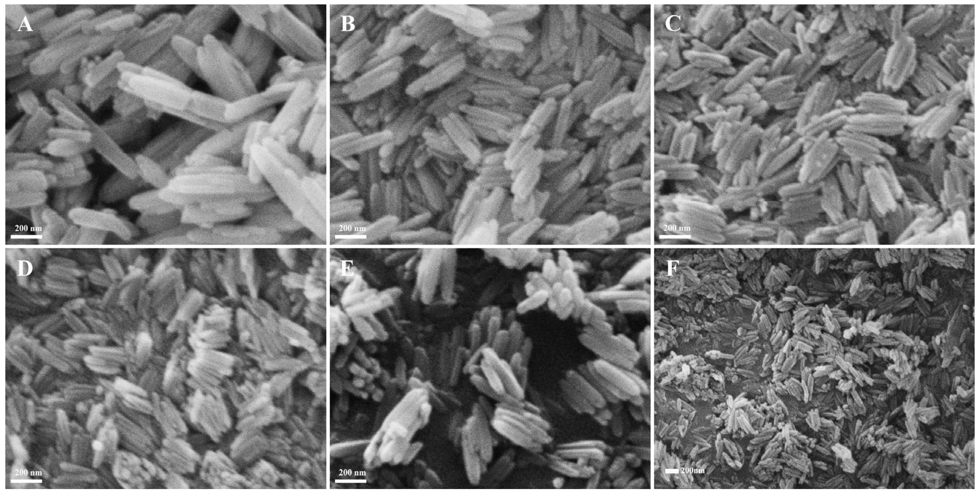
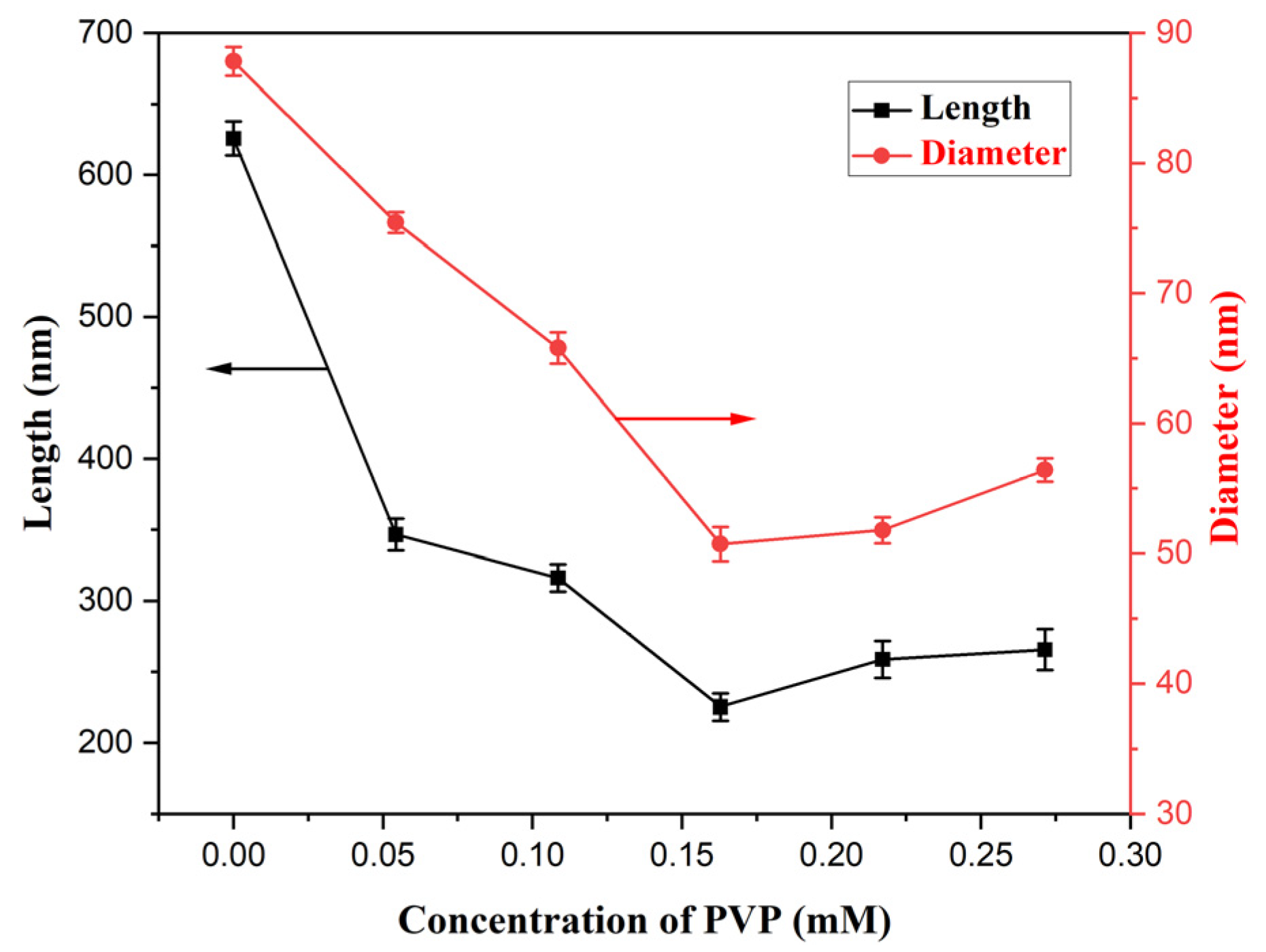
3.2.2. Optimization of PVP Concentration

The solutions containing 0.171 M FeCl3 and various PVP concentrations experienced hydrothermal reaction at 100 °C for 10 h; the SEM morphologies of the products are revealed in Figure 3. While all the products displayed a nanorod structure, their average diameter and length were largest at 87.8 nm and 625.7 nm without PVP in the solution (Figure 3A); with the PVP concentration increasing from 0.054 mM to 0.163 mM (Figure 3B–D), their average length decreased from 346.5 nm to 225.2 nm, while their average diameter also decreased from 75.4 nm to 50.7 nm, and their change trend is revealed in Figure 4, which obviously demonstrates the role of restrictive growth and the dispersion effect of PVP. With the continued increase in PVP concentration, their average diameter and length almost stayed immovable. The corresponding results likewise are listed in Table 1. Therefore, for the same purpose, the concentration of PVP was selected as 0.163 mM for the fabrication of β-FeOOH NRs.

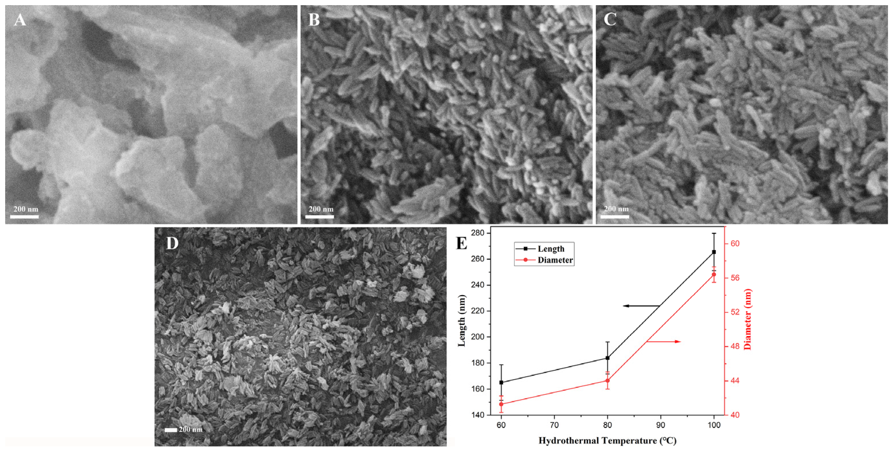
3.2.3. Optimization of Hydrothermal Temperature

The solutions of 70 mL containing 0.171 M FeCl3 and 0.163 mM PVP reacted at various temperatures for 10 h via the hydrothermal process; the SEM morphologies of the as-prepared products are revealed in Figure 5. From Figure 5A, when the hydrothermal treatment was 40 °C, the β-FeOOH NRs were unformed, while rod-shaped β-FeOOH precursors with a well-arranged and good morphology were formed, as in Figure 5B–D, as the hydrothermal temperature was enhanced from 60 °C to 100 °C. Their average length increased from 165.1 nm and 265.5 nm, while the average diameter increased from 41.3 nm and 56.4 nm, which is shown in Figure 5E; with the increase in hydrothermal temperature, the morphology of the nanorods became uniform, and the size increased gradually. The reason for this was that the solubility of the FeCl3 increased with the rise in hydrothermal temperature; then, the concentration of Fe3+ in the solution expanded, which was advantageous to the growth of the crystals. The yield (Table 1) of β-FeOOH NRs increased rapidly from 0.91% to 28.19% with the rise in hydrothermal temperature. According to the above the results, the optimum hydrothermal temperature was selected as 100 °C for the fabrication of β-FeOOH NRs.

3.2.4. Optimization of Hydrothermal Time

The SEM morphologies of the products fabricated at 100 °C for different hydrothermal times in each 70 mL solution containing 0.171 M FeCl3 and 0.163 mM PVP are shown in Figure 6; obviously, the products fabricated for hydrothermal times of 4–12 h were rod-structured, but they failed to form rod structures for hydrothermal time of 2 h. At the same time, the average diameter and length of the β-FeOOH NRs decreased with the time prolonging from 2 h to 8 h and then increased when the time continued to prolong; the experimental data are shown in Figure 7. The reason might be that the extension of time was advantageous to the dehydration reaction of Fe(OH)3, and β-FeOOH NRs with a short length and uniform morphology were produced. The yield of β-FeOOH NRs increased first and then decreased with the increase in hydrothermal time, as listed in Table 1. According to the yield and the size of the nanorods, the optimal hydrothermal time was 8 h.
In conclusion, for a larger yield, faster production speed, and smaller size, the optimal preparation conditions for β-FeOOH NRs were an FeCl3 concentration of 0.171 M, a PVP concentration of 0.163 mM, a hydrothermal time of 8 h, and a hydrothermal temperature of 100 °C.

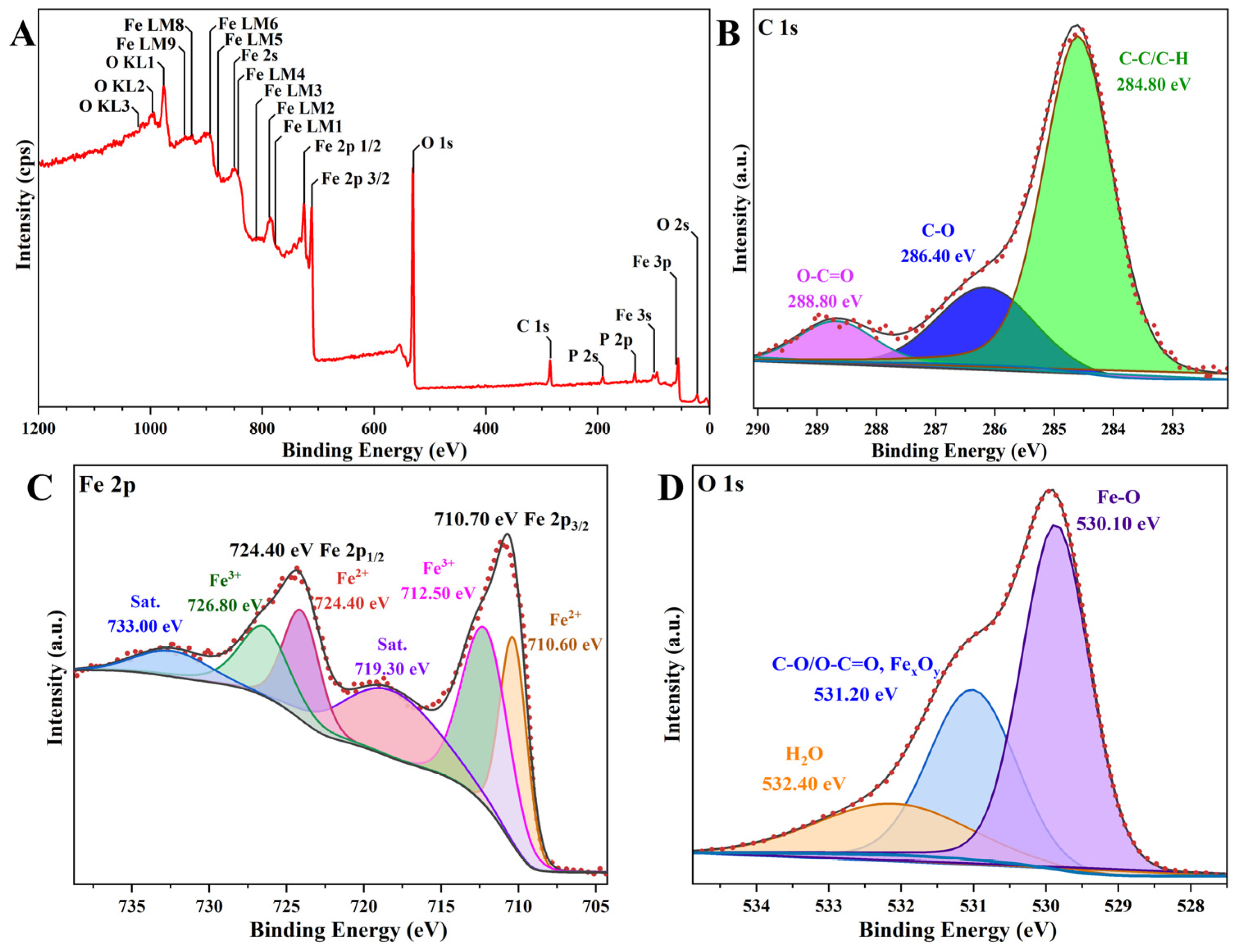
3.3. Characteristics and MTT Evaluation of α-Fe2O3/Fe3O4 MNCs

Taking β-FeOOH NRs as precursors, α-Fe2O3/Fe3O4 MNCs were prepared via the combustion-reduction process with galactose as a reducing agent. Figure 8 shows the XRD pattern and the TEM image of α-Fe2O3/Fe3O4 MNCs calcined at 300 °C for 1.5 h with a mass ratio of 1:2 for β-FeOOH NRs and galactose. The XRD pattern (Figure 8A) showed that the X-ray diffraction peaks at a 2θ of 30.1°, 35.4°, 62.5°, and 78.9° corresponded to the Fe3O4 standard card (JCPDS No. 19-0629), while the X-ray diffraction peaks at 24.1°, 33.2°, 35.6°, 40.8°, 49.5°, 54.1°, and 64.0° corresponded to the standard card of α-Fe2O3 (JCPDS No. 33-0664), which confirmed the existence of α-Fe2O3 and Fe3O4 phases in the product. The TEM image (Figure 8B) confirmed the rod-like structure of the α-Fe2O3/Fe3O4 MNCs, with an average diameter and length of 23.9 nm and 81.6 nm.
The surface element composition, chemical state, and electronic state of the α-Fe2O3/Fe3O4 MNCs were analyzed by the XPS method, and their XPS spectra are displayed in Figure 9. Firstly, all the peaks were calibrated using the binding energy (BE) of C 1s in the vacuum system (284.80 eV) as a reference [24]. As shown in Figure 9A, the BE of the C 1s peak is 284.85 eV; thus, the C 1s, Fe 2p, and O 1s peaks were calibrated with 0.05 eV of the chemical shift, as shown in Figure 9B–D. Figure 9C presents the high-resolution XPS spectrum of Fe 2p; the characteristic peaks appeared at BEs of approximately 710.70 eV and 724.40 eV, corresponding to Fe 2p3/2 and Fe 2p1/2, respectively [25], and the peaks around 719.10 eV and 732.80 eV belonged to the satellite peaks, which arose from the charge transfer screening. Upon deconvolving the Fe 2p characteristic peaks via Gaussian–Lorentz profile patterns, it was determined that the splitting peaks at high BEs of around 712.30 eV and 726.60 eV were attributed to Fe(III), while the splitting peaks at approximately 710.4 eV and 724.2 eV were ascribable to Fe(II). All the results demonstrated the formation of α-Fe2O3/Fe3O4 MNCs due to the existences of Fe(II) and Fe(III) [26,27]. The narrow spectrum of O 1s is presented in Figure 9D [28]. All the above results demonstrated again the successful formation of α-Fe2O3/Fe3O4 MNCs.
Figure 10 displays the cell viabilities of LO2 affected by various concentrations of α-Fe2O3/Fe3O4 MNCs (0–1000 μg/mL) for 24 h. Compared with the blank group (0 μg/mL of α-Fe2O3/Fe3O4 MNCs suspension), all the cell viabilities of LO2 cells affected by α-Fe2O3/Fe3O4 MNCs suspension of 100–1000 μg/mL were not lower than 95.6% of those of LO2 cells affected by a suspension without α-Fe2O3/Fe3O4 MNCs, which revealed α-Fe2O3/Fe3O4 MNCs showed excellent biocompatibility and lower toxicity.

3.4. Optimization of Preparation Conditions for α-Fe2O3/Fe3O4 MNCs

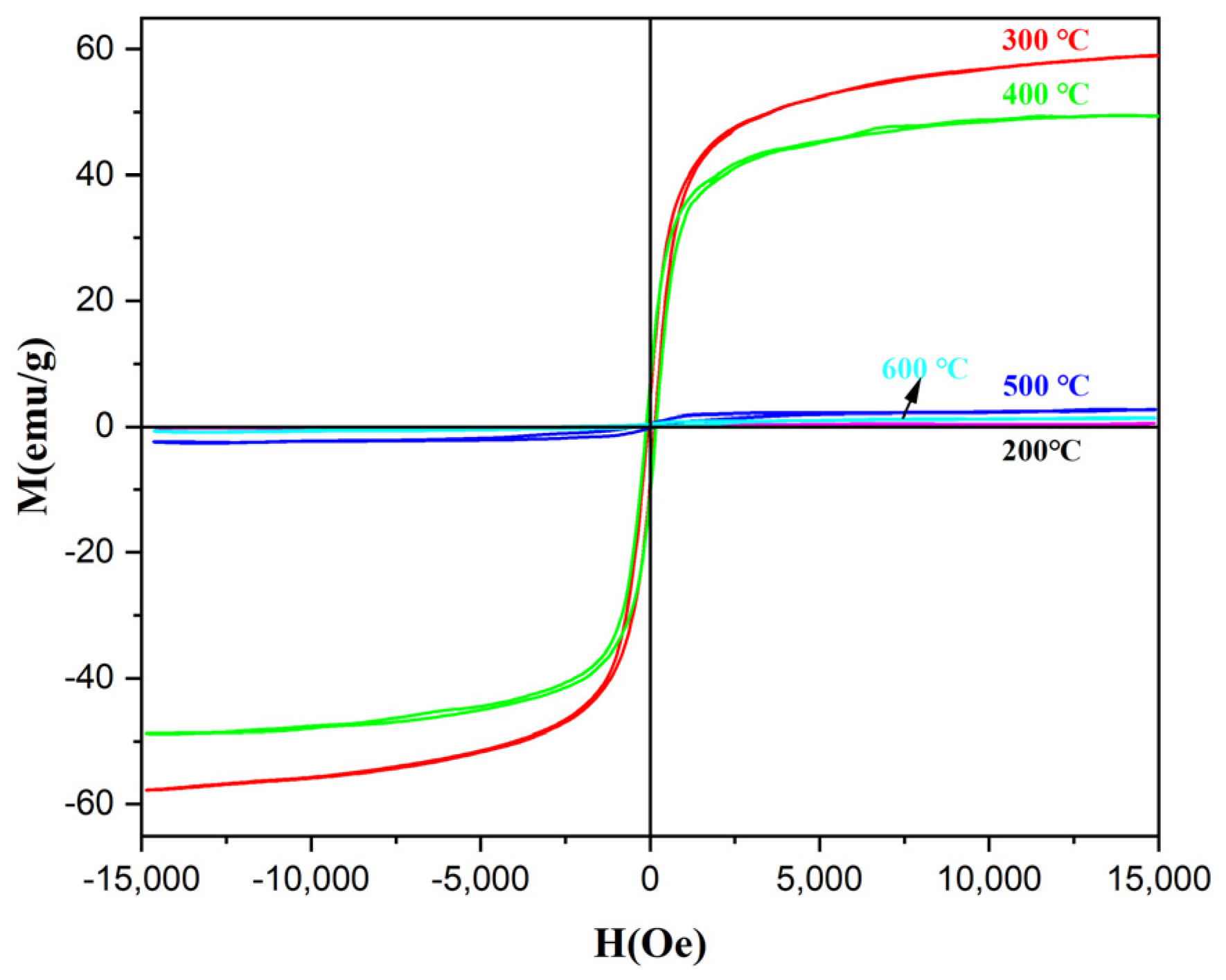
3.4.1. Optimization of Calcination Temperature

Figure 11 displays the hysteresis loops of α-Fe2O3/Fe3O4 MNCs calcined at different temperatures for 2 h with a 1:6 mass ratio for β-FeOOH NRs and galactose; the α-Fe2O3/Fe3O4 MNCs began to reveal the characteristic of superparamagnetism when the calcination temperature achieved 200 °C, which suggested that α-Fe2O3/Fe3O4 MNCs began to form. However, for the lowest the saturation magnetization (Ms) for α-Fe2O3/Fe3O4 MNCs calcined at 200 °C, the reason might be that the calcination temperature was below the thermal decomposition temperature of galactose, which affected the growth of the crystals. Therefore, the reduction of galactose did not take place completely. As the calcination temperature increased to 300 °C, the reduction of galactose was more obvious, the content of the Fe3O4 phase in α-Fe2O3/Fe3O4 MNCs increased gradually, and the color of the product changed from red to black; the Ms of α-Fe2O3/Fe3O4 MNCs achieved the maximum value of 59.0 emu/g. However, as the calcination temperature exceeded 300 °C, galactose began to decompose at express speed; when the galactose was exhausted, the reduction effect decreased, the reduced Fe3O4 began to be oxidized to α-Fe2O3, and the content of the α-Fe2O3 phase in α-Fe2O3/Fe3O4 MNCs increased gradually, so their Ms value decreased. When the temperature exceeded 500 °C, Fe3O4 was almost oxidized to α-Fe2O3 completely due to the enhancement of oxidation at high temperature, and then single-phase α-Fe2O3 nanorods with a low saturation magnetization were obtained. For larger Ms and better applications in biomedicine, the optimal calcination temperature was selected as 300 °C.

3.4.2. Optimization of Calcination Time

Figure 12 displays the hysteresis loops of α-Fe2O3/Fe3O4 MNCs calcined at 300 °C for various times, with a mass ratio of 1:6 for β-FeOOH NRs and galactose. Obviously, with the calcination time prolonging from 0.5 h to 2.0 h, the Ms of α-Fe2O3/Fe3O4 MNCs continually decreased, which revealed that the extension of the calcination time resulted in the deep oxidation of the nanorods and the phase transition from Fe3O4 to α-Fe2O3. Therefore, 0.5 h was employed as the optimal calcination time, and the maximal Ms of α-Fe2O3/Fe3O4 MNCs could reach 69.4 emu/g.

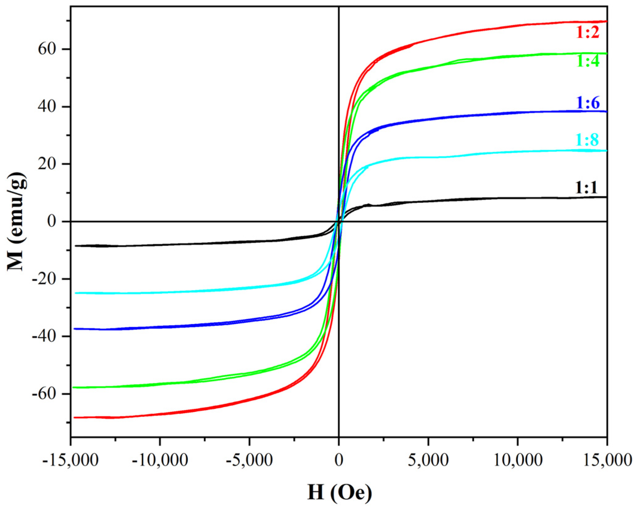
3.4.3. Optimization of Mass Ratio for β-FeOOH NRs and Galactose

Figure 13 reveals the hysteresis loops of α-Fe2O3/Fe3O4 MNCs calcined at 300 °C for 0.5 h with diverse mass ratios for β-FeOOH NRs and galactose. The Ms were enhanced with the mass ratio decreasing from 1:1 to 1:2. However, when the mass ratio continued to decrease to 1:8, galactose was excessive due to the limitations of the calcination temperature and time, the content of Fe3O4 in α-Fe2O3/Fe3O4 MNCs reduced, so the saturation magnetization also decreased. Therefore, for playing the role of a magnetic property in applications, the mass ratio 1:2 was selected as the optimal ratio for the preparation of α-Fe2O3/Fe3O4 MNCs, while the Ms of the prepared α-Fe2O3/Fe3O4 MNCs under optimal conditions reached 69.8 emu/g.
To sum up, for smaller size and larger Ms, the mixture of β-FeOOH NRs and galactose could be calcined at 300 °C for 0.5 h with a mass ratio of 1:2 to prepare rod-like α-Fe2O3/Fe3O4 MNCs.

4. Conclusions

A hydrothermal process was developed to fabricate β-FeOOH NRs, and the optimal fabrication conditions were an FeCl3 concentration of 0.171 M, a PVP concentration of 0.163 mM, a hydrothermal time of 8 h, and a hydrothermal temperature of 100 °C; the average diameter and length of the fabricated β-FeOOH NRs were 43.2 nm and 193.1 nm, respectively.
α-Fe2O3/Fe3O4 MNCs were prepared via the combustion-reduction process with galactose as a reducing agent. For a smaller size and larger Ms, the mixture of β-FeOOH NRs and galactose could be calcined at 300 °C for 0.5 h with a mass ratio of 1:2 to prepare rod-like α-Fe2O3/Fe3O4 MNCs, and the average diameter and length of the as-prepared rod-like α-Fe2O3/Fe3O4 MNCs were 23.9 nm and 81.6 nm, and their Ms could reach 69.8 emu/g.

Author Contributions

All authors contributed to the study conception and design. Investigation, Data curation, Methodology, Formal analysis, Visualization, and Writing—original draft were performed by Y.B.; Resources, Project administration, Writing—review and editing, and Supervision were executed by Z.W. and Y.L. All authors have read and agreed to the published version of the manuscript.

Funding

This research received no external funding.

Institutional Review Board Statement

Not applicable.

Informed Consent Statement

Informed consent was obtained from all subjects involved in the study.

Data Availability Statement

The original contributions presented in this study are included in the article. Further inquiries can be directed to the corresponding author.

Conflicts of Interest

The authors declare no conflicts of interest.

References

  1. Wang, X.Y.; Zhang, X.J.; Zhao, S.H.; Zhou, J.Y.; Wu, L.P.; Liu, R.J. Fabrication and Characterization of Magnetic Fe3O4/α-Fe2O3 Heterogeneous Nanorods. J. Inorg. Organomet. Polym. 2024, 34, 2163–2175. [Google Scholar] [CrossRef]
  2. Zhu, Z.Y.; Ouyang, H.Z.; Ling, C.; Ma, M.Y.; Wang, J.; Yu, X.; Li, Y.J. Fabrication of Magnetic α-Fe2O3/Fe3O4 Heterostructure Nanorods via the Urea Hydrolysis-Calcination Process and Their Biocompatibility with LO2 and HepG2 Cells. Nanotechnology 2023, 34, 505711. [Google Scholar] [CrossRef] [PubMed]
  3. Ju, Q.; Huang, R.; Hu, R.M.; Fan, J.J.; Zhang, D.L.; Ding, J.; Li, R. Phytic Acid-Modified Manganese Dioxide Nanoparticles Oligomer for Magnetic Resonance Imaging and Targeting Therapy of Osteosarcoma. Drug Deliv. 2023, 30, 2181743. [Google Scholar] [CrossRef] [PubMed]
  4. Liu, L.; Yang, S.P.; Zheng, Z.L.; Li, Q.S.; Liu, C.C.; Hu, D.H.; Liu, Z.; Zhang, X.P.; Zhang, R.P.; Gao, D.Y. Biomimetic Theranostic Agents with Superior NIR-II Photoacoustic and Magnetic Resonance Imaging Performance for Targeted Photothermal Therapy of Prostate Cancer. Pharmaceutics 2023, 15, 1617. [Google Scholar] [CrossRef]
  5. Ni, Y.; Deng, P.; Yin, R.T.; Zhu, Z.Y.; Ling, C.; Ma, M.Y.; Wang, J.; Li, S.S.; Liu, R.J. Effect and Mechanism of Paclitaxel Loaded on Magnetic Fe3O4@mSiO2-NH2-FA Nanocomposites to MCF-7 Cells. Drug Deliv. 2023, 30, 64–82. [Google Scholar] [CrossRef]
  6. Trucillo, P. Biomaterials for Drug Delivery and Human Applications. Materials 2024, 17, 456. [Google Scholar] [CrossRef]
  7. Koshev, N.; Kapralov, P.; Evstigneeva, S.; Lutsenko, O.; Shilina, P.; Zharkov, M.; Pyataev, N.; Darwish, A.; Timin, A.; Ostras, M.; et al. Yttrium-Iron Garnet Film Magnetometer for Registration of Magnetic Nano- and Submicron Particles: In Vitro and In Vivo Studies. IEEE T. Biomed. Eng. 2024, 71, 122–129. [Google Scholar] [CrossRef]
  8. Ahmadi, M.; Ghoorchian, A.; Dashtian, K.; Kamalabadi, M.; Madrakian, T.; Afkhami, A. Application of Magnetic Nanomaterials in Electroanalytical Methods: A Review. Talanta 2021, 225, 121974. [Google Scholar] [CrossRef]
  9. Yue, Y.; Zhang, X.J.; Zhao, S.H.; Wang, X.Y.; Wang, J.; Liu, R.J. Construction of a Label-Free Electrochemical Biosensing System Utilizing Fe3O4/α-Fe2O3@Au with Magnetic-Induced Self-Assembly for the Detection of EGFR Glycoprotein. Vacuum 2024, 222, 112975. [Google Scholar] [CrossRef]
  10. Zhang, Y.L.; Wang, J.; Liu, M.; Ni, Y.; Yue, Y.; He, D.W.; Liu, R.J. Magnetically Induced Self-Assembly Electrochemical Biosensor with Ultra-Low Detection Limit and Extended Measuring Range for Sensitive Detection of HER2 Protein. Bioelectrochemistry 2024, 155, 108592. [Google Scholar] [CrossRef]
  11. Rytov, R.A.; Usov, N.A. Specific Absorption Rate of Randomly Oriented Magnetic Nanoparticles in a Static Magnetic Field. Beilstein J. Nanotechnol. 2023, 14, 485–493. [Google Scholar] [CrossRef] [PubMed]
  12. Wang, J.; Liu, M.; Tang, J.W.; Yang, Y.P.; He, N.; Li, S.S.; Liu, R.J. Construction of Electrochemical Biosensor Based on Magnetic Fe3O4/α-Fe2O3 Heterogeneous Nanorods for the Sensitive Detection of MUC1 Mucoprotein. Ceram. Int. 2024, 50, 10706–10714. [Google Scholar] [CrossRef]
  13. Wang, J.; Ouyang, H.Z.; Ni, Y.; Zhang, H.D.; Sun, L.; Liu, R.J.; Li, S.S. Magnetic Self-Assembled Label-Free Electrochemical Biosensor Based on Fe3O4/α-Fe2O3 Heterogeneous Nanosheets for the Detection of Tau Proteins. Bioelectrochemistry 2024, 157, 108678. [Google Scholar] [CrossRef] [PubMed]
  14. Yue, Y.; Zhang, X.J.; Xu, Z.H.; Sun, L.; Li, S.S.; Liu, R.J. Ultrasensitive Detection of PSA in Human Serum Using Label-Free Electrochemical Biosensor with Magnetically Induced Self-Assembly Based on α-Fe2O3/Fe3O4@Au Nanocomposites. Microchem. J. 2024, 201, 110487. [Google Scholar] [CrossRef]
  15. Nagare, A.; Dhadage, A.; Baithy, M.; Bhuyan, P.M.; Gogoi, P.; Athare, A.; Navgire, M. Sol-Gel Assisted β-Cyclodextrin Coated MoO3-Fe2O3 Nanocomposite for Photodegradation of Methylene Blue Dye. J. Sol-Gel Sci. Technol. 2024, 110, 304–318. [Google Scholar] [CrossRef]
  16. Kumar, N.; Banerjee, A.M.; Pai, M.R.; Meena, S.S.; Patra, A.K.; Sastry, P.U.; Jagannath; Tripathi, A.K. Sol-Gel Mediated Synthesis and Characterization of Hierarchically Porous Fe2O3/SiO2 Monolithic Catalyst for High Temperature Sulfuric Acid Decomposition. Catal. Commun. 2023, 179, 106686. [Google Scholar] [CrossRef]
  17. Kanimozhi, G.; Nibagani, N.; Nair, D.S.; Kumar, H.; Satyanarayana, N. A Synergic Effect of N-Graphene Wrapped α-Fe2O3 Nanofacets Prepared by Microwave-Assisted Solvothermal Method for Lithium-Ion Battery. J. Phys. Chem. Solids 2022, 169, 110885. [Google Scholar]
  18. Li, P.H.; Zhuang, X.Y.; Xu, J.H.; Ruan, L.X.; Jiang, Y.F.; Lin, J.X.; Zhang, X.M. Enhanced Photo-Fenton Activity of SnO2/α-Fe2O3 Composites Prepared by a Two-Step Solvothermal Method. Materials 2022, 15, 1743. [Google Scholar] [CrossRef]
  19. Srivastava, N.; Srivastava, M.; Alhazmi, A.; Mohammad, A.; Khan, S.; Pal, D.B.; Haque, S.; Singh, R.; Mishra, P.K.; Gupta, V.K. Sustainable green approach to synthesize Fe3O4/α-Fe2O3 nanocomposite using waste pulp of Syzygium cumini and its application in functional stability of microbial cellulases. Sci. Rep. 2021, 11, 24371. [Google Scholar] [CrossRef]
  20. Leduc, J.; Goenuellue, Y.; Ghamgosar, P.; You, S.; Mouzon, J.; Choi, H.; Vomiero, A.; Grosch, M.; Mathur, S. Electronically-Coupled Phase Boundaries in α-Fe2O3/Fe3O4 Nanocomposite Photoanodes for Enhanced Water Oxidation. ACS Appl. Nano Mater. 2019, 2, 334–342. [Google Scholar] [CrossRef]
  21. Alkanad, K.; Hezam, A.; Shekar, G.C.S.; Drmosh, Q.A.; Kala, A.L.A.; AL-Gunaid, M.Q.A.; Lokanath, N.K. Magnetic recyclable α-Fe2O3–Fe3O4/Co3O4–CoO nanocomposite with a dual Z-scheme charge transfer pathway for quick photo-Fenton degradation of organic pollutants. Catal. Sci. Technol. 2021, 11, 3084. [Google Scholar] [CrossRef]
  22. Liu, R.J.; Huang, W.; Pan, S.; Li, Y.; Yu, L.L.; He, D.W. Covalent Immobilization and Characterization of Penicillin G Acylase on Magnetic Fe2O3/Fe3O4 Heterostructure Nanoparticles Prepared via a Novel Solution Combustion and Gel Calcination Process. Int. J. Biol. Macromol. 2020, 162, 1587–1596. [Google Scholar] [CrossRef] [PubMed]
  23. Hong, J.Z.; Yang, F.; Sun, Z.P. Hexagonal Bi-Pyramid α-Fe2O3 Microcrystals: Unusual Formation, Characterization and Application for Gas Sensing. J. Alloy. Compd. 2021, 889, 161515. [Google Scholar] [CrossRef]
  24. Xu, Z.H.; Lv, Z.X.; Yang, H.J.; Zhang, J.S.; Sun, Z.J.; He, D.W.; Liu, R.J. Label-Free Electrochemical Biosensor with Magnetic Self-Assembly Constructed via PNA-DNA Hybridization Process on α-Fe2O3/Fe3O4 Nanosheets for APOE Ε4 Genes Ultrasensitive Detection. Bioelectrochemistry 2025, 161, 108847. [Google Scholar] [CrossRef]
  25. Zhang, S.H.; Fan, X.L.; Xue, J. A Novel Magnetic Manganese Oxide Halloysite Composite by One-Pot Synthesis for the Removal of Methylene Blue from Aqueous Solution. J. Alloy. Compd. 2023, 930, 167050. [Google Scholar] [CrossRef]
  26. Yamashita, T.; Hayes, P. Analysis of XPS Spectra of Fe2+ and Fe3+ Ions in Oxide Materials. Appl. Surf. Sci. 2008, 254, 2441–2449. [Google Scholar] [CrossRef]
  27. Zhu, Z.Q.; Huang, Q.S. In-Plane Structured Fe3O4/FeS Composite Loaded on Reduced Graphene Oxide as a Stabilized Anode Material for Lithium-Ion Batteries. Appl. Phys. A 2022, 128, 563. [Google Scholar] [CrossRef]
  28. Li, L.; Ma, P.; Hussain, S.; Jia, L.J.; Lin, D.; Yin, X.; Lin, Y.; Cheng, Z.H.; Wang, L.Y. FeS2/Carbon Hybrids on Carbon Cloth: A Highly Efficient and Stable Counter Electrode for Dye-Sensitized Solar Cells. Sustain. Energy Fuels 2019, 3, 1749–1756. [Google Scholar] [CrossRef]
Figure 1. The XRD pattern (A) and SEM morphology (B) of β-FeOOH NRs fabricated at 100 °C for 8 h in 70 mL solution containing FeCl3 of 0.171 M and PVP of 0.163 mM.
Materials 18 01014 g001
Figure 2. The SEM morphologies of β-FeOOH NRs fabricated at 100 °C for 10 h in each 70 mL solution containing FeCl3 of 0.171 M (A), 0.342 M (B), 0.513 M (C), 0.684 M (D), and 0.855 M (E) and PVP of 0.271 mM, and the corresponding broken-line graph of dimension change (F).
Materials 18 01014 g002
Figure 3. The SEM morphologies of β-FeOOH NRs fabricated at 100 °C for 10 h in each 70 mL solution containing FeCl3 of 0.171 M and PVP of 0 mM (A), 0.054 mM (B), 0.109 mM (C), 0.163 mM (D), 0.217 mM (E), and 0.271 mM (F).
Materials 18 01014 g003
Figure 4. Broken-line graph of dimension change with various PVP concentrations.
Materials 18 01014 g004
Figure 5. The SEM morphologies of β-FeOOH NRs fabricated at 40 °C (A), 60 °C (B), 80 °C (C), and 100 °C (D) for 10 h in each 70 mL solution containing FeCl3 of 0.171 M and PVP of 0.271 mM, and the corresponding broken-line graph of dimension change (E).
Materials 18 01014 g005
Figure 6. The SEM morphologies of β-FeOOH NRs experiencing a hydrothermal reaction at 100 °C for 2 h (A), 4 h (B), 6 h (C), 8 h (D), 10 h (E), and 12 h (F) with FeCl3 of 0.171 M and PVP of 0.217 mM.
Materials 18 01014 g006
Figure 7. Broken-line graph of dimension change against hydrothermal time.
Materials 18 01014 g007
Figure 8. XRD pattern (A) and TEM image (B) of α-Fe2O3/Fe3O4 MNCs.
Materials 18 01014 g008
Figure 9. The XPS spectrum (A) and high-resolution XPS spectra (BD) of F α-Fe2O3/Fe3O4 MNCs calcined at 300 °C for 1.5 h with a mass ratio of 1:2 for β-FeOOH NRs and galactose.
Materials 18 01014 g009
Figure 10. The cell viabilities of LO2 cells incubated with various concentrations of α-Fe2O3/Fe3O4 MNCs for 24 h (n = 3).
Materials 18 01014 g010
Figure 11. The hysteresis loops of α−Fe2O3/Fe3O4 MNCs calcined at diverse temperatures for 2 h with a mass ratio of 1:6 for β−FeOOH NRs and galactose.
Materials 18 01014 g011
Figure 12. The hysteresis loops of α−Fe2O3/Fe3O4 MNCs calcined at 300 °C for diverse times with a mass ratio of 1:6 for β−FeOOH NRs and galactose.
Materials 18 01014 g012
Figure 13. The hysteresis loops of α−Fe2O3/Fe3O4 MNCs calcined at 300 °C for 0.5 h with diverse mass ratios of β−FeOOH NRs and galactose.
Materials 18 01014 g013
Table 1. Productivities of β-FeOOH precursors fabricated under various conditions.
FeCl3 (M)PVP (mM)Hydrothermal Time (h)Hydrothermal Temperature (°C)Yield (%)
0.1710.2711010028.19
0.3420.2711010014.63
0.5130.271101008.90
0.6840.271101007.27
0.8550.271101005.25
0.1710.271108024.30
0.1710.27110606.91
0.1710.27110400.91
0.1710.0001010025.19
0.1710.0541010028.10
0.1710.1091010028.56
0.1710.1631010030.13
0.1710.2171010029.31
0.1710.16321001.47
0.1710.163410023.87
0.1710.163610026.66
0.1710.163810033.31
0.1710.1631210033.19
Disclaimer/Publisher’s Note: The statements, opinions and data contained in all publications are solely those of the individual author(s) and contributor(s) and not of MDPI and/or the editor(s). MDPI and/or the editor(s) disclaim responsibility for any injury to people or property resulting from any ideas, methods, instructions or products referred to in the content.

