Article

Treatment of Aqueous Amoxicillin Solutions with Sunlight Using a Pelletized Macrocomposite Photocatalyst

React! Group, Department of Chemistry, Faculty of Sciences & CICA, Universidade da Coruña, E-15071 A Coruña, Spain
*
Author to whom correspondence should be addressed.
Materials 2025, 18(7), 1394; https://doi.org/10.3390/ma18071394
Submission received: 13 February 2025 / Revised: 7 March 2025 / Accepted: 10 March 2025 / Published: 21 March 2025

Abstract

:
We report on the preparation and characterization of a cost-effective, durable, and reusable macrocomposite, prepared in the form of pellets and designed for the photodegradation of water pollutants, using amoxicillin (AMX) as a model compound. Using the wet impregnation method, kaolin clay and TiO2-P25 composites were doped with copper (Cu2+) and cobalt (Co2+). The produced materials were characterized by SEM-EDS, XRD, XRF, textural property analysis, and their potential lixiviation of components by ICP-MS. The photodegradation efficiency under solar irradiation was evaluated by varying the acidity of the medium, the concentration of AMX, and the amount of catalyst. The performance of the recycled photocatalysts was also studied. The photodegradation of AMX was monitored by UV–Vis and UV–Vis/HPLC spectrophotometry. The optimal formulations, Cu (0.1%)/TiO2 and Co (0.1%)/TiO2, achieved up to 95% degradation of 5 mg·L−1 AMX in 3 h at pH 5.9, with a catalyst loading of 1 g·L−1. The Cu-doped material showed a slightly faster reaction rate and higher total-organic-carbon removal (80.4%) compared to the Co-doped material (59%) under identical conditions. The same photodegradation intermediates were identified by LC-MS/MS for both doped macrocomposites, and a reaction mechanism is proposed. These macrocomposites showed efficient and consistent recyclability over more than five reuse cycles, showing their potential to be used for antibiotic pollution abatement in water-treatment facilities.

1. Introduction

The preservation of water, an essential resource for life on Earth, is a global priority. However, human activities have significantly deteriorated water quality, with pollution by antibiotic drugs emerging as a critical concern because of the possible emergence of antimicrobial resistances, among others. Hospital, pharmaceutical, and residential wastewater often contain residues of antibiotics that adversely affect aquatic ecosystems and pose potential risks to human health. To mitigate the environmental and health impacts of a growing pollution, advanced techniques have been developed for the oxidation and degradation of antibiotics in water. These methods aim to transform antibiotics into safer molecules or, ideally, eliminate them, thereby reducing their concentration in water sources. Among these strategies, advanced oxidation processes (AOPs) have demonstrated effectiveness in removing a broad range of organic contaminants from water and wastewater through reaction with hydroxyl radicals (HO) [1,2].
Photocatalysis, one of the most promising AOPs, has been widely investigated using various photocatalysts such as Al2O3, ZnO, Fe2O3, and TiO2 [3]. Among them, TiO2 stands out due to its chemical stability, non-toxicity, high photocatalytic activity, and cost effectiveness. TiO2 is typically used in the form of an anatase–rutile mixture, commercially known as TiO2-P25 (Evonik), which is particularly efficient [4]. TiO2-P25, activated by solar irradiation, is a reliable solution for degrading persistent organic pollutants in water through HO radical generation. Efforts to enhance the photocatalytic performance of TiO2-P25 under solar irradiation have included modifications with metal dopants.
We have previously shown that transition metal-impregnated TiO2 is effective in photocatalytic applications. Cu-modified TiO2 demonstrated improved photocatalytic activity under Vis light, achieving over 95% total organic carbon (TOC) removal for phenol degradation. This improvement is due to a reduced bandgap energy and better charge-carrier separation [5]. Similarly, the Co-modified TiO2 performed exceptionally well under near-UV–Vis radiation, achieving 96% TOC removal for phenol degradation. This success can be attributed to the creation of oxygen vacancies and inter-band states that allowed for greater light absorption in the visible spectrum [6]. These results underscore the potential of metal-modified TiO2 as highly effective photocatalysts for environmental processing.
Here, we summarize our investigations on the photocatalytic degradation of amoxicillin (AMX), a widely used beta-lactam antibiotic, under sunlight irradiation. AMX, (2S,5R,6R)-6-[[(2R)-2-amino-2-(4-hydroxyphenyl)acetyl]amino]-3,3-dimethyl-7-oxo-4-thia-1-azabicyclo[3.2.0]heptane-2-carboxylic acid, belongs to the penicillin family and contains a beta-lactam ring and a thiazolidine ring (Scheme 1). It is effective against a broad spectrum of Gram-positive and Gram-negative bacteria [7].
To enhance the efficiency of AMX degradation, we prepared a series of macrocomposite photocatalysts based on kaolin and TiO2-P25 (90% TiO2-P25 and 10% kaolin), impregnated with different weight percentages of copper (Cu2+) and cobalt (Co2+) (0.1%, 0.5%, 1%). These materials were characterized using X-ray diffraction (XRD), diffuse reflectance spectroscopy (DRS), scanning electron microscopy (SEM), and other techniques. We optimized key parameters influencing amoxicillin photodegradation, such as pH, amoxicillin concentration, and photocatalyst dosage. The degradation process was monitored using high-performance liquid chromatography (HPLC), UV–Vis spectroscopy, and total organic carbon (TOC) analysis, while photodegradation products were identified via HPLC-MS.
Promising results were achieved with both the Cu2+ and Co2+-modified photocatalytic materials, attaining approximately 86% AMX degradation within 180 min under sunlight irradiation. Verma and Haritash, studying the photocatalyzed degradation of AMX, found a degradation of 75% [8]. These findings highlight the potential of metal-doped TiO2-P25 macrocomposites for efficient antibiotic degradation in water-treatment applications [9,10,11].

2. Experimental Procedure

2.1. Materials

TiO2-P25 was acquired from Evonik (ca. 70% anatase, 30% rutile, and a tiny proportion of amorphous phase), with a surface area of 55 ± 15 m2·g−1 [12] and a particle size of ~30 nm [13]. Copper (II) sulfate pentahydrate (CuSO4·5H2O) (≥98%, Sigma, St. Louis, MO, USA), Cobalt (II) sulfate heptahydrate (CoSO4·7H2O) (≥97%, Aldrich), and AMX (C16H19N3O5S) (99.5% Sigma-Aldrich, St. Louis, MO, USA) were purchased and used without further purification. Acetonitrile (HPLC grade) was purchased from J.T. Baker (Center Valley, PA, USA). Some studies employed O2 gas with a purity of at least 99.995%. The distilled water was taken from a Millipore device (Milli-Q water, Burlington, MA, USA), with a resistivity of 18.2 MΩ at 298.0 K and a total organic carbon (TOC) of less than 5 μg/L.

2.2. Catalyst Synthesis

The immobilization of metals onto the photocatalyst was achieved using the incipient wetness impregnation method. A kaolin clay and TiO2-P25 composite was prepared by dissolving the required amount of metal salt in distilled water; the clay-TiO2 mixture was then gradually added to the solution under constant stirring. The resulting suspension was stirred continuously for 24 h at 50 °C to ensure a uniform distribution of the metal ions throughout the composite.
The mixture was then manually extruded through a syringe with a 2 mm inner-diameter tip to form elongated, spaghetti-like rolls, and deposited on a clean aluminum sheet. These rolls were dried in an oven at 90 °C for 24 h. Subsequently, the dried photocatalysts were calcined at 600 °C for 4 h, with a ramp rate of 10 °C·min−1. The calcined materials, hereafter called M (X%)/TiO2, were finely ground, where M (X%) denotes the specific metal (M = Cu or Co) and its mass percentage (X = 0.1%, 0.5%, or 1%).
To remove loose particles, the photocatalysts were rinsed thoroughly with distilled water and then dried again at 90 °C. To evaluate Cu2+ and Co2+ potential leaching, the samples M (0.1%)/TiO2 were immersed in water with stirring for 24 h. The resulting solution was filtered and analyzed using inductively coupled plasma mass spectrometry (ICP-MS) to determine Cu2+ and Co2+ concentration, 1476 μg/L and 10,950 μg/L at pH 5.9, respectively, indicating a slight leaching process. Copper was found in the leaching water below the limit of 2,0 mg/L established by the EU, which would allow for the use of the Cu (0.1%)/TiO2 macrocomposite for water treatment [14]. Though no maximum admissible limit of cobalt has been established by the UE or WHO, the US EPA has established a reference maximum admissible limit at 0.1 mg/L, well below the lixiviated concentration achieved in this experiment, a clear indication that the Co (0.1%)/TiO2 macrocomposite is not mechanically stable and further work on its stabilization would be needed prior to scaling up its use [15].

2.3. Photocatalyts Characterization

The morphology of TiO2-P25, Cu (X%)/TiO2, and Co (X%)/TiO2 photocatalysts was analyzed using a JEOL JSM-6400FEG scanning electron microscope (JEOL Ltd., Tokyo, Japan) equipped with an energy-dispersive X-ray spectroscopy (EDS) system. Prior to imaging, the samples were coated with a thin layer of gold (~2 nm) and mounted on small pieces of amorphous carbon.
The surface morphology of the photocatalysts was further examined using a Jeol-JEM-1010 microscope (JEOL Ltd., Japan) operated at an accelerating voltage of 80 kV. For TEM analysis, a few drops of a nanoparticle suspension were deposited onto carbon-coated copper grids (200 mesh) and allowed to air-dry before imaging.
X-ray diffraction (XRD) analysis was conducted using a Bruker Siemens D5000 diffractometer (Bruker Corporation, Billerica, MA, USA) equipped with Bragg–Brentano geometry and a θ/2θ configuration. The system incorporates a graphite monochromator and features advanced optics, including 2° primary and secondary Soller slits, a variable divergence slit, a 1 mm receiving slit, a 0.2 mm monochromator slit, and a 0.6 mm detector slit. A scintillation counter served as the detector. Data acquisition was performed over a 2θ range of 2–80° with a step size of 0.050° and a dwell time of 2.5 s per step. The DiffracPlus software (v. 8.0.0.2, Socabim, Billerica, MA, USA) was employed for data processing and analysis.
N2 adsorption/desorption BET isotherms were obtained at 77.4 K (Tristar II Plus-Micromeritics, Norcross, GA, USA), between P/P0 = 0.1 and 1.0. The relative pressure range used to calculate the BET-specific surface area was P/P0 = 0.05–0.3. “MicroActive for TriStar II Plus” v. 2.03 (Micromeritics, Norcross, GA, USA) software was utilized for instrument control, data acquisition, and data processing.
The UV–Vis diffuse reflectance spectra (200–800 nm) of solid photocatalysts were measured using a JASCO V-560 UV-Vis spectrophotometer (Hachioji, Tokyo, Japan) with a double monochromator and double-beam optical system, as well as an integrating sphere attachment (JASCO ISV-469, Hachioji, Tokyo, Japan). Reflectance spectra were transformed into equivalent absorption Kubelka-Munk units.

2.4. Photocatalytic Degradation Procedure

Photocatalytic degradation experiments were carried out on sunny days from 10:30 a.m. to 2:30 p.m. at A Coruña, Spain (43°19′36″ N, 8°24′33″ W). AMX solution was initially prepared by adding 5 mg of AMX to 1 L of deionized water. In the photocatalytic degradation experiment, the effect of operational parameters such as initial pH (3.3, 5.9, and 8.6), catalyst dose (0.1 and 1.0 g/L), and initial AMX concentration (1–9 mg/L) were studied. The experiment was conducted at temperatures ranging from 19 to 24 °C, and the solution pH was modified to the desired level, when needed, using 1M HCl(aq) and 1M NaOH(aq).
The photocatalytic degradation experiment was carried out as follows: 1 g of the photocatalyst was dispersed in 50 mL of an aqueous AMX solution with an initial concentration of 5 mg/L. The suspension was then exposed to sunlight to initiate the photocatalytic reaction. Samples were collected at specific time intervals during irradiation to monitor AMX photodegradation. The collected samples were analyzed using HPLC to determine the extent of AMX degradation. The degradation efficiency (% degradation) was calculated as follows:
%   A M X   d e g r a d a t i o n = [ A M X ] 0 [ A M X ] t [ A M X ] 0 · 100
where [AMX]0 represents the initial AMX concentration and [AMX]t is the remaining antibiotic concentration at time (t) [16,17]. Fitting the first-order equation to the kinetic data allowed us to obtain the corresponding pseudo-first-order rate constants.
The photocatalytic degradation efficiency was calculated based on the initial AMX concentration. AMX was monitored by measuring the UV–Vis absorbance at 226 nm and 272 nm, using a Biochrom Libra S70 spectrophotometer (Cambridge, UK), and by UV–Vis HPLC analysis also at 226 and 272 nm, in a Thermo Fisher apparatus equipped with a 6000 LP UV detector (Waltham, MA, USA), an AS 3000 automatic sample, and a P4000 solvent pump (Waltham, MA, USA). The Kromaphase C18 column (Waltham, MA, USA) is 4.6 mm × 150 mm × 5 µm was used, with an injected volume of 50 µL, a flow rate of 1.0 mL·min−1, at 25 °C, with acetonitrile buffer solution in pH = 4.45 (25:75, v/v) as mobile phase. The TOC removal efficiency was determined by a Shimadzu TOC-5000A analyzer (Kyoto, Japan).
Photoproducts were detected by HPLC/MS (Thermo Scientific LTQ Orbitrap Discovery tools (Waltham, MA, USA) using an electrospray interface in positive ion mode (ESI+). Kromaphase 100 C18 column (Waltham, MA, USA) (150 mm × 5 μm) was used, operated at 25 °C, with elution solvents A (H2O 0.1% H-COOH) and B (ACN 0.1% H-COOH) at flow rate of 0.4 mL/min. The gradient was as follows: 0–2 min, 97–97% A and 3–3% B; 2–5 min, 97–80% A and 3–20% B; 5–12 min, 80–20% A and 20–80% B; 12–14 min, 20–20% A and 80–80% B; 14–15 min, 20–97% A and 80–3% B; 15–20 min, 97–97% A and 3–3% B.

3. Results and Discussion

3.1. Catalyst Characterization

The efficiency of a photocatalyst depends on the surface and structural properties of the semiconductor, such as its crystalline structure (facets exposed), surface area, particle-size distribution, porosity, band gap, and the density of hydroxyl groups on the surface [18]. The use of doped metals such as copper (Cu) and cobalt (Co) in photocatalysts for the removal of AMX improve the photocatalytic activity of the base material, TiO2, by altering the electronic and structural characteristics. Low doping concentrations (e.g., 0.1% to 1%) are generally enough to produce desirable changes in the photocatalyst (narrowed bandgap, improved charge separation, and increased ROS generation) without overloading the system [19,20,21]. Over-doping (>1%) generates an excess of defect sites within the photocatalyst lattice, that can promote e/h+ recombination, thus reducing the overall efficiency of the photocatalytic process [22,23,24].

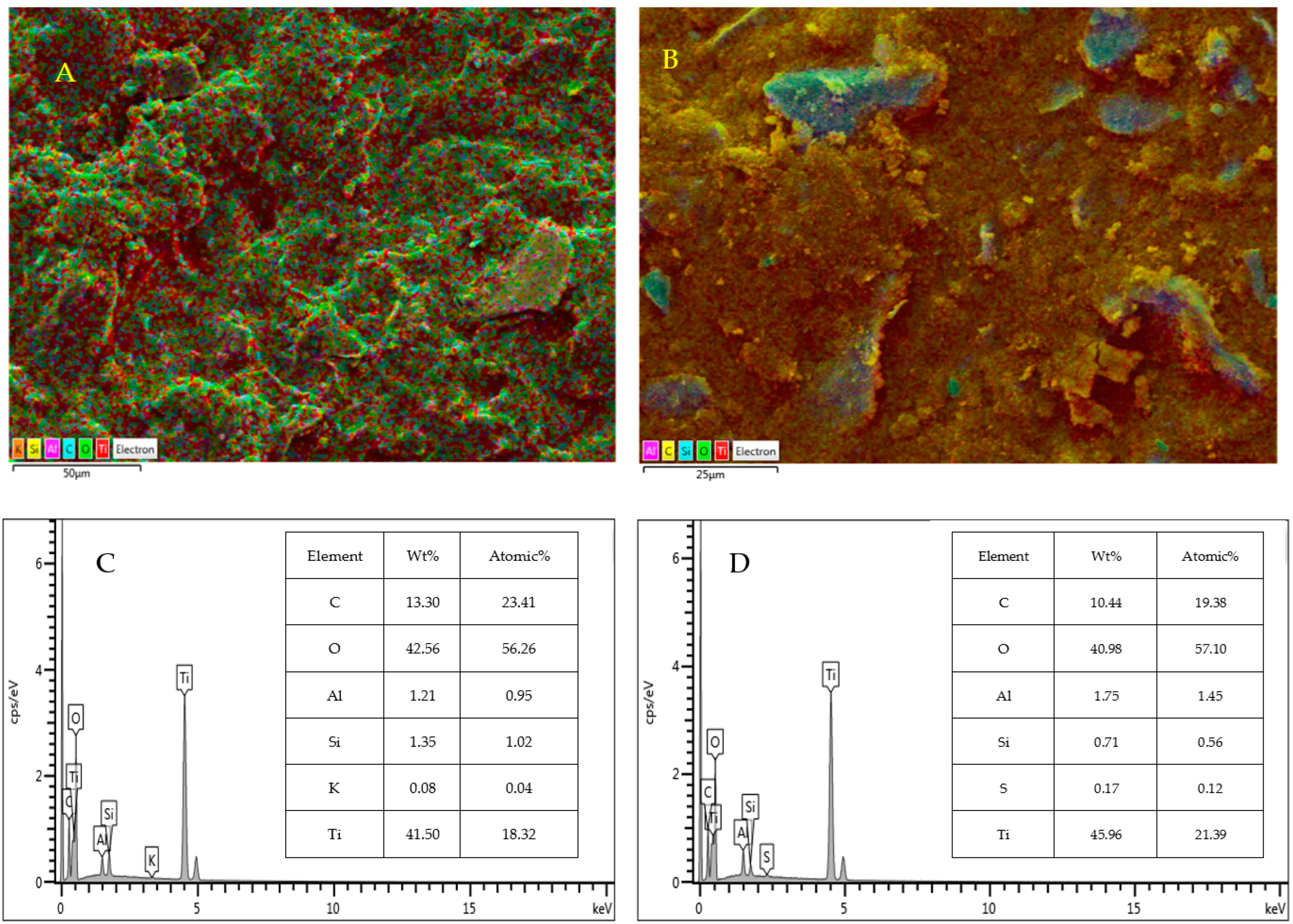
3.1.1. Scanning Electron Microscopy (SEM) and Energy-Dispersive Spectroscopy (EDS)

The SEM and EDS analyses of M (0.1%)/TiO2 are displayed in Figure 1. The micrographs of Cu (0.1%)/TiO2, Co (0.1%)/TiO2 show the presence of irregularly shaped particles with different sizes (Figure 1A), and of some large crystalline particles (Figure 1B). M (0.1%)/TiO2 surfaces remain stable, as they exhibit no cracks and are characterized solely by the presence of cavities. EDS analysis of M (0.1%)/TiO2 shows the dominance of Ti, O, and C, and to a lesser extent Al and S were also detected, but in lower amounts (Figure 1C,D).

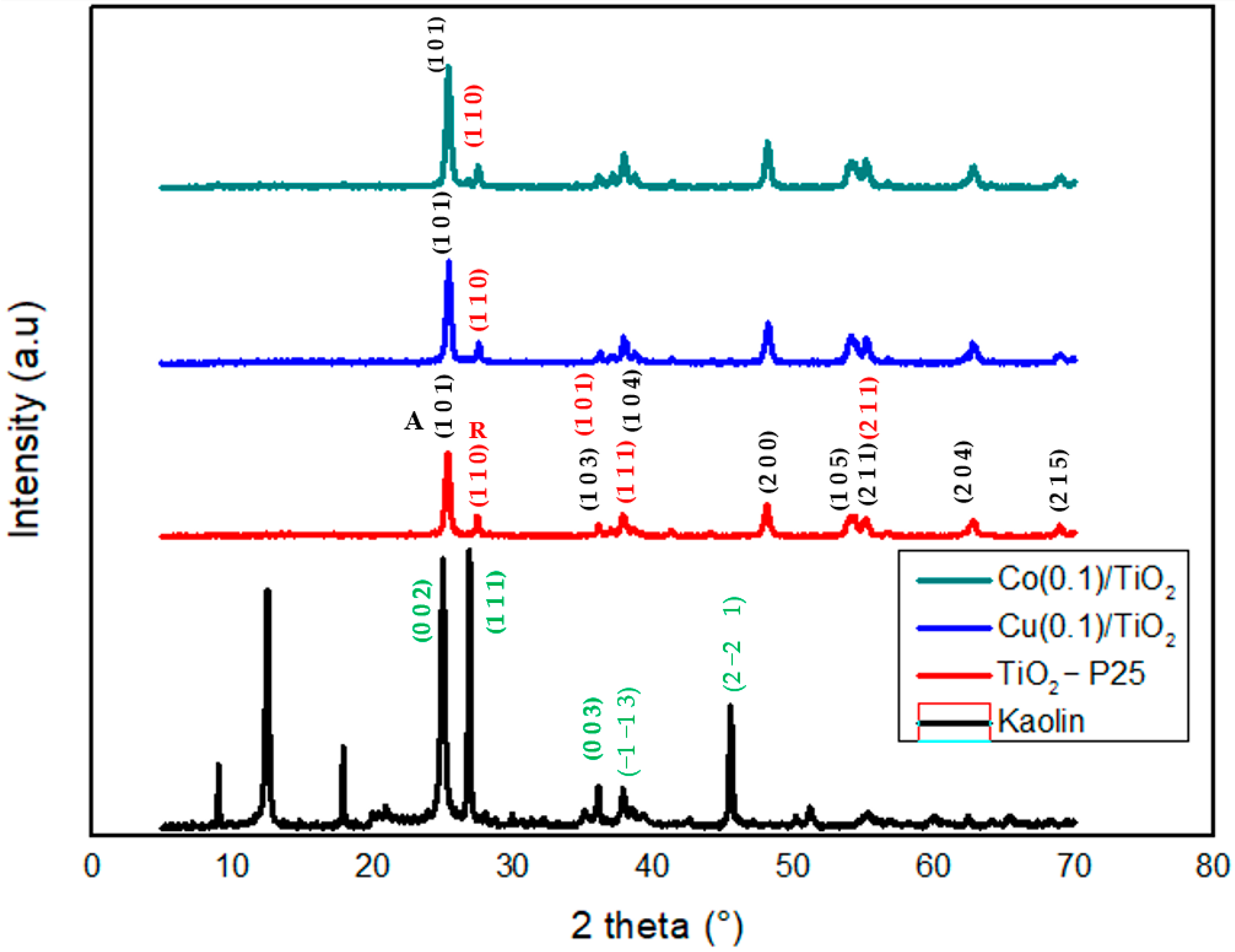
3.1.2. X-Ray Diffraction (XRD) and Fluorescence (XRF)

The crystalline structure of the as-synthesized M (0.1%)TiO2 samples was evaluated using XRD analysis (Figure 2). The seven prominent diffraction peaks (2θ: 25.25°, 37.52°, 48.02°, 53.58°, 54.88°, 62.61°, and 75.07°—red diffractogram) are attributed to the anatase phase of TiO2 in pristine TiO2-P25, as identified by JCPDS No. 00-021-1272 [25,26]. The anatase phase predominates (80% anatase and 20% rutile) at low processing temperatures, whereas the rutile one becomes dominant at higher temperatures, typically above 600 °C (it is commonly formed through high-temperature calcination of the anatase phase) [27].
Cu (0.1%)/TiO2 and Co (0.1%)/TiO2 diffractograms (Figure 2) do not show new peaks due to the presence of Cu and Co after impregnation, which may indicate that the low amount of Cu and Co is well disseminated into TiO2 lattice. Some researchers [26,28] suggested that no dopant peaks are detected due to the low dopant load. In spite of the absence of new peaks, doping with Cu and Co increases the intensity of the existing ones (Figure 2). Some studies showed similar behavior, but the increased intensity was related to an improvement in crystallinity [29] a phenomenon that may be attributed to a size mismatch between Cu or Co and TiO2, with a beneficial effect on the TiO2 crystallinity.
Table 1 shows the result of the fluorescence X-ray analysis (XRF) of the as-prepared photocatalytic materials. Cu (0.1%)/TiO2 and Co (0.1%)/TiO2 are predominantly composed of TiO2, SiO2, Al2O3, and Fe2O3, which account for 97.70% and 98.85%, respectively, of the total weight of the photocatalysts. The small percentage of Fe2O3 and SiO2 observed is attributed to the presence of kaolin -Al2Si2O5(OH)4-.

3.1.3. Textural Properties

The N2 adsorption–desorption isotherms of Cu (0.1% TiO2) and Co (0.1% TiO2) and the corresponding pore-size distribution are shown in Figure 3; both isotherms correspond to type IV according to the IUPAC classification, featuring an H3-type hysteresis loop observed near the high-relative-pressure region (0.9 < P/P0 < 1), characteristic of a typical mesoporous structure [30].
The porous microsphere M (X%)/TiO2 stacking forms cavities with a pore-size distribution ranging from 1 to 5 nm, consistent with the SEM analysis. The isotherm was utilized to calculate the BET surface area (SBET, multipoint) and to evaluate the textural properties based on the Barrett, Joyner, and Halenda (BJH) model [31] (Table 2).
The Cu-doped photocatalyst exhibits a slightly higher BET surface area and a larger BET constant (C), reflecting an increased affinity for N2. While the median pore width remains comparable between the two photocatalysts, the Cu-doped sample shows a slightly greater maximum pore volume. On the other hand, the Co-doped photocatalyst has a marginally larger average particle size, despite both materials displaying similar median pore widths. These properties suggest potential effectiveness in pollutant adsorption and UV light absorption.
The observed textural characteristics differ from those observed in Cu and Co samples prepared via hydrothermal process (Table 2), where the type IV isotherm exhibits a significant H3 hysteresis loop, but a lower P/P0 range (0.4–0.8) [32,33].
The pore-size distribution (PSD) reveals a maximum at 23 Å for Cu (0.1%)/TiO2 and 22 Å for Co (0.1%)/TiO2, both at the lower limit of the mesoporous range.
Adsorption of AMX on both types of pelletized composites was studied in the dark. The adsorption–desorption equilibrium was attained at 120 and 180 min, respectively, for Cu (0.1)/TiO2 and Co (0.1)/TiO2 (Table 3). The adsorption followed first-order kinetics, with rate constants kads,Cu (0.1)/TiO2 = 2.54 × 10⁻³ s−1 and kads,Co (0.1)/TiO2 = 1.79 × 10⁻³ s−1, respectively. The adsorption experiment also revealed that, under dark conditions, Cu (0.1%)/TiO2 and Co (0.1%)/TiO2 adsorbed 19% and 13% of the initial AMX concentration, respectively, after 180 min, from which the corresponding adsorption–desorption equilibrium constants can be estimated as Ka-d Cu (0.1)/TiO2 = 0.23 L·g−1 and Ka-d Co (0.1)/TiO2 = 0.15 L·g−1, respectively. These values confirm the system is not far from equilibrium, as expected in the framework of the Langmuir approach.

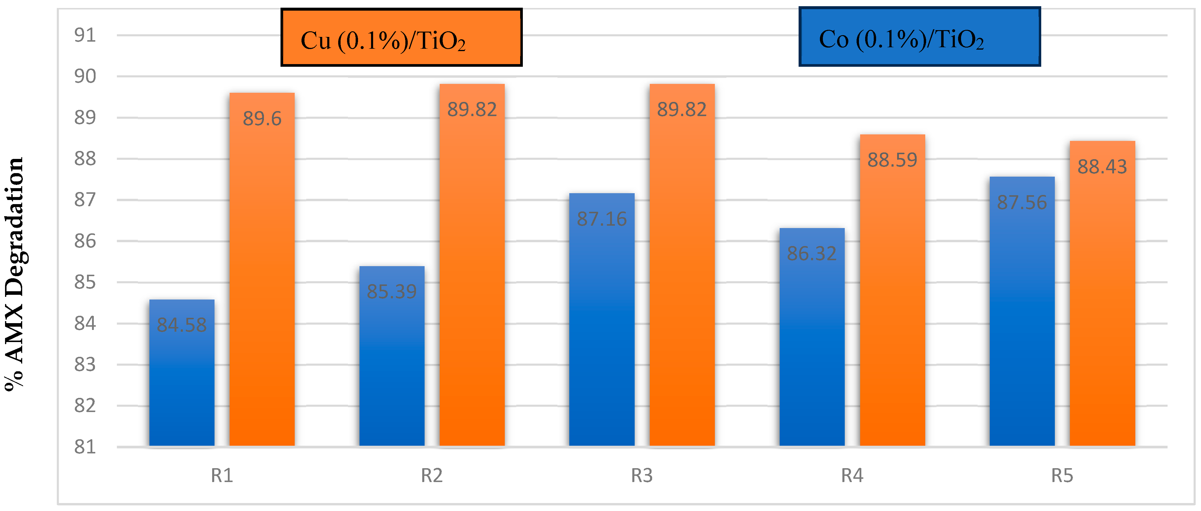
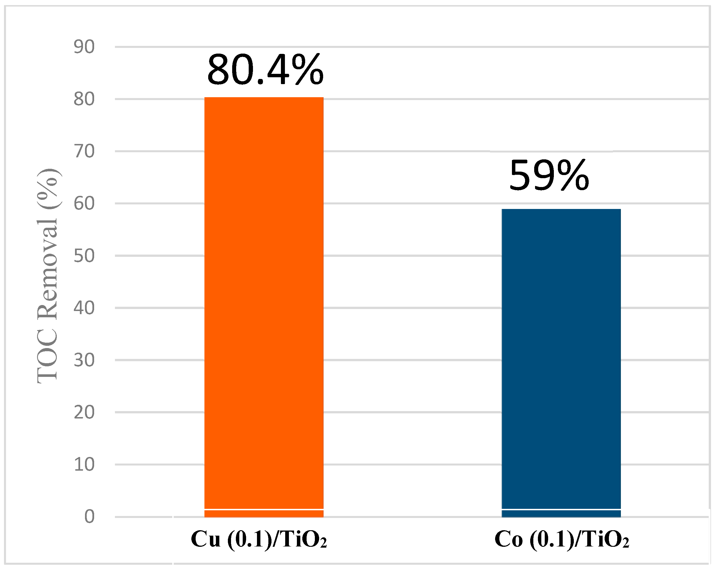
3.2. Photodegradation of AMX upon Sunlight Irradiation

The photocatalytic degradation of AMX in aqueous solution ([AMX]0) = 5 mg/L) under solar irradiation was monitored using Cu (X)/TiO2 and Co (X)/TiO2 macrocomposite pellets with different metal concentrations (X = 0.1%, 0.5%, 1.0%).
Cu (X) and Co (X) catalysts, with varying metal concentrations, were tested for their efficiency in degrading AMX under sunlight exposure [34]. The most effective concentrations were 0.1% and 0.5% for Co, and 0.1% and 1% for Cu (Figure 4 and Table 4). Both catalysts exhibited similar degradation trends, achieving comparable efficiencies at 120 min and beyond, ultimately reaching ca. 86% AMX removal after 180 min. This highlights their significant potential for AMX degradation.

3.3. Parameters Affecting Photocatalytic Degradation of AMX

3.3.1. Direct Photolysis vs. Photocatalysis

AMX solutions were exposed to sunlight without a catalyst and, after 180 min, only 29.9% of AMX was degraded (Figure 4 and Table 4). When macrocomposite photocatalysts of different compositions were added, both the reaction rate and the final percentage of degradation increased largely. The observed processes followed pseudo-first-order kinetics. In all cases, the M (0.1%)/TiO2 materials outperformed with respect to other with higher percentages of M. Based on this, we selected these as reference materials for the optimization of the parameters affecting the process.
Although the results are promising within the context of how to easily recycle this catalyst, it must be noted that such results cannot be easily translated into real-world applications. The long-term stability of the doped catalysts, such as the leaching of Cu and Co metals, is one of the concerns. However, stability tests of this catalyst showcase continuous degradation efficiency for amoxicillin after several cycles of photodegradation (Figure 5). This shows that the material retains integrity after time, and multiple uses directly correlate to cost effectiveness and actual use in the field.

3.3.2. pH Effect

Table 5 shows the effect of the acidity (3.3 < pH < 8.6) on the degradation efficiency of AMX under sunlight by the prepared photocatalyst macrocomposites. The largest AMX percentage of photodegradation was observed at pH 3.3 for both photocatalysts.
The dependence observed on the acidity can be attributed to the relative charges of both AMX and the photocatalyst surface. The following acid-base equilibria take place at the surface of M (0.1%)/TiO2 [35]:
TiOH + H+ D TiOH2+
TiOH + HO D TiO + H2O
Below the point of zero charge (pHPZC), the photocatalyst surface carries a net positive charge, while at pH > pHPZC, it becomes negatively charged. For the prepared macrocomposites, the pHPZC values were determined to be 4.5 for Cu (0.1%)/TiO2 and 4.4 for Co (0.1%)/TiO2, indicating that the surface remains positively charged only at pH levels below approximately 4.4. Regarding AMX, three pKa values have been reported [36]: 2.7 (-CO2H), 7.5 (-NH2), and 9.6 (phenolic ring). As the pH increases from 3.3 to 8.6, both the photocatalyst surface and AMX molecule acquire negative charges. The increasing electrostatic repulsion between the negatively charged surface and AMX molecules contribute to diminishing the adsorption efficiency, resulting in reduced AMX removal as pH increases. Additionally, Bougarrani et al. [37,38] observed that TiO2 particles tend to form agglomerates in alkaline aqueous media. These agglomerates can further contribute to the observed reduction in AMX removal rates, as they decrease the available surface area for photocatalytic activity.

3.3.3. Effect of AMX Concentration

The effect of the initial AMX concentration on its photocatalytic degradation is compiled in Table 6. Degradation experiments were conducted with varying [AMX]0 = (1, 3, 5, 7 and 9) mg/L under sunlight irradiation for 180 min. The degradation efficiency changes from 95% for [AMX]0 = 1 mg/L to 75% at 3 mg/L for Cu (0.1%)/TiO2 and Co (0.1%)/TiO2. However, when the concentration of AMX was further increased to [AMX]0 = 9 mg/L, the degradation efficiency stabilized, reaching a plateau for both catalysts. This trend can be explained by the accumulation of organic substances on the photocatalyst surface as the initial concentration of AMX increases. Once all active sites on the photocatalyst surface are occupied, the degradation efficiency becomes constant, indicating saturation. AMX degradation with a photocatalyst loading of 1 g/L (Cu (0.1%)/TiO2 and Co (0.1%)/TiO2 achieves higher degradation at higher concentrations, particularly at pH = 5.9.

4. Degradation Pathways

Under sunlight irradiation, M (0.1%)/TiO2 macrocomposites show a high AMX removal efficiency (~85% after 180 min, Figure 4). However, AMX mineralization, measured as TOC removal, is less pronounced, achieving 80% with Cu (0.1)/TiO2 and 59% with Co (0.1)/TiO2 (Figure 6 and Figure S1, Table S1). LC-MS/MS analysis was performed to elucidate the reaction mechanism, revealing identical products for both macrocomposites; the differences lie in its reaction rate and stability.
Two primary decomposition pathways were identified (Figures S2 and S3, Figure 7). Pathway 1 involves the opening of the β-lactam ring (Figure 7), leading to the formation of amoxicillin penicilloic acid (A1, m/z = 383.12). Subsequent decarboxylation and deamination reactions generate the intermediates A2 (m/z = 357) and A3 (m/z = 431) [39].
Pathway 2 involves cleavage in the central section of AMX, producing intermediates B1 (m/z = 205.08) and B2 (m/z = 185). Intermediates C1 and C2 emerge from both pathways; specifically, C2 (m/z = 176.06) is derived from A3 and A2 through -NH-CO- bond cleavage, while phenol (C1, m/z = 95.04) originates from B2 and/or A2. As the reaction progresses, small carboxyl groups and aliphatic compounds are gradually formed, eventually mineralizing into CO2 and H2O.
The improved charge separation and transfer dynamics of Cu (0.1%)/TiO2 can account for its enhanced performance. Doping TiO2 with Cu creates extra energy levels within the bandgap of TiO2, working as electron traps that decrease the recombination rate of photoinduced e/h+ pairs. This leads to an increase in reactive species (HO and O2•−) available for reaction with amoxicillin. Regarding cobalt (Co) doping, although it enhances charge separation, it may give rise to recombination centers in higher concentrations, and this can be detrimental for photocatalytic activities. Moreover, the redox potentials of Cu+/Cu2+ species are more advantageous in reactive oxygen species (ROS) production than Co2+/Co³+, leading to the higher mineralization efficiency that we observed [40,41,42].

5. Conclusions

This study focused on preparing and characterizing efficient macrocomposites (in the form of pellets) for the abatement of water pollutants, specifically targeting the photodegradation of AMX under sunlight irradiation. AMX was selected as a model compound due to its widespread use and frequent detection in aquatic environments. Recent reviews have highlighted the environmental risks associated with AMX contamination and the effectiveness of photocatalytic degradation as a remediation strategy [43,44].
Kaolin clay and TiO2-P25 composites were doped with Cu and Co using the incipient wetness impregnation method. The resulting macrocomposites were characterized using SEM-EDS, XRD, XRF, textural property analysis, and their lixiviation through ICP-MS.
The effects of medium acidity, AMX concentration, catalyst percentage, and reuse on AMX degradation under sunlight were monitored via UV–Vis spectrophotometry and UV–Vis HPLC analysis at 226 and 272 nm. A pseudo-first-order kinetic model fitted the observed data for AMX photodegradation. Optimal formulations, Cu (0.1%)/TiO2 and Co (0.1%)/TiO2, achieved up to 95% photodegradation of 5 mg·L−1 AMX in 180 min at pH 5.9 with a catalyst load of 1 g·L−1. While both doped macrocomposites exhibited similar photodegradation percentages, the Cu-doped composite demonstrated a slightly faster reaction rate and higher TOC removal (80.4%) compared to the Co-doped composite (59%) after 180 min of sunlight irradiation under the same conditions (vide supra). The results indicate that Cu and Co-doped kaolin clay and TiO2-P25 composites are effective photocatalysts for AMX degradation in water under solar irradiation.
The main photodegradation intermediates were identified via LC-MS, revealing consistent intermediates regardless of the doped metal, and a reaction mechanism was proposed. These cost-effective and durable macrocomposites exhibit good durability with efficient and consistent recycling over more than five cycles, making them ideal candidates for mitigating antibiotic pollution in water-treatment facilities.

Supplementary Materials

The following supporting information can be downloaded at: https://www.mdpi.com/article/10.3390/ma18071394/s1, Figure S1: Degradation %, COD and BOD of Amoxicillin Over Time using Cu (0.1)/TiO2 and Co (0.1)/TiO2. [AMX]0 = 15 mg/L; catalyst dose = 1 g/L; pH = 5.9; T = 19 °C; Figure S2: Chromatogram of Amoxicillin; Figure S3: Mass spectra of the byproducts during the photocatalytic degradation of Amoxicillin using Cu (0.1%)/TiO2 under sunlight. Table S1: Residual COD, BOD and BOD/COD Ratio for Cu (0.1)/TiO2, Co (0.1%)/TiO2 Over Time. [AMX]0 = 15 mg/L; catalyst dose = 1 g/L; pH=5.9; T = 19 °C.

Author Contributions

Conceptualization, M.C. and J.A.S.; methodology, M.C. and J.A.S.; software, M.C. and J.A.S.; validation, M.C., S.S.T., Z.M. and J.A.S.; formal analysis, M.C., S.S.T., Z.M. and J.A.S.; investigation, M.C., S.S.T., Z.M. and J.A.S.; resources, M.C. and J.A.S.; data curation, M.C., S.S.T., Z.M. and J.A.S.; writing—original draft preparation, M.C., S.S.T. and Z.M.; writing—review and editing, M.C., S.S.T. and J.A.S.; visualization, M.C., S.S.T., Z.M. and J.A.S.; supervision, M.C. and Z.M.; project administration, M.C. and J.A.S.; funding acquisition, M.C. and J.A.S. All authors have read and agreed to the published version of the manuscript.

Funding

PID2021-127898OB-I00 (WAntRed), funded by MICIU/AEI and “ERDF: A way of makingEurope” Xunta de Galicia through project GRC/ED431C 2023/33.

Institutional Review Board Statement

Not applicable.

Informed Consent Statement

Not applicable.

Data Availability Statement

The original contributions presented in this study are included in the article/Supplementary Material. Further inquiries can be directed to the corresponding author.

Acknowledgments

S.S.T. is grateful to the Erasmus+ program for a mobility grant.

Conflicts of Interest

The authors declare no conflict of interest.

References

  1. Christensen, A.; Gurol, M.D.; Garoma, T. Treatment of persistent organic compounds by integrated advanced oxidation processes and sequential batch reactor. Water Res. 2009, 43, 3910–3921. [Google Scholar] [CrossRef] [PubMed]
  2. Martín de Vidales, M.J.; Millán, M.; Saez, C.; Canizares, P.; Rodrigo, M.A. Irradiated-assisted electrochemical processes for the removal of persistent pollutants from real wastewater. Sep. Purif. Technol. 2017, 175, 428–434. [Google Scholar] [CrossRef]
  3. Miwa, T.; Kaneco, S.; Katsumata, H.; Suzuki, T. Photocatalytic hydrogen production from aqueous methanol solution with CuO/Al2O3/TiO2 nanocomposite. Int. J. Hydrogen Energy 2010, 35, 6554–6560. [Google Scholar] [CrossRef]
  4. Jiang, X.; Manawan, M.; Feng, T.; Qian, R.; Zhao, T.; Zhou, G.; Kong, F.; Wang, Q.; Dai, S.; Pan, J.H. Anatase and rutile in Evonik Aeroxide P25: Heterojunctioned or individual nanoparticles? Catal. Today 2018, 300, 12–17. [Google Scholar] [CrossRef]
  5. Belekbir, S.; El Azzouzi, M.; El Hamidi, A.; Rodríguez-Lorenzo, L.; Santaballa, J.A.; Canle, M. Improved photocatalyzed degradation of phenol, as a model pollutant, over metal-impregnated nanosized TiO2. Nanomaterials 2020, 10, 996. [Google Scholar] [CrossRef]
  6. Belekbir, S.; El Azzouzi, M.; Rodríguez-Lorenzo, L.; El Hamidi, A.; Santaballa, J.A.; Canle, M. Cobalt impregnation on titania photocatalysts enhances Vis phenol photodegradation. Materials 2023, 16, 4134. [Google Scholar] [CrossRef]
  7. Gaim, Y.T.; Yimanuh, S.M.; Kidanu, Z.G. Enhanced photocatalytic degradation of amoxicillin with Mn-doped Cu2O under sunlight irradiation. J. Compos. Sci. 2022, 6, 317. [Google Scholar] [CrossRef]
  8. Verma, M.; Haritash, A.K. Photocatalytic degradation of Amoxicillin in pharmaceutical wastewater: A potential tool to manage residual antibiotics. Environ. Technol. Innov. 2020, 20, 101072. [Google Scholar] [CrossRef]
  9. Sddiqa, A.; Masih, D.; Anjum, D.; Siddiq, M. Cobalt and sulfur co-doped nano-size TiO2 for photodegradation of various dyes and phenol. J. Environ. Sci. (China) 2015, 37, 100–109. [Google Scholar] [CrossRef]
  10. Andreozzi, R.; Canterino, M.; Marotta, R.; Paxeus, N. Antibiotic removal from wastewaters: The ozonation of amoxicillin. J. Hazard. Mater. 2005, 122, 243–250. [Google Scholar] [CrossRef]
  11. Yu, J.; Lv, L.; Lan, P.; Zhang, S.; Pan, B.; Zhang, W. Effect of effluent organic matter on the adsorption of perfluorinated compounds onto activated carbon. J. Hazard. Mater. 2012, 225–226, 99–106. [Google Scholar] [CrossRef]
  12. Konstantinou, I.K.; Albanis, T.A. TiO2-assisted photocatalytic degradation of azo dyes in aqueous solution: Kinetic and mechanistic investigations: A review. Appl. Catal. B Environ. 2004, 49, 1–14. [Google Scholar] [CrossRef]
  13. Romeiro, A.; Freitas, D.; Azenha, M.E.; Canle, M.; Burrows, H.D. Effect of the calcination temperature on the photocatalytic efficiency of acidic sol-gel synthesized TiO2 nanoparticles in the degradation of alprazolam. Photochem. Photobiol. Sci. 2017, 16, 935–945. [Google Scholar] [CrossRef] [PubMed]
  14. Directive (EU) 2020/2184 of the European Parliament and of the Council of 16 December 2020 on the Quality of Water Intended for Human Consumption. Available online: http://data.europa.eu/eli/dir/2020/2184/oj (accessed on 2 January 2025).
  15. Lace, A.; Cleary, J. A review of microfluidic detection strategies for heavy metals in water. Chemosensors 2021, 9, 60. [Google Scholar] [CrossRef]
  16. Olama, N.; Dehghani, M.; Malakootian, M. The removal of amoxicillin from aquatic solutions using the TiO2/UV-C nanophotocatalytic method doped with trivalent iron. Appl. Water Sci. 2018, 8, 1–12. [Google Scholar] [CrossRef]
  17. Balarak, D.; Mostafapour, F.K. Photocatalytic degradation of amoxicillin using UV/Synthesized NiO from pharmaceutical wastewater. Indones. J. Chem. 2019, 19, 211–218. [Google Scholar] [CrossRef]
  18. Carp, O.; Huisman, C.L.; Reller, A. Photoinduced reactivity of titanium dioxide. Prog. Solid State Chem. 2004, 32, 33–177. [Google Scholar] [CrossRef]
  19. Zhang, X.; Chen, Y.L.; Liu, R.; Tsai, D.P. Plasmonic photocatalysis. Rep. Prog. Phys. 2013, 76, 046401. [Google Scholar] [CrossRef]
  20. Wang, H.; Zhang, L.; Chen, Z.; Hu, J.; Li, S.; Wang, Z.; Liu, J.; Wang, X. Semiconductor heterojunction photocatalysts: Design, construction, and photocatalytic performances. Chem. Soc. Rev. 2014, 43, 5234–5244. [Google Scholar] [CrossRef]
  21. Li, X.; Yu, J.; Jaroniec, M. Hierarchical photocatalysts. Chem. Soc. Rev. 2016, 45, 2603–2636. [Google Scholar] [CrossRef]
  22. Schneider, J.; Matsuoka, M.; Takeuchi, M.; Zhang, J.; Horiuchi, Y.; Anpo, M.; Bahnemann, D.W. Understanding TiO2 photocatalysis: Mechanisms and materials. Chem. Rev. 2014, 114, 9919–9986. [Google Scholar] [CrossRef] [PubMed]
  23. Etacheri, V.; Di Valentin, C.; Schneider, J.; Bahnemann, D.; Pillai, S.C. Visible-light activation of TiO2 photocatalysts: Advances in theory and experiments. J. Photochem. Photobiol. C Photochem. Rev. 2015, 25, 1–29. [Google Scholar] [CrossRef]
  24. Liu, G.; Wang, L.; Yang, H.G.; Cheng, H.-M.; Lu, G.Q. Titania-based photocatalysts—Crystal growth, doping and heterostructuring. J. Mater. Chem. 2010, 20, 831–843. [Google Scholar] [CrossRef]
  25. Zhao, X.W.; Jin, W.Z.; Cai, J.G.; Ye, J.F.; Li, Z.H.; Ma, Y.R.; Xie, J.L.; Qi, L.M. Shape- and Size-Controlled Synthesis of Uniform Anatase TiO2 Nanocuboids Enclosed by Activity {100} and {001}. Adv. Funct. Mater. 2011, 21, 3554–3563. [Google Scholar]
  26. Shieh, D.L.; Lin, Y.S.; Yeh, J.H.; Chen, S.C.; Lin, B.C.; Lin, J.L. N-doped, porous TiO2 with rutile phase and visible light sensitive photocatalytic activity. Chem. Commun. 2012, 48, 2528–2530. [Google Scholar] [CrossRef]
  27. Pongwan, P.; Wetchakun, K.; Phanichphant, S.; Wetchakun, N. Enhancement of visible-light photocatalytic activity of Cu-doped TiO2 nanoparticles. Res. Chem. Intermed. 2016, 42, 2815–2830. [Google Scholar] [CrossRef]
  28. Ganesh, I.; Kumar, P.P.; Annapoorna, I.; Sumliner, J.M.; Ramakrishna, M.; Hebalkar, N.Y.; Padmanabham, G.; Sundararajan, G. Preparation and characterization of Cu-doped TiO2 materials for electrochemical, photoelectrochemical, and photocatalytic applications. Appl. Surf. Sci. 2014, 293, 229–247. [Google Scholar] [CrossRef]
  29. Ohtani, B.; Prieto-Mahaney, O.O.; Li, D.; Abe, R. What is Degussa (Evonik) P25? Crystalline composition analysis, reconstruction from isolated pure particles and photocatalytic activity test. J. Photochem. Photobiol. A Chem. 2010, 216, 179–182. [Google Scholar] [CrossRef]
  30. Thommes, M.; Kaneko, K.; Neimark, A.V.; Olivier, J.P.; Rodríguez-Reinoso, F.; Rouquerol, J.; Sing, K.S.W. Physisorption of gases, with special reference to the evaluation of surface area and pore size distribution (IUPAC technical report). Pure Appl. Chem. 2015, 87, 1051–1069. [Google Scholar] [CrossRef]
  31. Emmett, P.H. Gases in multimolecular layers. J. Am. Chem. Soc. 1936, 58, 407. [Google Scholar]
  32. Chiang, K.; Amal, R.; Tran, T. Photocatalytic degradation of cyanide using titanium dioxide modified with copper oxide. Adv. Environ. Res. 2002, 6, 471–485. [Google Scholar]
  33. Tianping, L.V.; Zhao, J.; Chen, M.; Shen, K.; Zhang, D.; Zhang, J.; Zhang, G.; Liu, Q. Boosted visible-light photodegradation of methylene blue by V and Co co-doped TiO2. Materials 2018, 11, 1946. [Google Scholar] [CrossRef] [PubMed]
  34. Bougarrani, S.; Skadell, K.; Arndt, R.; El Azzouzi, M.; Glaser, R. Novel CaxMnOy/TiO2 composites for efficient photocatalytic degradation of methylene blue and the herbicide imazapyr in aqueous solution under visible light irradiation. J. Environ. Chem. Eng. 2018, 6, 1934–1942. [Google Scholar] [CrossRef]
  35. Kristin, H.; Bettina, S.; Gerrit, S.; Thorsten, R. New hydrolysis products of the beta-lactam antibiotic amoxicillin, their pH-dependent formation and search in municipal wastewater. Water Res. 2016, 88, 880–888. [Google Scholar] [CrossRef]
  36. Trovó, A.G.; Nogueira, R.F.P.; Agüera, A.; Fernandez-Alba, A.R.; Malato, S. Degradation of the antibiotic amoxicillin by photo-Fenton process—Chemical and toxicological assessment. Water Res. 2011, 45, 1394–1402. [Google Scholar] [CrossRef]
  37. Ali, M.A.; Maafa, I.M. Photodegradation of amoxicillin in aqueous systems: A review. Int. J. Mol. Sci. 2024, 25, 9575. [Google Scholar] [CrossRef]
  38. Li, X.; Zhang, Q.; Guo, Y.; Shi, H.; Wang, J.; Cheng, X. Synthesis and characterization of Cu and Co doped TiO2/kaolin composites for photocatalytic degradation of organic pollutants. J. Environ. Chem. Eng. 2017, 5, 4876–4884. [Google Scholar] [CrossRef]
  39. Elmolla, E.S.; Chaudhuri, M. Photocatalytic degradation of amoxicillin, ampicillin and cloxacillin antibiotics in aqueous solution using UV/TiO2 and UV/H2O2/TiO2 photocatalysis. Desalination 2010, 252, 46–52. [Google Scholar] [CrossRef]
  40. Kümmerer, K. Antibiotics in the aquatic environment—A review—Part I. Chemosphere 2009, 75, 417–434. [Google Scholar] [CrossRef]
  41. Wang, X.; Li, S.; Yu, H.; Yu, J.; Liu, S. Ag2O as a new visible-light photocatalyst: Self-stability and high photocatalytic activity. Chem. Eur. J. 2011, 17, 7777–7780. [Google Scholar]
  42. Linic, S.; Christopher, P.; Ingram, D.B. Plasmonic-metal nanostructures for efficient conversion of solar to chemical energy. Nat. Mater. 2011, 10, 911–921. [Google Scholar] [CrossRef] [PubMed]
  43. Zhang, J.; Xu, Q.; Feng, Z.; Li, M.; Li, C. Importance of the relationship between surface phases and photocatalytic activity of TiO2. Angew. Chem. Int. Ed. 2008, 47, 1766–1769. [Google Scholar] [CrossRef]
  44. Norte, T.H.; Marcelino, R.B.P.; Medeiros, F.H.A.; Moreira, R.P.L.; Amorim, C.C.; Lago, R.M. Ozone oxidation of β-lactam antibiotic molecules and toxicity decrease in aqueous solution and industrial wastewaters heavily contaminated. Ozone Sci. Eng. 2018, 40, 385–391. [Google Scholar] [CrossRef]
Scheme 1. Amoxicillin structure.
Materials 18 01394 sch001
Figure 1. SEM-EDS micrographs of raw Cu (0.1%)/TiO2 (A,C) and Co (0.1%)/TiO2 (B,D) calcined at 600 °C.
Materials 18 01394 g001
Figure 2. X-ray diffraction patterns of clay (kaolin) (black),TiO2-P25 (red), Cu (0.1%)/TiO2, (blue), and Co (0.1%)/TiO2 (green). (A) anatase; (R) rutile.
Materials 18 01394 g002
Figure 3. N2 adsorption–desorption isotherm of (A) Cu (0.1%)/TiO2 and (B) Co (0.1%)/TiO2 photocatalyst. Inset: pore-size distribution.
Materials 18 01394 g003
Figure 4. AMX photocatalytic degradation using different Co (X%)/TiO2 and Cu (X%)/TiO2 macrocomposites (pellets) under sunlight. [AMX]0 = 5 mg/L; photocatalyst load = 1 g/L; pH = 5.9; T = 19 °C.
Materials 18 01394 g004
Figure 5. Macrocomposites reused five times with the same conditions. [AMX]₀ = 5 mg/L; photocatalyst load = 1 g/L; pH = 5.9; T = 19 °C.
Materials 18 01394 g005
Figure 6. Percentage of TOC removal in the AMX degradation under sunlight irradiation using the as-synthetized M (0.1%)/TiO2 photocatalysts. [AMX]0 = 5 mg/L; catalyst dose = 1 g/L; pH = 5.9; T =19 °C.
Materials 18 01394 g006
Figure 7. Proposed pathways for AMX degradation under sunlight irradiation using the as-synthetized macrocomposites (Cu (0.1%)/TiO2 and Co (0.1%)/TiO2.
Materials 18 01394 g007
Table 1. Chemical analysis Cu (0.1%)/TiO2, Co (0.1%)/TiO2 pellets measured by XRF.
%TiO2SiO2Al2O3Fe2O3K2OSO3CO2CaOCuOCoONiOCr2O3V2O5
Cu92.72.72.10.230.180.111.70.0390.14---0.0130.067---
Co93.52.92.20.220.190.120.510.022---0.070------0.25
Table 2. BET parameters and textural properties of Cu (0.1%)/TiO2 and Co (0.1%)/TiO2 photocatalysts measured by N2 adsorption–desorption. TiO2-P25, BET surface area = 51 m2·g−1.
PhotocatalystCu (0.1%)/TiO2Co (0.1%)/TiO2
PropertyRef. [32]This WorkThis WorkRef. [33]
SBET (m2·g−1)51 a47.0 ± 0.145.5 ± 0.122.8 a
Constant C 139.5119.7
Pore volume (cm3·g−1) 0.1010.0950.033
Average pore width (4 V/A) (nm) 96 b94 b65 c
Average particle size (A) 127.5131.7
Median pore width (nm) 0.77580.7788
a Hydrothermal synthesis; b D-H desorption; c average pore diameter.
Table 3. Dark adsorption of the AMX of the as-synthetized photocatalysts. [AMX]0 = 5 mg/L; photocatalyst load = 1 g/L; pH = 5.9; T = 19 °C.
Time/min153060120180
% AMX adsorption on Cu (0.1)/TiO20.542.565.428.4013.82
% AMX adsorption on Co (0.1)/TiO23.055.512.5014.5118.01
Table 4. Observed apparent first-order rate constant and AMX photocatalytic degradation under sunlight using the prepared M (X%)/TiO2 macrocomposites (pellets). [AMX]0 = 5 mg/L; photocatalyst load = 1 g/L; pH = 5.9; T = 19 °C.
Photocatalystk·103/min−1% Removal at 120 min% Removal at 180 min
Photolysis39.624.231.1
Cu (0.1%)/TiO2152.586.786.4
Cu (0.5%)/TiO2102.565.379.2
Cu (1.0%)/TiO2128.488.087.6
Co (0.1%)/TiO2107.283.484.4
Co (0.5%)/TiO276.981.782.3
Co (1.0%)/TiO228.656.769.1
Table 5. Influence of pH on the photocatalytic degradation of AMX under sunlight. [AMX]0 = 5 mg/L; catalyst dose = 1 g/L; T =19 °C.
PhotocatalystPropertypH
3.35.98.6
Cu (0.1%)/TiO2k·103/min−164.1152.59.2
% removal at 180 min90.986.486.1
Co (0.1%)/TiO2k·103/min−1133.7107.237.0
% removal at 180 min89.584.476.8
Table 6. The effect of [AMX]0 on its photocatalytic degradation. AMX percentage removal, after 180 min under sunlight, is reported. Catalyst load 1 g/L; pH = 5.9; T =19 °C.
[AMX]0/mg·L−113579
Cu (0.1%)/TiO295.473.986.487.882.0
Co (0.1%)/TiO296.475.384.466.982.0
Disclaimer/Publisher’s Note: The statements, opinions and data contained in all publications are solely those of the individual author(s) and contributor(s) and not of MDPI and/or the editor(s). MDPI and/or the editor(s) disclaim responsibility for any injury to people or property resulting from any ideas, methods, instructions or products referred to in the content.

