Article

Detection and Microscopy of Alnus glutinosa Pollen Fluorescence Peculiarities

by
Ingrida Šaulienė
1,*,
Laura Šukienė
1,
Gintautas Daunys
1,
Gediminas Valiulis
1,
Alfredas Lankauskas
1,
Inese Kokina
2,
Vjačeslavs Gerbreders
2 and
Inese Gavarāne
2
1
Institute of Regional Development, Šiauliai University, 76351 Šiauliai, Lithuania
2
Institute of Life Science and Technology, Daugavpils University, LV5401 Daugavpils, Latvia
*
Author to whom correspondence should be addressed.
Forests 2019, 10(11), 959; https://doi.org/10.3390/f10110959
Submission received: 30 September 2019 / Revised: 19 October 2019 / Accepted: 24 October 2019 / Published: 28 October 2019
(This article belongs to the Special Issue Study of Tree Pollen and Pollination)

Abstract

:
Alnus glutinosa is an important woody plant in Lithuanian forest ecosystems. Knowledge of fluorescence properties of black alder pollen is necessary for scientific and practical purposes. By the results of the study, we aimed to evaluate possibilities of identifying Alnus glutinosa pollen fluorescence properties by modeling ozone effect and applying two different fluorescence-based devices. To implement the experiments, black alder pollen was collected in a typical habitat during the annual flowering period in 2018–2019. There were three groups of experimental variants, which differed in the duration of exposure to ozone, conditions of pollen storage before the start of the experiment, and the exposure time. Data for pollen fluorescence analysis were collected using two methods. The microscopy method was used in order to evaluate the possibility of employing image analysis systems for investigation of pollen fluorescence. The second data collection method is related to an automatic device identifying pollen in real time, which uses the fluorescence method in the pollen recognition process. Data were assessed employing image analysis and principal component analysis (PCA) methods. Digital images of ozone-exposed pollen observed under the fluorescence microscope showed the change of the dominant green colour toward the blue spectrum. Meanwhile, the automatic detector detects more pollen whose fluorescence is at the blue light spectrum. It must be noted that assessing pollen fluorescence several months after exposure to ozone, no effect of ozone on fluorescence remains.

1. Introduction

Pollen, like any airborne particle of biological origin, can be identified using fluorescence examination methods. Application of these methods in the bioparticle identification process promotes designing and development of laser fluorescence–based devices for recognising airborne pollen in real time [1,2,3]. The topics of pollen fluorescence have been the focus of scientists for many years, and this issue is constantly readdressed. Identification of fossil pollen and spores in geological samples and the possibility of dating contaminated sediments became a strong impetus for gathering knowledge of pollen fluorescence [4,5]. Fluorescence-microscopical techniques and the physical and chemical nature of pollen walls have expanded the possibilities of palynological research. First, laboratory tests have shown that pollen fluoresced under ultraviolet light [4]. Also, fresh and subfossil pollen grains and spores showed various fluorescence colours, depending on type or species [5]. Studies on pollen in geological samples confirm that the exine of a pollen grain is naturally autofluorescent and the strength of the fluorescence varies with exine thickness [6]. Fluorescence microscopy turned into a tool for studying different pollen type morphology, while acquired knowledge became valuable in analysing not only fossil biological particles but also airborne particles [7,8,9,10,11].
Studies on the fluorescence of airborne particles (pollen and spores) were encouraged not by the aspiration to acquired additional knowledge of particle morphology but by assessment of practical possibilities of applying the method itself. A new stimulus of employing the fluorescence method is a growing demand for devices capable of automatic identification of the airborne pollen and spores. Fluorescence microscopy has been used to develop 3D volumetric imaging technology [7] for designing the online pollen monitoring system. At present, microscopy techniques for identification of pollen and spores still prevail worldwide [12]. These methods have become traditional in aerobiology [13,14], requiring that specialists should identify each particle present in air samples under the microscope. For more than 15 years, there have been attempts to develop semi-automatic [1,15] or automatic [7,10,16] pollen identification systems, in which the use of the fluorescence method for identification of airborne particles occupies an important place. However, to date, the results of collected data, gained by automatic real-time devices, differ [10,11,17,18], and these differences may be determined not only by peculiarities of devices but also by conditions of the environment affecting identified pollen [19,20,21,22]. This fact is confirmed by scientific studies that are targeted at long-distance pollen transport analysis [23,24,25,26,27,28,29]. It is assumed that pollen suspended in the air is affected by environmental conditions (air temperature, humidity, radiation, chemical compounds), and the effect may change pollen fluorescence results (intensity, colour, etc.). Laboratory tests showed that pollen has chemosensitivity to ozone and ozone could directly oxidize the constituents of the pollen wall [30,31], although the number of pollen grains produced per inflorescence was unaffected by ozone [32]. There is scientific evidence that ozone alters the allergen content in the pollen [33,34,35] reduces or destroys viability of the pollen [36,37,38] and changes fluorescence properties of fluorescent substances present in pollen [8,30,31]. Studies on pollen fluorescence properties are conducted employing various devices ranging from the microscope to devices enabling automatic identification. Their application in routinely performed monitoring of airborne pollen requires specific knowledge.
By the results of our study, we aimed to evaluate the possibilities of identifying Alnus glutinosa pollen fluorescence properties by modeling ozone effect and applying two different fluorescence-based devices.

2. Materials and Methods

2.1. Pollen Samples

Alnus glutinosa ((L.) Gaertn.) is an important woody plant in Lithuanian forest ecosystems. It is the fourth most abundant tree in Lithuanian forests with an occurrence of around 8.5% [39]. Black alder is one of the earliest flowering plants producing abundant content of allergenic pollen [40].
Catkins for the experiments were collected in the forest, in the typical habitat of Alnus glutinosa during the annual flowering period. Catkins were collected from the same plant at the beginning of April 2018 and again at the end of March 2019 because we aimed to test the influence of storage conditions on pollen fluorescence. Weather conditions (data taken from Lithuanian hydrometeorological service Šiauliai weather station) in 2018 and in 2019 from January until March were different (Table 1). The beginning of 2019 was colder, and the average air temperature in February and March was higher than in 2018.
In 2018, catkins were kept in a thermostatic convection oven in the laboratory for three days at 40 °C, and in 2019, at 30 °C. The procedure of pollen separation from catkins was performed according to Šaulienė et al. [11]. Until the start of the experiment in 2018, clean, impurity-free pollen was stored in the refrigerator at a temperature of 5 ± 1 °C and relative humidity of 57 ± 10%. In 2019, was kept indoors at 25 ± 5 °C and 45 ± 10%, respectively. These different temperatures were used to ascertain the effect of temperature on pollen fluorescence trait.

2.2. Pollen Exposures to O3

The experiments were carried out in laboratory conditions using an ozone generator operating on the high voltage discharge principle. Ozone is generated in a sealed plastic box containing a system of insulators and electrodes. The applied high voltage (approx. 25 kV) to the electrodes creates an ionisation current that passes through an air gap and gives high energy to electrons to break the oxygen molecule, allowing the formation of a 3-atom oxygen molecule—ozone (Figure 1).
Here, 5 mg of Alnus glutinosa pollen were dispersed in two 100-mm-diameter Petri dishes and placed in the generator box (Figure 1). The first sample was kept in a running generator for 3 h per day, and the second sample was kept running for 5 h. The experiment was run for five days. In total, the first sample was exposed to ozone for 15 h, and the second was exposed for 25 h. The average measured ozone concentration was 5.83 ppm. The measurements were performed using GV-100 gas sampling pump (Gastec Corporation, Ayase-Shi, Japan) with colorimetric tubes GASTEC Ozone 18L and GASTEC Ozone 18M (Gastec Corporation, Ayase-Shi, Japan). The measurement of ozone concentration was performed every 30 min.
Groups of experimental variants of pollen, formed this way (Table 2) differed in the duration of exposure (3 and 5 h) to ozone, pollen storage conditions (refrigerator or indoors) until the start of the experiment, and the time of their use in the experiment (1–5 months). After exposure, pollen was stored in room temperature conditions. Pollen of control group was not exposed to ozone therefore during the experiment stored in room conditions (C_1, C_2) or in the refrigerator (CS_1).

2.3. Pollen Fluorescence Data Collection

Data for performance of pollen fluorescence analysis were collected employing two methods. The microscopy method was used to evaluate the possibility of using image analysis systems for investigation of pollen fluorescence. The second data collection method is related to the automatic device identifying pollen in real time, which uses the fluorescence method in the pollen recognition process.

2.3.1. Fluorescence by Microscope

After exposure to ozone, pollen was stored in room conditions for 5–6 days until it was prepared for microscopy. Pollen samples were prepared for work with the fluorescence microscope having scattered pollen on slides. They were embedded into a solution of polyvinylalcohol (Gelvatol from Sigma-Aldrich, St. Louis, MO, USA) [41,42]. Pollen was photographed with Nikon ECLIPSE 80I fluorescence microscope (BioTek Instruments, Inc., Winooski, VT, USA), using 400× magnification. Samples were paced under UV light. To obtain the fluorescence image, three neutral-density (4, 8, 16) filters were used. Pollen images was digitized at random, but not less than 200 pollen per one type (pollen not affected by ozone/ozone-exposed pollen) sample. Collected digital pollen fluorescence images were analysed using image analysis techniques (see Section 2.4.1).

2.3.2. Fluorescence by Automatic Pollen Recognition Device

The study employed the device Rapid-E (Plair, Geneva, Switzerland) which has capabilities allowing real-time detection, counting and classification of airborne pollen. To identify airborne particles of biological origin, the device uses the fluorescence method. Fluorescence of particles is excited using the UV laser (Plair, Geneva, Switzerland) (320 nm light) [3]. Ozone-exposed black alder pollen and those not affected by ozone was blown into the device using the method described by Šaulienė et al. [11]. The experiment was carried out with Alnus glutinosa pollen, whose average size was less than 30 µm [43]. For pollen recognition, the device was set in the pollen mode when the range of identified particles was 5–100 µm. Not less than 10,000 fluorescent particles were analysed per one type (not exposed to ozone/ozone-exposed pollen) sample.

2.4. Data Analysis

2.4.1. Image Analysis

The RGB image digitized with the fluorescence microscope contained several or a dozen of pollen. In such cases, it is necessary to find and group pixels belonging to fluorescent pollen and count the statistics of obtained pixels. To achieve this, the following steps were made: image binarization, blob detection, morphological blob correction, turning blobs into masks, pixel selection by masks, conversion of pixel RGB values into HSV space, and calculation of statistics in HSV space for each pollen. The actions were implemented in Python programming language using modules matplotlib.image, scipy.ndimage.morphology, and scikit-image.
Image binarization requires a threshold value to distinguish the pollen from the background. The threshold value was manually selected for each photo searching for the best option. Blobs were found using the Difference of Gaussian (DoG) method implemented in the scikit-image module. To avoid erroneously detected small areas of the image, the morphological operation erosion was applied. Having applied dilation after erosion, pollen areas are restored to the original area. The obtained blobs served as masks in order to select pixels of individual pollen. Conversion of pixels into HSV space allows us to perform averaging of hue values and to obtain a generalized value for describing the fluorescence hue of the pollen. The hue value is expressed in the interval 0–1.

2.4.2. Principal Component Analysis

In previous research [11], the detection of pollen by fluorescence spectrum was performed employing artificial neural networks (ANNs). However, neural networks prevent us from finding out why one or another decision was made. Therefore, although neural networks are widely used today and often yield good recognition results, they can generate many errors if unfavourable conditions are formed. For this reason, in this study, the fluorescence spectrum analysis was performed applying the principal component analysis method (PCA).
Applying the PCA method, the correlated variables (in this case, fluorescence amplitudes of certain captured wavelengths of light) are replaced by their linear combinations that are uncorrelated with each other. Besides, these components are ranked according to the average power falling to them. The PCA method particularly serves the purpose when the highest power is borne only by a small share of components. In this case, the first component used to receive from 65% to 85% of the total power, and the first five components receive about 95% of the power.
Because of the variety of particles dispersion in the air, the excited laser of the Rapid-E device often illuminates particles other than pollen. Therefore, it is important to filter obtained data, leaving only the pollen spectra for further analysis. Pollen spectra compared to those of various artefacts are wide (the radiation power is distributed in the entire range of visible light rather than a narrow band). Therefore, the first step is to reject fluorescence events where only a narrow range light is radiated. To implement this step, the maximum value of the spectrum of one fluorescence event was taken, and its ratio with the sum of values of all components of the spectrum was calculated. The obtained ratio must not exceed the experimentally selected threshold value.
The remaining data were subjected to PCA analysis. To perform it, the Python programming language and the scikit-learn module were used. Once the principal components were obtained, fluorescence spectra emission by them was performed, thus obtaining emission coefficients.
The success of the method selection was confirmed by its repetition with the data of different samples when the results would replicate well.

3. Results

3.1. Recognition of Ozone Effect on Pollen under Fluorescence Microscope

Digitized images of Alnus glutinosa pollen fluorescence were analysed in order to evaluate whether high concentrations of ozone and duration of exposure could substantially alter fluorescence peculiarities. The results of the microscope used in this study highlighted inequalities of pollen properties in individual groups of experimental variants. Figure 2 demonstrates several cases illustrating fluorescence variations in groups.
The given examples show that walls of pollen that are not affected by ozone fluoresce more intensively than the walls of ozone-exposed pollen. The strength of the fluorescence signal also depends on the duration of ozone exposure. Pollen fluorescence images demonstrated that longer ozone exposure time weakened the fluorescence signal of pollen walls. This statement is verified by the results given in the column called “five-hour exposure.” The comparison of experimental variant groups with each other shows that the fluorescence signal of pollen that was exposed to ozone for the longest time is most altered. Systematized results given in Figure 3 demonstrate a shift of the change.
Digital images of fluorescent pollen captured under the microscope were analysed by decomposing RGB components. The tendency in the dispersion chart of the control group emerges that, by hue values, about 80% of digital image results of pollen not affected by ozone concentrate at the green spectral band. Compared to the control group, the fluorescence results of ozone-exposed pollen differ. Figure 3B shows the shift of results toward the blue portion of the spectrum. This trend is particularly pronounced in the group of experimental variants where pollen was exposed to ozone for 3 h per day. About 60% of cases cover the portion of the colour spectrum from 0.42 to 0.48. In the case of three-hour exposure, data scattering is greater than in the case of five-hour exposure. However, the fluorescence spectrum of pollen exposed to ozone for 5 h/day has a visible shift to the blue position of the spectrum (0.55–0.60). Here, most results of pollen fluorescence digital images concentrate at the green portion of the spectrum (0.33–0.43 hue values). To sum up, the assumption is formed that the fluorescence microscope used in the study showed slight changes in fluorescence of those pollen that were exposed to ozone for 3 h.

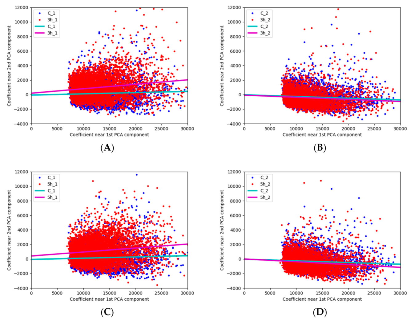
3.2. Possibilities of the Automatic Particle Detector in Assessing Ozone Effect on Pollen

Unlike the image analysis under the fluorescence microscope, the automatic particle detector allows for the evaluation of the fluorescence spectrum of airborne particles in real time, thus separating biological origin particles from the overall aerosol flow. Because the data array while capturing pollen by the automatic particle detector is usually large, the fluorescence spectrum analysis was performed applying the PCA method. In the PCA results, it is important to properly evaluate principal components. Figure 4 shows 15 principal (32 in total) PCA components of the conducted study.
The largest fluorescence amplitude of PCA component 1 includes wavelength from 400 to 550 nm, when the peak is at 450 nm. The peak of PCA component 2, expressed at 350 nm of fluorescence spectrum, is one of the few peaks of PCA components located in short wavelength. Meanwhile, PCA component 4 has several peaks located in the range of different wavelength. The first five PCA components receive about 95% of power. All combined PCA components form the fluorescence spectrum of pollen analysed. Combining all components, a more accurate image of the fluorescence spectrum is formed.
However, component 1 is principal and allows us to evaluate the shape of the spectrum. Such a situation is determined by wide scattering of experimental data; therefore, the chosen imaging method via component 1 (the dominant component that has 85% of the signal power) enables the grouping of data. Figure 5 shows the fluorescence spectrum of PCA component 1 for different groups of experimental variants.
The results given in Figure 5A show a similar pollen fluorescence spectrum of PCA component 1. This corresponds to the cases where pollen was not affected by ozone (the control group) but was stored in different conditions until the start of the experiment (1–5 months). Minor differences can be seen only in the wavelength range up to 500 nm of the Alnus glutinosa pollen fluorescence amplitude. For this reason, when analysing fluorescence peculiarities of ozone-exposed pollen, the mean of control group results was used. The fluorescence amplitude of PCA component 1 of pollen exposed to ozone is most pronounced at wavelengths up to 400 nm (Figure 5B). The tendency is observed that there are minor differences between experimental groups that substantially differ with regard to time from pollen collection until the start of the experiment. The results of PCA component 1 show that the duration of exposure to ozone in principal does not change the pollen fluorescence amplitude, which is important for the calibration of real-time pollen detectors. This means that, in the case of evaluating pollen fluorescence several months after exposure to ozone, the effect of ozone on fluorescence may not be detected. Summarising these research results, it can be seen that, essentially, the results of the principal PCA component in both control group and ozone-exposed pollen group do not differ (Figure 5). After conducting further analysis, dispersion charts of PCA component 1 and component 2 are given.
When assessing how changes in ozone-exposed pollen fluorescence are identified by real-time particle detector, we distinguished four main groups of results, which are presented in Figure 6. In cases when pollen was briefly stored in room conditions and was not affected by ozone before the start of the experiment (control C_1), PCA component coefficients do not depend on each other, as shown in Figure 6A,C. These graphs demonstrate that changes occur in cases when pollen is exposed to ozone. In cases of both three-hour exposure and five-hour exposure, direct dependence with regard to PCA component coefficients emerges. Dependence of coefficients shows that the dominant light wavelength of fluorescence of the part of pollen is decreasing. The dependence straight of C_2 coefficients is insignificantly different compared to that of C_1. The obtained results may be determined by pollen maturation, because in the case of the second control, experiments were carried out 5 months after pollen dispersal from Alnus glutinosa catkins, which is four months later than time of C_1 experiment. It should be noted that fluorescence PCA component coefficients of ozone-exposed pollen in the experiment performed after five months in principal do not differ from the control (Figure 6B,D). The same trend is observed in the results of experiments when pollen was exposed to ozone for 3 h and 5 h. The assumption is formed that not only ozone can affect pollen fluorescence—storage conditions and duration of pollen shed from catkins can also become modifying environmental factors for determining variation in fluorescence properties.

4. Discussion

This article analyses possibilities of identifying Alnus glutinosa pollen fluorescence peculiarities using manual (fluorescence microscopy) and automatic real-time devices. At present, there is an increasingly active search for tools and techniques enabling us to replace the microscopic identification of pollen in routine aerobiological monitoring with recognition by automatic real-time detectors. Operation of the latter is inseparable from fluorescence measurements of identified particles, which has been performed by spectrometers or fluorescence microscopes for many years. Various experiences have been performed analysing fluorescence properties of different types of pollen with tools enabling measurement of fluorescence peculiarities of particles and gathering plentiful valuable knowledge [7,8,9,30,31,36]. Often, available knowledge of fluorescence is difficult to apply in real-time measurements due to the different methods used so far (photo/electro-luminescence system with xenon lamps, various types of light-source-based fluorescence images, etc.) [8,31,36] or in measurement using automatic real-time devices (deep-UV laser, etc.) [10,11,18]. On the other hand, there is a lack of studies analysing pollen fluorescence characteristics of a particular plant species in various aspects, and searching for responses whether fluorescence peculiarities in a species can be modified or altered by environmental factors affecting pollen, e.g., air pollution [30,35], atmospheric condition [20,22,32], etc.
In this study, we used pollen collected from local Alnus glutinosa plant in the forest and did not process pollen with any chemical substances. The pure pollen fraction was as control. Accurately selected samples enabled us to obtain results that confirmed that the fluorescence properties of pollen changed over time. An obvious example is the disappearance of linear dependence of PCA component coefficients in the case of ozone-exposed pollen fluorescence, when samples were stored for a longer time. This result indicates that, when assessing pollen samples of different origin and preparation, qualitative parameters of pollen samples must be considered as well. We exposed Alnus glutinosa pollen to 5.83 ppm ozone to alter the fluorescence properties of pollen and analysed digital images obtained under the fluorescence microscope and pollen data captured by an automatic detector. Ozone changes the composition of the cell wall [31], increases the content of allergen in pollen [35], decreases viability [37], and otherwise modifies pollen development. Most researchers [30,31] who use fluorescence-based methods to assess ozone exposure note that fluorescence of pollen exposed to ozone is changing. Our research verifies this fact by colour changes that can be seen in the images of ozone-exposed Alnus glutinosa pollen taken under the fluorescence microscope. Blue hues appearing next to dominant green colour can be seen. The visual change is also confirmed by the analysis of digital images of pollen obtained under the fluorescence microscope. It shows that the results of pollen stored in the ozone environment shift toward the blue portion of the spectrum.
The use of a Rapid-E automatic particle detector, which identifies pollen by the fluorescence spectrum, in this study enabled us to ascertain that ozone at the concentrations tested affects the pollen fluorescence spectrum. Data analysis supplemented the results obtained under a fluorescence microscope. Rapid-E shows the dominance of blue colour in Alnus glutinosa pollen fluorescence spectrum. By analysing the pollen fluorescence spectral feature, we obtained the result that the sharp peak of PCA 1 fluorescence amplitude of pollen not affected by ozone and ozone exposed pollen was identical, i.e., was at 450 nm. Meanwhile, the study of pure alder pollen, conducted using a custom-built spectrometer, showed a sharp peak of Alnus glutinosa fluorescence at 420 nm [8]. Roshchina and Melnikova [30], who investigated changes in pollen fluorescence properties of other plant species due to ozone impact, found that fluorescence peaks of ozone-affected pollen had changed, e.g., control of Philadelphus grandiflorus (the variant that was not affected by ozone) had its peak at 465 nm, while having been exposed to 5 ppm ozone, the peak shifted toward 475–480 nm. The authors confirm that, in pollen whose fluorescence peak position was at blue or light blue colour, ozone treatment did not produce new peaks. In our experiments, there was no peak shift of fluorescence amplitude between ozone-exposed (neither 3 h or 5 h O3 exposure) pollen and pollen not affected by ozone. It is possible that the results of this investigation are typical only for Alnus glutinosa. Other researchers have shown [30] that the response to ozone exposure may be species-specific.
It should be noted that the Rapid-E device registers the fluorescence spectrum as pollen falls into the device. The fluorescence-induced laser is pulsed and emits the light beam as the pollen approaches. Therefore, the moment of light beam activation is important for successful illumination of the pollen. In addition, the pollen itself is heterogeneous. Different locations can emit light at different wavelengths. It is therefore also important to know at what angle the pollen intersects with the exciting laser beam.

5. Conclusions

Digital images of ozone-exposed pollen observed under the fluorescence microscope showed the change of dominant green colour toward the blue spectrum. Meanwhile, the automatic device detects more pollen whose fluorescence is in the blue light spectrum. It should be noted that, when evaluating pollen fluorescence several months after exposure to ozone, no effect of ozone on fluorescence remains. It has been shown that the conditions of pollen storage can influence the fluorescence spectrum of the pollen. This assumption should be confirmed by additional studies, focusing attention on the pollen peculiarities of different species.
Summarizing the obtained results, it can be stated that both fluorescence-based devices employed in this study generate similar results-generating data. Data collection by the fluorescence microscope takes significantly longer, and during the same time, less data is accumulated by the microscope than by automatic detector. However, both types of technologies can be useful for the analysis of pollen fluorescence characteristics.

Author Contributions

All the authors made significant contributions to this study. The conceptualization was performed by I.Š. and L.Š.; ozone generator created by A.L.; microscopy by I.Š., L.Š., I.K., V.G., and I.G.; experiments with particle detector were conducted by G.V.; software and validation by G.D. All authors contributed to the methodology, the interpretation of the results, and the editing of the paper.

Funding

This research has been supported by the European Social Fund (project no. 09.3.3-LMT-K-712-01-0066) under grant agreement with the Research Council of Lithuania (LMTLT).

Acknowledgments

The authors thank the Lithuanian Hydrometeorological Service for meteorological data.

Conflicts of Interest

The authors declare no conflict of interest.

References

  1. Boucher, A.; Hidalgo, P.J.; Thonnat, M.; Belmonte, J.; Galan, C.; Bonton, P.; Tomczak, R. Development of a semi-automatic system for pollen recognition. Aerobiologia 2002, 18, 195–201. [Google Scholar] [CrossRef]
  2. Mitsumoto, K.; Yabusaki, K.; Kobayashi, K.; Aoyagi, H. Development of a novel real-time pollen-sorting counter using species-specific pollen autofluorescence. Aerobiologia 2010, 26, 99–111. [Google Scholar] [CrossRef]
  3. Kiselev, D.; Bonacina, L.; Wolf, J.P. Individual bioaerosol particle discrimination by multi-photon excited fluorescence. Opt. Express 2011, 19, 24516–24521. [Google Scholar] [CrossRef] [PubMed]
  4. Asbeck, F. Fluoreszierender Blütenstaub. Naturwissenschaften 1955, 42, 632. [Google Scholar] [CrossRef]
  5. Van Gijzel, P. Autofluorescence of fossil pollen and spores with special reference to age determination and coalification. Leidse Geol. Meded. 1967, 40, 261–317. [Google Scholar]
  6. Sivaguru, M.; Urban, M.A.; Fried, G.; Wesseln, C.J.; Mander, L.; Punyasena, S.W. Comparative performance of airyscan and structured illumination superresolution microscopy in the study of the surface texture and 3D shape of pollen. Microsc. Res. Tech. 2018, 81, 101–114. [Google Scholar] [CrossRef] [PubMed]
  7. Ronneberger, O.; Schultz, E.; Burkhardt, H. Automated pollen recognition using 3D volume images from fluorescence microscopy. Aerobiologia 2002, 18, 107–115. [Google Scholar] [CrossRef]
  8. O’Connor, D.J.; Iacopino, D.; Healy, D.A.; O’Sullivan, D.; Sodeau, J.R. The intrinsic fluorescence spectra of selected pollen and fungal spores. Atmos. Environ. X 2011, 45, 6451–6458. [Google Scholar] [CrossRef]
  9. Stanley, W.R.; Kaye, P.H.; Foot, V.E.; Barrington, S.J.; Gallagher, M.; Gabey, A. Continuous bioaerosol monitoring in a tropical environment using a UV fluorescence particle spectrometer. Atmos. Sci. Lett. 2011, 12, 195–199. [Google Scholar] [CrossRef]
  10. Crouzy, B.; Stella, M.; Konzelmann, T.; Calpini, B.; Clot, B. All-Optical automatic pollen identification: Towards an operational system. Atmos. Environ. 2016, 140, 202–212. [Google Scholar] [CrossRef]
  11. Šaulienė, I.; Šukienė, L.; Daunys, G.; Valiulis, G.; Vaitkevičius, L.; Matavulj, P.; Brdar, S.; Panic, M.; Sikoparija, B.; Clot, B.; et al. Automatic pollen recognition with the Rapid-E particle counter: The first-level procedure, experience and next steps. Atmos. Meas. Tech. 2019, 2, 1–33. [Google Scholar] [CrossRef]
  12. Buters, J.T.M.; Antunes, C.; Galveias, A.; Bergmann, K.C.; Thibaudon, M.; Galán, C.; Schmidt-Weber, C.; Oteros, J. Pollen and spore monitoring in the world. Clin. Transl. Allergy 2018, 8, 9. [Google Scholar] [CrossRef] [PubMed]
  13. Hirst, J.M. An automatic volumetric spore trap. Ann. Appl. Biol. 1952, 39, 257–265. [Google Scholar] [CrossRef]
  14. Galán, C.; Smith, M.; Thibaudon, M.; Frenguelli, G.; Oteros, J.; Gehrig, R.; Berger, U.; Clot, B.; Brandao, R.; EAS QC Working Group. Pollen monitoring: Minimum requirements and reproducibility of analysis. Aerobiologia 2014, 30, 385–395. [Google Scholar] [CrossRef]
  15. Gottardini, E.; Rossi, S.; Cristofolini, F.; Benedetti, L. Use of Fourier transform infrared (FT-IR) spectroscopy as a tool for pollen identification. Aerobiologia 2007, 23, 211–219. [Google Scholar] [CrossRef]
  16. Mitsumoto, K.; Yabusaki, K.; Aoyagi, H. Classification of pollen species using autofluorescence image analysis. J. Biosci. Bioeng. 2009, 107, 90–94. [Google Scholar] [CrossRef]
  17. Oteros, J.; Pusch, G.; Weichenmeier, I.; Heimann, U.; Möller, R.; Röseler, S.; Traidl-Hoffmann, C.; Schmidt-Weber, C.; Buters, J.T. Automatic and online pollen monitoring. Int. Arch. Allergy Immunol. 2015, 167, 158–166. [Google Scholar] [CrossRef]
  18. Sofiev, M. On possibilities of assimilation of near-real-time pollen data by atmospheric composition models. Aerobiologia 2019, 35, 523–531. [Google Scholar] [CrossRef] [Green Version]
  19. Kasprzyk, I. Comparative study of seasonal and intradiurnal variation of airborne herbaceous pollen in urban and rural areas. Aerobiologia 2006, 22, 185–195. [Google Scholar] [CrossRef]
  20. Tormo Molina, R.; Silva Palacios, I.; Muñoz RodrÍguez, A.F.; Tavira Muñoz, J.; Moreno Corchero, A. Environmental factors affecting airborne pollen concentration in anemophilous species of Plantago. Ann. Bot. 2001, 87, 1–8. [Google Scholar] [CrossRef]
  21. García-Mozo, H.; Oteros, J.A.; Galán, C. Impact of land cover changes and climate on the main airborne pollen types in Southern Spain. Sci. Total Environ. 2016, 548, 221–228. [Google Scholar] [CrossRef] [PubMed]
  22. Lebourgeois, F.; Delpierre, N.; Dufrêne, E.; Cecchini, S.; Macé, S.; Croisé, L.; Nicolas, M. Assessing the roles of temperature, carbon inputs and airborne pollen as drivers of fructification in European temperate deciduous forests. Eur. J. For. Res. 2018, 137, 349–365. [Google Scholar] [CrossRef] [Green Version]
  23. Sofiev, M.; Siljamo, P.; Ranta, H.; Rantio-Lehtimäki, A. Towards numerical forecasting of long-range air transport of birch pollen: Theoretical considerations and a feasibility study. Int. J. Biometeorol. 2006, 50, 392–402. [Google Scholar] [CrossRef] [PubMed]
  24. Skjøth, C.A.; Sommer, J.; Stach, A.; Smith, M.; Brandt, J. The long-range transport of birch (Betula) pollen from Poland and Germany causes significant pre-season concentrations in Denmark. Clin. Exp. Allergy 2007, 37, 1204–1212. [Google Scholar] [CrossRef] [PubMed] [Green Version]
  25. Siljamo, P.; Sofiev, M.; Ranta, H. An approach to simulation of long-range atmospheric transport of natural allergens: An example of birch pollen. In Air Pollution Modeling and Its Application XVII; Springer: Boston, MA, USA, 2007; pp. 331–339. [Google Scholar] [CrossRef]
  26. Smith, M.; Skjøth, C.A.; Myszkowska, D.; Uruska, A.; Puc, M.; Stach, A.; Balwierz, Z.; Chlopek, K.; Piotrowska, K.; Kasprzyk, I.; et al. Long-Range transport of Ambrosia pollen to Poland. Agric. For. Meteorol. 2008, 148, 1402–1411. [Google Scholar] [CrossRef]
  27. Veriankaitė, L.; Siljamo, P.; Sofiev, M.; Šaulienė, I.; Kukkonen, J. Modelling analysis of source regions of long-range transported birch pollen that influences allergenic seasons in Lithuania. Aerobiologia 2010, 26, 47–62. [Google Scholar] [CrossRef]
  28. De Weger, L.A.; Pashley, C.H.; Šikoparija, B.; Skjøth, C.A.; Kasprzyk, I.; Grewling, Ł.; Thibaudon, M.; Magyar, D.; Smith, M. The long distance transport of airborne Ambrosia pollen to the UK and the Netherlands from Central and south Europe. Int. J. Biometeorol. 2016, 60, 1829–1839. [Google Scholar] [CrossRef] [Green Version]
  29. Sofiev, M. On impact of transport conditions on variability of the seasonal pollen index. Aerobiologia 2017, 33, 167–179. [Google Scholar] [CrossRef]
  30. Roshchina, V.V.; Melnikova, E.V. Pollen chemosensitivity to ozone and peroxides. Russ. J. Plant Physiol. 2001, 48, 74–83. [Google Scholar] [CrossRef]
  31. Roshchina, V.V.; Yashin, V.A.; Kuchin, A.V. Fluorescent analysis for bioindication of ozone on unicellular models. J. Fluoresc. 2015, 25, 595–601. [Google Scholar] [CrossRef]
  32. Albertine, J.M.; Manning, W.J.; DaCosta, M.; Stinson, K.A.; Muilenberg, M.L.; Rogers, C.A. Projected carbon dioxide to increase grass pollen and allergen exposure despite higher ozone levels. PLoS ONE 2014, 9, e111712. [Google Scholar] [CrossRef] [PubMed]
  33. Beck, I.; Jochner, S.; Gilles, S.; McIntyre, M.; Buters, J.T.; Schmidt-Weber, C.; Behrendt, H.; Ring, J.; Menzel, A.; Traidl-Hoffmann, C. High environmental ozone levels lead to enhanced allergenicity of birch pollen. PLoS ONE 2013, 8, e80147. [Google Scholar] [CrossRef] [PubMed]
  34. Kanter, U.; Heller, W.; Durner, J.; Winkler, J.B.; Engel, M.; Behrendt, H.; Pfeifer, M.; Mayer, K. Molecular and immunological characterization of ragweed (Ambrosia artemisiifolia L.) pollen after exposure of the plants to elevated ozone over a whole growing season. PLoS ONE 2013, 8, e61518. [Google Scholar] [CrossRef] [PubMed]
  35. Frank, U.; Ernst, D. Effects of NO2 and ozone on pollen allergenicity. Front. Plant Sci. 2016, 7, 91. [Google Scholar] [CrossRef]
  36. Gottardini, E.; Cristofolini, F.; Paoletti, E.; Lazzeri, P.; Pepponi, G. Pollen viability for air pollution bio-monitoring. J. Atmos. Chem. 2004, 49, 149–159. [Google Scholar] [CrossRef]
  37. Pasqualini, S.; Tedeschini, E.; Frenguelli, G.; Wopfner, N.; Ferreira, F.; D’Amato, G.; Ederli, L. Ozone affects pollen viability and NAD(P)H oxidase release from Ambrosia artemisiifolia pollen. Environ. Pollut. 2011, 159, 2823–2830. [Google Scholar] [CrossRef]
  38. Zhu, C.; Farah, J.; Choël, M.; Gosselin, S.; Baroudi, M.; Petitprez, D.; Visez, N. Uptake of ozone and modification of lipids in Betula Pendula pollen. Environ. Pollut. 2018, 242, 880–886. [Google Scholar] [CrossRef]
  39. Ministry of Environment. Lithuania’s Forests. Available online: http://www.amvmt.lt/index.php/nacionaline-misku-inventorizacija2/leidiniai/lietuvos-miskai (accessed on 6 September 2019).
  40. Šaulienė, I.; Šukienė, L.; Kainov, D.; Greičiuvienė, J. The impact of pollen load on quality of life: A questionnaire-based study in Lithuania. Aerobiologia 2016, 32, 157–170. [Google Scholar] [CrossRef]
  41. Mäkinen, Y.; Rantio-Lehtimaki, A. Diurnal variation of airborne fungal spores in Turku, Finland, in 1974. Rep. Aerobiol. Lab. Univ. Turku 1979, 1, 1–27. [Google Scholar]
  42. Jochner-Oette, S.; Simmons, M.; Jetschni, J.; Menzel, A. Impacts of land clearance by fire on spatial variation of mountain cedar pollen concentrations in Texas. Landsc. Urban Plan. 2017, 162, 178–186. [Google Scholar] [CrossRef]
  43. Sulmont, G. (Ed.) The Pollen Content of the Air: Identification Key; Studios Bouquet: Saint-Étienne, France, 2011. [Google Scholar]
Figure 1. Pollen sample exposure to O3 experimental set-up and high voltage electrical discharge system configuration.
Forests 10 00959 g001
Figure 2. Alnus glutinosa pollen image under the fluorescence microscope (400×). The columns show examples of images captured in photos in each of the experimental variant groups: Control—C_1; three-hour exposure—3 h_1; five-hour exposure—5 h_1.
Forests 10 00959 g002
Figure 3. Fluorescence colour dispersion chart of digital images of Alnus glutinosa pollen experimental variant groups by hue values: (A)—control (C_1); (B)—three-hour exposure (3 h_1); (C)—five-hour exposure (5 h_1). The x-axis represents hue values and the y-axis represents the number of cases.
Forests 10 00959 g003
Figure 4. Principal component analysis (PCA) component variety, 15 principal components. Y axis indicates dimensionless values in interval [0–1], values at 32 wavelength forms unit length vector.
Forests 10 00959 g004
Figure 5. Component 1 of the pollen fluorescence amplitude obtained by the PCA method: (A)—control group; (B)—group of ozone-exposed pollen and control average. Y axis indicates dimensionless values in interval [0–1], values at 32 wavelength forms unit length vector.
Forests 10 00959 g005
Figure 6. Dispersion charts with linear trends of PCA components of experimental variant groups: (A)—three-hour ozone exposure in comparison to control (pollen stored for one month before the start of the experiment); (B)—three-hour ozone exposure in comparison to control (pollen was stored for five months before the start of the experiment); (C)—five-hour ozone exposure in comparison to control (pollen was stored for one month before the start of the experiment); (D)—five-hour ozone exposure in comparison to control (pollen was stored for five months before the start of the experiment).
Forests 10 00959 g006
Table 1. Meteorological conditions in January–March of 2018–2019.
YearsAverage Monthly Air Temperature (°C) Total Precipitation Per Month (mm)
JanuaryFebruaryMarchJanuaryFebruaryMarch
2018−1.6−6.6−2.151.115.812.8
2019−4.01.33.154.038.431.8
Table 2. Groups of experimental variants of pollen.
Name of the Group of Experimental VariantAbbreviationExposure to OzoneStorageDuration of Storage Until the Start of the Experiment, Months
Cumulative Concentration, ppmExposure Time, h
Control CS_100In the refrigerator5
C_100Indoors1
C_25
3-h exposure3 h_120.53Indoors1
3 h_25
5-h exposure5 h_124.15Indoors1
5 h_25

