Article

Explant, Medium, and Plant Growth Regulator (PGR) Affect Induction and Proliferation of Callus in Abies koreana

1
Department of Horticulture, Division of Applied Life Science (BK4 Plus Program), Graduate School of Gyeongsang National University, Jinju 52828, Korea
2
Institute of Agriculture and Life Science, Gyeongsang National University, Jinju 52828, Korea
3
Research Institute of Life Science, Gyeongsang National University, Jinju 52828, Korea
*
Author to whom correspondence should be addressed.
Forests 2021, 12(10), 1388; https://doi.org/10.3390/f12101388
Submission received: 23 August 2021 / Revised: 30 September 2021 / Accepted: 5 October 2021 / Published: 11 October 2021
(This article belongs to the Special Issue Application of Biotechnology Techniques on Tree Species)

Abstract

:
Korean fir (Abies koreana E.H. Wilson) is a unique Pinaceae tree species endemic in Korea. In recent years, it is believed that climate change has caused many of them to die. Therefore, it has become extremely important to protect and preserve this tree species. In this study, the possibility of callus induction using different explants, media, and plant growth regulators (PGRs) was studied. After the dormancy period in May 2020, needles and stem segments that grew from the leaf buds as the explants were collected from one-year-old shoots. The explants were disinfected and subsequently transferred to culture media supplemented with different combinations of auxins and cytokinins. These explants were cultured in the dark in a culture room with a 16 h photoperiod, day/night temperature of 24/18 °C, and 80% relative humidity. After 8 weeks, significant differences were observed in the callus induction and proliferation, as affected by the explant type, basic medium, and PGR. The stem segments were more suitable as the explants for callus induction than needles were. Furthermore, fluffy calli suitable for differentiating the regeneration buds were observed on the calli induced from stem segments. The Murashige and Skoog (MS) medium was the most effective of the three media used in this study, namely MS, Douglas fir cotyledon revised (DCR), and Quoirin and Lepoivre (LP) media, with the highest callus induction ratio of stem segments being 100.0%. The highest fresh callus weight was also observed on the MS medium (819.3 mg). Moreover, the PGR combinations of α-naphthaleneacetic acid (NAA), 2,4-dichlorophenoxyacetic acid (2,4-D), and 6-benzylaminopurine (6-BA) consistently exerted a positive influence on callus induction throughout this study. In addition, the advantages of these two kinds of PGR were reflected in callus proliferation. The callus proliferation ratio reached 1147.6% as compared to the initial fresh weight, with a high concentration of 2,4-D (3.0 mg·L−1). In conclusion, the MS medium was optimal for callus induction on the stem segment explants, and 2,4-D promoted callus induction as well as an increased proliferation ratio of callus in A. koreana.

1. Introduction

Korean fir, also known as Abies koreana, is a conifer species endemic to South Korea. Fir is popularly used as a Christmas tree around the world and has high ornamental value [1]. Fir also has high economic and medical value; for example, its essential oil is bactericidal and anti-inflammatory, as well as able to improve memory by affecting the brain function through smell [2,3]. Korean fir was first discovered in 1907 by Father Urbain Faurie on Mt. Halla [4] and was officially named ‘A. koreana’ by Ernest Henry Wilson in 1915 [5]. The main natural distribution in South–Central Korea is in the alpine or sub-alpine zones, including Mts. Halla (≤1950 m), Gaya (1350–1420 m), Deogyu (1350–1590 m), and Jiri (≤1050 m) [5,6,7]. However, increased global warming and artificial destruction have caused A. koreana to suffer from dieback, and localized habitats have declined since the 1980s. A. koreana is an economic plant that prefers a wet soil with a low temperature and relatively weak radiation [7,8]. A. koreana has been listed on the Red List of Endangered species by the International Union for Conservation of Nature (IUCN) [9].
To prevent the decline of A. koreana, it is crucial to establish a sound reproduction system and promote its regeneration. According to previous studies, conifer forests are traditionally propagated artificially, mainly with seedlings [10], as genetic recombination from seed propagation renders maintaining the excellent genes of the parent plant too difficult. Moreover, the natural germination of conifer seeds is low and extremely unstable [11] (the germination ratio of A. koreana is between 8.0% and 52.0%) [12]. Compared to seed propagation, asexual reproduction is more stable, efficient, and sustainable.
However, contrary to traditional cutting propagation, tissue culture is a rapid asexual production method that produces sterile, virus-free seedlings [13,14]. Callus induction is undoubtedly the most effective in tissue culture. Callus induction can achieve tissue regeneration and diversity. In the process of callus development, not only adventitious buds but also embryoid tissues can be produced. In many previous studies, different schemes have been proposed for callus induction, leaf bud differentiation, or regeneration [13,14,15,16]. However, there are few studies on the establishment of a regeneration system for A. koreana.
Therefore, it is of great significance to select explants, optimize media, promote callus induction, and establish a good regeneration system to protect A. koreana as a species. In this study, different explants, media, and PGR combinations were used to induce callus in A. koreana, and it is discussed how these different factors affect the callus induction ratio, browning ratio, fresh weight (FW), and callus proliferation to optimize the regeneration system for A. koreana. The results will provide the basis for the large-scale propagation of and application to protect this important but endangered and related species.

2. Materials and Methods

2.1. Explant Materials and Sterilization

A mature tree was chosen as the material and purchased from Daelim Seedling Farm Co., Ltd., which was authorized by the Korea Rural Development Administration. The needles and stem segments from 7–10 cm explants were collected from annual shoots of A. koreana after the dormancy period from leaf buds in May 2020, in Jinju, Korea. Dust and pollen were washed from the surfaces of healthy and disease-free shoots with an experimental toothbrush. Then, the needles and stem segments were separated from the shoots. The surfaces of explants were rinsed with running tap water for 3 h, and the explants were subsequently placed in a 1 L graduated flask and stirred with 700 mL of distilled water and 5 mL of Tween 20 for 30 min. After washing the clean surfaces with foam and distilled water, the explants were transferred to a sterilized container placed in a clean bench and the surfaces of explants were disinfected with 0.5% (v/v) NaClO for 5 min and then with 70% (v/v) EtOH for 1 min, and finally rinsed 3–4 times with sterilized distilled water.

2.2. Experimental Design of Media and PGR Combinations for Callogenesis

The Murashige and Skoog (MS) [17], Douglas fir cotyledon revised (DCR) [18], and Quoirin and Lepoivre (LP) [19] media were used as the basic media, to which 3.0% (w/v) sucrose and 0.8% (w/v) plant agar were added in this study for callogenesis from needle and stem segment explants. The combinations and concentrations were used according to Huang [20], with auxins (2,4-dichlorophenoxyacetic acid (2,4-D) and α-naphthaleneacetic acid (NAA)) and cytokinins (6-benzylaminopurine (6-BA), kinetin (KT), and thidiazurone (TDZ)) supplemented to the basic medium (Table 1). The pH of the medium was adjusted to 5.8 or 5.9 before agar was added and autoclaved at 121 °C for 15 min. All explants were cultured on the surface of 20 mL medium in 50 mL Petri dishes (90.0 mm × 15.0 mm Cat. No. 11090, SPL Life Sciences, Pocheon, Korea). The cultures were then placed in the dark in a culture room with a 16 h photoperiod, day/night temperature of 24/18 °C, and 80% relative humidity.

2.3. Measurements of Callus Growth Parameters and Statistical Analysis of the Data

After 4 weeks, the callus tissues from needle and stem segment explants were observed with a stereoscopic microscope (Leica Stereozoom S9i, Bensheim, Germany), and the induction ratio (%, Equation (1)) and browning ratio (%, Equation (2)) were calculated according to the following formulas:
Callus   induction   ratio = No .   of   explants   with   callus   No .   of   incubated   explants × 100.0   ( % )
Callus   browning   ratio = No .   of   calli   with   browning   No .   of   induced   calli × 100.0   ( % )
The callus fresh weight (FW) was measured with an electronic balance (ENTRIS224I-1S, Sartorius, Goettingen, Germany) after 8 weeks. All treatments in this study were repeated three times, with ten explants per replication.
Duncan’s multiple range tests were performed with the Statistical Analysis System (SAS, version 8, SAS Institute Inc., Cary, NC, USA) at a 5% probability level, and significant differences between the treatments were determined by analysis of variance (ANOVA). The F-test was also executed based on Fisher’s test of least significant difference multi-comparison test (p < 0.05). The OriginPro 9.0 software (OringinLab Co., Northampton, MA, USA) was used for graphing.

2.4. Orthogonal Design Test of Callus Proliferation

An L16(4)4 orthogonal design was used in this study and designed using a statistical package (SPSS 21.0, IBM SPSS Inc., Chicago, IL, USA); 16 treatments of PGR combinations with 2 auxins (2,4-D and NAA) and 2 cytokinins (6-BA and KT) at 4 concentrations were researched (Table 2). The pH of the MS medium was adjusted to 5.8 or 5.9 before agar was added and autoclaved at 121 °C for 15 min. All explants were cultured on the surface of 20 mL MS medium in 50 mL Petri dishes (90.0 mm × 15.0 mm, Cat. No. 11090, SPL Life Sciences, Pocheon, Korea). The cultures were then placed in the dark in a culture room with a 16 h photoperiod, day/night temperatures of 24/18 °C, and 80% relative humidity.

2.5. Measurements of Callus Proliferation and Statistical Analysis of the Data

After 4 weeks, the proliferation ratio (%, Equation (3)) and browning ratio (%, Equation (4)) of callus were calculated using the following formulas:
Proliferation   ratio   ( % ) = FW   of   proliferated   callus FW   of   initial   callus   FW   of   initial   callus × 100.0
Browning   ratio   ( % ) = No .   of   calli   with   browning   No .   of   incubated   calli × 100.0
The FW of proliferative calli were measured with an electronic balance (ENTRIS224I-1S, Sartorius, Goettingen, Germany). All treatments in this study were repeated three times, with five fluffy calli induced from stem segments (each callus was 30 ± 5 mg) per replication.
Range analysis was conducted to study the optimal level of factors affecting callus proliferation. Equation (5) was used, as follows:
R = max { K i x } min { K i x }
where K i x is the average of the variables A, B, C, and D at level 1, 2, 3, and 4, respectively; x is A, B, C, or D; and i is 1, 2, 3, or 4 (Table 2). A large value of R manifested a more significant effect of the factor.

3. Results

3.1. Induction of Calli

Varying degrees of callus formation were observed in both needles and stem segments. Calli were observed at different locations in the needles, including the interfaces between the needles, as well as in the middle of needles (Figure 1A). Calli on stem segments were concentrated mainly on the cross-sections of both sides (Figure 1B). The callus induction of needles and stem segments showed that the callogenesis was much more pronounced on the stem segments.
No obvious callus induction was observed on the MS medium without PGRs (control). Overall, calli were better induced on the stem segment than on the needle. However, on the LP medium without any PGRs, the callogenesis ratio of needles (13.3%) was higher than that of stem segments (10.0%). On the DCR medium without PGRs, the callus induction ratio of the needle and the stem segment was 26.7% and 16.7%, respectively. Furthermore, the highest ratio of callus induction was 100% in stem segments and 56.7% in needles among all treatments (Figure 2A,B).
Among all treatments of callus induction of stem segments, 17 treatments led to a 100% callus induction ratio, and two PGR combinations of formulas were simultaneously found in the three different basic media (Figure 2B). These PGR combinations were T5 (2.0 mg·L−1 NAA + 2.0 mg·L−1 6-BA + 0.2 mg·L−1 TDZ) and T7 (1.0 mg·L−1 2,4-D + 2.0 mg·L−1 NAA + 1.0 mg·L−1 KT). We then analyzed the proportion of each PGR in 17 treatments with an induction ratio of 100%. NAA was added in 13 treatments, while 2,4-D and 6-BA were used in 11 and 12 treatments, respectively, and KT and TDZ accounted for a smaller proportion (Figure 2B).

3.2. Browning of Calli

Differences in the browning ratio of callus were mainly observed on the basic medium. As shown in Figure 3, there was a great difference in the browning ratio of needle explants. LP was the basic medium with the greatest number (10) of treatments leading to browning, and the highest ratio of browning was 87.5% with T13 (1.0 mg·L−1 2,4-D + 2.0 mg·L−1 6-BA + 0.2 mg·L−1 TDZ). The second greatest number (4) of treatments leading to browning was observed on the DCR medium, where the highest browning ratio was 55.6% with T12 (1.0 mg·L−1 2,4-D + 2.0 mg·L−1 6-BA + 0.1 mg·L−1 TDZ). Only three treatments on the MS medium led to browning. The T12 treatment showed callus browning on all three basic media, and this PGR combination was not favorable for callogenesis of A. koreana. As displayed in Figure 3, calli induced on the stem segment explants displayed no browning on the MS medium. For the T1 treatment (control), the browning ratio on the DCR and LP media reached the highest value of 100% (Figure 3B). It is likely that the MS medium helps to inhibit callus browning of A. koreana. All PGR combinations led to varying degrees of callus browning. However, on all three media, calli induced on the needle explants in the control displayed no browning. Calli on the stem segments displayed no browning only on the MS medium. All calli on the DCR and LP media showed browning.

3.3. Fresh Weight of Calli

The FW of calli induced on the two different explants significantly differed. With all treatments of PGR combinations, the FW of calli induced by needles did not exceed 100 mg, and the maximum callus FW was 87.0 mg from the T2 treatment (2.0 mg·L−1 NAA + 2.0 mg·L−1 6-BA + 0.5 mg·L−1 KT). The maximum FW of calli induced on the stem segments was 819.3 mg from the T4 treatment (2.0 mg·L−1 NAA + 2.0 mg·L−1 6-BA + 0.1 mg·L−1 TDZ) (Figure 4A,B). The maximum FW of calli of needle and stem segments were observed with treatments on the MS medium. By observing the sorted results of needle and stem segments, it was obvious that the MS medium induced a greater callus FW than DCR and LP media did. The calli on the needle and stem segments without any PGR applications (T1 treatment) were too small to be weighed. The proportion of each PGR in the 12 treatments that led to callus FW greater than 400 mg were also analyzed. The results were similar to those of the induction ratio of calli, where greater proportions of 2,4-D, NAA, and 6-BA compared to those of KT and TDZ were observed. The 2,4-D, NAA, and 6-BA were added in eight treatments, while KT and TDZ were used in only six treatments.
Fisher’s F-test analysis showed that the explant type, medium, and PGR significantly affected the callus induction and browning. However, the growth media and PGR did not significantly affect the callus FW (Table 3).

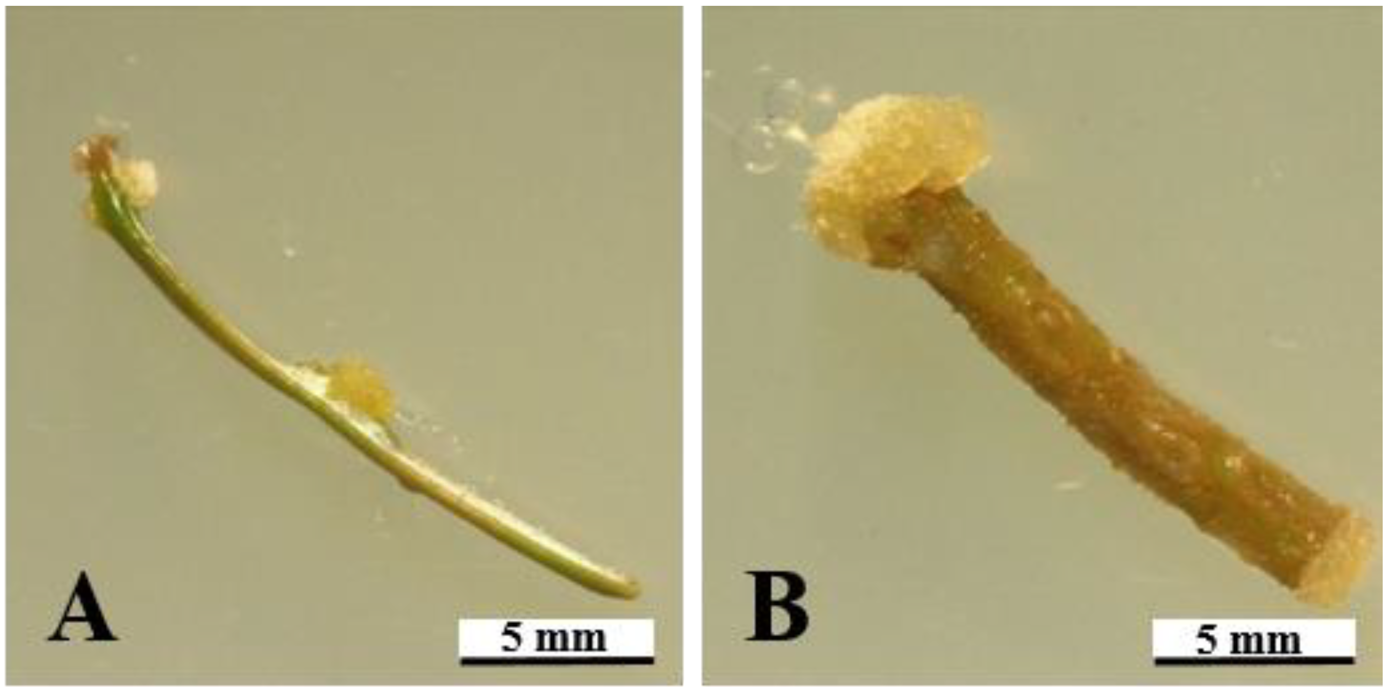
3.4. Morphology of Calli

After 4 weeks of culture, several different types of calli were observed. Compared with the stem, the calli induced on the needle had more varying forms. Four types of calli were observed on the needle: the watery tissue (which mainly occurs in the middle of the needle); the fluffy tissue (mainly concentrated at the top of the needle); the dry brown tissue (with the browning of explants); and the crystalline tissue (observed only at the needle junction). Only two types of callus tissues were found on stem explants. The watery tissue and the fluffy tissue were mainly concentrated on the cross-section of the stem segments (Figure 5).

3.5. Proliferation and Browning of Callus

The proliferation ratio and browning ratio of calli were significantly different among PGR combinations (Figure 6A,B). The greatest proliferation ratio was 1147.6%, measured in the P14 (A4B1C2D3), followed by 1146.7% in the P15 (A4B4C3D2). There were three PGR combinations with a 0.0% callus browning ratio, the P9 (A3B4C1D3), P15 (A4B4C3D2), and P16 (A4B3C4D1).
According to the results of the range analysis, 2,4-D had the strongest effect on callus proliferation and callus browning suppression (Table 4). The most effective to least effective PGRs for proliferation (%) and browning (%) were A > C > D > B, and A > B > C > D, respectively. Meanwhile, the optimal levels of proliferation (%) and browning (%) were A4B4C3D3 and A4B2C2D1, respectively (Table 4).

4. Discussion

Different explants have different levels of callus induction, which depends on the different totipotency expressions of the callus cells [21]. In many past studies on callus induction in conifers, researchers tended to use the axillary bud, immature or mature zygotic embryos, megagametophyte, and dormant apical buds as explants [18,22,23,24,25]. There are also some cases where researchers used the shoot and needles to induce calli [18,24]. In this study, the stem segments and needles developed from dormant buds were selected as the experimental materials. The results showed that the callus induction frequency of the stem segment was greater than that of the needle. Fett-Neto et al. [26], Hussain et al. [27], and Bhat et al. [28] also concluded that the stem segment was the best explant for callus induction in conifer species, which may be related to the vegetative reproduction ability of the explants and the maturity of the tissue.
According to the results of previous studies, the basic medium has a direct effect on callus induction in tissue culture. The composition of the basic medium was also one of the factors affecting callus induction in conifer species. In this study, the callus induction ratio of A. koreana on the MS, DCR, and LP media could reach 100%. However, when comparing the FW of calli, that on the MS medium was much higher than those on the LP medium and DCR medium. The MS medium also had a more obvious inhibitory effect on browning. This was similar to the results of Huang [20]. When the callus induction experiments on MS, DCR, LP, and N6 media in Cunninghamia Lanceolata were compared, the highest callus induction ratio was obtained on the MS medium (95.7%), followed by the DCR medium (69.5%) and the LP medium (42.7%). Moreover, the effects of the MS medium in a callus induction experiment on Pinus Sylvestris [29] were also very significant. Many studies have shown that the contents of NH4+ and NO3 in the MS medium are higher than those in the DCR medium, and that NH4+ and NO3 play a major role in somatic generation [30]. In the case where KNO3 is eliminated, the ratio of callus induction will be reduced, while omission of NH4NO3 has insignificant effects on the callus induction. In general, the combination of NH4+ and NO3 can aid in the growth of conifer species in tissue culture [30,31]. Therefore, in the subsequent orthogonal experiment of callus proliferation, only the MS medium was used as the basic medium due to limitations of calli available.
The different PGR combinations had significant effects on the induction and FW of calli. As mentioned previously, the PGR combination with the addition of NAA, 2,4-D, and 6-BA promoted callus induction and increased their FW. This is similar to findings by other researchers. Bhat et al. [28] found that NAA (3.0 mg·L−1) had the best effects on callus induction and proliferation in Abies pinfrow Royle. Furthermore, adding different concentrations of 6-BA and auxins (2,4-D and NAA) to different media had a great influence on the callus induction and tissue quality [30]. Gao et al. [32] demonstrated that when 2,4-D and 6-BA were added to the culture medium, the calli of Pinus koraiensis were well maintained; a similar effect was achieved with the addition of NAA and 6-BA. These two groups have potential for callus maintenance and proliferation. This viewpoint was also confirmed in the subsequent experiments on callus proliferation, where a more significant effect of PGR was found with the supplementation of 2,4-D as compared to 6-BA, NAA, and KT.
Several different forms of calli observed were found following the different treatments in this study, indicating the heterogeneity of A. koreana explants. According to Santana et al. [33] and Sivachandran et al. [34], these are non-embryonic calli. Huang [20] pointed out, in a study on callus re-differentiation, that different calli differ in their ability to regenerate buds, and the fluffy callus has a higher induction rate of regenerated buds. The stem segments can induce even more fluffy callus tissues and were more suitable for use in the experiment on subsequent callus proliferation or induction of regeneration buds.

5. Conclusions

This experiment examined the effects of appropriate explants, basic media, and PGR combinations in callus induction and proliferation of A. koreana. Different from previous studies on the micropropagation of A. koreana, in which mature zygotic embryos or winter buds were used as the explants, in this experiment, needles and stem segments, which are easier to collect, were selected as the explants. This eliminated the cumbersome extraction process, high failure ratios, and seasonal restrictions. The MS medium had the best effect on callus induction on the stem segments. The PGR combinations of NAA, 2,4-D, and 6-BA showed a very positive influence throughout this callus induction study in A. koreana. Moreover, it was also proven that 2,4-D had a significant effect on callus proliferation in A. koreana and also had a good inhibitory effect on callus browning. This study filled the gaps in previous research in A. koreana by using needles and stem segments as the explants for the induction and proliferation of calli, thereby realizing the possibility of inducing indirect embryogenesis and/or organogenesis. On this basis, the indirect induction of embryogenesis and/or organogenesis using calli will be the future direction of research in A. koreana. More in-depth research will be needed to establish a complete micropropagation system of A. koreana and further application for the mass propagation of this important but endangered plant species.

Author Contributions

Conceptualization, B.R.J. and G.G.; methodology, B.R.J. and G.G.; software, G.G; validation, B.R.J.; formal analysis, B.R.J. and G.G.; investigation, G.G.; resources, B.R.J.; data curation, G.G.; writing—original draft preparation, G.G.; writing—review and editing, B.R.J. and G.G.; supervision, B.R.J.; project administration, B.R.J.; funding acquisition, B.R.J. All authors have read and agreed to the published version of the manuscript.

Funding

This research was funded by the National Research Foundation of Korea (Project No. NRF-2019R1C1C1010573).

Data Availability Statement

Data sharing is not applicable to this article.

Acknowledgments

Ge Guo was supported by the BK 4 Program (BK21 Four), Ministry of Education, Republic of Korea.

Conflicts of Interest

The authors declare no conflict of interest.

References

  1. Farjon, A. A Handbook of the World’s Conifers (2 Vols.); Brill: Boston, MA, USA, 2010. [Google Scholar]
  2. Seo, M.; Sowndhararajan, K.; Kim, S. Influence of binasal and uninasal inhalations of essential oil of Abies koreana twigs on electroencephalographic activity of human. Behav. Neurol. 2016, 2016, 9250935. [Google Scholar] [CrossRef] [PubMed]
  3. Yoon, W.J.; Kim, S.S.; Oh, T.H.; Lee, N.H.; Hyun, C.G. Abies koreana essential oil inhibits drug resistant skin pathogen growth and LPS-induced inflammatory effects of murine macrophage. Lipids 2009, 44, 471–476. [Google Scholar] [CrossRef] [PubMed]
  4. Lee, T.B. Abies koreana and its new forms discovered. J. Korean For. Soc. 1970, 10, 5–6. [Google Scholar]
  5. Lee, Y.W.; Hong, S.C. Ecological studies on the vegetational characteristics of the Abies koreana forest. J. Korean Soc. For. Sci. 1995, 84, 247–257. [Google Scholar]
  6. Chang, C.S.; Jeon, J.I.; Hyun, J.O. An analysis of morphological variation in Abies koreana Wilson and A. nephrolepis (Traut.) Maxim. of Korea (Pinaceae) and their phylogenetic problems. J. Korean Soc. For. Sci. 1997, 86, 378–390. [Google Scholar]
  7. Koo, K.A.; Kong, W.S.; Park, S.U.; Lee, J.H.; Kim, J.; Jung, H. Sensitivity of Korean fir (Abies koreana Wils.), a threatened climate relict species, to increasing temperature at an island subalpine area. Ecol. Modell. 2017, 353, 5–16. [Google Scholar] [CrossRef]
  8. Woo, S.Y. Forest decline of the world: A linkage with air pollution and global warming. Afr. J. Biotechnol. 2009, 8, 7409–7414. [Google Scholar]
  9. Kim, Y.S.; Chang, C.S.; Kim, C.S.; Gardner, M. Abies koreana. The IUCN Red List of Threatened Species 2011: E.T31244A9618913. 2011. Available online: https://www.iucnredlist.org/search?query=Abies%20koreana&searchType=species (accessed on 22 August 2021). [CrossRef]
  10. Kwak, M.; Hong, J.K.; Park, J.H.; Lee, B.Y.; Suh, M.H.; Kim, C.S. Genetic assessment of Abies koreana (Pinaceae), the endangered Korean fir, and conservation implications. Conserv. Genet. 2017, 18, 1165–1176. [Google Scholar] [CrossRef]
  11. Noland, T.L.; Parker, W.C.; Morneault, A.E. Natural variation in seed characteristics of eastern white pine (Pinus strobus L.). New For. 2006, 32, 87–103. [Google Scholar] [CrossRef]
  12. Song, J.H.; Jang, K.H.; Hur, S.-D. Variation of seed and germination characteristics of natural populations of Abies koreana Wilson, a Korean endemic species. J. Korean Soc. For. Sci. 2010, 99, 849–854. [Google Scholar]
  13. Nunes, S.; Santos, C.; Moutinho-Pereira, J.; Correia, C.; Oliveira, H.; de Oliveira, J.M.F.; Pereira, V.T.; Almeida, T.; Marum, L.; Dias, M.C. Physiological characterization and true-to-typeness evaluation of in vitro and ex vitro seedlings of Pinus elliottii: A contribution to breeding programs. Plant Physiol. Biochem. 2016, 107, 222–227. [Google Scholar] [CrossRef]
  14. Nunes, S.S.C. Characterization, Micropropagation and Preservation of Pinus Genotypes. Ph.D. Thesis, Universidade de Aveiro, Aveiro, Portugal, 2016. [Google Scholar]
  15. Gupta, P.K.; Durzan, D.J. Shoot multiplication from mature trees of Douglas-fir (Pseudotsuga menziesii) and sugar pine (Pinus lambertiana). Plant Cell Rep. 1985, 4, 177–179. [Google Scholar] [CrossRef] [PubMed]
  16. Lelu-Walter, M.A.; Bernier-Cardou, M.; Klimaszewska, K. Clonal plant production from self-and cross-pollinated seed families of Pinus sylvestris (L.) through somatic embryogenesis. Plant Cell Tissue Organ Cult. 2008, 92, 31–45. [Google Scholar] [CrossRef]
  17. Salaj, T.; Klubicová, K.; Matusova, R.; Salaj, J. Somatic embryogenesis in selected conifer trees Pinus nigra Arn. and Abies hybrids. Front. Plant Sci. 2019, 10, 13. [Google Scholar] [CrossRef] [PubMed] [Green Version]
  18. Murasnige, T.; Skoog, F. A revised medium for rapid growth and bio assays with tobacco tissue cultures. Physiol. Plant 1962, 15, 473–497. [Google Scholar] [CrossRef]
  19. Quoirin, M.; Lepoivre, P. Improved media for in vitro culture of Prunus sp. Acta Hortic. 1977, 78, 437–442. [Google Scholar] [CrossRef]
  20. Huang, J. Efficiency of Callus and Regeneration Bud Induction from Different Explants of Cunninghamia lanceolata. Master’s Thesis, Fujian Agriculture and Forestry University, Fujian, China, 2017. [Google Scholar]
  21. Bonga, J.M. Adventitious shoot formation in cultures of immature female strobili of Larix decidua. Physiol. Plant 1984, 62, 416–421. [Google Scholar] [CrossRef]
  22. Li, X.; Huang, F.; Gbur, E., Jr. Effect of basal medium, growth regulators and Phytagel concentration on initiation of embryogenic cultures from immature zygotic embryos of loblolly pine (Pinus taeda L.). Plant Cell Rep. 1998, 17, 298–301. [Google Scholar] [CrossRef]
  23. Szczygiel, K.; Hazubska-Przybyl, T.; Bojarczuk, K. Somatic embryogenesis of selected coniferous tree species of the genera Picea, Abies and Larix. Acta Soc. Bot. Pol. 2007, 76, 7–15. [Google Scholar]
  24. Nawrot-Chorabik, K. Embryogenic callus induction and differentiation in silver fir (Abies alba Mill.) tissue cultures. Dendrobiology 2008, 59, 31–40. [Google Scholar]
  25. De Diego, N.; Montalbán, I.; Moncaleán, P. In vitro regeneration of adult Pinus sylvestris L. trees. S. Afr. J. Bot. 2010, 76, 158–162. [Google Scholar] [CrossRef] [Green Version]
  26. Fett-Neto, A.G.; DiCosmo, F.; Reynolds, W.; Sakata, K. Cell culture of Taxus as a source of the antineoplastic drug taxol and related taxanes. Bio/technology 1992, 10, 1572–1575. [Google Scholar] [CrossRef] [PubMed]
  27. Hussain, A.; Qarshi, I.A.; Nazir, H.; Ullah, I.; Rashid, M.; Shinwari, Z.K. In vitro callogenesis and organogenesis in Taxus wallichiana ZUCC, The Himalayan Yew. Pak. J. Bot. 2013, 45, 1755–1759. [Google Scholar]
  28. Bhat, S.; Gangoo, S.; Geelani, S.; Qasba, S.; Parray, A. Callus culture and organogenesis in fir (Abies pindrow Royle). J. Cell Tissue Res. 2014, 14, 4653–4658. [Google Scholar]
  29. Hohtola, A. Seasonal changes in explant viability and contamination of tissue cultures from mature Scots pine. Plant Cell Tissue Organ Cult. 1988, 15, 211–222. [Google Scholar] [CrossRef]
  30. Shanjani, P.S. Nitrogen effect on callus induction and plant regeneration of Juniperus excelsa. Int. J. Agric. Biol. 2003, 5, 419–422. [Google Scholar]
  31. Rout, G.; Mohapatra, A.; Jain, S.M. Tissue culture of ornamental pot plant: A critical review on present scenario and future prospects. Biotechnol. Adv. 2006, 24, 531–560. [Google Scholar] [CrossRef]
  32. Gao, F.; Peng, C.; Wang, H.; Shen, H.; Yang, L. Selection of culture conditions for callus induction and proliferation by somatic embryogenesis of Pinus koraiensis. J. For. Res. 2021, 32, 483–491. [Google Scholar] [CrossRef]
  33. Santana, M.G.; Velásquez, S.R.; Mata, J. Carbon source effect on cacao organogenesis and somatic embryogenesis. Cytogenetic analysis. Agron. Trop. 2010, 60, 193–202. [Google Scholar]
  34. Sivachandran, R.; Gnanam, R.; Sudhakar, D.; Suresh, J.; Ram, S.G.; Selvi, R.G. Development of in vitro gynogenesis system in cocoa (Theobroma cacao L.). Chem. Sci. Rev. Lett. 2017, 6, 2169–2177. [Google Scholar]
Figure 1. Microscopic observation of calli induced on various locations of the needle and stem segment explants after 4 weeks of culture: (A) callogenesis on the interfaces between the needle and the stem, and the middle of the needle; and (B) callogenesis on the cut surfaces of the stem segment.
Forests 12 01388 g001
Figure 2. Effect of medium and PGR combination on callus induction ratio from needle (A) and stem segments (B) after 4 weeks of culture. Different letters above the bars indicate significant differences by Duncan’s multiple range test at p ≤ 0.05. Vertical bars indicate the standard errors (n = 3).
Forests 12 01388 g002aForests 12 01388 g002b
Figure 3. Effect of basic medium and PGR combination on browning ratio of callus from the needle (A) and stem segments (B) after 8 weeks of culture. Different letters above the bars indicate significant differences by Duncan’s multiple range test at p ≤ 0.05. Vertical bars indicate the standard errors (n = 3).
Forests 12 01388 g003
Figure 4. Effect of basic medium and PGR combination on FW of callus from needle (A) and stem segments (B) after 8 weeks of culture. Different letters above the bars indicate significant differences by Duncan’s multiple range test at p ≤ 0.05. Vertical bars indicate the standard errors (n = 3).
Forests 12 01388 g004
Figure 5. Microscopic observation of morphological heterogeneity of calli induced from needle and stem segment explants after 4 weeks of culture: (A) Watery tissue; (B) Fluffy tissue; (C) Crystalline tissue induced from needles; (D) Dry brown tissue; (E) Watery tissue; and (F) Fluffy tissue induced from stem segments).
Forests 12 01388 g005
Figure 6. Effect of PGR combinations on callus proliferation (A) and browning (B) of calli after 4 weeks. Different letters above the bars indicate significant differences calculated by Duncan’s multiple range test at p ≤ 0.05. Vertical bars indicate the standard error (n = 3).
Forests 12 01388 g006
Table 1. The PGR combinations and concentrations used for callogenesis from the needle and stem segment explants of A. koreana.
Treatment CodeAuxin (mg·L−1)Cytokinin (mg·L−1)
2,4-DNAAKTTDZ6-BA
T10.00.00.00.00.0
T20.02.00.50.02.0
T30.02.01.00.02.0
T40.02.00.00.12.0
T50.02.00.00.22.0
T61.02.00.50.00.0
T71.02.01.00.00.0
T81.02.00.00.10.0
T91.02.00.00.20.0
T101.00.00.50.02.0
T111.00.01.00.02.0
T121.00.00.00.12.0
T131.00.00.00.22.0
Table 2. L16(4)4 orthogonal design used in this study.
Treatment CodeAuxin (mg·L−1)Cytokinin (mg·L−1)
2,4-D (A)NAA (B)6-BA (C)KT (D)
P10.0A10.0B10.5C10.0D1
P20.0A10.5B21.0C20.5D2
P30.0A11.0B31.5C31.0D3
P40.0A11.5B42.0C41.5D4
P51.0A21.0B30.5C10.5D2
P61.0A21.5B41.0C20.0D1
P71.0A20.0B11.5C31.5D4
P81.0A20.5B22.0C41.0D3
P92.0A31.5B40.5C11.0D3
P102.0A31.0B31.0C21.5D4
P112.0A30.5B21.5C30.0D1
P122.0A30.0B12.0C40.5D2
P133.0A40.5B20.5C11.5D4
P143.0A40.0B11.0C21.0D3
P153.0A41.5B41.5C30.5D2
P163.0A41.0B32.0C40.0D1
Table 3. Effects of the explant type, medium, and PGR combination on the callus induction (%), browning (%), and FW (mg) of calli induced from the needle and stem segments.
Explant
(A)
Medium
(B)
PGR
(C, mg·L−1)
Induction (%)Browning (%)FW
(mg)
F-test zA*********
B*********
C********
A × B*********
A × C*********
B × C*****NS
A × B × C******NS
z NS, **, and ***: Nonsignificant or significant at p ≤ 0.05, 0.01, or 0.001, respectively.
Table 4. The range analysis of L16(4)4 for proliferation (%) and browning (%) of calli. The optimal level is shown in bold.
VariableProliferation (%) *Browning (%)
ABCDABCD
K1252.3722.0617.5682.390.053.846.330.8
K2853.7722.2788.3758.833.824.630.036.3
K3895.2709.0811.5786.39.636.334.638.8
K4954.7802.6738.6728.58.827.531.336.3
R702.493.6194.0103.981.329.216.37.9
Optimal levelA4B4C3D3A4B2C2D1
* The greatest value of proliferation ratio and the lowest value of browning ratio are the optimal levels.
Publisher’s Note: MDPI stays neutral with regard to jurisdictional claims in published maps and institutional affiliations.

