Article

Drought Stress Can Induce the Pathogenicity of Cryptostroma corticale, the Causal Agent of Sooty Bark Disease of Sycamore Maple

Slovenian Forestry Institute, Večna pot 2, SI-1000 Ljubljana, Slovenia
*
Author to whom correspondence should be addressed.
Forests 2021, 12(3), 377; https://doi.org/10.3390/f12030377
Submission received: 16 February 2021 / Revised: 12 March 2021 / Accepted: 18 March 2021 / Published: 21 March 2021
(This article belongs to the Section Forest Health)

Abstract

:
Reports of sooty bark disease of maples caused by the fungus Cryptostroma corticale have recently been emerging from across Europe. The aims of our study were to describe the first report of sooty bark disease in Slovenia, to determine the pathogenicity of C. corticale, to confirm the optimum temperature for the growth of the fungus, and to determine the mass loss of Acer pseudoplatanus wood inoculated by C. corticale. We confirmed the presence of C. corticale on A. pseudoplatanus via morphological and molecular analysis. The optimal growth of C. corticale was measured in vitro on potato dextrose agar and was determined to occur at 25 °C. Pathogenicity tests were performed on 30 saplings of A. pseudoplatanus under two treatments, humid and drought stress, and the fungus was pathogenic in both treatments. The mean length of bark lesions and wood discoloration of the drought-stressed saplings was significantly greater than that in the humid treatment. Re-isolations of C. corticale were successful from all inoculated saplings, and thus Koch’s postulates were confirmed. The mass loss of A. pseudoplatanus wood was determined by mini-block test in a period of 10 weeks and was observed as minimal. Based on the results, we conclude that C. corticale is a weak and opportunistic pathogen that most likely expresses itself intensively under hot and dry conditions.

1. Introduction

Sooty bark disease of maples is caused by the fungus Cryptostroma corticale (Ellis & Everh.) P.H. Greg. & S. Waller [1]. It is distributed in North America [2] and in most countries of Western and Central Europe, i.e., Great Britain [3], France [4], the Netherlands [5], Belgium [6], Germany [7,8], the Czech Republic [9,10], Austria [11,12], Italy [13], Switzerland [14], and Bulgaria [15]. Most of the countries surrounding Slovenia have already reported the disease, but it has not been reported in Slovenia until now.
The hosts of C. corticale are maples (Acer spp.). It most frequently infects sycamore maple (A. pseudoplatanus L.), field maple (A. campestre L.), Norway maple (A. platanoides L.) and box elder (A. negundo L.) [16]. Maples are one of the most important hardwood tree species in Slovenia, and therefore the presence and spread of sooty bark disease would raise serious concerns.
Wounds serve as entry points for the fungus, which quickly invades the wood and later also the phloem [9,16,17]. Cryptostroma corticale is recognized as an endophyte, latent invader, saprophyte, and weak pathogen that favors hot and dry periods [9,10,13,15,17,18]. Because of its preference for hot and dry weather, some studies predict more frequent outbreaks of sooty bark disease in the future due to climate change [9,10,19].
The first suspicious case of sooty bark disease was observed in October 2019 on a chunk of Acer pseudoplatanus that was collected in February 2019 in the central part of Slovenia (46.13369° N, 14.77848° E) and displayed in the hallway of the Slovenian Forestry Institute for educational purposes because it had typical symptoms of Eutypella canker of maple caused by Eutypella parasitica R.W. Davidson & R.C. Lorenz. Surprisingly, after seven months under room temperature and natural daylight, the sample developed typical symptoms of sooty bark disease caused by C. corticale (Figure 1): the bark cracked and peeled off, revealing dark brown masses of ovoid conidia which measured 4.9–6.1 × 3.1–4.0 µm.
The aims of the study were (1) to investigate the first report of sooty bark disease of sycamore maple in Slovenia, (2) to determine the optimum temperature for growth of the fungus in pure culture, (3) to test its pathogenicity under two watering treatments simulating humid and drought conditions, and (4) to determine the mass loss of sycamore wood samples exposed to C. corticale.
The first report of sooty bark disease of sycamore maple in Slovenia was accompanied by pathogenicity trials. Pathogenicity tests and mass loss of sycamore wood were performed under optimal temperature for growth of C. corticale. Therefore, the optimal temperature was obtained prior to performing those experiments. Furthermore, the collected samples for first report showed wood decay; therefore, the additional aim was added to investigate the mass loss of sycamore wood.

2. Materials and Methods

2.1. Identification

Three 1-m-long and 12–15-cm-thick chunks of A. pseudoplatanus were collected in November 2019 in the central part of Slovenia, in a managed, mixed forest stand (46.13369° N, 14.77848° E) from which the first suspected case also originated. One chunk was cut from a visually healthy tree, and the other two from trees that had old, but healed, wounds. No visible symptoms of the disease were observed at the location. The end parts of the chunks were sealed with wax to slow down desiccation and were incubated at room temperature under natural daylight.
After the symptoms were expressed, the samples were morphologically examined, and the identity of the causal agent was determined. Morphological analysis was done with an Olympus SZX16 stereomicroscope and an Olympus BX53 microscope, both equipped with an Olympus UC90 digital camera and Olympus CellSens Standard 1.18 software.
The conidia of C. corticale were aseptically transferred on the tip of a needLe to growing medium containing 3.9% (w/v) potato dextrose agar (PDA; Becton Dickinson, Sparks, MD, USA) and incubated in a Kambič I-190 CK chamber (Kambič, Semič, Slovenia) at 23.9 ± 0.3 °C. During this process, a representative pure culture of C. corticale was obtained, and the isolate was deposited at the culture collection of the Laboratory of Forest Protection at the Slovenian Forestry Institute (ZLVG 859). This isolate was used in all further experiments. Furthermore, a sample of bark and conidia was stored in the Mycotheca and Herbarium of the Slovenian Forestry Institute (LJF 7437).
The morphological identification of the ZLVG 859 isolate was also confirmed by molecular analysis, i.e., by sequencing the ITS-rDNA region. DNA extraction and PCR conditions followed the procedure described by Brglez et al. [20], except that the mycelium was scraped from the PDA plates.
The obtained PCR product was cleaned using a Wizard SV Gel and PCR Clean-Up System (Promega, Madison, WI, USA) and sequenced at a sequencing facility (Eurofins, Köln, Germany) in both forward and reverse directions using the same primers as for the PCR (ITS1 and ITS4). Sequences were visualized and manually edited using Geneious Prime® v.2019.0.3. (Biomatters Ltd., Auckland, New Zealand). The obtained consensus sequence was used to perform an individual nucleotide–nucleotide search with the megablast algorithm on the NCBI website (http://www.ncbi.nlm.nih.gov/BLAST/ (accessed on 3 March 2019)). The sequence was deposited in the GenBank database (accession no. MW575287).

2.2. Optimum Temperature

Other studies have already determined the optimum temperature for growth of C. corticale in pure culture to be 25 °C [17,21]. However, the studies also acknowledged high variability, and those studies tested growth on malt extract agar (MEA) but not on potato dextrose agar (PDA). Therefore, our ZLVG 859 isolate of C. corticale was verified for optimum temperature: the fungus was inoculated with a 5-mm diameter agar plug taken from a 7-day-old culture onto a 1-day-old 3.9% (w/v) PDA agar plate in five replicates. The cultures were incubated in a Kambič I-190 CK chamber at a temperature range of 10–35 °C, at 5 °C intervals. Additionally, two temperatures of 26 and 27 °C were checked after the first series. The actual temperature in the chamber was measured with an independent sensor at 30-min intervals (Voltcraft DL-120TH, Conrad Electronic SE, Hirschau, Germany). Radial growth rate was checked and measured daily in four directions along two perpendicular lines drawn on the back of the Petri dish and originating from the center of the inoculated agar plug. For the calculation of the mean growth rate, only the linear phase of growth was considered.

2.3. Pathogenicity Tests

For the pathogenicity tests, 30 two-year-old saplings of A. pseudoplatanus planted in 17-cm diameter flowerpots were used. The saplings were acquired in November 2019 and left in the field under natural conditions until July 2020 when they were moved to an RK2 plant growth chamber (Kambič, Semič, Slovenia). The following settings were used in the plant growth chamber: daylight 16 h with maximum illumination setting, night 8 h, temperature 25 °C, relative humidity 60%, air exchange every 55 min for 300 s. The plant growth chamber was equipped with six fluorescent lamps OSRAM L 58W/77 FLUORA (OSRAM GmbH, Munich, Germany) with luminous flux of 2250 lm. The actual temperature and relative air humidity in the plant growth chamber were controlled with three independent sensors at 30-min intervals (Voltcraft DL-120TH, Conrad Electronic SE, Hirschau, Germany) and measured 24.54 ± 0.01 °C, 59.9 ± 0.1%, respectively. The saplings were left in the plant growth chamber for four days to acclimatize to the conditions prior to inoculation. During the acclimatization period all saplings were watered once with 2 dL of distilled water (two days after they were transferred to the chamber).
The inoculation procedure consisted of the following steps: (1) the stem was surface-sterilized by 70% ethanol; (2) the bark was wounded and removed with a 5-mm diameter sterile cork borer; (3) an agar plug of the same diameter taken from an 11-day-old culture of the ZLVG 859 C. corticale isolate growing on PDA was inserted into the wound; (4) the agar plug was covered with a sterile and moist piece of cotton (1 × 1 cm); and (5) the inoculation point was wrapped with aluminum foil and Parafilm (Sigma-Aldrich, Saint Louis, MO, USA). For the control, the same procedure was used except for the agar plug in the third step, which was sterile. Twenty saplings were inoculated with the ZLVG 859 isolate and 10 saplings were used as a control.
Two watering treatments were used: (1) 2 dL twice per week (designated as humid, H), and (2) 2 dL once per week (designated as drought, D). Distilled water was used for watering. Fifteen saplings per treatment were used, i.e., 10 inoculated saplings and five controls.
The saplings were checked daily. Two C. corticale-inoculated saplings were killed after 25 days and another one after 31 days. For these three saplings, re-isolations were made immediately when they were killed. For the rest of the 27 saplings, re-isolations were made after 38 days. All re-isolations were made from the margin of the lesion in the bark onto PDA supplemented with Streptomycin (Sigma-Aldrich, Saint Louis, MO, USA). The re-isolations were checked regularly, and any outgrowing cultures were transferred to fresh PDA. Isolates were grouped by morphotypes and sapling ID. Each morphotype per sapling was identified by the molecular technique as described in 2.1.
The length of the necrosis in the bark and discoloration of sapwood was measured. Differences in mean lengths between the treatments were checked by a t-test. Equality of variances were checked by an F-test. The statistics were performed in the R software environment for statistical computing [22]. Charts were drawn in Microsoft Excel version 16.0.

2.4. Mass Loss of Wood

The wood of sycamore maple was used in the experiment. The wood originated from the southeastern part of Slovenia (45.8491° N, 15.6113° E), where this species grows naturally. Cross-sections were taken approximately 1 m above the ground from visually healthy trees. Until further processing, they were stored in an LTH ZO 700 BEZ freezer cabinet (Loška hladilna tehnika, Škofja Loka, Slovenia) at −20 °C for 20 days. Sixty wood samples (30 × 10 × 5 mm) were made and air-dried for a year. The initial dry mass of the samples was determined using a Kern ABJ220-4NM analytical balance (Kern & Sohn, Balingen, Germany) after oven drying the samples at 103 °C for 24 h in a Kambič SP-250 oven (Kambič, Semič, Slovenia). Thereafter, samples were steam-sterilized (30 min, 121 °C, 0.12 MPa) in a Kambič A-65 V autoclave (Kambič, Semič, Slovenia) and further used in the decay test.
The decay test was performed according to the modified EN 113 standard [23,24,25]. Disposable Petri dishes (Ø = 90 mm, h = 15 mm) containing 3.9% (w/v) PDA (Becton Dickinson, Sparks, MD, USA) were inoculated with C. corticale isolate ZLVG 859 and with a reference fungus Trametes versicolor (L.) Lloyd (ZIM L057, culture collection of industrial microorganisms at the University of Ljubljana). After one week of fungal growth, the wood samples were exposed to the fungi. Two wood samples were placed on sterilized plastic mesh, which was used to avoid direct contact between the samples and the nutrient medium in the Petri dish. Twenty replicates per fungal isolate were used. For comparison, twenty control samples were placed in Petri dishes with sterile agar plugs. The assembled test dishes were incubated in a Kambič I-190 CK chamber (Kambič, Semič, Slovenia) at 23.9 ± 0.01 °C for 10 weeks. After incubation, the fungal mycelium was carefully removed from the samples. The final dry mass was determined and the respective relative loss in mass was calculated after 24 h of drying at 103 °C in a Kambič SP-250 oven (Kambič, Semič, Slovenia) using the following equation:
Mass loss (%) = ((m0 − m1)/m0) × 100,
where m0 represents the initial dry mass and m1 the final dry mass of the sample.
Because the assumptions of normal distribution and homogeneity of variances had not been met, a non-parametric Kruskal–Wallis test was used to compare average mass loss between fungal isolates. Afterwards, a post-hoc multiple comparison Dunn test with Bonferroni correction was used. A paired t-test was used to compare the initial and final mean dry mass of samples exposed to different fungal isolates.

3. Results

3.1. Identification

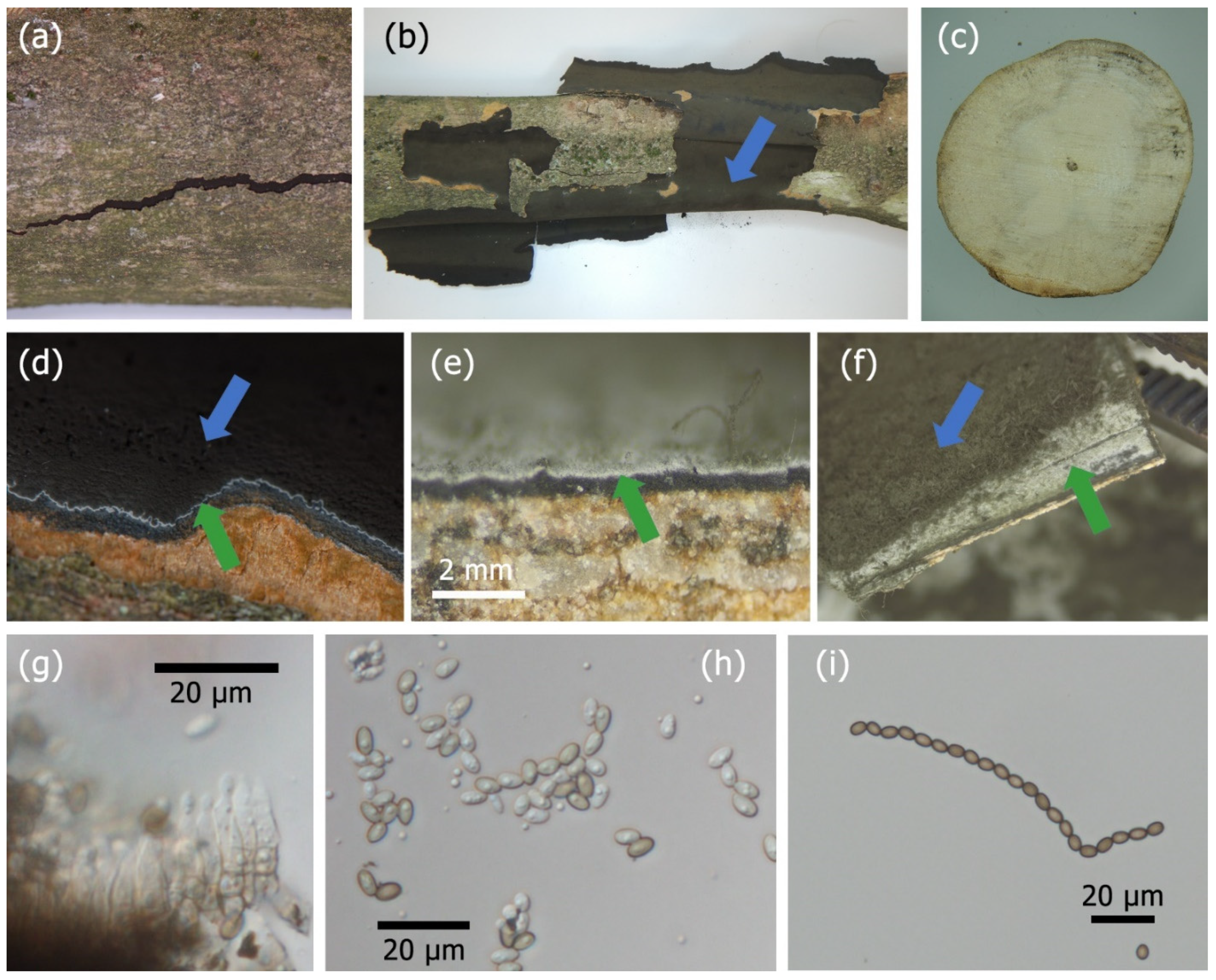
All three chunks of A. pseudoplatanus incubated under room temperature and daylight started to show typical symptoms of C. corticale after nine weeks. The bark started to show cracks (Figure 2a) and was easily peeled off (Figure 2b); the sapwood and heart wood were discolored, degraded and visually decayed (Figure 2c); dense masses of dark gray to dark brown conidia were observed under the bark (Figure 2b,d,f); the conidiomata had black 0.48-mm-thick stroma and a whitish to bluish upper layer of conidiophores that produced conidia in chains (Figure 2d,e,f); ovoid conidia arose endogenously from short, erect, unbranched phialides (Figure 2g); and young conidia were hyaline to light brown (Figure 2g,h), while mature conidia were dark brown, measuring 4.9–6.1 × 3.4–4.0 µm (Figure 2i). All morphological characteristics corresponded to the description of C. corticale [1,16,17].
The ITS-rDNA sequence (Gen Bank Acc. No. MW575287) of a representative C. corticale isolate (ZLVG 859) showed 100% identity with reference sequences of C. corticale (e.g., MH857008 from isolate CBS 216.52).
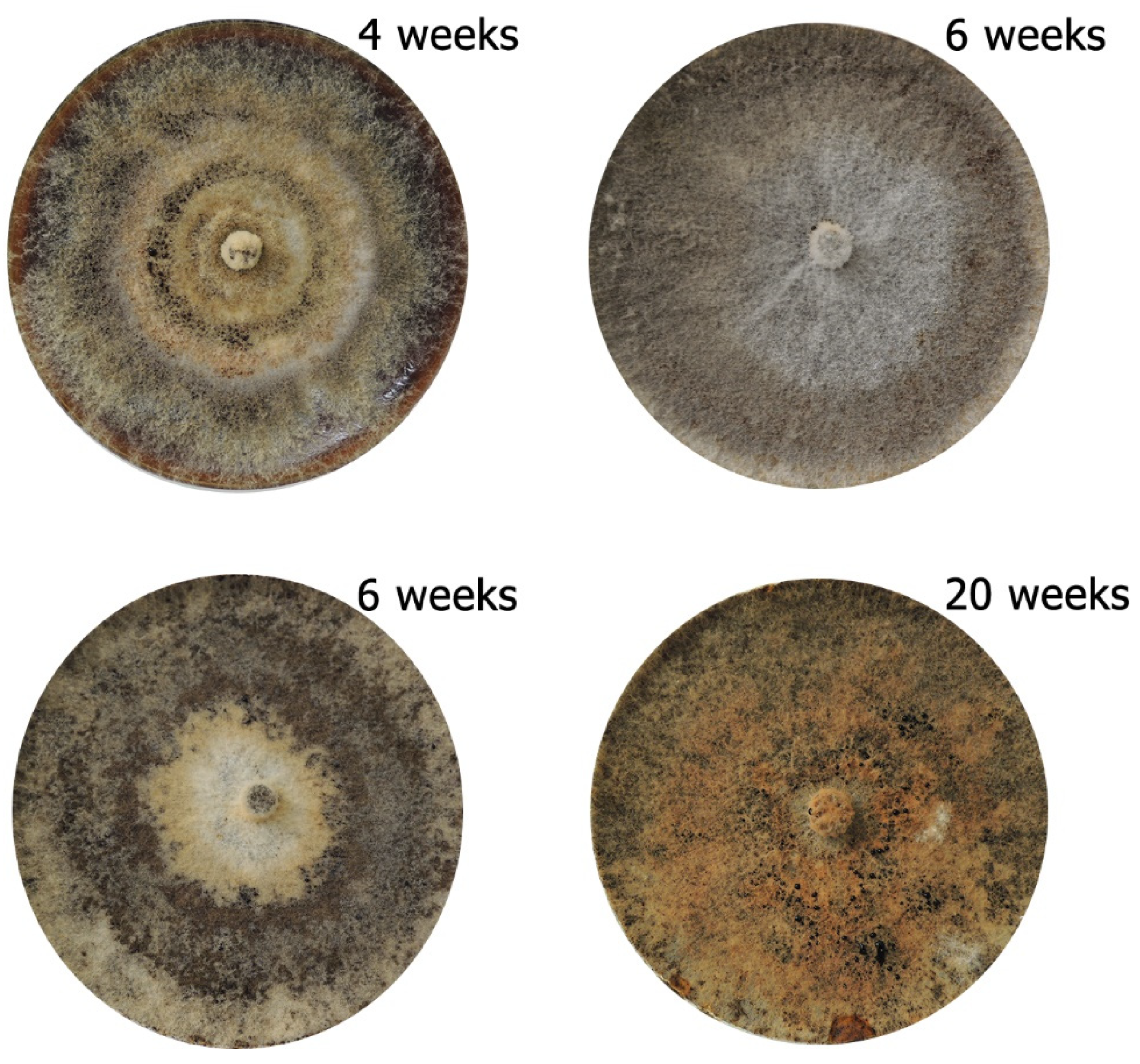
The color of the ZLVG 859 isolate of C. corticale differed between the cultures depending on the time exposed to natural daylight (Figure 3): from white and slightly yellow, to dark brown, to yellowish orange, to different intensities of grey, and, after 20 weeks, to totally black at the back and ochre brown at the front. No pigment production was observed.

3.2. Optimum Temperature

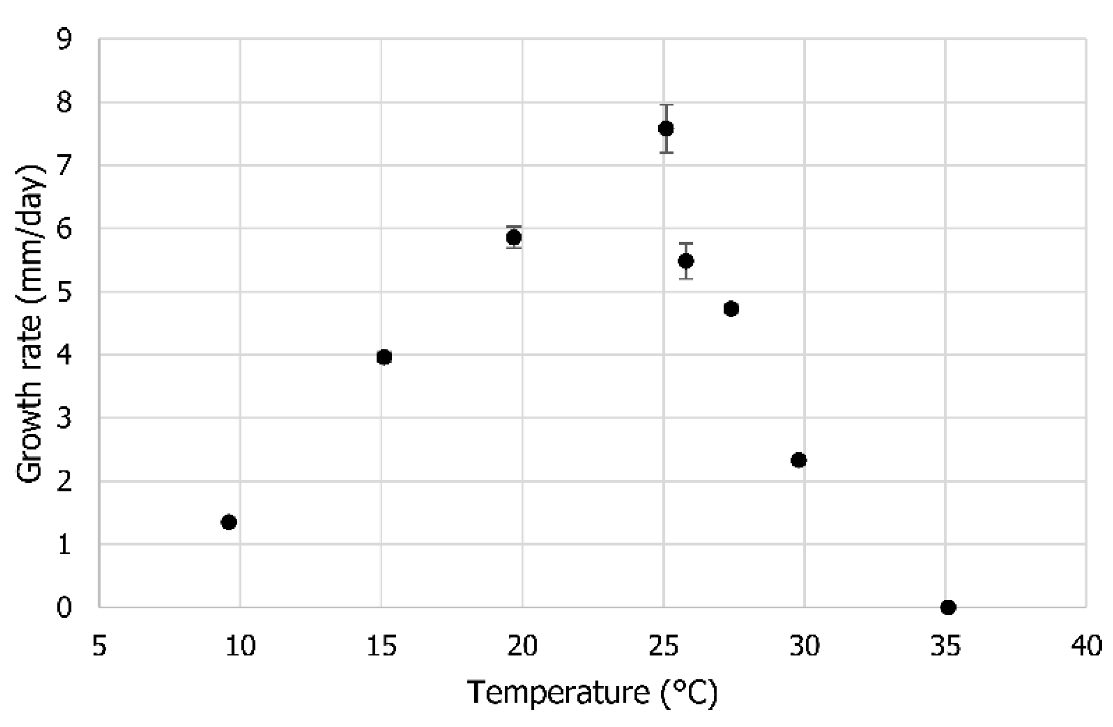
The growth rate of the C. corticale isolate gradually increased from 9.6 °C to 25.1 °C (Figure 4, Table S1). Growth quickly slowed above 25.1 °C and completely stopped at 35.1 °C.

3.3. Pathogenicity Tests

Inoculation points on control saplings in both watering treatments healed completely or started to heal after 38 days (Figure 5a). Saplings inoculated with C. corticale developed lesions in the bark (Figure 5b,g), the sapwood was discolored (Figure 5c,h), and in two cases, typical conidiomata of sooty bark disease developed (Figure 5d–f). Wilting was observed at nine of ten C. corticale-inoculated saplings under drought stress, and only one sapling under the humid treatment.
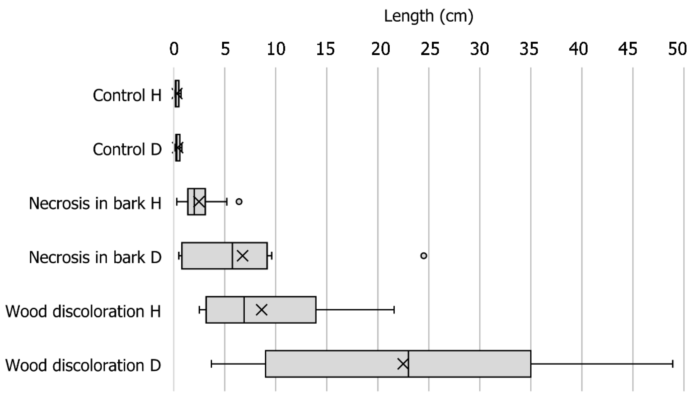
The mean length of discoloration in control saplings was significantly different from the mean length of lesions in the bark (p = 0.005) and mean length of the discoloration in the sapwood of C. corticale-inoculated saplings (p = 0.002, Figure 6, Table S2).
The mean length of necrosis in the bark of C. corticale-inoculated saplings, which were under the water deficit treatment, was significantly greater than the mean length of the necrosis that developed under the humid treatment (p = 0.05; Figure 6). Similarly, the mean length of the wood discoloration of the drought-stressed saplings was significantly greater than that of the humid-treated saplings (p = 0.03). The maximum length of wood discoloration reached 48.9 cm in the drought stress treatment (Figure 6). The mean length of wood discoloration was significantly greater than the mean length of the bark necrosis (p = 0.007 for H, p = 0.02 for D).
Re-isolations of C. corticale were successful from all 20 inoculated saplings. The identity of re-isolated C. corticale cultures was confirmed by sequence analysis of ITS-rDNA. Re-isolations from controls did not yield any C. corticale isolate. Thus, the pathogenicity of C. corticale was confirmed in both studied treatments.
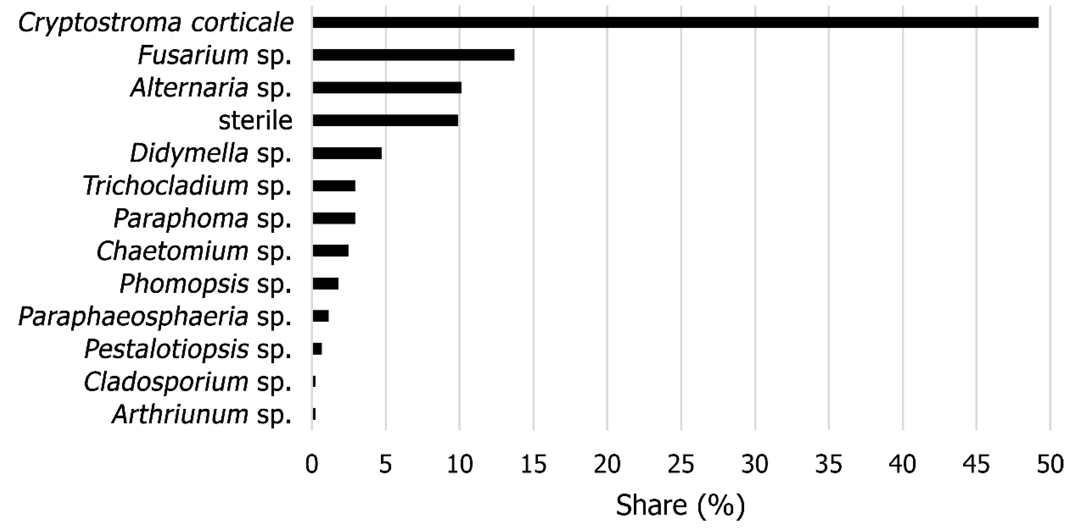
Besides C. corticale, other fungal species were isolated from the necrosis in the inoculated saplings, representing altogether 19 morphotypes. From 445 obtained cultures, 49.2% of the cultures were identified as C. corticale (Figure 7), more frequently from drought-stressed saplings (136) than from saplings undergoing the humid treatment (83). From the margins of developed lesions in the bark, Fusarium sp. (13.7%) and Alternaria sp. (10.1%) were also isolated. Other cultures belonged to the genera Didymella, Trichocladium, Paraphoma, Chaetomium, Phomopsis, Paraphaeosphaeria, Pestalotiopsis, Cladosporium, and Arthriunum (Figure 7, Table S3).

3.4. Mass Loss of Wood

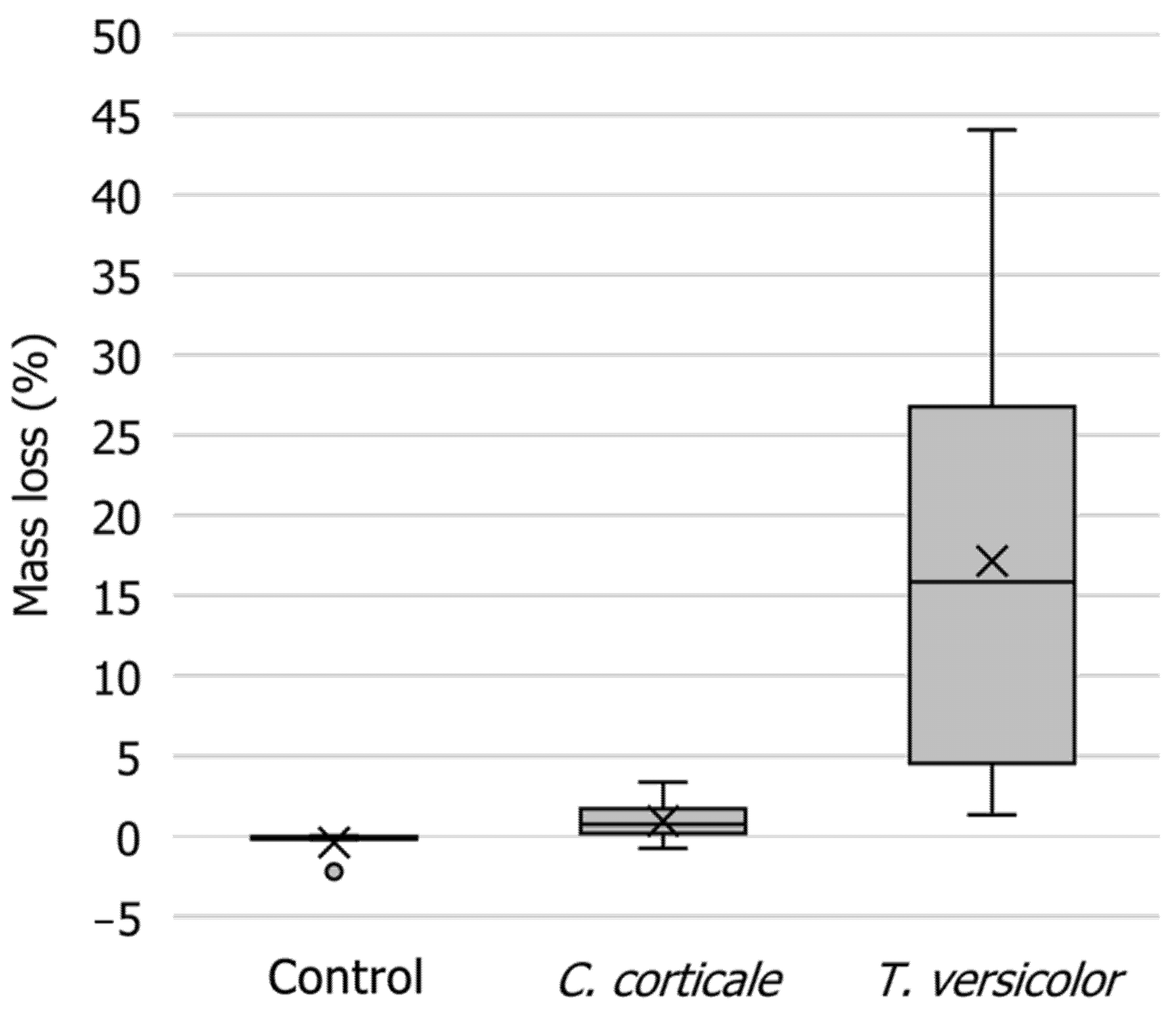
The mass loss of wood after 10 weeks averaged 9.0% in A. pseudoplatanus samples. Pairwise comparisons of mass loss in A. pseudoplatanus revealed statistically significant differences between the control and C. corticale (p < 0.05) and between the control and T. versicolor (p < 0.001). C. corticale caused an average mass loss of 0.9%. The values ranged from −0.7% to 3.4%. For comparison, T. versicolor was significantly more effective (p < 0.001), causing mass loss ranging from 1.3% to 44.0% after 10 weeks of exposure (Figure 8, Table S4). Differences between average initial and final dry mass were significant for both fungal isolates (p < 0.01 for C. corticale and p < 0.001 for T. versicolor) and the control (p < 0.05).

4. Discussion

4.1. Identification

The identity of C. corticale was confirmed for the first time in A. pseudoplatanus in Slovenia. However, the disease has only been identified in a single location in the central part of the country. Further studies should focus on the actual extent of disease distribution. We believe that sooty bark disease of maple is likely to be widely distributed but has gone unnoticed due to its cryptic and endophytic nature [3], its relation to dry periods [10,15,17,18,26], and the difficulty of detecting it on the upper branches. The environmental preferences of the disease match IPPC climate change scenario RCP8.5 for the Mediterranean [27] relating to drought stress, and therefore it is likely to become more frequent in the future in these areas. However, its sporadic occurrence and dependency on environmental stress indicate that the pathogen does not pose a fundamental risk to the natural sycamore maple population in Central Europe in the near future [10].
The fungus can cause severe hypersensitivity pneumonitis in humans [19,28,29,30]; therefore, persons who have intensive occupational contact with infested trees or wood, e.g., woodsmen, foresters, sawyers, and paper mill workers, are at particular risk [19]. However, the disease can frequently inhabit maples in public areas [9]. To protect the public, infested maples must be carefully removed—it is imperative that persons working on infested wood or trees wear personal protective equipment, especially a mask. Because of this, it is crucial that we identify the actual distribution of the disease.
The first suspicious case of sooty bark disease of sycamore maple in Slovenia developed on a sample of A. pseudoplatanus with Eutypella canker. As we know that C. corticale is a wound pathogen, we can speculate that C. corticale entered the tree diseased by E. parasitica through the canker wound. Another possibility is that C. corticale was already present in the bark endophytically. However, this was beyond the scope of the study.

4.2. Optimum Temperature

The optimal growth of C. corticale isolate ZLVG 859 on PDA was achieved at 25 °C. This result is in agreement with other studies [16,17,21]. The growth of C. corticale stopped at 35 °C on PDA plates. Nevertheless, Bulman and Stretton [31] showed that C. corticale can survive and grow slowly at 37 °C in an experimental animal (rabbit). However, Emanuel, Wenzel, and Lawton [30] stated that C. corticale did not grow at 37 °C. The maximum temperature for C. corticale in maple wood is difficult to extrapolate from the mentioned experiments and thus need to be addressed in future studies.

4.3. Pathogenicity Tests

The pathogenicity of C. corticale was confirmed. The C. corticale-inoculated saplings under drought stress produced significantly longer lesions in the bark and discoloration in the sapwood than saplings under the humid treatment, which indicates that C. corticale is a weak or an opportunistic pathogen of wounds and expresses its pathogenicity under hot and dry weather conditions. Our conclusions agree with those of previous studies [17,26].
Cryptostroma corticale most likely infects plants through wounds [9,16,17] and then progressively colonizes the wood. We confirmed that C. corticale can colonize sapwood very quickly under optimal temperatures and drought stress. However, the spread of mycelium in the bark is distinctly slower than in the sapwood. This gives the fungus an advantage in its biology that enables it to infect large portions of bark from sapwood once under favorable (i.e., hot and dry) conditions. The results of our experiments are consistent with those of previous studies showing that C. corticale is a latent invader of maples [8,9,10].

4.4. Mass Loss of Wood

Both fungal isolates used in the experiment caused significantly greater mass loss compared to control samples. The mass loss of samples exposed to T. versicolor was higher than 15%, indicating that the test was performed correctly. The mass loss of samples exposed to C. corticale was lower than 3% and thus considered insignificant according to EN 113 [32]. As expected, T. versicolor was more efficient in the colonization and decay of wood samples compared to C. corticale. Our results confirm the low durability of maple wood and high degrading ability of the respective white rot fungi.
To our knowledge, this is the first attempt to assess the ability of C. corticale to decay sycamore maple wood. The fungus is known to cause a greenish brown wood stain, but until now there has only been a single report of wood decay in naturally occurring C. corticale lesions [33]. The present study provides an overview of the degradation activity of C. corticale on A. psedoplatanus wood. As expected, the rate and degree of wood degradation by C. corticale was much slower and lower in comparison with T. versicolor. Although inoculations were successful, and wood samples exhibited good mycelial overgrowth, we suspect that C. corticale is not very effective in wood decay. Furthermore, because the variability of C. corticale is high [3,21], further studies of the mass loss of wood and type of rot should consider this while also including a broader number of C. corticale isolates.
Interestingly, C. corticale formed typical black stromata on the lower sides of the wood samples. Microscopy revealed thick, brown hyphae, while conidia were missing. Obviously, the fungus does not need bark to form stromata. This is consistent with the findings of Dickenson [17], who reported sporulation of C. corticale on different tested wood samples within two to five months in laboratory conditions, similar to our experiment.

5. Conclusions

This is the first report of C. corticale causing sooty bark disease of sycamore maple in Slovenia. Many aspects of C. corticale biology were consistent with previous studies: it exhibits optimal growth in pure culture at 25 °C; it is a latent invader of wounds; it is a weak and opportunistic pathogen that expresses itself under hot and dry periods; and it quickly invades wood. The mass loss of sycamore maple wood due to C. corticale activity was measured for the first time. The rate and degree of wood degradation by C. corticale was much slower and lower in comparison with T. versicolor, showing that C. corticale is not a good wood degrader.

Supplementary Materials

The following are available online at https://www.mdpi.com/1999-4907/12/3/377/s1, Table S1: Growth rate of Cryptostroma corticale on PDA; Table S2: Necrosis and wood discoloration lengths of two-year-old Acer pseudoplatanus saplings inoculated with Cryptostroma corticale isolate ZLVG 859 after 38 days at 25 °C under two watering treatments (H—humid, D—drought); Table S3: GenBank Accession Numbers of representative fungal isolates from two-year-old Acer pseudoplatanus saplings inoculated with Cryptostroma corticale isolate ZLVG 859 after 38 days at 25 °C; Table S4: Mass loss (%) of Acer pseudoplatanus samples after 10 weeks of exposure to Cryptostroma corticale and Trametes versicolor.

Author Contributions

Conceptualization, N.O.; methodology, N.O., A.B. and B.P.; validation, N.O., A.B. and B.P.; formal analysis, N.O., A.B. and B.P.; investigation, N.O., A.B. and B.P.; resources, N.O., A.B. and B.P.; data curation, N.O., A.B. and B.P.; writing—original draft preparation, N.O.; writing—review and editing, N.O., A.B. and B.P.; visualization, N.O.; supervision, N.O. and B.P.; funding acquisition, N.O. and B.P. All authors have read and agreed to the published version of the manuscript.

Funding

This research was funded by the Slovenian Research Agency (Research Program P4-0107 Forest Biology, Ecology and Technology; Young Researcher Scheme for A.B.) and the Ministry of Agriculture, Forestry and Food (Public Forestry Service). The APC was funded by the Slovenian Research Agency. In the study we used a plant growth chamber that was funded by the project EUFORINNO (Grant Agreement No. 315982, REGPOT-2020-2013-1 EU FP7).

Data Availability Statement

The data presented in this study are available in the supplementary material here.

Acknowledgments

We thank Špela Jagodic, Tašja Cvelbar, Zina Devetak, Nina Šramel, and Marko Bajc from the Slovenian Forestry Institute for their valuable technical support. We are grateful to Marija Kolšek, Barbara Slabanja, and Boštjan Šneberger from the Slovenia Forest Service for reporting the first observation of sooty bark disease of sycamore in Slovenia and for help in gathering samples. We are thankful to two reviewers for their constructive comments, which improved the manuscript.

Conflicts of Interest

The authors declare no conflict of interest. The funders had no role in the design of the study; in the collection, analyses or interpretation of the data; in the writing of the manuscript; or in the decision to publish the results.

References

  1. Gregory, P.H.; Waller, S. Cryptostroma corticale and sooty bark disease of sycamore (Acer pseudoplatanus). Trans. Br. Mycol. Soc. 1951, 34, 579–597. [Google Scholar] [CrossRef]
  2. Goree, H. Occurrence of Cryptostroma corticale in northwestern United States. Plant Dis. Rep. 1969, 53, 87. [Google Scholar]
  3. Bevercombe, G.P.; Rayner, A.D.M. Population structure of Cryptostroma corticate, the causal fungus of sooty bark disease of sycamore. Plant Pathol. 1984, 33, 211–217. [Google Scholar] [CrossRef]
  4. EPPO. EPPO Global Database. Available online: https://gd.eppo.int/ (accessed on 3 February 2021).
  5. NPPO. Quick Scan Number: QS. Myc 417, Cryptostroma corticale. Available online: https://pra.eppo.int/getfile/d67aa42f-b317-4a89-97e3-9b14aaf42408 (accessed on 2 February 2021).
  6. OWSF. Observatoire Wallon de la Sante des Forets. La News de l’OWSF (Avril 2019). Available online: http://owsf.environnement.wallonie.be/fr/26-04-2019-owsf-news-avril-2019.html?IDC=5792&IDD=6073 (accessed on 2 February 2021).
  7. Robeck, P. Die Russrindenkrankheit (Cryptostroma corticale) des Ahorns in Deutschland; GRIN Verlag: München, Germany, 2007; p. 35. [Google Scholar]
  8. Bork, K. Rußrindenkrankheit an Ahorn–Erstfund in Bayern. AZV Der Wald 2018, 20, 40–41. [Google Scholar]
  9. Kelnarová, I.; Černý, K.; Zahradník, D.; Koukol, O. Widespread latent infection of Cryptostroma corticale in asymptomatic Acer pseudoplatanus as a risk for urban plantations. For. Pathol. 2017, 47, e12344. [Google Scholar] [CrossRef]
  10. Koukol, O.; Kelnarová, I.; Černý, K. Recent observations of sooty bark disease of sycamore maple in Prague (Czech Republic) and the phylogenetic placement of Cryptostroma corticale. For. Pathol. 2015, 45, 21–27. [Google Scholar] [CrossRef]
  11. Cech, T.L. Remarkable pathogens in 2004. Forstsch. Aktuell 2004, 32, 31–34. [Google Scholar]
  12. Cech, T.L. Sooty bark disease endangers stands of Sycamore maple in Lower Austria. Forstsch. Aktuell 2019, 65, 23–28. [Google Scholar]
  13. Oliveira Longa, C.M.; Vai, N.; Maresi, G. Cryptostroma corticale in the northern Apennines (Italy). Phytopathol. Mediterr. 2016, 55, 136–138. [Google Scholar] [CrossRef]
  14. Cochard, B.; Crovadore, J.; Bovigny, P.Y.; Chablais, R.; Lefort, F. First reports of Cryptostroma corticale causing sooty bark disease in Acer sp. in Canton Geneva, Switzerland. New Dis. Rep. 2015, 31, 8. [Google Scholar] [CrossRef] [Green Version]
  15. Bencheva, S. First report of Cryptostroma corticale (Ellis & Everh.) P.H. Greg. & S. Waller on Acer platanoides L. in Bulgaria. Silva Balc. 2014, 15, 101–104. [Google Scholar]
  16. Abbey, S.D. The Morphology and Physiology of Cryptostroma corticale. Ph.D. Thesis, Loughborough University of Technology, Loughborough, UK, 1978; p. 177. [Google Scholar]
  17. Dickenson, S.J. Biology of Cryptostroma corticale and the sooty bark disease of sycamore. Ph.D. Thesis, Faculty of Science of the University of London, Berkshire, UK, 1980. [Google Scholar]
  18. Metzler, B. Cryptostroma corticale on Acer pseudoplatanus in Germany after the drought period of 2003. Mitt. Biol. Bundesanst. Land Forstw. 2006, 400, 161–162. [Google Scholar]
  19. Braun, M.; Klingelhöfer, D.; Groneberg, D.A. Sooty bark disease of maples: The risk for hypersensitivity pneumonitis by fungal spores not only for woodman. J. Occup. Med. Toxicol. 2021, 16, 2. [Google Scholar] [CrossRef] [PubMed]
  20. Brglez, A.; Piškur, B.; Ogris, N. Eutypella parasitica and other frequently isolated fungi in wood of dead branches of young sycamore maple (Acer pseudoplatanus) in Slovenia. Forests 2020, 11, 467. [Google Scholar] [CrossRef] [Green Version]
  21. Alcock, S.; Wheeler, B.E.J. Variability of Cryptostroma corticale, the causal fungus of sooty bark disease of sycamore. Plant Pathol. 1983, 32, 173–176. [Google Scholar] [CrossRef]
  22. Team, R.C. R: A Language and Environment for Statistical Computing; R Foundation for Statistical Computing: Vienna, Austria, 2019; Available online: https://www.R-project.org/ (accessed on 31 January 2021).
  23. Bravery, F. A miniaturised wood-block test for the rapid evaluation of wood preservative fungicides. In Proceedings of the International Research Group on Wood Protection, Peebles, UK, 18–22 September 1978; pp. 57–65. [Google Scholar]
  24. Sutter, H.P. A new technique for screening fungicides for wood preservatives. Int. Biodeterior. 1987, 14, 95–99. [Google Scholar]
  25. Brown, J.; Caswell, S.; Williams, G.R. Development of miniblock test method for rapid evaluation of preservative performance against Basidiomycete fungi. In Proceedings of the International Research Group on Wood Preservation, Kyoto, Japan, 20–24 May 1991; pp. 1–11. [Google Scholar]
  26. Dickenson, S.; Wheeler, B.E.J. Effects of temperature, and water stress in sycamore, on growth of Cryptostroma corticale. Trans. Br. Mycol. Soc. 1981, 76, 181–185. [Google Scholar] [CrossRef]
  27. Stocker, T.F.; Qin, D.; Plattner, G.-K.; Tignor, M.; Allen, S.K.; Boschung, J.; Nauels, A.; Xia, Y.; Bex, V.; Midgley, P.M. (Eds.) Climate Change 2013: The Physical Science Basis. Contribution of Working Group I to the Fifth Assessment Report of the Intergovernmental Panel on Climate Change; Cambridge University Press: Cambridge, UK; New York, NY, USA, 2013; p. 1535. [Google Scholar]
  28. Towey, J.W.; Sweany, H.C.; Huron, W.H. Severe bronchial asthma apparently due to fungus spores found in maple bark. J. Am. Med Assoc. 1932, 99, 453–459. [Google Scholar] [CrossRef]
  29. Kessler, K.J.; Ohman, J.H. Cryptostroma corticale-allergen, plant pathogen, saprophyte. Phytopathology 1965, 55, 811–812. [Google Scholar]
  30. Emanuel, D.A.; Wenzel, F.J.; Lawton, B.R. Pneumonitis due to Cryptostroma corticale (Maple-Bark Disease). N. Engl. J. Med. 1966, 274, 1413–1418. [Google Scholar] [CrossRef]
  31. Bulman, R.A.; Stretton, R.J. Lesions experimentally produced by fungi implicated in extrinsic allergic alveolitis. J. Hyg. 1974, 73, 369–374. [Google Scholar] [CrossRef] [PubMed] [Green Version]
  32. CEN. EN 113. Wood Preservatives-Test Method for Determining the Protective Effectiveness against Wood Destroying Basidiomycetes-Determination of the Toxic Values; European Committee for Standardization: Brussels, Belgium, 2002; p. 34. [Google Scholar]
  33. Pearce, R.B.; Fisher, B.J.; Carpenter, T.A.; Hall, L.D. Water distribution in fungal lesions in the wood of sycamore, Acer pseudoplatanus, determined gravimetrically and using nuclear magnetic resonance imaging. New Phytol. 1997, 135, 675–688. [Google Scholar] [CrossRef]
Figure 1. The first suspicious case of sooty bark disease of sycamore maple (Acer pseudoplatanus) caused by Cryptostroma corticale in Slovenia: (a) The bark cracked and peeled off, revealing dark brown masses of conidia; (b) masses of dark brown conidia; (c) ovoid, dark brown conidia.
Forests 12 00377 g001
Figure 2. Symptoms and morphological characteristics of Cryptostroma corticale: (a) Crack in the bark; (b) bark easily peeled off where dark brown masses of spores were extruding; (c) sapwood and heartwood were discolored and quite degraded; (d) white edge of conidiomata; (e) stroma, white layer which produced conidia, and dark brown conidia; (f) perspective view of whitish layer of conidiophores with dense layer of dark conidia above it; (g) phialides that produced ovoid, hyaline conidia; (h) young, nearly hyaline ovoid-shaped conidia which became gradually darker; (i) ovoid-shaped, mature, dark brown conidia produced in a chain. Blue arrows show masses of conidia, green arrows show conidiomata.
Forests 12 00377 g002
Figure 3. Variation in color of Cryptostroma corticale isolate ZLVG 859 on potato dextrose agar.
Forests 12 00377 g003
Figure 4. Growth rate of Cryptostroma corticale on PDA. Bars = standard error.
Forests 12 00377 g004
Figure 5. Pathogenicity tests of Cryptostroma corticale on Acer pseudoplatanus after 38 days at 25 °C under two watering treatments: (a) Healed control; (b) necrosis in the bark of a sapling under the drought treatment, marked by the blue arow; (c) discoloration of the sapwood, marked by the blue arow; (d) conidiomata under the bark of a sapling under the drought treatment, marked by the blue arow; (e) black areas of stroma (red arrow), whitish areas with a layer of conidiogenous cells (blue arrow), brownish areas with masses of conidia (green arrow); (f) masses of ovoid, young hyaline (blue arrow), and darker brown older conidia (green arrow); (g) lesion in the bark of a sapling under the humid treatment, marked by the blue arow; (h) discoloration of the wood, marked by the blue arow. White bar = 2 mm, black bar = 1 cm.
Forests 12 00377 g005
Figure 6. Boxplots showing necrosis and wood discoloration lengths of two-year-old Acer pseudoplatanus saplings inoculated with Cryptostroma corticale isolate ZLVG 859 after 38 days at 25 °C under two watering treatments (H—humid, D—drought). The boxplot shows the minimum, first quartile, median, third quartile, and maximum values; × designates average length; and the dots show outlier points.
Forests 12 00377 g006
Figure 7. Fungi isolated from two-year-old Acer pseudoplatanus saplings inoculated with Cryptostroma corticale isolate ZLVG 859 after 38 days at 25 °C (% of cultures, n = 445).
Forests 12 00377 g007
Figure 8. Boxplot showing mass loss (%) of Acer pseudoplatanus samples after 10 weeks of exposure to Cryptostroma corticale and Trametes versicolor. The boxplot represents the minimum, first quartile, median, third quartile, and maximum values; × designates average value; the dots show outlier points.
Forests 12 00377 g008
Publisher’s Note: MDPI stays neutral with regard to jurisdictional claims in published maps and institutional affiliations.