Article

The Separation Mechanism of Bamboo Bundles at Cellular Level

1
College of Mechanical and Electrical Engineering, Northeast Forestry University, Harbin 150006, China
2
Research Institute of Wood Industry, Chinese Academy of Forestry, Beijing 100091, China
*
Author to whom correspondence should be addressed.
Forests 2022, 13(11), 1897; https://doi.org/10.3390/f13111897
Submission received: 13 October 2022 / Revised: 1 November 2022 / Accepted: 8 November 2022 / Published: 11 November 2022
(This article belongs to the Special Issue Wood-Based Composites: Innovation towards a Sustainable Future)

Abstract

:
Bamboo bundles with linear cracks were produced using mechanical treatments that were more environmentally friendly and more efficient than chemical decomposition and steam explosion. This study presented the separation mechanism by analyzing the structure, micro-mechanical properties and chemical constituent of bamboo bundles at the cellular level. The micro X-ray tomography technology (u-CT) morphology of bamboo and bamboo bundles presented that the separation of bamboo bundles was caused by crack propagation, which was related to the structure of the cell types in bamboo. Field emission scanning microscopy (SEM) was performed to observe the appearance of bamboo bundles at the cellular level, which illustrated that the cracks were prone to grow in the middle lamella (ML) in fiber cells and parenchymal cells. The nanoindentation technique and Raman microscopy was used to illustrate that the middle lamella (ML)with low indentation moduli and high lignin content was the weak structure in bamboo. This is interpreted as how the structure and mechanical properties contributed to the separation of the bamboo.

1. Introduction

As an environmentally friendly and sustainable natural resource, bamboo is the fastest-growing and highest-yielding material, includes 1642 species and covers more than 32 million hectares. Therefore, bamboo is widely used in producing bamboo-based composites, such as bamboo plywood, laminated bamboo lumber, bamboo particleboard and bamboo fiber resin-forced composite [1,2,3]. Bamboo scrimber is composed of bamboo bundles arranged in parallel with PF resin under cold molding, hot curing and hot-pressing and offers good mechanical properties and good water resistance. The modulus of rupture (MOR) of the bamboo scrimber reaches 253 MPa, with a water resistance of 2.35%, which makes it possible to be widely used for indoor and outdoor decorations and structural applications [4,5]. Uniform loosing of bamboo bundles is one of the necessary requirements for manufacturing high-quality bamboo scrimber [6,7,8,9]. Uneven loosing of bamboo bundles will cause an uneven distribution of PF resin, moisture content and bulk density in bamboo scrimber, which results in defects in the bamboo scrimber, such as warping and deformation [7].
The crucial technology for making bamboo bundles is producing many potted/linear cracks on the surface of bamboo via mechanical treatment. Thus, the bamboo bundles are separated in situ exhibiting loosened horizontally and continuous longitudinally [7]. Notably, earlier efforts showed that the cracks in bamboo bundles create abundant pathway for adhesive penetration, which developed into water resistance and dimensional stability in bamboo scrimber [10,11,12,13]. For example, the findings by Wei and Li reported that thinner bamboo bundles had a significant benefit of 114% water absorption and 142.3% in the distribution of adhesives, which resulted in 20% and 50% increments in water resistance and mechanical properties of bamboo scrimber, respectively [11,12]. Nevertheless, the separation mechanism of bamboo bundles has been unclear, especially at the cellular level.
As a functionally graded bio-composite, bamboo consists of vascular bundles and a parenchyma matrix at a macroscopic scale. At the microscopic level, the bamboo contains fibers, parenchymal cells and vessels, exhibiting different abilities to resist external damage due to structure and component differences [14,15,16,17,18,19]. Fiber cells are the strongest, which support the whole bamboo culm along the longitudinal direction, whereas parenchymal cells act as a load transfer. Hollow vessels reduce the weight of bamboo, which have not been analyzed at the cellular level in this study [20,21,22,23,24,25]. Chen showed that the multilayer cell wall of fibers governed the fracture mode [25], and Lu revealed that cracks were prone to grow in the interfacial areas, along with parenchymal cells in the hierarchical cellular structure of bamboo, which was the comparatively weaker phase [17]. Yu illustrated that the cracks had little effect on the mechanical properties of the fiber bundles, though the mechanism was not investigated further [7]. In light of above, we supposed that the separation mechanism of bamboo bundles was highly attributed to the structure-property relationships in bamboo at cellular level.
In this paper, X-ray micro-computed tomography (u-CT) and scanning electron microscopy (SEM) were performed to characterize the morphology and the structure of crack propagation in bamboo bundles at the cellular level. Then, the cell wall mechanical properties of fiber cells and parenchymal cells were investigated by nanoindentation and using the modulus mapping test. In parallel, the cellulose and lignin distribution in the cell wall were visualized by the Raman microscopy analysis to illustrate the fracture mechanism. By combining the structure of bamboo bundles, mechanical properties and chemical components, we intended to illustrate the separation mechanism of bamboo bundles.

2. Materials and Methods
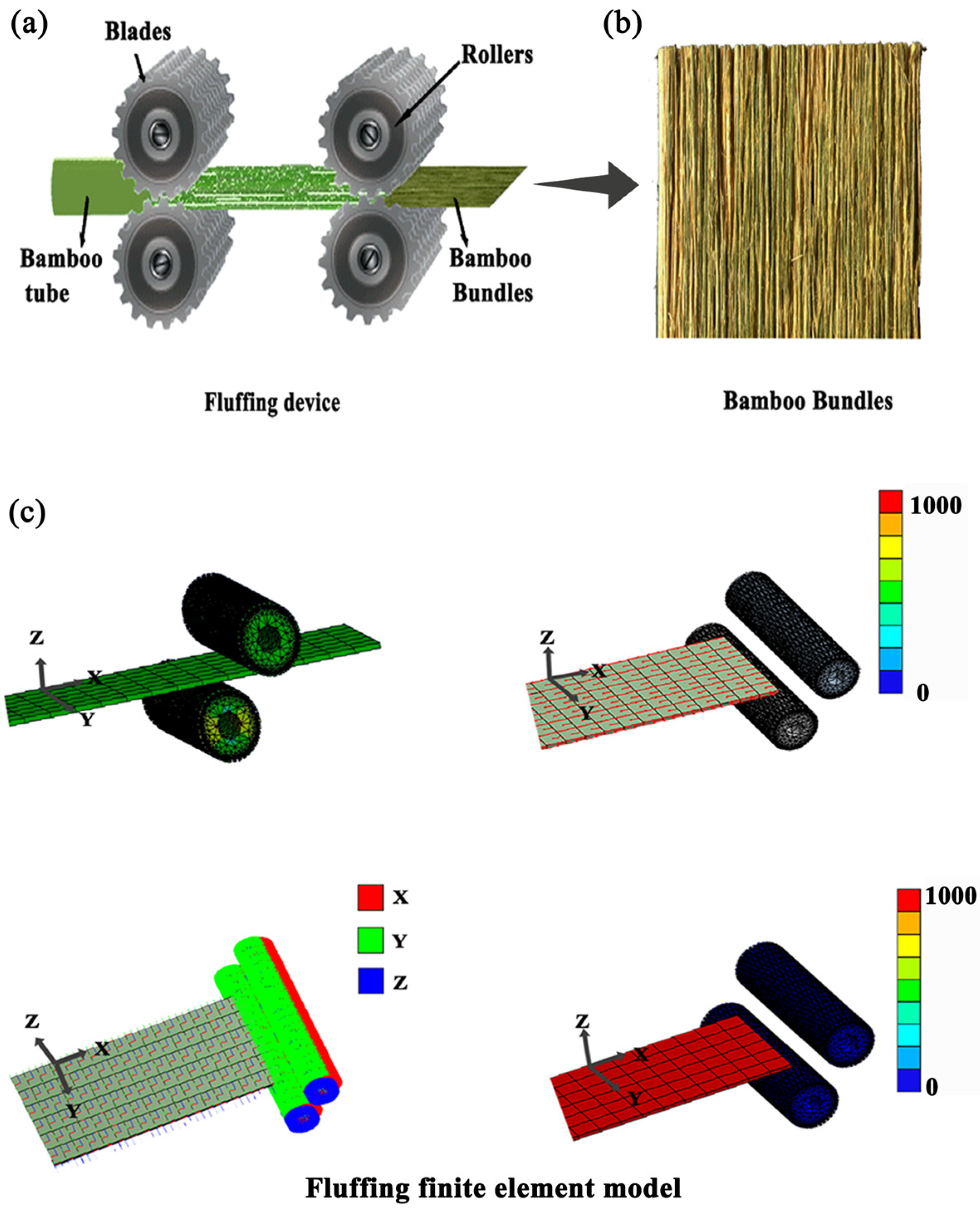
2.1. Materials

This study utilized samples from 4 to 5-year-old moso bamboo (Phyllostachys pubescens Mazel ex H.de Lehaie) with breast heights of 50–60 mm and wall ply of 8–12 mm collected from Zhejiang province in China. All of the samples with 0.65 g/cm3 were mostly air-dried until an EMC of 10%–12%, prepared from internodes with 30 mm longitudinal length and 5 mm radial width.
Bamboo bundles (2600 mm × 150 mm × 5 mm) were produced by a self-developed multifunctional fluffer machine (Bamboo flattening and dispersing machine, Anji Tianyou Machinery Technology Co., Ltd., HuZhou, China). The number of rollers was 13 pairs with a blade spacing of 12 mm, and the blades depth was 4 mm. A diagram of the simplified representation composed of a pair of fluffing rollers with blades and bamboo bundles is presented in Figure 1a,b. A 3D fluffing finite element method (FEM) model of bamboo and a pair of rollers with blades are presented in Figure 1c, which showed cracks that occurred in the bamboo under shear force and compression.

2.2. Three-Dimensional Reconstructed Geometries

A nondestructive method to reconstitute the three-dimensional image of samples was conducted using Micro X-ray tomography technology (u-CT, Skyscan 1172, Bruker, Madison, WI, USA) with a 0.5 µm micron pixel size. Bamboo blocks and bamboo bundles with sizes of 6 mm × 2 mm × 2 mm were fixed on the sample table, and the cross section faced upwards. Five replicates were used for three measurements at room temperature.

2.3. Morphology Characterization

To observe the morphology of bamboo bundles at the cellular level, a field emission scanning microscope (SEM, S-4800, Hitachi, Japan) has been conventionally used. Bamboo bundles with sizes of 3 mm (L) × 3 mm (T) × 5 mm (R) were bonded with double-sided glue and then sprayed with several coats of gold. Five replicate samples were tested for three times at a 10 kV acceleration voltage in a normal temperature condition.

2.4. Nanoindentation and Modulus Mapping Test

A nanoindentation test and dynamic modulus maps were used to measure the mechanical properties and to show the cell wall layer of the fiber cells and parenchymal cells. Bamboo blocks with sizes of 5 mm (L) × 1 mm (R) × 1 mm (T) for the nanoindentation test were ground and polished to obtain a smooth surface. Mechanical experiments were conducted with a nanoindenter (Hysitron TI 980 TriboIndenter, Bruker, Germany). Load control tests were employed to indent the cell wall of fiber cells and parenchymal cells. The indent tip was loaded on 10 mN at a rate of 20 µN·s−1, and the hold time at the peak load was 5 s. The hardness and reduced elastic modulus of each wall layer and middle lamellae of fiber cells and parenchymal cells were measured, respectively, to obtain a quantitative understanding of mechanical strength in the graded structure in bamboo. After indentation, the scanning probe microscopy images were obtained to observe the position and morphological characteristics of the cell wall layer and the middle lamellae. Three measurements were repeated for two samples at room temperature.

2.5. Raman Microscopy Analysis

To understand the chemical composition and distribution of different cell types, the sample blocks for Raman mapping were conducted by the Micro-FTIR (Spotlight 400 FT-IR Imaging System, PerkinElmer, Cambridge, MA, USA) experiment. For the Raman test, transverse sections of 5 µm thickness were cut using a sliding microtome and then immediately placed upon a glass slide. Then, a drop of distilled water was added to the glass, and a cover slip was placed on the bamboo slice. Resin was used to seal the edge of bamboo slice to avoid evaporation. Raman measurements were performed with a Renishaw in Via confocal Raman microscope and motorized XYZ stage with the following parameters: an excitation wavelength of 532 nm, a step size of 0.3 µm and an integration of 0.66 s. Two replicates were used for three tests at room temperature.

3. Results

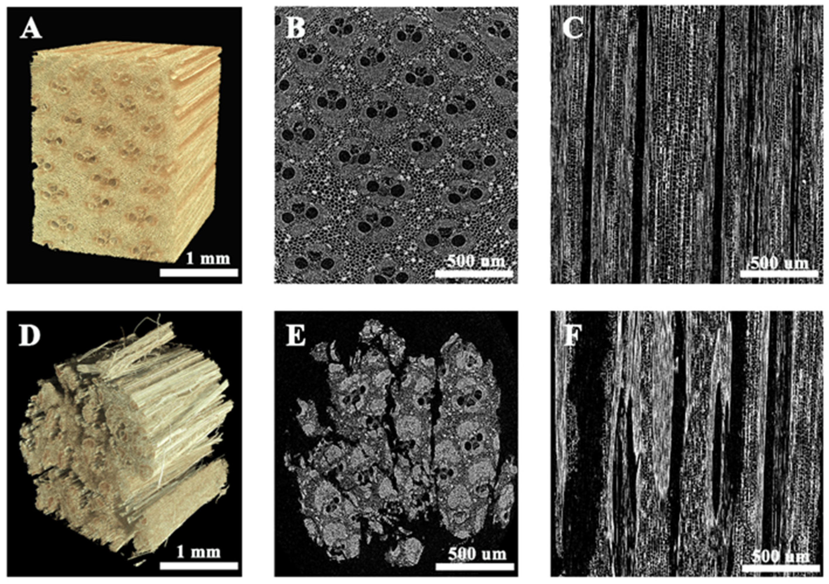
3.1. The 3D Morphology of Bamboo Bundles

The morphology and crack propagation of vascular bundles and parenchyma tissues in bamboo and bamboo bundles were acquired by X-ray microtomography tests. The 3D morphologies of bamboo and bamboo bundles are presented in Figure 2A,D. As Figure 2A presents, the bamboo was arranged regularly in the longitudinal direction, which was not conducive to the adhesive immersion. The bamboo culm was composed of vascular bundles embedded in the basic tissues (Figure 2B), which were arranged in the longitudinal direction (Figure 2C). As shown in Figure 2D, the bamboo bundles, after fluffing, were dispersed horizontally and separated in the longitudinal direction, which increased the penetration pathway of adhesives. Figure 2E presents that the fracture, once initiated, propagated in a tortuous path in the vascular bundles and in a straight path in the parenchyma, which separated the section of bamboo into several parts. Figure 2F illustrates that the fracture that propagated in the bamboo proceeded along the grain. The longitudinal fiber apparently prevented the cracks from transversely growing. The fracture type of the bamboo bundles in the longitudinal and radial directions were related to the structure of the cells in bamboo.

3.2. Characteristics of Cells in the Bamboo Bundles

Figure 3 shows the morphology and crack propagation pathway within vascular bundles, fibers and parenchymal cells of bamboo bundles in the radial and longitudinal directions. As shown in the radial section of a vascular bundle in Figure 3A, a series of irregular cracks were produced in the fiber sheath, which divided it into several fiber bundles of different sizes. Figure 3C shows that the cell wall was well-preserved and that the fiber cells were separated along the middle lamella interface. The fracture surface of the parenchyma in Figure 3D shows similar tearing characteristics to the fracture surface of the fiber sheath, and the cracks expanded along the middle lamella layer in the parenchyma cells.
As displayed in Figure 3B, the fracture images demonstrated that the vascular bundles were separated into bamboo bundles and that the cracks extended longitudinally. Figure 3E shows that the cracks grew along the middle lamella interface of the fiber cells without cell wall damage. The parenchyma was broken down in a ladder pattern and most of the long-axis parenchyma cell wall layer was intact, while a few short-axis parenchyma cells had their walls cut off to expose the lumen (Figure 3F).
Micrographs of the fracture appearance in the bamboo bundles were taken to show that cracks run mainly by cell wall peeling along the middle lamella interface in fiber cells and parenchymal cells in the bamboo bundles.

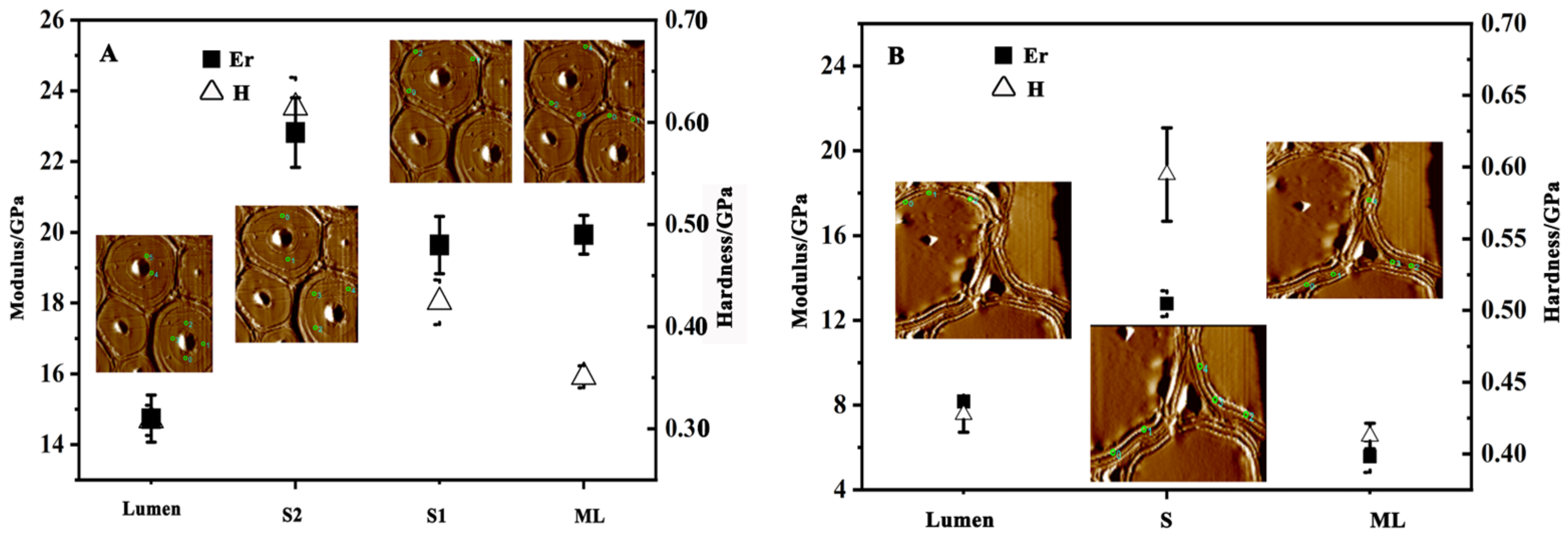
3.3. Micro-Mechanical Properties of Cell Walls

In this study, the reduced nanoindentation modulus and hardness across individual cell wall lamellae and middle lamella were tested to obtain a quantitative understanding at the cellular and subcellular levels. Figure 4A presents the micromechanical properties and corresponding position of mapping images from the lumen to the middle lamella (ML) in fiber cells. As Figure 4A shows, the middle lamella (ML) and s1 were narrow layers due to their limited width, whereas s2 was a thick layer. The elastic modulus presented a peak in the s2 (23.52 GPa) and declines in s1 (18.02 GPa), ML (15.91 GPa) and lumen (14.63 GPa), in that order. However, the hardness gave a gradual upward trend of lumen (0.31 GPa), s1 (0.48 GPa), ML (0.49 GPa) and s2 (0.59 GPa) in the fiber cells. The indentation modulus and hardness values of the fiber cells indicated that the middle lamella (ML) was the weaker interface, which tended to fracture under an external force. Similar to fiber cell walls, parenchymal cell walls are generally described as a polylamellate structure consisting of a primary cell wall, a secondary cell wall and a middle lamella. The contact force mapping images in Figure 4B showed the arrangement of the cell wall layers and individual reduced elastic moduli and hardness. As presented in Figure 4B, the parenchymal cells did not exhibit alternating broad and narrow lamellae. The uniform thickness of the cell wall resulted in small changes in the elastic modulus values from s1 (8.17 GPa) to s2 (12.78 GPa), presented in the Figure 4B. The elastic modulus and hardness in the parenchymal cell layers exhibited a downward trend of s2 (12.78 GPa, 0.59 GPa), s1(8.17 GPa, 0.42 GPa) and ML (5.57 GPa, 0.41 GPa). Similar to the fiber cells, the middle lamella (ML) was also the weaker interface in parenchymal cells.
When compared to the reduced modulus value in fiber cells (Figure 4A), the s2 layer in parenchymal cells (Figure 4B) was much lower than that of the fiber cells. This may account for the fiber cells that were the strongest interface in the graded bamboo structure, which prevented fiber cells from bucking under the external force.

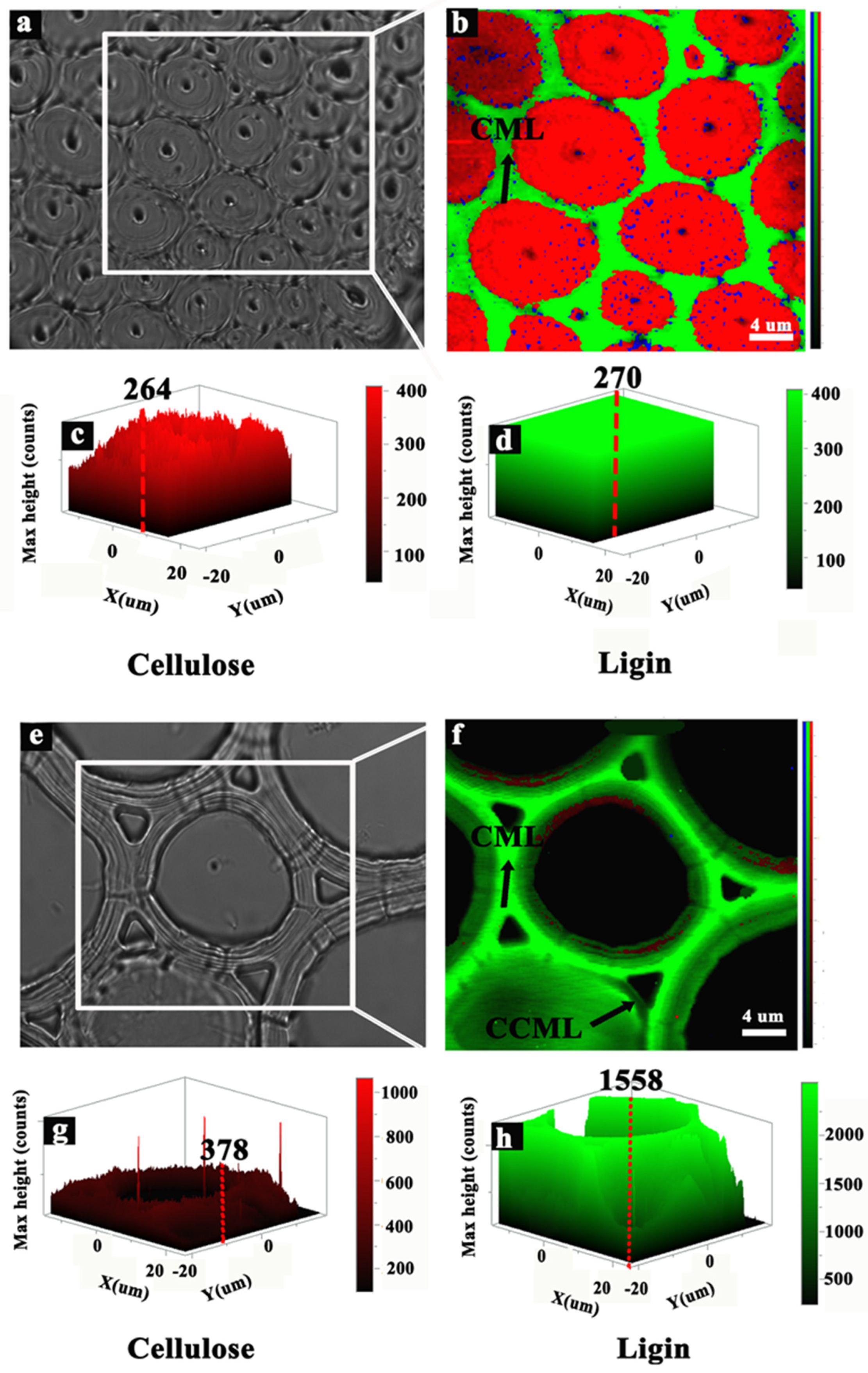
3.4. Chemical Components of the Cells

Owing to the chemical components determined by the mechanical properties of the cells, the Raman microscope was used to quantitatively characterize the chemical components. Since the cell wall of vessel cells is too thin to have any meaning for this study, we measured the chemical distribution map and Raman FTIR spectra of fiber cells and parenchymal cells described in Figure 5. The relative concentration of cellulose and lignin in the fibers, middle lamella (ML) and parenchyma cells were also analyzed using the plane scanning method. Examining Figure 5a,b, it was apparent that cellulose was mainly distributed in the secondary wall of fibers and that a considerable lignin concentration was found in the ML. According to the plane-scanning results, the cellulose content in the fiber walls was the highest (Figure 5c); nevertheless, the ML showed the higher amount of lignin (Figure 5d). As shown in Figure 5e,f, parenchymal cells were composed of plenty of lignin distributed in ML and a small quantity of cellulose distributed in cell wall. The relative concentrations of both cellulose and lignin showed that the max height of lignin was about five times that of cellulose. Consequentially, cellulose concentrated in the secondary wall of fiber cells, while lignin was distributed in the ML of fiber cells and parenchyma cells.
Compared with the secondary wall of fibers, the weak interface attributed to crack initiation and propagation in the course of fluffing was strongly related to the high content of lignin, which was a mesh-like structure cross-linked with hemicelluloses and cellulose. Thus, the fluffing process disrupts the cross-linked structure of highly lignified middle lamella (ML), which is crucial in the process of selective separation in bamboo bundles.

4. Discussion

Bamboo bundles with a series of dotted and linear cracks exhibited a facile netlike structure. This study used u-CT, SEM observation, nanoindentation technology and Raman tests to study the separation mechanism of bamboo bundles at the cellular level in depth. ML in fiber cells and parenchymal cells was the prominent fracture ultrastructure in bamboo bundles due to low mechanical properties and high lignin content.
Previous studies have focused on the anatomical and morphological characteristics of cracks in bamboo bundles at the macroscopic level by investigating the cross section and quantity of vascular bundles [8,10]. In this paper, the crack propagation path in bamboo bundles was analyzed at the cellular level and the fracture mechanism was illustrated by analyzing the subcellular structure and component characteristics of bamboo. The weak interface in bamboo was attributed to the cracks growing, which highlight the separation mechanism of bamboo bundles. For bamboo, the cells are arranged longitudinally, which are useful to observe and investigate the fracture behavior under fluffing with the help of instruments. However, other biomaterials with hierarchical structures, such as wood, require further research.

5. Conclusions

Compared with bamboo, the morphology of bamboo bundles showed that fracture propagation was related to the structure of the cells. CML in fiber, parenchymal and vessel cells were the prominent fracture ultrastructure in bamboo bundles owing to their thick walls and lower elastic moduli. Fiber cells remain almost intact in the longitudinal direction due to their higher elastic moduli and cellulose aggregation. On the contrary, the break in CML with more lignin led to arborization cracks and formed a network link among fiber cells. Overall, the bamboo bundles were characterized by horizontal interweaving, vertical orientations and spatial patterns. Therefore, a complete analysis on the cells’ structure–property relationship shows the theoretical mechanism at cellular level and illustrates the morphology of bamboo bundles under fluffing.

Author Contributions

X.H.: Data curation, Investigation, Writing—original draft. X.T.: Finite element method analysis. S.L.: Supervision, Writing—review and editing. C.Y.: supervision, Writing—review and editing. Y.Y.: Formal analysis. W.Y.: Conceptualization, Funding acquisition, Methodology, Project administration. All authors have read and agreed to the published version of the manuscript.

Funding

The authors gratefully acknowledge the financial support of the Fundamental Research Funds for the Central Universities (No. 2572020AW40), Nature Science Foundation of China (No. 31870550) and Guangdong Province Important Project Foundation (No. 2020B020216001).

Data Availability Statement

The data presented in this study are available on request from the corresponding author.

Conflicts of Interest

The authors declare that they have no known competing financial interest or personal relationships that could have appeared to influence the work reported in this paper.

References

  1. Lugt, V.; Dobbelsteen, V.D. An environmental, economic and practical assessment of bamboo as a building material for supporting structures. Constr. Build. Mater. 2006, 20, 648–656. [Google Scholar] [CrossRef]
  2. Sharma, B.; Gatoo, A.; Bock, M.; Ramage, M. Engineered bamboo for structural applications. Constr. Build. Mater. 2015, 81, 66–73. [Google Scholar] [CrossRef]
  3. Ahmad, M.; Kamke, F.A. Analysis of Calcutta bamboo for structural composite materials: Physical and mechanical properties. Wood Sci. Technol. 2005, 39, 448–459. [Google Scholar] [CrossRef]
  4. Yu, W.; Yu, Y. Development and prospect of wood and bamboo scrimber industry in China. China Wood Ind. 2013, 27, 5–8. [Google Scholar]
  5. Zhang, J.; Wang, K.H.; Li, Q. Prospects of efficient and comprehensive utilization of bamboo and wood resources in China. China Timber 2006, 005, 24–26. [Google Scholar]
  6. Zhang, Y.; Huang, Y.; Zhang, Y.; Yu, W. Scrimber board (sb) manufacturing by a new method and characterization of sb’s mechanical properties and dimensional stability. Holzforschung 2018, 72, 283–289. [Google Scholar] [CrossRef]
  7. Yu, Y. Manufacturing Technology and Mechanism of High Performance Bamboo-Based Fiber Composites. Ph.D. Dissertation, Chinese Academy of Forestry, Beijing, China, 2014. [Google Scholar]
  8. Belayachi, N.; Hoxha, D.; Slaimia, M. Impact of accelerated climatic aging on the behavior of gypsum plaster-straw material for building thermal insulation. Constr. Build. Mater. 2015, 81, 66–73. [Google Scholar] [CrossRef]
  9. Bao, M.; Huang, X.; Zhang, Y.; Yu, W.; Yu, Y. Effect of density on the hygroscopicity and surface characteristics of hybrid poplar compreg. Wood Sci. Technol. 2016, 62, 441–451. [Google Scholar] [CrossRef] [Green Version]
  10. Qi, J.; Yu, W.; Xie, J.; Yu, Y.; Zhang, Y.; Sun, H. Morphologies of crushed bamboo veneer used for bamboo-based fiber composites. China Wood Ind. 2012, 26, 6–9. [Google Scholar]
  11. Wu, B.; Yu, Y.; Qi, J.; Yu, W. Effects of bamboo bundles treated with fine fluffing and carbonized treatment on the properties of bamboo scrimber. J. Nanjing For. Univ. (Nat. Sci. Ed.) 2014, 38, 115–120. [Google Scholar]
  12. Li, J.; Wu, Z.; He, S.; Huang, C.; Chen, Y. Effect of bundle diameter on water absorption properties of bamboo scrimber. J. Zhejiang For. Sci. Technol. 2016, 36, 67–68. [Google Scholar]
  13. Wei, J.; Rao, F.; Zhang, Y.; Li, C.; Yu, W. Effect of veneer crushing process on physical and mechanical properties of scrimbers made of Pinus radiata and Populus tomentosa. China Wood Ind. 2019, 33, 55–58. [Google Scholar]
  14. Sutnaun, S.; Srisuwan, S.; Jindasai, P.; Cherdchim, B.; Matan, N. Macroscopic and microscopic gradient structures of bamboo culms. Walailak J. Sci. Technol. 2005, 2, 31–39. [Google Scholar]
  15. Lian, C.; Liu, R.; Zhang, S.; Yuan, J.; Luo, J.; Yang, F.; Fei, B. Ultrastructure of parenchyma cell wall in bamboo (Phyllostachys edulis) culms. Cellulose 2020, 27, 7321–7329. [Google Scholar] [CrossRef]
  16. Dixon, P.G.; Gibson, L.J.; Bock, M. The structure and mechanics of moso bamboo material. Sci. Rep. 2014, 11, 20140321. [Google Scholar] [CrossRef]
  17. Habibi, M.; Lu, Y. Cracks propagation in bamboo’s hierarchical cellular structure. Sci. Rep. 2014, 4, 5598. [Google Scholar] [CrossRef] [Green Version]
  18. Shao, Z.; Fang, C.; Huang, S. Tensile properties of moso bamboo (Phyllostachys pubescens) and its components with respect to its fiber-reinforced composite structure. Wood Sci. Technol. 2010, 44, 655–666. [Google Scholar] [CrossRef]
  19. Wang, F.; Shao, Z.; Bock, M. Study on the variation law of bamboo fibers’ tensile properties and the organization structure on the radial direction of bamboo stem. Ind. Crops Prod. 2020, 152, 112521. [Google Scholar] [CrossRef]
  20. Wang, X.; Ren, H.; Zhang, B.; Fei, B.; Burgert, I. Cell wall structure and formation of maturing fibres of moso bamboo (Phyllostachys pubescens) increase buckling resistance. J. R. Soc. Interface 2012, 9, 988–996. [Google Scholar] [CrossRef] [Green Version]
  21. Wang, D.; Lin, L.; Fu, F. Fracture mechanisms of moso bamboo (Phyllostachys pubescens) under longitudinal tensile loading. Ind. Crops Prod. 2020, 153, 112574. [Google Scholar] [CrossRef]
  22. Parameswaran, N.; Liese, W. On the fine structure of bamboo fibres. Wood Sci. Technol. 1976, 10, 231–246. [Google Scholar] [CrossRef]
  23. Obataya, E.; Kitin, P.; Yamauchi, H. Bending characteristics of bamboo (Phyllostachys pubescens) with respect to its fiber–foam composite structure. Wood Sci. Technol. 2007, 41, 385–400. [Google Scholar] [CrossRef]
  24. Liu, X.; Tian, G.; Shang, L.; Yang, S.; Jiang, Z. Compression properties of vascular boundles and parenchyma of rattan (Plectocomia assamica Griff). Holzforschung 2014, 68, 927–932. [Google Scholar] [CrossRef]
  25. Chen, M.; Ye, L.; Wang, G.; Fang, C.; Dai, C.; Fei, B. Fracture modes of bamboo fiber bundles in three-point bending. Cellulose 2019, 26, 8101–8108. [Google Scholar] [CrossRef]
Figure 1. Schematic for fluffing process, product and fluffing finite element model. (a) A pair of fluffing rollers with blades; (b) bamboo bundles image; (c) fluffing finite element model.
Forests 13 01897 g001
Figure 2. CT images of bamboo and bamboo bundles. (A,D) Three-dimensionally reconstructed images; (B,E) two-dimensional X-ray tomography slice in the radial direction; (C,F) two-dimensional X-ray tomography slice in the longitudinal direction.
Forests 13 01897 g002
Figure 3. SEM microscope images of bamboo bundles. (A,C,D) Vascular bundles, fiber cells and parenchymal cells in a radial section; (B,E,F) vascular bundles, fiber cells and parenchymal cells in a longitudinal section.
Forests 13 01897 g003
Figure 4. Nanoindentation and modulus mapping tests in the fiber cells (A) and parenchymal cells (B).
Forests 13 01897 g004
Figure 5. Raman spectra of a fiber cell and a parenchyma cell. (a,b) Raman image of the cellulose and lignin contents of a fiber cell. (c,d) Chemical components of the fiber cell and the compound middle lamella (CML). (e,f) Raman images of the parenchymal cell. (g,h) Chemical components of the parenchymal cell.
Forests 13 01897 g005
Publisher’s Note: MDPI stays neutral with regard to jurisdictional claims in published maps and institutional affiliations.

