Article

Comparison of Endophytic and Epiphytic Microbial Communities in Surviving and Dead Korean Fir (Abies koreana) Using Metagenomic Sequencing

1
Department of Biological Sciences, Chungnam National University, Daejeon 34134, Republic of Korea
2
Division of Forest Bio-Information, National Institute of Forest Science, Suwon 16631, Republic of Korea
3
Department of Biology, Jeju National University, Jeju 63243, Republic of Korea
4
Department of Agricultural Life Science, Sunchon National University, Suncheon 57922, Republic of Korea
5
Department of Biology, Chungbuk National University, Cheongju 28644, Republic of Korea
*
Author to whom correspondence should be addressed.
Forests 2022, 13(11), 1932; https://doi.org/10.3390/f13111932
Submission received: 30 September 2022 / Revised: 8 November 2022 / Accepted: 10 November 2022 / Published: 16 November 2022
(This article belongs to the Special Issue Plant-Microbe Interactions in Forests)

Abstract

:
Plant endophyte and epiphyte communities cooperatively interact with their host plants and play crucial roles in sustaining plant fitness. In Korea, a variety of studies have been conducted to elucidate the reasons for the declining population of the endangered Korean fir (Abies koreana), but the relationship between microbiota and the healthy condition of trees remains unclear. Here, we conducted bacterial 16S rRNA gene and fungal ITS sequence analyses to dissect the composition of endophytic and epiphytic microbiota in both live and dead trees located in the same Mt. Jiri habitat. In the live trees, the bacterial class Armatimonadia and the lichenized fungi groups were significantly dominant, whereas many bacterial and fungal taxa mainly found in rotten wood were enriched in the dead trees. Functional prediction of the microbial communities in live trees suggested the possibility that bacterial endophytes and epiphytes play a role in inorganic nutrient metabolism and fungal endophytes and epiphytes produce biologically active secondary metabolites, thereby contributing to the healthy condition of Korean fir trees. The ecological function of endophytes and epiphytes in dead trees was predicted to be involved in the decomposition of wood for nutrient recycling. Our analyses revealed a distinct difference in microbial communities depending on the health condition of Korean fir trees. The results from this study would be useful for understanding the ecological function of endophytic and epiphytic microorganisms to conserve and manage this endangered species from ecologically vulnerable environments.

1. Introduction

The Korean fir (Abies koreana) is an endemic coniferous tree species in the Republic of Korea and distributed throughout subalpine areas of tall mountains in the southern province, which has altitude ranges of 1000 to 1900 m above sea level [1]. The Korean fir tree species are valuable biological resources. For example, the shape of Korean fir trees has high ornamental and commercial values, thereby cultivating them for Christmas tree plantations [2]. Several studies reported that the phytochemicals extracted from the trees have several medical functions, including anti-cancer activity and memory-enhancing effect [3,4,5]. However, the population of Korean fir trees was rapidly declined since the 1980s, resulting in the declaration of endangered species by the International Union of Conservation of Nature (IUCN) [6].
A variety of studies have been conducted to explore the reasons for the declining population of Korean Fir trees. Although the precise mechanisms of the decline are largely unknown, many studies suggest that tree mortality is highly correlated with increased temperature from global warming [7]. Both temperature and warm index in the subalpine habitats of Korean fir trees were increased in the last few decades [8], which could facilitate to decrease in the growth of Korean fir trees by reducing photosynthesis efficiency [9]. Furthermore, Korean fir trees from ecologically vulnerable habitats exhibited higher expression levels of heat stress-induced genes than those from ecologically stable habitats, suggesting that the declining population of Korean fir trees is likely experiencing heat stress [10]. In addition, other environmental factors such as strong winds from typhoons, soil moisture, drought, and precipitation might be attributed to the declining population of Korean Fir trees [11,12].
Plants live in cooperative interactions with microorganisms, collectively forming plant microbiota. Plant microbiota is involved in plant stress tolerance and nutrition, thereby affecting plant growth and survival [13]. For example, the microbial diversity in horse chestnut (Aesculus hippocastanum) trees is negatively correlated with bleeding canker disease symptom severity [14]. In Pinus monticola, exogenous inoculation of fungal endophytes to seedlings confers resistance to the pathogen Cronartium ribicola, the fungal causal agent of white pine blister rust disease [15]. Hence, understanding the composition of plant microbiota and deciphering the interplay between plant and plant microbiota would be important for maintaining plant fitness. However, our understanding of microbiota in Korean fir trees and their interaction related to fitness is rudimentary compared to our understanding of interactions between abiotic stresses and the mortality of Korean fir trees [7]. A previous analysis of soil fungal communities between dead and live Korean fir trees revealed significant differences in the saprophyte and the mycorrhizal Clavulina spp. [16]. Recently, the rhizosphere microbiome analysis of live and dead Korean fir trees reported that bacteria involved in plant growth and stress tolerance were significantly more abundant in the rhizosphere of live trees than in dead trees [17]. These two microbiome studies suggest that structure of fungal and bacterial rhizosphere communities is highly affected by the fitness of Korean fir trees. However, no endophytic and epiphytic communities in Korean fir trees have been studied to date.
In this study, we analyzed the composition of endophytes and epiphytes of live and dead Korean fir trees located in the same Mt. Jiri habitat to understand how the health conditions of trees affect microbial communities and predict their ecological roles. We found a significant increase in the diversity of bacterial communities in the dead Korean fir trees but not in the fungal communities. Both bacterial and fungal microbes between live and dead trees were relatively distinct. Functional prediction of endophytic and epiphytic bacteria suggested that bacterial communities in live trees might play a role in inorganic nutrient metabolism to sustain the fitness of Korean fir trees. In contrast, those in dead trees might be involved in decomposing wood for nitrogen recycling. Furthermore, fungal endophytes and epiphytes in live trees were predicted to be involved in the production of secondary metabolites to enhance tree fitness, whereas the ecological role of fungal endophytes and epiphytes in dead trees may contribute to nutrient recycling in the forest ecosystem. Our findings would be useful for understanding the potential role of endophytic and epiphytic microorganisms in the endangered Korean fir trees and improving the sustainability of trees from ecologically vulnerable environments.

2. Materials and Methods

2.1. Study Site and Tree Sampling

The Korean fir trees were obtained from a Korean fir tree forest in Mt. Jiri at an altitude of 1490–1730 m above sea level. We randomly selected three dead and live trees with at least 25 cm of diameter at breast height in the same sites at intervals of >10 m to choose trees in similar growth environmental conditions (Supplementary Figure S1). Since the causal agents of the declining Korean fir trees are largely unknown, we determined the health conditions of trees based on the apparent conditions of leaves on the branches. We chose three live trees that had vivid greenish leaves on all branches of trees. For the selection of dead trees, we carefully chose three dying trees that had both greenish and yellowish leaves on 10% of branches whereas the remaining 90% of the branches had no leaves. To extract microbial DNA from the Korean fir trees, we collected sawdust samples from three live (A1–A3) and three dead (D1–D3) trees by drilling the 6 cm of four holes on the trunks at four different directional spots with a clean appearance. The sawdust samples were mainly composed of sapwood and trace amounts of cambium and bark.

2.2. DNA Extraction and Next-Generation Sequencing of 16S rRNA Genes and ITS Sequences

Total DNA was extracted from the sawdust samples using a Bacteria DNA Purification Kit (26-008, BioD, Gwangmyeong, Korea) according to the manufacturer’s instructions. The quality and quantity of the extracted gDNA were determined using the Agilent 4150 TapeStation (Agilent, Inc., Santa Clara, CA, USA). For bacterial 16S rRNA gene (V3–V4 region) amplification, we performed PCR using the following mixture: 10 μL of 2× Dr. MAX Master Mix Solution (Doctor Protein Corp., Seoul, Korea), 1 μM of 341F (5′-CCTACGGGNGGCWGCAG-3′) and 805R (5′-GACTACHVGGGTATCTAATCC-3′) primer set [18,19], and ~10 ng of the extracted DNA as a template. For eukaryotic fungal ITS region amplification, we performed the same protocol as with bacterial 16 rRNA PCR except for the primers (1 μM of ITS3 (5′-GCATCGATGAAGAACGCAGC-3′) and ITS4 (5′-TCCTCCGCTTATTGATATGC-3′)) [20]. PCR amplification was performed with an initial denaturation at 95 °C for 7 min, followed by 30 cycles at 95 °C for 30 s, 55 °C for 30 s, and 72 °C for 30 s, and a final extension at 72 °C for 10 min. The amplicons were sequenced using the Illumina Mi-Seq platform (Illumina Inc., San Diego, CA, USA) at Macrogen (Seoul, Korea).

2.3. Diversity Analysis and Functional Prediction of 16S rRNA Genes and ITS Sequences

Quantitative Insights Into Microbial Ecology 2 (QIIME2) software package was used for subsequent bioinformatic analysis [21]. The obtained raw paired-end sequences were filtered, trimmed, denoised, and merged using Cutadapt and DADA2 [22,23], yielding Amplicon Sequence Variants (ASVs). To improve the performance of a taxonomic classifier, we trained the Silva database for 16S rRNA [24] and the UNITE fungal database for fungal ITS [25] using a scikit-learn naïve Bayes machine-learning taxonomy classifier [26], resulting in our own taxonomic classifiers based on primer sequences used for PCR amplification. ASVs were classified at the species level using the q2-feature-classifier plugin [27] against the trained taxonomic classifiers. Sequences assigned to chloroplasts, mitochondria, or non-identified were filtered out from the dataset. The filtered sequences were aligned with MAFFT [28] and a rooted phylogenetic tree was constructed using FastTree 2 [29]. Alpha-diversity and beta-diversity analyses were performed using the q2-diversity plugin at a rarefied sampling depth of 52,530. The Principal Coordinate Analysis (PCoA) plot was generated using the Bray–Curtis dissimilarity between the live and dead tree samples [30].
The functional abundances of the bacterial communities were predicted using Phylogenetic Investigation of Communities by Reconstruction of Unobserved States2 (PICRUSt2) [31] based on the ASV table from QIIME2. The prediction was carried out with EPA-ng placement [32], the hidden-state prediction [33], and the prediction of gene pathways using MinPath [34]. The predicted MetaCyc pathway abundances were collapsed into MetaCyc pathway ontologies using SmartTable for further analysis [35]. The trophic modes of the fungal communities were predicted using FUNGuild [36].

2.4. Statistical Analysis

We tested significant effects for Faith’s Phylogenetic Diversity (PD) using a pairwise Kruskal–Wallis test [37]. Differential abundance analysis between live and dead trees was performed using linear discriminant analysis (LDA) effect size (LEfSe) [38]. The alpha value for the factorial Kruskal–Wallis test was set to lower than 0.05 for all analyses. The threshold on the LDA score was set to higher than either 2.0 or 3.0, as indicated in the figure legends. The results of LEfSe analyses were visualized in either bar graphs or cladograms.

3. Results

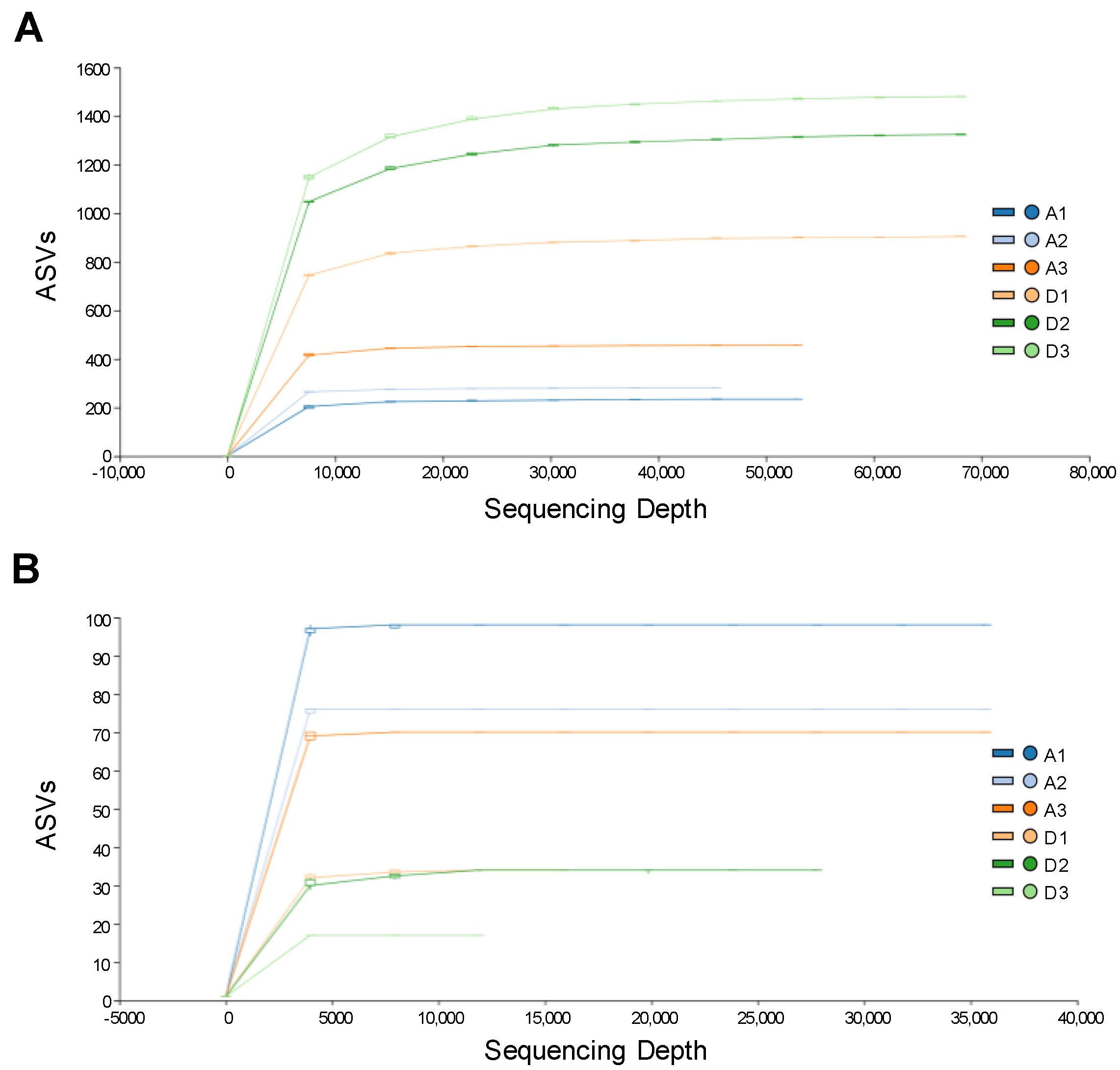
3.1. 16S rRNA and ITS Metagenomic Sequencing Analyses

The bacterial and fungal communities between live and dead Korean fir trees at the same site were dissected using 16S rRNA and ITS metagenomic sequencing analyses. After filtering low-quality, short, and chimera sequences, 413,873 high-quality 16S rRNA reads and 220,411 high-quality ITS reads from six tree samples were obtained (Supplementary Table S1). The 16S rRNA and ITS reads were clustered into 3658 ASVs and 223 ASVs, respectively. The coverage of detected bacterial and fungal ASVs in the six tree samples was estimated by rarefaction analyses (Figure 1). The rarefaction curves of bacterial and fungal communities were about to reach saturation at approximately 8000 and 3800 sequencing depths, respectively, suggesting that the bacterial and fungal communities associated with the tree endophyte and epiphyte represent an acceptable proportion of the biological species richness [39]. The number of bacterial ASVs in the dead trees was higher than in live trees (Figure 1A), whereas the dead trees exhibited a lower number of fungal ASVs than live trees (Figure 1B).

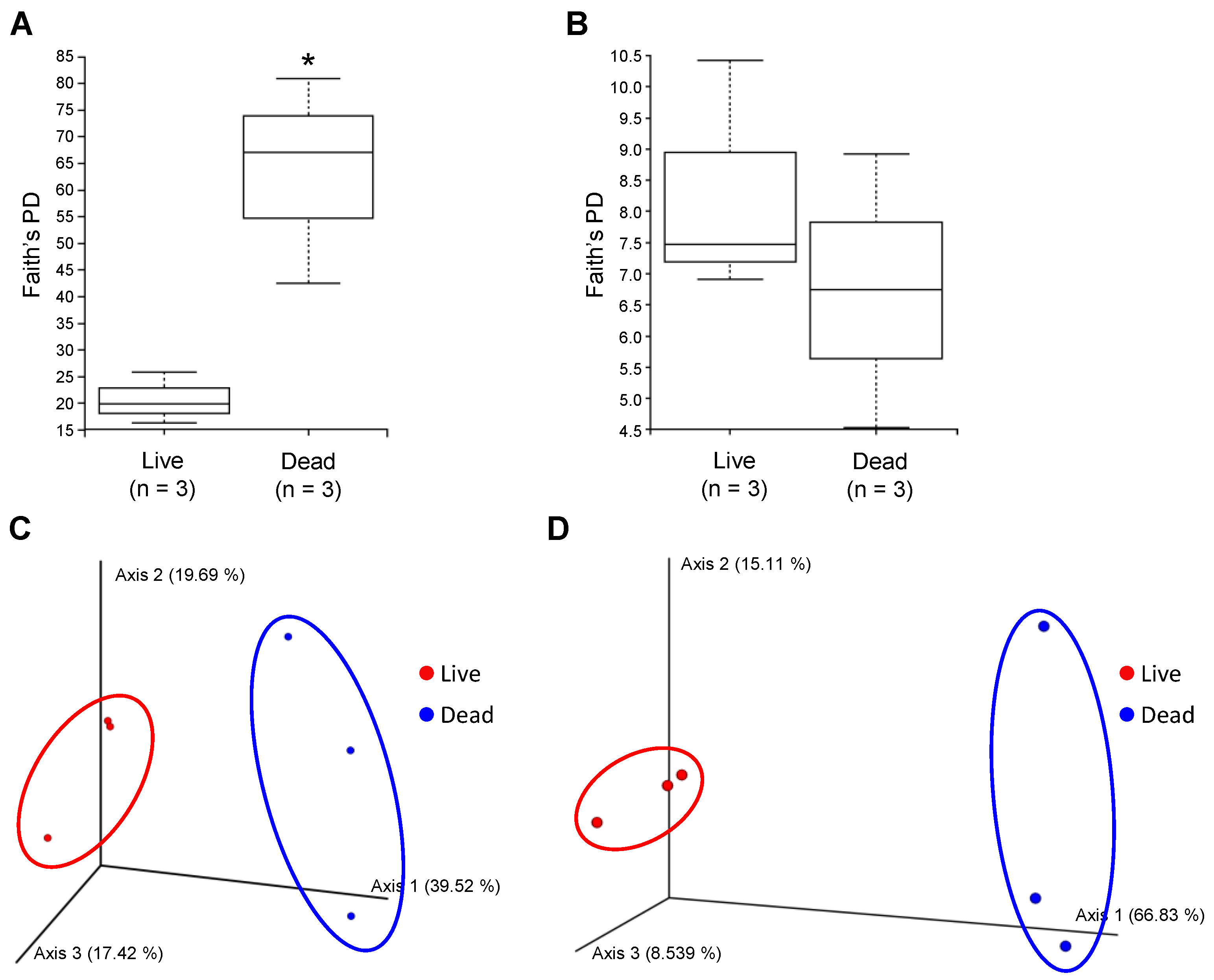
3.2. Diversity of Endophytic and Epiphytic Communities in Live and Dead Korean Fir Trees

To evaluate the richness of bacterial and fungal communities in live and dead trees, we measured Faith’s phylogenetic diversity (PD) of the samples. We found that the bacterial communities in the dead trees have significantly higher ASV richness than the ones in live trees (Figure 2A) but not for the fungal communities (Figure 2B). Next, we explored overall similarities and differences between live and dead tree microbial communities using the Principal Coordinate Analysis (PCoA) based on the Bray–Curtis distance matrix. Both bacterial and fungal communities from the live trees were grouped in a relatively distinct cluster from the dead trees (Figure 2C,D), indicating a difference in the bacterial and fungal composition between the live and dead Korean fir trees.

3.3. Taxonomic Profiling and Comparative Analysis of the Bacterial Communities between Live and Dead Korean Fir Trees

The phylum-level endophytic and epiphytic bacterial composition was examined to compare bacterial communities between live and dead trees. The bacterial ASVs were grouped into 26 phyla based on phylogenetic analysis, excluding the unknown phylum, and the relative abundance of recurrent phyla was represented in Figure 3A. There were three major phyla in the bacterial endophyte and epiphyte community of the live trees accounting for an average of 91.12% of the total identified taxa abundance; the average abundance of Proteobacteria, Acidobacteriota, and Actinobacteriota were 73.91%, 11.17%, and 6.03%, respectively. The bacterial communities in dead trees were dominated by six major phyla accounting for an average of 90.00% of the total identified taxa abundance; the average abundance of Proteobacteria, Actinobacteriota, Acidobacteriota, Bacteroidota, Patescibacteria, and Verrucomicrobiota was 36.30%, 18.63%, 13.92%, 12.27%, 4.47%, and 4.44%, respectively.
To identify differentially abundant bacterial taxa between live and dead Korean fir trees, we performed LEfSe analysis at the phylum and class levels. Cladogram generated from LEfSe analysis revealed that seven phyla and eleven classes were found to be significantly enriched in the dead trees; Actinobacteria and Thermoleophilia belonging to Actinobacteriota, Bacteroidia belonging to Bateroidota, Ktedonobacteria belonging to Chloroflexi, Babeliae belonging to Dependentiae, Polyangia belonging to Myxococcota, Parcubacteria and Saccharimonadia belonging to Patescibacteria, Chlamydiae and Verrucomicrobiae belonging to Verrucomicrobiota, and Phycisphaerae. However, only the class Armatimonadia (Armatimonadota phylum) was significantly enriched in the live trees at the class level (Figure 3B).

3.4. The Functional Inference of Bacterial Communities between Live and Dead Korean Fir Trees

The MetaCyc pathway prediction of bacterial communities using PICRUSt2 suggested that the pathways associated with lipopolysaccharide biosynthesis, cofactor biosynthesis, amino acid degradation, phosphorous metabolism, siderophores biosynthesis, and fermentation were significantly enriched in the bacterial endophyte and epiphyte communities of live Korean fir trees (Figure 4, red). The abundances of the pathways involved in nucleotide metabolism, nitrogen degradation, cofactor biosynthesis, lipid biosynthesis, and terpenoid biosynthesis were significantly increased in the bacterial endophyte and epiphyte communities of dead Korean fir trees (Figure 4, green).

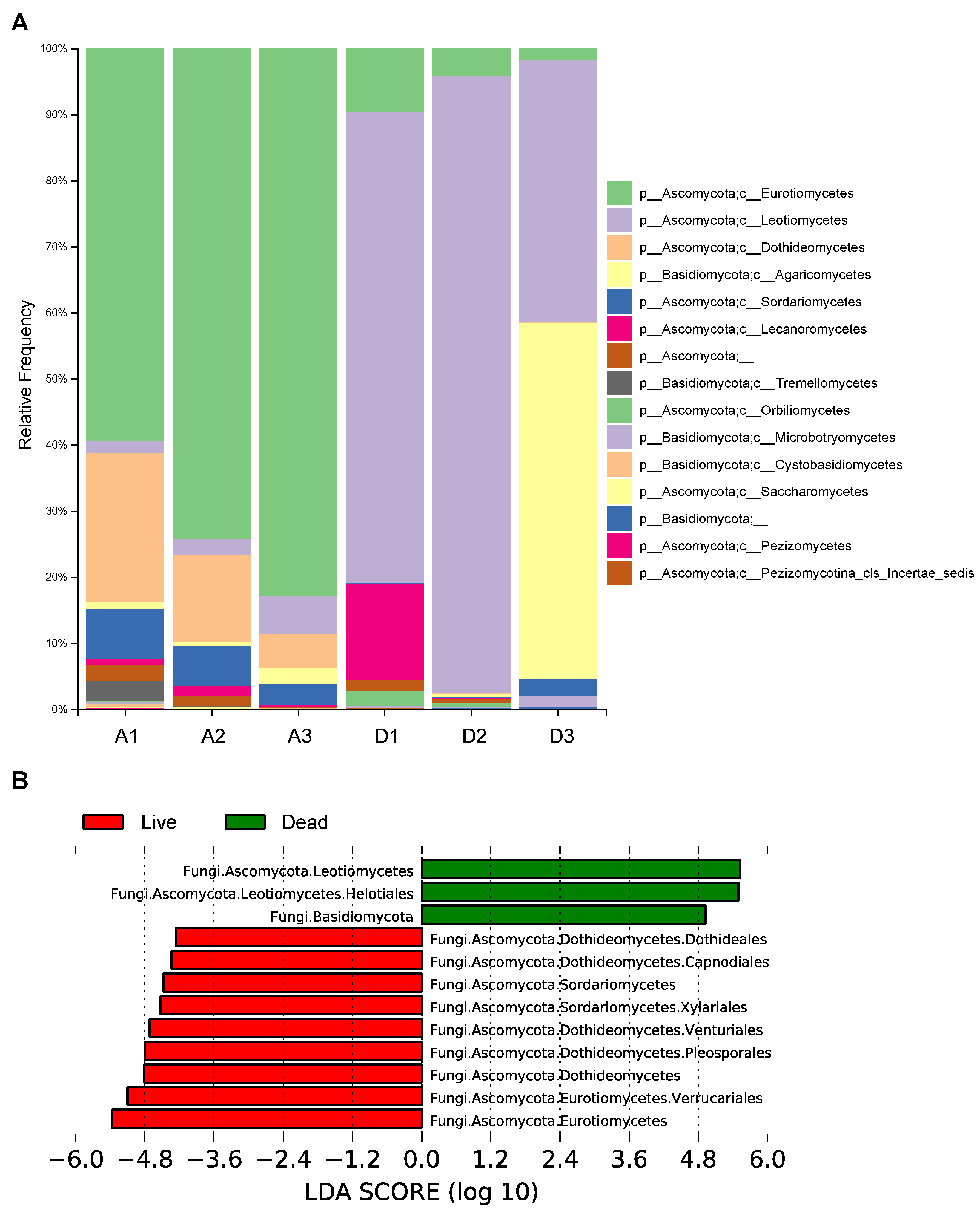
3.5. Taxonomic Profiling and Comparative Analysis of the Fungal Communities between Live and Dead Korean Fir Trees

The fungal composition of endophyte and epiphyte communities was examined at the phylum and class levels in order to compare fungal communities between live and dead trees. The fungal ASVs were grouped into 2 phyla and 13 classes, which exhibited a distinct difference in endophytic and epiphytic fungal communities between live and dead trees at the class level (Figure 5A). The fungal endophyte and epiphyte communities were dominated by Eurotiomycetes, Dothideomycetes, and Sordariomycetes belonging to Ascomycota accounting for an average of 72.32%, 13.71%, and 5.62% of the total identified fungal taxa abundance in the live trees, respectively. The dead tree endophyte and epiphyte fungal communities were dominated by Leotiomycetes belonging to Ascomycota, accounting for an average of 68.08% of the total identified taxa abundance in the live trees.
Next, we identified differentially abundant fungal taxa between live and dead Korean fir trees using LEfSe analysis. Three fungal taxa were significantly enriched in the dead Korean fir trees, including Basidiomycota, Leotiomycetes, and Helotiales (Figure 5B, green). However, the fungal endophyte and epiphyte communities in live trees were significantly enriched by six orders of fungi belonging to the phylum Ascomycota; Dothideales, Capnodiales, Venturiales, pleosporales, Xylariales, and Verrucariales, which were affiliated with Dothideomycetes, Sordariomycetes, and Eurotiomycetes at the class level (Figure 5B, red).

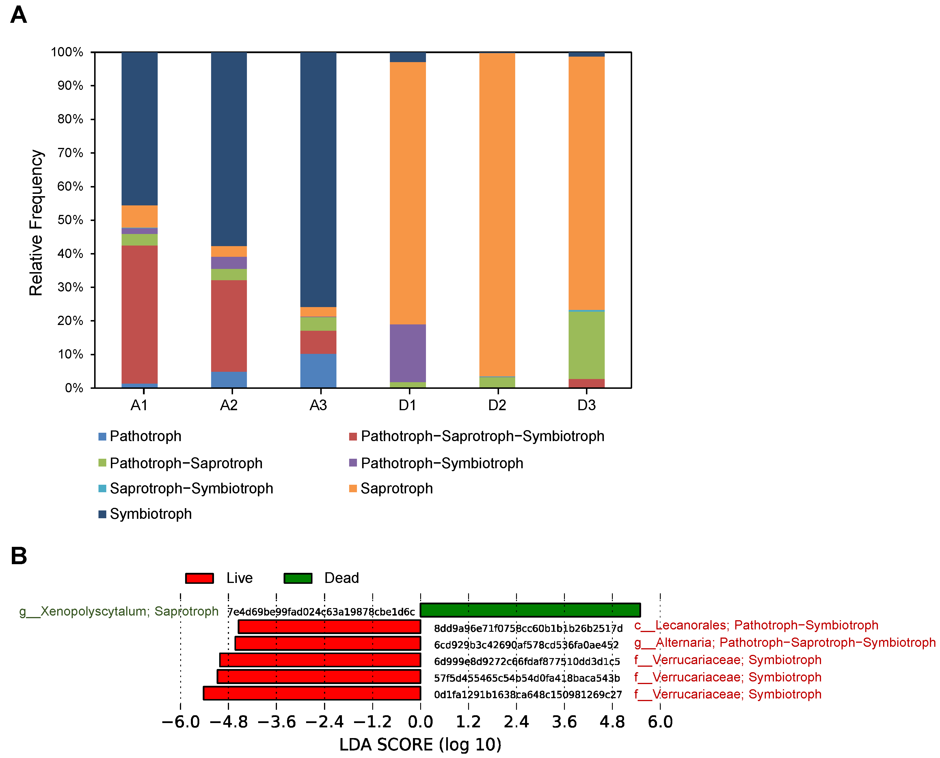
3.6. The Functional Prediction of Fungal Communities between Live and Dead Korean Fir Trees

The difference in the trophic mode of fungi in endophyte and epiphyte communities of live and dead trees was analyzed (Figure 6A). We found that the fungal endophyte and epiphyte communities in the live trees were dominated by three trophic mode groups; symbiotroph, pathotroph–saprotroph–symbiotroph, and saprotroph, which account for an average of 59.68%, 25.13%, and 4.19% of the total identified trophic mode groups, respectively. However, an average of 83.28% of the total identified fungal trophic mode groups in the dead trees were saprotroph.
To identify differentially abundant fungal ASVs between live and dead Korean fir trees, we performed LEfSe analysis using 223 fungal ASVs from the six tree samples. We found that five fungal ASVs were significantly enriched in the live trees (Figure 6B). Among five ASVs, three were grouped into the family Verrucariaceae, which were predicted to be lichenized fungi belonging to the symbiotroph group. The other two were grouped into the order Lecanorales and the genus Alternaria. They were predicted to be lichen parasite-lichenized fungi belonging to the pathotroph–symbiotroph group and animal pathogen–endophyte–plant pathogen–wood saprotroph fungi belonging to the pathotroph–saprotroph–symbiotroph group, respectively. However, only one ASV, significantly enriched in the dead trees, was grouped into the genus Xenopolyscytalum, which was predicted to be undefined saprotroph fungi.

4. Discussion

Plant endophyte and epiphyte communities interact with the host plant and play a major role in plant growth, development, and tolerance to multiple stresses [13,40], thus understanding the interplay between plant and microbe is crucial for sustaining plant fitness. However, in the endangered Korean fir tree, only two recent studies characterized the composition of the rhizosphere microbial communities and reported the possible correlation between the soil microbiome and the fitness of Korean fir trees [16,17]. In this study, we dissected the composition of endophytic and epiphytic microbial communities in the Korean fir tree and compared the difference between healthy and dead Korean fir trees using metagenomic high-throughput sequencing analyses. Our results revealed a significant increase in the species richness of bacterial communities in dead trees compared to live ones (Figure 2A). The diversity of bacteria was previously reported to be strongly influenced by the status of the host plant and environmental factors [41,42]. A previous study reported that the diseased oilseed rape has a higher diversity and total taxonomic species of bacterial endophyte community compared to the healthy one [43]. Our data on the bacterial endophyte and epiphyte in Korean fir trees also support the impact of plant fitness on the diversity of bacterial communities and suggest that the illness of plants might increase the diversity of bacterial communities. In contrast to our findings, the severity of bleeding canker disease negatively correlated with the diversity of bark-associated microbiota in horse chestnut trees [14]. The soil bacterial diversity in the Korean fir tree was positively correlated with the fitness of trees [17], implying that the increasing diversity of the rhizosphere microbiome could sustain the growth of plants by enhancing disease suppression potential [44]. These results suggest the possibility that microbial diversity in the plant might have different effects on plant fitness. Further metagenomic analyses of plant microbiota are necessary to determine the correlation between the diversity of microbial communities and the health condition of plants.
Our 16S rRNA amplicon analyses revealed that Proteobacteria was a major phylum in both live and dead Korean fir trees, in line with the findings of previous reports showing that Proteobacteria is the most predominant endophytic bacterial phylum from plants [45,46]. LEfSe analysis of endophytic and epiphytic bacterial metagenome data provided the differentially abundant bacterial taxa that might be affected by the health condition of Korean fir trees (Figure 3B). At the class level, dead trees had significantly more Actinobacteria, Thermoleophilia, Bacteroidia, Ktedonobacteria, Babeliae, Polyangia, Parcubacteria, Saccharimonadia, Chlamydiae, Verrucomicrobiae, and Phycisphaerae than live trees (Figure 3B). Among them, Actinobacteria, Thermoleophilia, Bacteroidia, Polyangia, and Verrucomicrobiae were previously reported to be abundant taxa in decomposing dead trees [47,48,49], indicating that the decomposition of dead Korean fir trees is likely to have proceeded. In contrast to dead trees, only Armatimonadia were significantly enriched in live trees (Figure 3B), which have a limited functional study; they have a soil-based ecological niche and are aerobic oligotrophs [50]. Although the ecological role of some rare taxa cannot be directly connected with the fitness of Korean fir trees due to limited knowledge, rare taxa can play a unique role in ecosystem function [51].
The functional inference of endophytic and epiphytic bacterial communities suggested that microbial communities in live and dead Korean fir trees might have distinct ecological functions (Figure 4). More specifically, bacterial communities in live trees might be involved in the inorganic nutrient metabolism, as phosphorous metabolism and siderophores biosynthesis were highly enriched pathways in live trees compared to dead trees, thereby contributing to the health condition of trees (Figure 4). The correlation between inorganic nutrients and the fitness of Korean fir trees was suggested by a previous study showing that available phosphorus content, cation exchange capacity, and cation content at the site of declining trees were lower than at the site of healthy trees [52]. In dead trees, the enrichment of bacterial functional pathways involved in nitrogen degradation was consistent with previous studies suggesting that the decomposition of wood can act as a significant nitrogen reservoir in forest nitrogen cycling [53,54].
Based on our fungal ITS amplicon sequencing, the endophytic and epiphytic communities in both live and dead Korean fir trees were highly dominated by Ascomycota (Figure 5A), which is the most prevalent endophyte fungi in plants [55]. The functional classification of endophytic and epiphytic fungi in Korean Fir trees predicted that symbiotroph was a dominant trophic mode in live trees, whereas saprotroph was dominant in dead trees (Figure 6A). LEfSe analysis using fungal ASVs revealed that Verrucariaceae, the lichenized fungi family, were significantly enriched in live Korean fir trees (Figure 6B), suggesting the possibility that lichenized fungi might contribute to the health condition of Korean Fir trees. Endophytic lichenized fungi were reported to form symbiotic relationships with plants and speculated to enhance the fitness of plants by producing a variety of biologically active secondary metabolites, including steroids, quinones, terpenoids, peptides, xanthones, and sulfur-containing chromenones [56]. In dead Korean fir trees, only Xenopolyscytalum predicted to be saprotrophic fungi was significantly abundant ASV (Figure 6B), suggesting that Xenopolyscytalum might be important for the decomposition of dead Korean Fir trees for nutrient cycling in the forest ecosystem. Two previous studies supported the possibility that Xenopolyscytalum might be involved in nutrient recycling by decomposing dead trees. It was speculated that Xenopolyscytalum could cause decay in wood materials in the Arctic [57]. Similar to our findings, Xenopolyscytalum was found in dead poplar woods in northern Poland [58]. Since there are a lot of missing pieces in the prediction of fungal function due to limitations of literature and data, it is necessary further to study the function of endophytic and epiphytic fungi in the ecosystem.

5. Conclusions

A lot of studies have been conducted to discover abiotic factors involved in the declining population of endangered Abies koreana species, but the correlation between biotic factors and the healthy condition of trees is largely unknown. Here, we dissected the composition of the endophytic and epiphytic microbiota in both live and dead trees located in the same Mt. Jiri habitat to understand the relationship between the fitness of trees and microorganisms. We found that the bacterial class Armatimonadia and lichenized fungi were significantly enriched in live trees, while dead trees had bacterial and fungal taxa mainly found in dead wood. The functional inference of microorganisms suggested that endophyte and epiphyte communities in live trees may be involved in inorganic nutrient metabolism and production of secondary metabolites, thus promoting a healthy state of Korean fir trees. Based on our findings, deciphering the structure of endophytes and epiphytes in Korean fir trees could be useful to inspect the fitness of trees since healthy conditions of trees profoundly affect the endophytic and epiphytic microbial communities. Lastly, understanding the ecological functions of endophytes and epiphytes in tree physiology might contribute to protecting endangered Korean fir trees and their ecosystems with other abiotic and biotic parameters.

Supplementary Materials

The following supporting information can be downloaded at: https://www.mdpi.com/article/10.3390/f13111932/s1, Figure S1: Sampling sites in Abies koreana forest in Mt. Jiri; Table S1. Number of high-quality 16S rRNA and ITS reads after filtering.

Author Contributions

B.Y.C. and D.S. conceived the ideas and designed the study. J.-H.K. provided the materials of Abies koreana in Mt. Jiri. B.Y.C., S.L., J.K., H.P., H.R. and D.S. collected and managed the data. B.Y.C., S.L., M.K., S.-J.P. and D.S. performed the statistical analysis and interpreted data. B.Y.C., K.-T.K., H.R. and D.S. wrote and revised the manuscript. All authors have read and agreed to the published version of the manuscript.

Funding

This research was supported by a Korea Forestry Promotion Institute (KOFPI) grant (2021368A00-2123-BD01) funded by the Korea Forest Service (KFS).

Data Availability Statement

The data presented in this study are openly available in the National Center for Biotechnological Information under the accession number PRJNA897855.

Conflicts of Interest

The authors declare no conflict of interest.

References

  1. Kwak, M.; Hong, J.-K.; Park, J.H.; Lee, B.Y.; Suh, M.H.; Kim, C.S. Genetic assessment of Abies koreana (Pinaceae), the endangered Korean fir, and conservation implications. Conserv. Genet. 2017, 18, 1165–1176. [Google Scholar] [CrossRef]
  2. Nzokou, P.; Leefers, L.A. Costs and Returns in Michigan Christmas Tree Production, 2006; Michigan State University Extension: East Lansing, MI, USA, 2007. [Google Scholar]
  3. Kim, H.J.; Choi, E.H.; Lee, I.-S. Two lanostane triterpenoids from Abies koreana. Phytochemistry 2004, 65, 2545–2549. [Google Scholar] [CrossRef] [PubMed]
  4. Kim, K.; Bu, Y.; Jeong, S.; Lim, J.; Kwon, Y.; Cha, D.S.; Kim, J.; Jeon, S.; Eun, J.; Jeon, H. Memory-Enhancing Effect of a Supercritical Carbon Dioxide Fluid Extract of the Needles of Abies koreana on Scopolamine-Induced Amnesia in Mice. Biosci. Biotechnol. Biochem. 2006, 70, 1821–1826. [Google Scholar] [CrossRef] [PubMed] [Green Version]
  5. Kim, H.J.; Le, Q.K.; Lee, M.H.; Kim, T.S.; Lee, H.-K.; Kim, Y.H.; Bae, K.; Lee, I.-S. A cytotoxic secocycloartenoid fromAbies koreana. Arch. Pharmacal Res. 2001, 24, 527–531. [Google Scholar] [CrossRef] [PubMed]
  6. Kim, Y.; Chang, C.; Kim, C.; Gardner, M. Abies Koreana. The IUCN Red List of Threatened Species 2011:E. T31244A9618913; IUCN: Gland, Switzerland, 2011. [Google Scholar]
  7. Koo, K.A.; Kim, D.-B. Review Forty-year Studies of Korean fir(Abies koreana Wilson). Korean J. Environ. Ecol. 2020, 34, 358–371. [Google Scholar] [CrossRef]
  8. Kim, N.-S.; Lee, H.-C. A study on changes and distributions of Korean fir in sub-alpine zone. J. Korean Soc. Environ. Restor. Technol. 2013, 16, 49–57. [Google Scholar] [CrossRef]
  9. Woo, S.Y.; Lim, J.-H.; Lee, D.K. Effects of Temperature on Photosynthetic Rates in Korean Fir (Abies koreana) between Healthy and Dieback Population. J. Integr. Plant Biol. 2008, 50, 190–193. [Google Scholar] [CrossRef]
  10. Kim, D.W.; Jeong, D.Y.; Park, H.C. Identification of Molecular Markers for Population Diagnosis of Korean Fir (Abies koreana) Vulnerable to Climate Change. Proc. Natl. Inst. Ecol. Repub. Korea 2020, 1, 68–73. [Google Scholar]
  11. Seo, J.-W.; Choi, E.-B.; Park, J.-H.; Kim, Y.-J.; Lim, H.-I. The Role of Aging and Wind in Inducing Death and/or Growth Reduction in Korean Fir (Abies Koreana Wilson) on Mt. Halla, Korea. Atmosphere 2021, 12, 1135. [Google Scholar] [CrossRef]
  12. Ahn, U.S.; Yun, Y.S. Causes of Decline in the Korean Fir Based on Spatial Distribution in the Mt. Halla Region in Korea: A Meta-Analysis. Forests 2020, 11, 391. [Google Scholar] [CrossRef] [Green Version]
  13. Vandenkoornhuyse, P.; Quaiser, A.; Duhamel, M.; Le Van, A.; Dufresne, A. The importance of the microbiome of the plant holobiont. New Phytol. 2015, 206, 1196–1206. [Google Scholar] [CrossRef] [PubMed]
  14. Koskella, B.; Meaden, S.; Crowther, W.J.; Leimu, R.; Metcalf, C.J.E. A signature of tree health? Shifts in the microbiome and the ecological drivers of horse chestnut bleeding canker disease. New Phytol. 2017, 215, 737–746. [Google Scholar] [CrossRef] [PubMed] [Green Version]
  15. Ganley, R.J.; Sniezko, R.A.; Newcombe, G. Endophyte-mediated resistance against white pine blister rust in Pinus monticola. For. Ecol. Manag. 2008, 255, 2751–2760. [Google Scholar] [CrossRef]
  16. Kim, C.S.; Jo, J.W.; Lee, H.; Kwag, Y.-N.; Cho, S.E.; Oh, S.H. Comparison of Soil Higher Fungal Communities between Dead and Living Abies koreana in Mt. Halla, the Republic of Korea. Mycobiology 2020, 48, 364–372. [Google Scholar] [CrossRef]
  17. Han, G.; Mannaa, M.; Jeon, H.; Jung, H.; Kim, J.-C.; Park, A.R.; Seo, Y.-S. Dysbiosis in the Rhizosphere Microbiome of Standing Dead Korean Fir (Abies koreana). Plants 2022, 11, 990. [Google Scholar] [CrossRef]
  18. Herlemann, D.P.R.; Labrenz, M.; Jürgens, K.; Bertilsson, S.; Waniek, J.J.; Andersson, A.F. Transitions in bacterial communities along the 2000 km salinity gradient of the Baltic Sea. ISME J. 2011, 5, 1571–1579. [Google Scholar] [CrossRef] [Green Version]
  19. Klindworth, A.; Pruesse, E.; Schweer, T.; Peplies, J.; Quast, C.; Horn, M.; Glöckner, F.O. Evaluation of general 16S ribosomal RNA gene PCR primers for classical and next-generation sequencing-based diversity studies. Nucleic Acids Res. 2012, 41, e1. [Google Scholar] [CrossRef]
  20. White, T.J.; Bruns, T.; Lee, S.; Taylor, J. Amplification and direct sequencing of fungal ribosomal RNA genes for phylogenetics. PCR Protoc. Guide Methods Appl. 1990, 18, 315–322. [Google Scholar]
  21. Bolyen, E.; Rideout, J.R.; Dillon, M.R.; Bokulich, N.A.; Abnet, C.C.; Al-Ghalith, G.A.; Alexander, H.; Alm, E.J.; Arumugam, M.; Asnicar, F.; et al. Reproducible, interactive, scalable and extensible microbiome data science using QIIME 2. Nat. Biotechnol. 2019, 37, 852–857. [Google Scholar] [CrossRef]
  22. Callahan, B.J.; McMurdie, P.J.; Rosen, M.J.; Han, A.W.; Johnson, A.J.A.; Holmes, S.P. DADA2: High-resolution sample inference from Illumina amplicon data. Nat. Methods 2016, 13, 581–583. [Google Scholar] [CrossRef] [Green Version]
  23. Martin, M. Cutadapt removes adapter sequences from high-throughput sequencing reads. EMBnet J. 2011, 17, 3. [Google Scholar] [CrossRef]
  24. Quast, C.; Pruesse, E.; Yilmaz, P.; Gerken, J.; Schweer, T.; Yarza, P.; Peplies, J.; Glöckner, F.O. The SILVA ribosomal RNA gene database project: Improved data processing and web-based tools. Nucleic Acids Res. 2012, 41, D590–D596. [Google Scholar] [CrossRef] [PubMed]
  25. Nilsson, R.H.; Larsson, K.-H.; Taylor, A.F.S.; Bengtsson-Palme, J.; Jeppesen, T.S.; Schigel, D.; Kennedy, P.; Picard, K.; Glöckner, F.O.; Tedersoo, L.; et al. The UNITE database for molecular identification of fungi: Handling dark taxa and parallel taxonomic classifications. Nucleic Acids Res. 2018, 47, D259–D264. [Google Scholar] [CrossRef] [PubMed]
  26. Pedregosa, F.; Varoquaux, G.; Gramfort, A.; Michel, V.; Thirion, B.; Grisel, O.; Blondel, M.; Prettenhofer, P.; Weiss, R.; Dubourg, V.; et al. Scikit-learn: Machine Learning in Python. J. Mach. Learn. Res. 2011, 12, 2825–2830. [Google Scholar]
  27. Bokulich, N.A.; Kaehler, B.D.; Rideout, J.R.; Dillon, M.; Bolyen, E.; Knight, R.; Huttley, G.A.; Caporaso, J.G. Optimizing taxonomic classification of marker-gene amplicon sequences with QIIME 2’s q2-feature-classifier plugin. Microbiome 2018, 6, 90. [Google Scholar] [CrossRef]
  28. Katoh, K.; Standley, D.M. MAFFT Multiple Sequence Alignment Software Version 7: Improvements in Performance and Usability. Mol. Biol. Evol. 2013, 30, 772–780. [Google Scholar] [CrossRef] [Green Version]
  29. Price, M.N.; Dehal, P.S.; Arkin, A.P. FastTree 2—Approximately Maximum-Likelihood Trees for Large Alignments. PLoS ONE 2010, 5, e9490. [Google Scholar] [CrossRef]
  30. Bray, J.R.; Curtis, J.T. An Ordination of the Upland Forest Communities of Southern Wisconsin. Ecol. Monogr. 1957, 27, 325–349. [Google Scholar] [CrossRef]
  31. Douglas, G.M.; Maffei, V.J.; Zaneveld, J.R.; Yurgel, S.N.; Brown, J.R.; Taylor, C.M.; Huttenhower, C.; Langille, M.G.I. PICRUSt2 for prediction of metagenome functions. Nat. Biotechnol. 2020, 38, 685–688. [Google Scholar] [CrossRef]
  32. Barbera, P.; Kozlov, A.M.; Czech, L.; Morel, B.; Darriba, D.; Flouri, T.; Stamatakis, A. EPA-ng: Massively Parallel Evolutionary Placement of Genetic Sequences. Syst. Biol. 2018, 68, 365–369. [Google Scholar] [CrossRef]
  33. Louca, S.; Doebeli, M. Efficient comparative phylogenetics on large trees. Bioinformatics 2017, 34, 1053–1055. [Google Scholar] [CrossRef] [PubMed] [Green Version]
  34. Ye, Y.; Doak, T.G. A Parsimony Approach to Biological Pathway Reconstruction/Inference for Genomes and Metagenomes. PLoS Comput. Biol. 2009, 5, e1000465. [Google Scholar] [CrossRef] [PubMed] [Green Version]
  35. Caspi, R.; Altman, T.; Billington, R.; Dreher, K.; Foerster, H.; Fulcher, C.A.; Holland, T.A.; Keseler, I.M.; Kothari, A.; Kubo, A.; et al. The MetaCyc database of metabolic pathways and enzymes and the BioCyc collection of Pathway/Genome Databases. Nucleic Acids Res. 2013, 42, D459–D471. [Google Scholar] [CrossRef] [PubMed]
  36. Nguyen, N.H.; Song, Z.; Bates, S.T.; Branco, S.; Tedersoo, L.; Menke, J.; Schilling, J.S.; Kennedy, P.G. FUNGuild: An open annotation tool for parsing fungal community datasets by ecological guild. Fungal Ecol. 2016, 20, 241–248. [Google Scholar] [CrossRef]
  37. Benjamini, Y.; Hochberg, Y. Controlling the False Discovery Rate: A Practical and Powerful Approach to Multiple Testing. J. R. Stat. Soc. Ser. B Methodol. 1995, 57, 289–300. [Google Scholar] [CrossRef]
  38. Segata, N.; Izard, J.; Waldron, L.; Gevers, D.; Miropolsky, L.; Garrett, W.S.; Huttenhower, C. Metagenomic biomarker discovery and explanation. Genome Biol. 2011, 12, R60. [Google Scholar] [CrossRef] [Green Version]
  39. Pii, Y.; Borruso, L.; Brusetti, L.; Crecchio, C.; Cesco, S.; Mimmo, T. The interaction between iron nutrition, plant species and soil type shapes the rhizosphere microbiome. Plant Physiol. Biochem. 2016, 99, 39–48. [Google Scholar] [CrossRef]
  40. Compant, S.; Saikkonen, K.; Mitter, B.; Campisano, A.; Mercado-Blanco, J. Editorial special issue: Soil, plants and endophytes. Plant Soil 2016, 405, 1–11. [Google Scholar] [CrossRef] [Green Version]
  41. Shi, Y.; Yang, H.; Zhang, T.; Sun, J.; Lou, K. Illumina-based analysis of endophytic bacterial diversity and space-time dynamics in sugar beet on the north slope of Tianshan mountain. Appl. Microbiol. Biotechnol. 2014, 98, 6375–6385. [Google Scholar] [CrossRef]
  42. Peñuelas, J.; Rico, L.; Ogaya, R.; Jump, A.S.; Terradas, J. Summer season and long-term drought increase the richness of bacteria and fungi in the foliar phyllosphere of Quercus ilex in a mixed Mediterranean forest. Plant Biol. 2012, 14, 565–575. [Google Scholar] [CrossRef]
  43. Schmidt, C.S.; Mrnka, L.; Lovecká, P.; Frantík, T.; Fenclová, M.; Demnerová, K.; Vosátka, M. Bacterial and fungal endophyte communities in healthy and diseased oilseed rape and their potential for biocontrol of Sclerotinia and Phoma disease. Sci. Rep. 2021, 11, 3810. [Google Scholar] [CrossRef] [PubMed]
  44. Hu, J.; Wei, Z.; Friman, V.-P.; Gu, S.-H.; Wang, X.-F.; Eisenhauer, N.; Yang, T.-J.; Ma, J.; Shen, Q.-R.; Xu, Y.-C.; et al. Probiotic Diversity Enhances Rhizosphere Microbiome Function and Plant Disease Suppression. mBio 2016, 7, e01790-16. [Google Scholar] [CrossRef] [PubMed] [Green Version]
  45. Miliute, I.; Buzaite, O.; Baniulis, D.; Stanys, V. Bacterial endophytes in agricultural crops and their role in stress tolerance: A review. Zemdirb. Agric. 2015, 102, 465–478. [Google Scholar] [CrossRef]
  46. Santoyo, G.; Moreno-Hagelsieb, G.; Orozco-Mosqueda, M.D.C.; Glick, B.R. Plant growth-promoting bacterial endophytes. Microbiol. Res. 2016, 183, 92–99. [Google Scholar] [CrossRef]
  47. Tláskal, V.; Zrůstová, P.; Vrška, T.; Baldrian, P. Bacteria associated with decomposing dead wood in a natural temperate forest. FEMS Microbiol. Ecol. 2017, 93, fix157. [Google Scholar] [CrossRef] [PubMed] [Green Version]
  48. Moll, J.; Kellner, H.; Leonhardt, S.; Stengel, E.; Dahl, A.; Bässler, C.; Buscot, F.; Hofrichter, M.; Hoppe, B. Bacteria inhabiting deadwood of 13 tree species are heterogeneously distributed between sapwood and heartwood. Environ. Microbiol. 2018, 20, 3744–3756. [Google Scholar] [CrossRef] [Green Version]
  49. Garcia, R.; Müller, R. The Family Polyangiaceae. In The Prokaryotes: Deltaproteobacteria and Epsilonproteobacteria; Rosenberg, E., DeLong, E.F., Lory, S., Stackebrandt, E., Thompson, F., Eds.; Springer: Berlin/Heidelberg, Germany, 2014; pp. 247–279. [Google Scholar]
  50. Lee, K.C.Y.; Dunfield, P.F.; Stott, M.B. The Phylum Armatimonadetes. In The Prokaryotes: Other Major Lineages of Bacteria and The Archaea; Rosenberg, E., DeLong, E.F., Lory, S., Stackebrandt, E., Thompson, F., Eds.; Springer: Berlin/Heidelberg, Germany, 2014; pp. 447–458. [Google Scholar]
  51. Fuhrman, J.A. Microbial community structure and its functional implications. Nature 2009, 459, 193–199. [Google Scholar] [CrossRef]
  52. Lim, J.-H.; Woo, S.-Y.; Kwon, M.J.; Kim, Y.K. Antioxidant enzyme activities and soil properties of healthy and declining Abies koreana (Wils.) in Mt. Halla. J. Korean Soc. For. Sci. 2007, 96, 14–20. [Google Scholar]
  53. Palviainen, M.; Laiho, R.; Mäkinen, H.; Finér, L. Do decomposing Scots pine, Norway spruce, and silver birch stems retain nitrogen? Can. J. For. Res. 2008, 38, 3047–3055. [Google Scholar] [CrossRef]
  54. Rinne, K.T.; Rajala, T.; Peltoniemi, K.; Chen, J.; Smolander, A.; Mäkipää, R. Accumulation rates and sources of external nitrogen in decaying wood in a Norway spruce dominated forest. Funct. Ecol. 2017, 31, 530–541. [Google Scholar] [CrossRef]
  55. Terhonen, E.; Blumenstein, K.; Kovalchuk, A.; Asiegbu, F.O. Forest Tree Microbiomes and Associated Fungal Endophytes: Functional Roles and Impact on Forest Health. Forests 2019, 10, 42. [Google Scholar] [CrossRef] [Green Version]
  56. Kellogg, J.J.; Raja, H.A. Endolichenic fungi: A new source of rich bioactive secondary metabolites on the horizon. Phytochem. Rev. 2017, 16, 271–293. [Google Scholar] [CrossRef] [Green Version]
  57. Pedersen, N.B.; Matthiesen, H.; Blanchette, R.A.; Alfredsen, G.; Held, B.W.; Westergaard-Nielsen, A.; Hollesen, J. Fungal attack on archaeological wooden artefacts in the Arctic—Implications in a changing climate. Sci. Rep. 2020, 10, 14577. [Google Scholar] [CrossRef] [PubMed]
  58. Kwaśna, H.; Szewczyk, W.; Baranowska, M.; Gallas, E.; Wiśniewska, M.; Behnke-Borowczyk, J. Mycobiota Associated with the Vascular Wilt of Poplar. Plants 2021, 10, 892. [Google Scholar] [CrossRef]
Figure 1. Rarefaction analyses of the metagenomic 16S rRNA (A) and fungal ITS (B) sequences from Korean fir trees. A1 to A3 indicate live trees and D1 to D3 indicate dead trees.
Forests 13 01932 g001
Figure 2. Alpha and beta diversity comparisons of microbial and fungal communities from live and dead trees. (A,B) Faith’s Phylogenetic Diversity (PD) of the bacterial (A) and fungal (B) communities from live and dead Korean fir trees. Boxes represent the interquartile range (IQR) of 25th to 75th percentiles. The median value is shown as a horizontal line within the box. Whiskers represent the most extreme value within 1.5 times the IQR. Statistical analyses were carried out using a pairwise Kruskal–Wallis test relative to the Live trees (* p < 0.05). (C,D) Principal Coordinate Analysis (PCoA) plot using Bray–Curtis distance matrix on bacterial (C) and fungal (D) communities from live (Red circles) and dead (Blue circles) Korean fir trees.
Forests 13 01932 g002
Figure 3. Abundance and taxonomic composition of the bacterial communities from live and dead Korean fir trees. (A) Relative abundances of bacterial phyla in live and dead trees. A1 to A3 and D1 to D3 indicate live and dead trees, respectively. (B) Cladogram representation of differentially abundant bacterial taxa between live and dead trees. Differentially abundant bacterial taxa were analyzed using LEfSe with linear discriminant analysis (LDA) score > 2.0 and a p < 0.05. Taxonomic level of phylum is labeled while class is abbreviated.
Forests 13 01932 g003
Figure 4. Cladogram representation of differentially abundant predicted MetaCyc pathway ontologies of bacterial communities between live and dead trees. MetaCyc pathway abundance were predicted using PICRUSt2. The overlap of MetaCyc pathways are classified by the MetaCyc pathway ontologies. Differentially abundant MetaCyc pathway ontologies were analyzed using LEfSe with linear discriminant analysis (LDA) score > 3.0 and a p < 0.05.
Forests 13 01932 g004
Figure 5. Abundance and taxonomic composition of the fungal communities from live and dead Korean fir trees. (A) Relative abundances of fungal taxa in live and dead trees. A1 to A3 indicate live tree and D1 to D3 indicate dead tree. “p__” indicates phylum and “c__” indicates class. (B) Differentially abundant fungal taxa between live and dead trees. Differentially abundant fungal taxa were analyzed using LEfSe with a linear discriminant analysis (LDA) score > 2.0 and a p < 0.05.
Forests 13 01932 g005
Figure 6. Relative abundance of predicted functional fungal communities from live and dead Korean fir trees. (A) Relative abundances of fungal trophic modes in live and dead trees. A1 to A3 indicate live tree and D1 to D3 indicate dead tree. (B) Differentially abundant fungal ASVs between live and dead trees. Differentially abundant fungal ASVs were analyzed using LEfSe with a linear discriminant analysis (LDA score) > 2.0 and a p < 0.05. “c__” indicates Class, “f__” indicates Family, and “g__” indicates Genus. Fungal trophic modes were predicted using FUNguild.
Forests 13 01932 g006
Publisher’s Note: MDPI stays neutral with regard to jurisdictional claims in published maps and institutional affiliations.

