Article

Rapid Detection of Phytophthora cambivora Using Recombinase Polymerase Amplification Combined with CRISPR/Cas12a

1
Co-Innovation Center for the Sustainable Forestry in Southern China, Nanjing Forestry University, Nanjing 210037, China
2
Nanjing Institute of Surveying, Mapping & Geotechnical Investigation, Co., Ltd., Nanjing 210005, China
3
School of Food Science, Nanjing Xiaozhuang University, 3601 Hongjin Avenue, Nanjing 211171, China
*
Authors to whom correspondence should be addressed.
Forests 2023, 14(11), 2141; https://doi.org/10.3390/f14112141
Submission received: 15 August 2023 / Revised: 12 October 2023 / Accepted: 24 October 2023 / Published: 27 October 2023
(This article belongs to the Section Forest Health)

Abstract

:
Phytophthora cambivora is a major quarantine pathogen that devastates economically important plants across the globe. P. cambivora causes ink disease in chestnut trees and root and stem rot in various fruit trees, resulting in significant yield reductions and plant death. Given the potential dangers of P. cambivora, effective detection methods are needed for both disease management and prevention. In this study, based on the whole-genome screening of specific target genes, a combination of the recombinase polymerase amplification technique (RPA) and CRISPR/Cas12 was established to detect P. cambivora. The RPA-CRISPR/Cas12a assay was able to specifically detect 7 target isolates of P. cambivora but did not detect the following 68 non-target isolates, including 28 isolates of Phytophthora, 3 isolates of Pythium, 3 isolates of Phytopythium, 32 isolates of fungi, and 2 isolates of Bursaphelenchus. The RPA-CRISPR/Cas12a detection method was able to detect 10 pg·μL−1 of P. cambivora genomic DNA at 37 °C within a short time span (60 min). Additionally, this method can identify the presence of P. cambivora in artificially inoculated apple fruits. In summary, compared with conventional detection techniques, the RPA-CRISPR/Cas12a detection method eliminates the need for expensive instruments, long reaction times, and high amounts of raw materials and can detect P. cambivora in imported plants at entry ports, enabling instant prevention and detection.

1. Introduction

The genus Phytophthora consists of more than 300 oomycete species [1] that are well-known as important soil-borne plant pathogens [2]. Phytophthora species can cause symptoms such as rot, wilting, and ulceration, resulting in significant yield losses and even plant death [3]. Phytophthora cambivora is a soil-borne pathogen [4] that can survive in soil in the form of oospores and chlamydospores for many years and pass through the initial stages of infection without causing significant damage to the plant, making it difficult to control effectively [5]. In 2011, P. cambivora was isolated for the first time from European beech (Fagus sylvatica) in the largest beech forest of Larvik, Norway [6]. Together with Phytophthora cinnamomi, P. cambivora is considered to be the most pathogenic species associated with chestnut ink throughout Europe [7]. P. cambivora also causes root and stem rot in many fruit trees worldwide including apricot, peach, plum, almond, cherry (Prunus), and apple (Malus), causing severe damage [8]. In China, P. cambivora has been listed as an important quarantine pathogen of Malus spp. [9,10]; however, there have been no reports of Malus root rot and stem rot caused by P. cambivora to date in China. With the increase in the demand for fruit, the import volume of fruit in China has continued to increase significantly in recent years [11] which, unfortunately, has increased the risk of P. cambivora invasion. Therefore, there is an urgent need for a reliable and accurate diagnostic tool for ensuring the safety of the fruit industry in China.
Conventional methods of pathogen isolation and taxonomic confirmation are time-consuming and require prior knowledge and extensive experience in fungal pathology and taxonomy [12,13,14,15,16]. Therefore, these methods hardly meet the requirements of rapid detection at customs. Advances in the field of molecular diagnostics have led to the development of rapid DNA-based detection methods, which has revolutionized the detection of Phytophthora pathogens. DNA-based detection methods such as the conventional PCR [17] and its many variants, such as the nested PCR [18], multiplex PCR [19], real-time PCR [20], multiplex real-time PCR [21], loop-mediated isothermal amplification (LAMP) [22], and recombinase polymerase amplification (RPA) [23], have been widely used for the rapid detection of Phytophthora pathogens [24]. However, these detection techniques also have certain disadvantages, such as high rates of false negatives and positives, as well as low sensitivity and poor specificity.
The clustered regularly interspaced short palindromic repeat (CRISPR)/CRISPR-associated protein (Cas) system uses a small guide RNA (gRNA) and a specific spacer sequence [25] which direct the Cas nuclease to recognize and cleave specific nucleic acid sequences, further improving the specificity and sensitivity of the RPA assay [26,27,28]. Given its simple design, convenient operation, cleavage activity, and high biocompatibility, the CRISPR/Cas9 system has been widely used in the fields of transcriptional regulation and genome editing [29]. In recent years, however, the CRISPR/Cas12a gene diagnosis technology has emerged rapidly, providing an efficient tool for the gene identification of genetic targets such as pathogens and viruses [30]. Cas12a is a type II V CRISPR RNA (CvRNA)-guided endonuclease [31]. Target DNA located 18–25 nt downstream of the protospacer adjacent motif (PAM) sequence (TTTN) is under a CRISPR RNA (crRNA) control. When the Cas12a/crRNA complex binds to nucleic acid substrates, the trans-cleavage activity of non-specific ssDNA (with fluorescent groups and quencher labels) is activated [32]. The cleavage products can then be visualized using a fluorescence reader [33]. However, the ability of the CRISPR/Cas12a system to detect pathogen-specific nucleic acids needs to be improved [34,35]. To solve this problem, a combination of the RPA detection method and CRISPR/Cas12a (RPA-CRISPR/Cas12a) was recently developed and was demonstrated to successfully detect viral, bacterial, and fungal pathogens [36].
In this study, a novel RPA-CRISPR/Cas12a method was developed specifically for P. cambivora via the design of primers based on a target gene, g2339. The reaction times for both RPA- and CRISPR/Cas12a-based editing were 30 min each. Green fluorescence was observed using a blue LED transilluminator (wavelength: 470 nm) or UV lamp, and results were obtained according to whether fluorescence was produced. To verify the reliability of this detection method, the fluorescence values were observed at the corresponding fluorescence intensity and measured using a multifunctional microplate reader (excitation wavelength (λex): 485 nm; emission wavelength (λem): 520 nm). A variety of oomycetes, fungi, and other microorganisms were used as a basis for the specific detection and evaluation of P. cambivora. The feasibility of this method detecting P. cambivora was further determined via the artificial inoculation of host plants. In addition, the sensitivity of the target was tested according to the designed primers, which provided more detailed technical support for the detection of P. cambivora.

2. Materials and Methods

2.1. Isolate Preservation and DNA Extraction

All oomycete isolates were grown on 10% clear 70 mm V8 juice agar (cV8A) plates at 18–25 °C in the dark. The fungal isolates were grown on 70 mm potato dextrose agar (PDA) plates in the dark at 25 °C. After 5 days, the mycelium on the surface of the medium was scraped, transferred into a 1.5 mL microporous filter tube, and stored at −20 °C. Bursaphelenchus xylophilus was co-cultured with Botrytis cinerea mycelium at 25 °C for 4–5 days. B. xylophilus were extracted using the Baermann funnel technique, rinsed three times with sterile water, and frozen at 4 °C. Genomic DNA (gDNA) was extracted using the DNAsecure Plant Kit (Tiangen Biotech, Beijing, China) and quantified using a Nanodrop ND-1000 spectrophotometer (NanoDrop Technologies, Wilmington, DE, USA). All extracted DNA samples were stored at −20 °C for future use.

2.2. Screening Phytophthora cambivora Target Genes

To select candidate target genes suitable for the most specific response for subsequent experiments, the annotated genomic sequences of Phytophthora cambivora (GenBank accession number AUVH00000000) were retrieved from (https://www.ncbi.nlm.nih.gov/nuccore/AUVH00000000, accessed on: 10 May 2022). The sequences of all P. cambivora genes were compared with the genome sequences of Phytophthora, Phytopythium, and Pythium published in the NCBI database (E-value cutoff: 1 × 10⁵), using BLASTn (Table S1).

2.3. Designing Primers, crRNA, and ssDNA Reporter

To amplify the selected target genes, the RPA primers were designed using Primer Premier 6.0 (Premier Biosoft, Palo Alto, CA, USA), according to the Twist Amp DNA Amplification Kit’s instructions (https://www.twistdx.co.uk/wp-content/uploads/2021/04/ta01cmanual-combined-manual_revo_v1-3b.pdf, accessed on: 12 January 2023) The selected target genes with the best specificity were used to design RPA primers. The primers used to amplify g2339 are listed in Table S2. The CHOPCHOP web tool (http://chopchop.cbu.uib.no/, accessed on: 12 January 2023) was used to design the crRNA probe (UAAUUUCUACUAAGUGUAGAUCGAACAUUCGUGCAGAUGAA), as described previously [37], while ensuring that the crRNA sequence did not overlap with the RPA primers and targets the conserved region of the RPA amplicon (Figure S1). To construct the ssDNA reporter plasmid, the ssDNA was labeled with 6-FAM at its 5′ end and with the BHQ-1 quencher at its 3′ end (5′ 6-FAM-TTATT-BHQ-1 3′) [34,38]. The crRNA and ssDNA reporter were synthesized by GenScript (Nanjing, China) and stored at −80 °C until use.

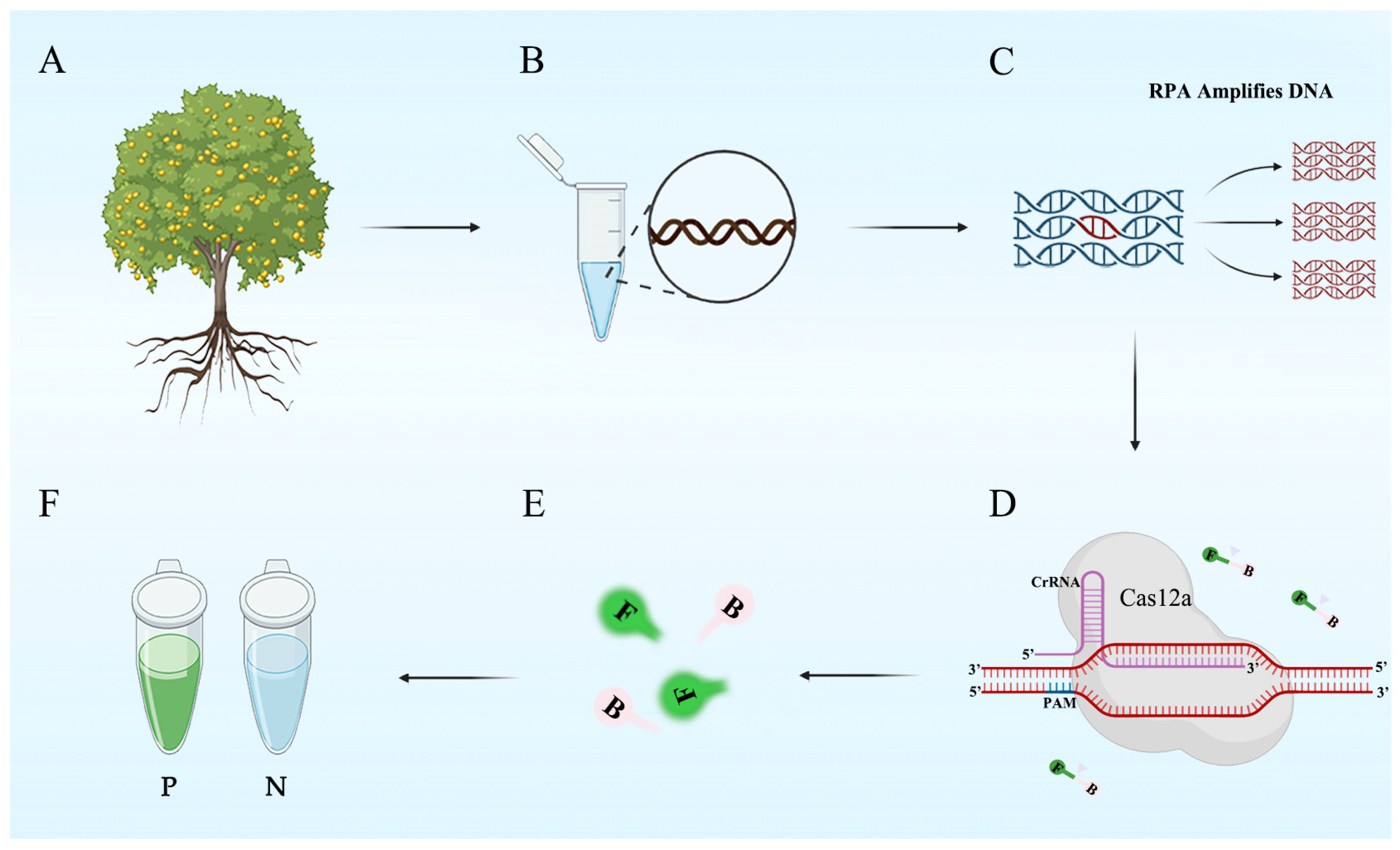
2.4. RPA-CRISPR/Cas12a Assay

The RPA-CRISPR/Cas12a assay consisted of two steps: RPA reaction and CRISPR/Cas12a-based detection (Figure 1). In the RPA reaction, the g2339 gene of P. cambivora was amplified using the g2339-RPA-F/-R primer pair over a period of 30 min. In the CRISPR/Cas12a-based detection, the green fluorescence signal was visualized within 30 min.
RPA experiments were performed using the test strip kit (Leshang Co., Ltd., Wuxi, China). First, several unit tubes required for the experiment were centrifuged in a small centrifuge at 4000× g rpm for 5 s. Then, an equal amount of experimental reagent was added to each unit tube. Each 50 μL reaction contained 25 μL of rehydrated buffer (provided in the kit), 16 μL of double-distilled water (ddH2O), 2 μL each of forward and reverse primers (g2339-RPA-F/g2339-RPA-R, 10 μM), and 2 μL of gDNA (100 ng/mL). After centrifugation at 4000× g rpm for 5 s, 3 μL of an initiator (provided in the kit) was added to the lid of the reaction unit. The reaction unit was sealed and then manually vortexed for 3 s so that the mixture was completely mixed with the initiator. Subsequently, the reaction mixture was centrifuged at 4000× g rpm for 5 s. After 4 min, the reaction was manually vortexed, centrifuged at 4000× g rpm for 5 s, and incubated at 37 °C for 20 min. The RPA products were then analyzed using the CRISPR/Cas12a system.
The FAM- and BHQ-labeled ssDNA reporter vector was added to the RPA-CRISPR/Cas12a reaction system. When the Cas12a/crRNA complex recognizes the PAM sequence in the target gene, it exhibits “collateral cleavage” activity, and the cleaved ssDNA reporter vector emits fluorescence. The concentrations of Cas12a, crRNA, and ssDNA reporter (1, 2, 5, 8, 10, 12.5, and 15 μM) (Table S3). The optimal duration of the RPA reaction and Cas12a cleavage was determined at eight time points (5, 10, 15, 20, 25, 30, 35, and 40 min). The CRISPR/Cas12a reaction was carried out in a 50 µL volume containing 38 μL of sterile ultrapure water, 5 μL of 10× Reaction Buffer (Magigen, Guangzhou, China), 3 μL of crRNA (10 μM), 1 μL of Cas12a (2 μM) (Magigen, Guangzhou, China), 1 μL of ss DNA reporter vector (10 μM), and 2 μL of the RPA product (added in this order). After centrifugation at 4000× g rpm for 5 s, the reaction mixture was immediately incubated at 37 °C. At the end of the reaction, fluorescence signal was detected using the multifunctional microplate reader or blue LED spectrophotometer at a wavelength of 470 nm. The above experiments were repeated three times. The data of the three repeats (Nos.1, 2, and 3) were recorded and analyzed using the STDEVP function, and the standard deviation was calculated. A statistical analysis was performed using Graph Pad Prism 8 (Graph Pad Software Inc., San Diego, CA, USA). Student’s t-test was used to compare the experimental and control groups, and only the differences with p-value < 0.05 were considered statistically significant [39].

2.5. Specificity and Sensitivity of the RPA-CRISPR/Cas12a Assay

The specificity of the RPA-CRISPR/Cas12a assay was tested using 7 isolates of P. cambivora, 68 isolates of 28 other Phytophthora species, 6 isolates of other oomycetes, 32 isolates of fungal species, and 2 isolates of Bursaphelenchus species (Table 1). The sensitivity of the RPA-CRISPR/Cas12a assay was tested using a concentration gradient of P. cambivora gDNA (100, 10, 1 ng·µL−1, 100, 10, 1 pg·µL−1, and 100 fg·µL−1). Each reaction included NC (ddH2O). The above experiment was repeated three times.

2.6. Detection of P. cambivora in Artificially Inoculated Apple Fruits Using the RPA-CRISPR/Cas12a Detection System

To inoculate apple fruits with P. cambivora, nine mature and disease-free apple fruits were selected, washed with sterile water, and wiped with a sterile cotton cloth sprayed with 75% alcohol. A 5-mm × 5-mm hole was punched into the surface of each apple, using a sterilized puncher, and filled with a P. cambivora plug or sterile cV8 plug (control) of the same size. The wound site was wrapped with sterile cotton dipped in pure sterile water for moisturization. After three days of incubation at room temperature (25 °C), 10 mg of pulp was sampled from the inoculation site, and DNA was extracted using DNA-safe plant kit (Tiangen Biochemical Technology Co., Ltd., Beijing, China). Then, the RPA-CRISPR/Cas12a experiment was performed using the extracted DNA, along with P. cambivora (100 ng·µL−1) and ddH2O as positive and negative controls, respectively. A data analysis was performed as described above.

3. Results

3.1. Optimization of the RPA-CRISPR/Cas12a Assay for the Detection of Phytophthora cambivora

More than 1000 genes of Phytophthora cambivora were found to have no homology with the genome sequences of other species. Ten genes (g7300, g5979, g5507, g2339, g33104, g28349, g26580, g18006, g16271, and g32133) were selected from among these putative P. cambivora-specific genes to design the RPA primers (Table S2). The RPA forward and reverse primers (g2339-RPA-F/R) and the positions of the crRNA sequences in the P. cambivora gene g2339 are shown in Figure S1. The RPA-CRISPR/Cas12a experiments were performed with different concentrations (Table S1) of crRNA and ssDNA reporter to determine the optimal concentration combination. The results showed that the higher the concentration, the stronger the fluorescence intensity. To minimize the time and cost involved in the detection of P. cambivora using the RPA-CRISPR/Cas12a assay, 10 μM was selected as the optimal concentration for both the crRNA and ssDNA reporter (Figure 2A,B).
Subsequently, the optimal RPA reaction time RPA was selected according to the optimal crRNA and ssDNA reporter concentration combination (Figure 3A,B). At 30 min, the blue LED transilluminator could detect strong green fluorescence, and the fluorescence intensity began to increase rapidly. Therefore, 30 min was selected as the optimal time for the RPA. Next, the time required for Cas12a-mediated cleavage was investigated using the products of the 30-min RPA reaction, and it was found that the optimal cleavage time was also 30 min (Figure 3C,D).

3.2. Verification of the Specificity of the RPA-CRISPR/Cas12a Assay

The g2339-RPA-F/-R primer pair was used to perform a conventional PCR-based detection of P. cambivora gDNA. As a result, a 219 bp PCR product was amplified. No PCR products were detected in the reaction containing Phytophthora nicotianae, Phytophthora cinnamomi, Phytophthora cryptogea, Phytophthora capsici, Phytophthora citrophthora, Phytopythium vexans, Pythium aphanidermatum, Pythium spinosum, Pythium ultimum, Fusarium asiaticum, Fusarium oxysporum, Bursaphelenchus xylophillus, Bursaphelenchus mucronatus, or Botrytis cinerea gDNA and NC (Figure S2). Similarly, in the RPA-CRISPR/Cas12a assay, the multifunctional microplate reader detected a bright fluorescent signal in the reaction containing P. cambivora gDNA but not in reactions containing other oomycetes and nematodes (Figure 4A,C). Additionally, the blue LED transilluminator could detect green fluorescence in the reaction containing the gDNA of P. cambivora but not in reactions containing the gDNA of other oomycetes, fungi, and nematodes (Figure 4B,D).

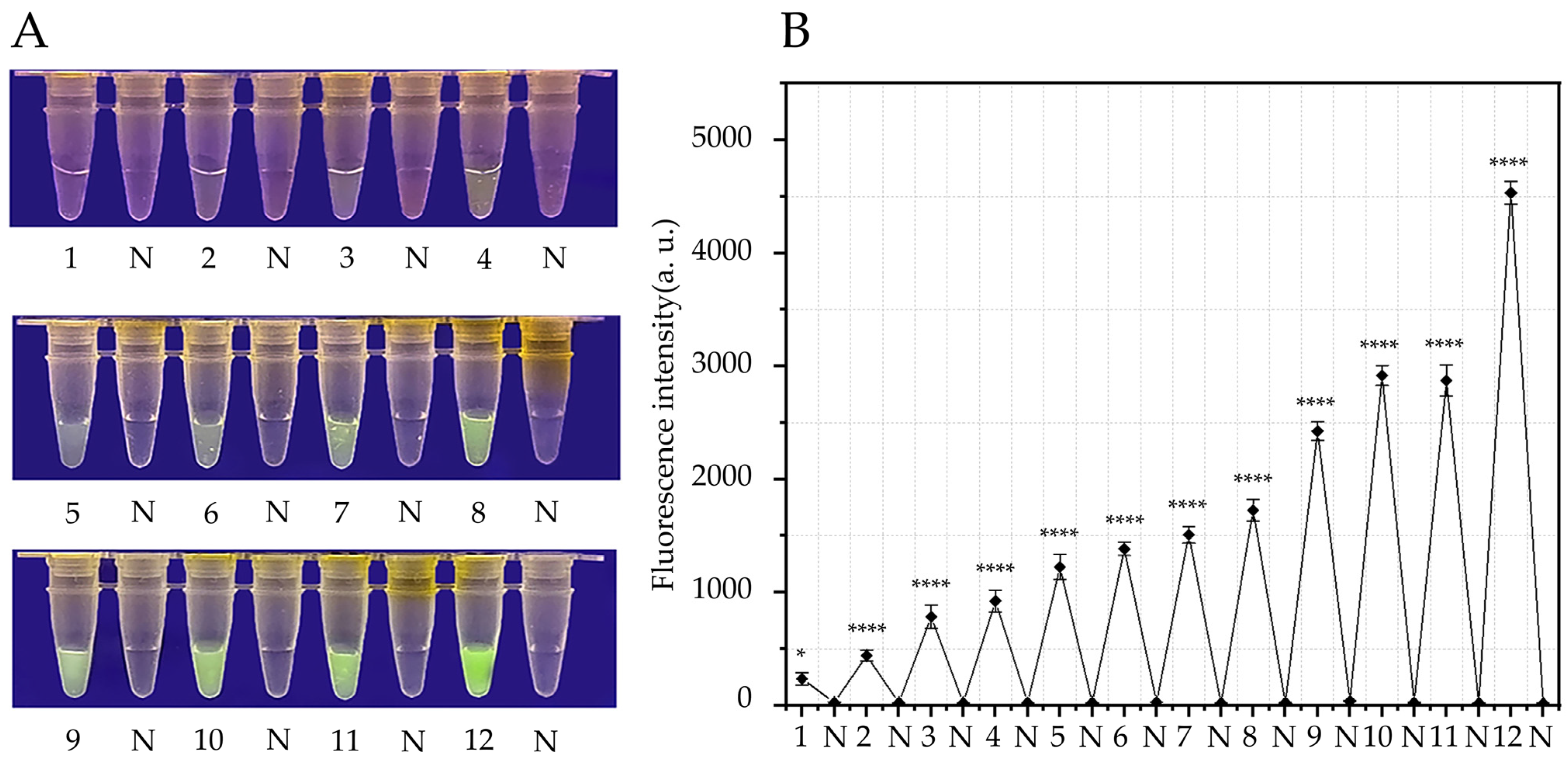
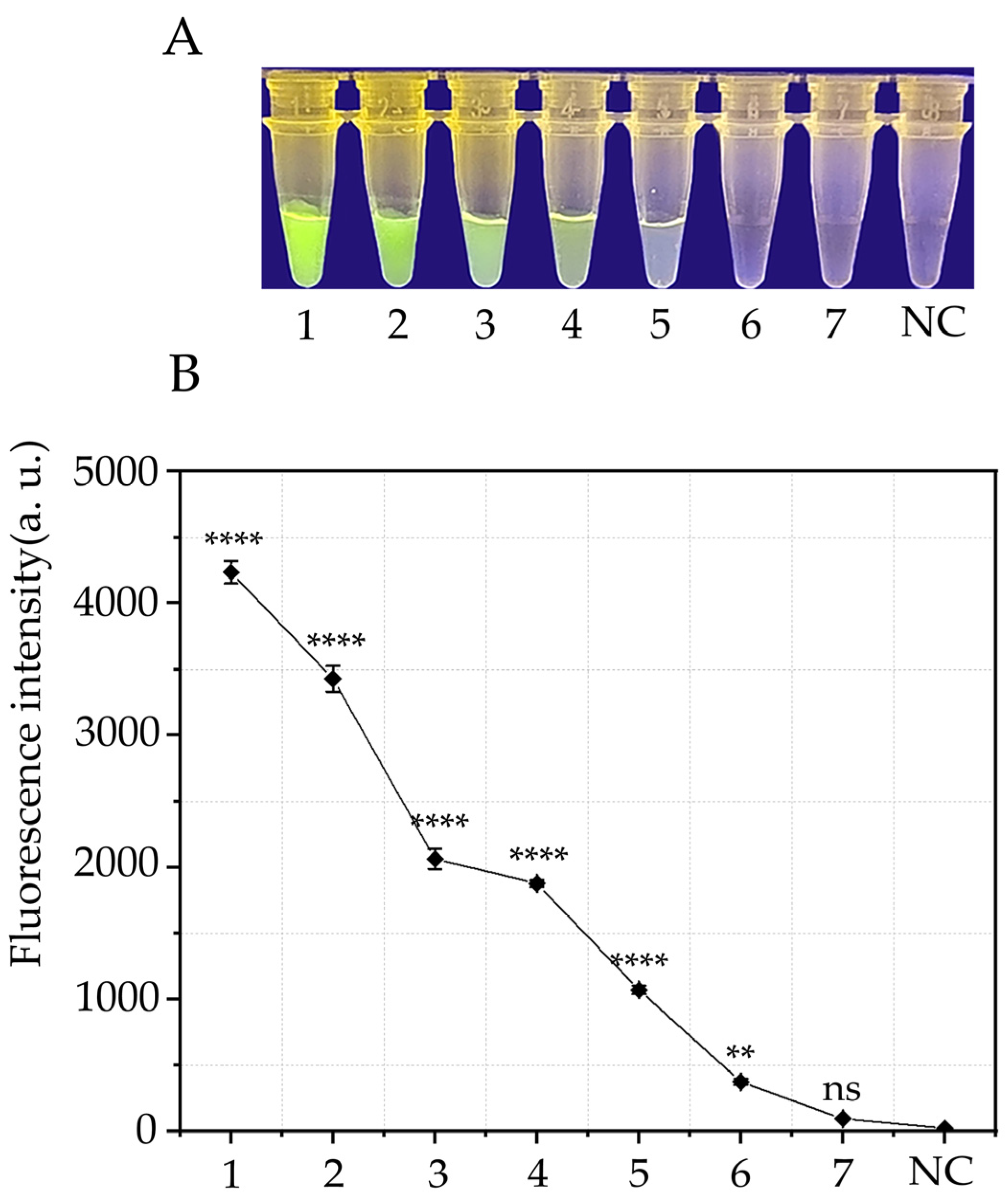
3.3. Sensitivity of the RPA-CRISPR/Cas12a Assay

A 10-fold dilution series of P. cambivora gDNA (100, 10, 1 ng·µL−1, 100, 10, 1 pg·µL−1, and 100 fg·µL−1) was amplified using the g2339-RPA-F/-R pair, and the experiment was repeated three times. Consequently, 10 pg·μL−1 was identified as the minimum P. cambivora gDNA concentration that could be detected by the RPA-CRISPR/Cas12a assay (Figure 5A,B).

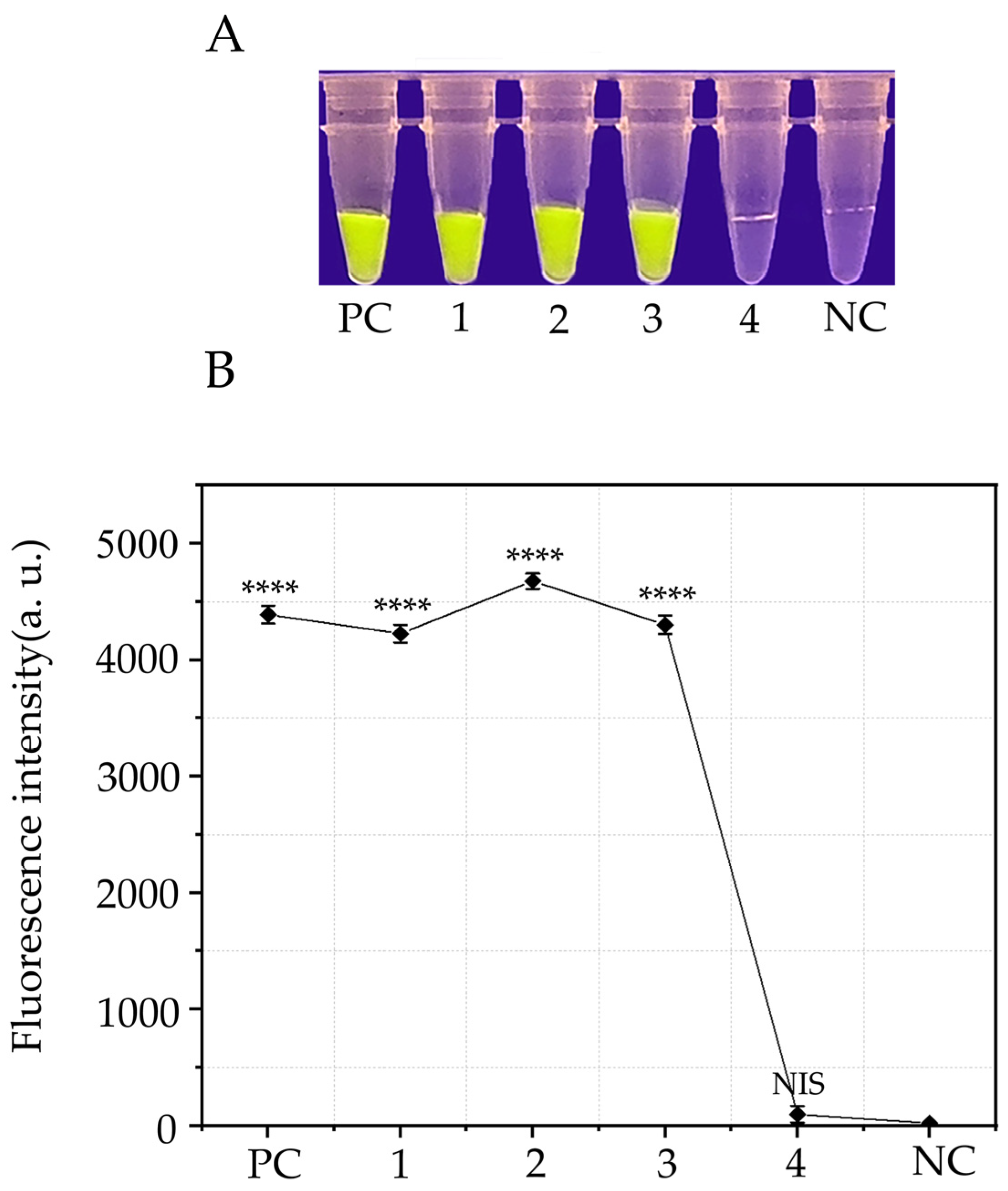
3.4. Detection of P. cambivora in Artificially Inoculated Apples Using the RPA-CRISPR/Cas12a Detection System

Healthy apples were artificially inoculated with P. cambivora. Three days later, rot was observed around the wounds in the inoculated apples but not in uninoculated apples (negative control). In the RPA-CRISPR/Cas12a assays, green fluorescence was detected using both the blue LED transilluminator and multifunctional microplate reader in samples containing DNA extracted from positive controls and the P. cambivora-inoculated apple samples. However, no fluorescence was detected in samples containing DNA extracted from uninoculated apples or NCs (Figure 6A,B).

4. Discussion

As a high-risk quarantine pathogen, Phytophthora cambivora mainly causes ink disease in chestnut trees and root and stem rot in various fruit trees, leading to severe losses in the agricultural industry. Therefore, attention should be paid to the quarantine of imported plants. At present, except for a report of leaf and stem blight and root rot on Prunus campanulata in Taiwan, China [40], the occurrence of this pathogen has not been reported in inland China. Given the potential danger of this pathogen, a rapid, accurate, and early detection technology needs to be established to prevent the introduction and spread of quarantine pests.
Unlike traditional methods such as the conventional PCR, real-time fluorescence PCR, nested PCR, multiplex PCR, and multiplex real-time fluorescence PCR, which usually require a cyclic heater and therefore cannot be adopted for rapid in-field detection [41], the RPA-CRISPR/Cas12a assay requires a temperature of 37 °C, which can be provided by inexpensive devices such as a USB-powered insulation box or a constant-temperature heater, and has a short amplification time [42]. LAMP usually requires a reaction temperature of approximately 60 °C [24]; the primers are more complex; and the number of false positives is higher. On the other hand, RPA has been optimized for the LAMP reaction temperature and primers; requires only one set of primers, each 30–35 nt in length; and can be performed at 37–42 °C. However, the RPA reaction is performed under constant-temperature conditions. Specific recognition can only be based on base nucleobase differences, and the species specificity is lower than that of PCR technology [42]. The combined use of RPA and CRISPR/Cas12a technology for the detection of P. cambivora can greatly improve the specificity of detection.
In this study, an RPA-CRISPR/Cas12a assay was established for the detection of P. cambivora, and the results could be determined based on fluorescence intensity. However, the RPA-CRISPR/Cas12a assay was also affected by the following factors: crRNA and ssDNA reporter concentrations, RPA reaction time, and Cas12a cleavage time. Therefore, the experimental conditions were optimized, and the optimal concentration and reaction time were investigated. Considering the overall cost, the combination of 10 μM of crRNA with 10 μM of a ssDNA reporter gene was chosen. The results showed that the longer the RPA reaction time, the more amplification products were obtained and the stronger the final signal. Therefore, both the RPA reaction time and CRISPR/Cas12a reaction time were selected to be 30 min. In addition, 10 pg·μL−1 was the lowest concentration of P. cambivora gDNA that could be detected by the RPA-CRISPR/Cas12a technology. The feasibility of this technique was investigated using apple samples inoculated with P. cambivora (Figure 6). The results showed that this method is highly accurate, indicating its potential for the early diagnosis of P. cambivora.
Although the RPA-CRISPR/Cas12a technology was fast, inexpensive, and highly accurate at detecting P. cambivora, there are still some problems that limit its application for pathogen detection. For example, the storage of reagents at room temperature is a major problem [43]. Some of the reagents in the CRISPR reaction experiment, such as the probe, will degrade after multiple freeze–thaw cycles and exposure to light, which will affect the experiment. The storage of the reagents needs further investigation. In addition, the 3′ end of the crRNA is essential for the accurate recognition of the PAM sequence in the target DNA. The base changes and deletions may affect the recognition of crRNA, thereby affecting cleavage by Cas12a [44].
In summary, this study established an RPA-CRISPR/Cas12a protocol for the detection of P. cambivora and verified its specificity and sensitivity. Our results demonstrate that this technology can be applied in-field.

5. Conclusions

A number of specific genes were screened from the genome sequence alignment of Phytophthora, Phytophthora and Pythium, and a new specific target gene of Phytophthora cambivora, g2339, was identified via the further design of primers (18–20 bp’s) according to a conventional PCR. A detection method based on RPA-CRISPR/Cas12a technology was established to detect P. cambivora. This technology has high specificity and sensitivity, and the detection time is shorter than that of traditional detection technology. Moreover, this technology enables visual detection without the need for a large number of expensive instruments and reagents, which is convenient for the on-port detection of P. cambivora.

Supplementary Materials

The following supporting information can be downloaded at https://www.mdpi.com/article/10.3390/f14112141/s1. Figure S1: The g2339 gene sequence of P. cambivora. Arrows indicate the directions of amplification of the g2339-RPA-F and g2339-RPA-R primers and the crRNA sequence and the direction of its amplification. Figure S2: Screening of the g2339-RPA-F and g2339-RPA-R primers using the conventional PCR method. Gel electrophoresis showed that the 219 bp amplicon was detected only in P. cambivora, indicating that the primer was highly specific for the detection of P. cambivora using different detection methods. The different lanes contain the following (from left to right): Marker DL500 (Takara Shuzo, Shiga, Japan), P. cambivora, P. nicotianae, P. cinnamomi, P. cryptogea, P. capsici, P. citrophthora, Phytopythium vexans, Pythium aphanidermatum, Py. spinosum, Py. ultimum, Fusarium asiaticum, F. oxysporum, Bursaphelenchus xylophillus, Bursaphelenchus mucronatus, Botrytis cinerea, negative control (NC), and Marker DL500. Table S1: Published genome sequences of 35 Phytophthora species, 8 Pythium species, and Phytopythium vexans. Table S2: Ten putative P. cambivora-specific genes used to design primers. Table S3: Screening of crRNA and ssDNA reporter concentrations for the RPA-CRISPR/Cas12a-based detection of P. cambivora.

Author Contributions

Conceptualization, J.Z. and T.D.; Methodology, J.Z.; Software, J.Z. and H.D.; Validation, J.Z.; Formal analysis, J.Z.; Investigation, J.Z. and H.D.; Resources, T.D.; Data curation, J.Z.; Writing—original draft, J.Z.; Writing—review & editing, T.D.; Visualization, J.Z.; Supervision, T.L.; Project administration, T.D. and T.L.; Funding acquisition, T.D. All authors have read and agreed to the published version of the manuscript.

Funding

This work was supported by the National Key R&D Program of China (No. 2021YFD1400100, No. 2021YFD1400103), the Natural Science Foundation of Jiangsu Province (BK20221426), the Jiangsu University Natural Science Research Major Project (21KJA220003), the Qinglan Project of 2020 and the Priority Academic Program Development of Jiangsu Higher Education Institutions.

Data Availability Statement

All data generated or analyzed during this study are included in this article.

Conflicts of Interest

The authors declare no conflict of interest.

References

  1. Zhou, J.; Xu, T.; Xu, X.; Dai, T.; Liu, T. The New Report of Root Rot on Fatsia jaonica Caused by Phytophthora nicotianae in China. Forests 2023, 14, 1459. [Google Scholar] [CrossRef]
  2. Nagy, Z.A.; Bakonyi, J.; Ersek, T.T. Standard and Swedish variant types of the hybrid alder Phytophthora attacking alder in Hungary. Pest Manag. Sci. 2003, 59, 484–492. [Google Scholar] [CrossRef] [PubMed]
  3. Félix-Gastélum, R.; Mircetich, S.M. Influence of flooding duration on the development of root and crown rot of Lovell peach [Prunus persica (L.) Bstsch] caused by three different Phytophthora species. Rev. Mex. Fitopatol. 2005, 23, 33–41. Available online: https://www.redalyc.org/pdf/612/61223105 (accessed on 8 April 2022).
  4. Blair, J.E.; Coffey, M.D.; Park, S.Y.; Geiser, D.M.; Kang, S. A multi-locus phylogeny for Phytophthora utilizing markers derived from complete genome sequences. Fungal Genet. Biol. 2008, 45, 266–277. [Google Scholar] [CrossRef] [PubMed]
  5. Dunstan, W.A.; Rudman, T.; Shearer, B.L. Containment and spot eradication of a highly destructive, invasive plant pathogen (Phytophthora cinnamomi) in natural ecosystems. Biol. Invas. 2010, 12, 913–925. [Google Scholar] [CrossRef]
  6. Vannini, A.; Breccia, M.; Bruni, N.; Tomassini, A.; Vettraino, A.M. Behaviour and survival of Phytophthora cambivora inoculum in soil-like substrate under different water regimes. For. Pathol. 2012, 42, 362–370. [Google Scholar] [CrossRef]
  7. Zambounis, A.; Xanthopoulou, A.; Madesis, P.; Tsaftaris, A.; Vannini, A.; Bruni, N.; Tomassini, A.; Chilosi, G.; Vettraino, A.M. A tool to assess genetic diversity of Phytophthora cambivora isolates. J. Plant Pathol. 2016, 9, 611–616. Available online: http://www.jstor.org/stable/44280509 (accessed on 8 April 2022).
  8. Mullett, M.S.; Van, P.K.; Haegeman, A.; Focquet, F.; Cauldron, N.C.; Knaus, B.J.; Horta, J.M.; Kageyama, K.; Hieno, A.; Masuja, H.; et al. Phylogeography and population structure of the global, wide host-range hybrid pathogen Phytophthora × cambivora. IMA Fungus 2023, 14, 4. [Google Scholar] [CrossRef]
  9. Feng, L.P.; Liu, H.M.; Wang, Y.C.; Li, Y.; Wu, X.H.; Ji, Y.; Wu, P.S. The Entry-Exit Inspection and Quarantine Industry Standards of the People’s Republic of China-Quarantine and Identification of Phytophthora cambivora (Petri) Buisman; China Standards Press: Beijing, China, 2011. [Google Scholar]
  10. Liao, F.; Huang, G.M.; Zhu, L.H.; Lv, D.J.; Zhang, D.D.; Luo, J.F.; Li, G.R. Quadruplex PCR detection of three quarantine Phytophthora pathogens of berries. Eur. J. Plant Pathol. 2019, 154, 1041–1049. [Google Scholar] [CrossRef]
  11. Zhu, L.H. Molecular Detection of the Ouarantine Phytophthora Pathogens of the Customs-Entry Fruits and Seedlings. Southwest University. 2016. Available online: https://kns.cnki.net/kcms/detail/detail.aspx?FileName=1015337659.nh&DbName=CMFD2016 (accessed on 8 April 2022).
  12. Hüberli, D.; Tommerup, I.C.; Hardy, G.E.S.J. False-negative isolations or absence of lesions may cause mis-diagnosis of diseased plants infected with Phytophthora cinnamomi. Australas. Plant Pathol. 2000, 29, 164–169. [Google Scholar] [CrossRef]
  13. Lane, C.R.; Hobden, E.; Walker, L.; Barton, V.C.; Inman, A.J.; Hughes, K.J.D.; Swan, H.; Colyer, A.; Barker, I. Evaluation of a rapid diagnostic field test kit for identification of Phytophthora species, including P. ramorum and P. kernoviae at the point of inspection. Plant Pathol. 2007, 56, 828–835. [Google Scholar] [CrossRef]
  14. Jung, T. Beech decline in Central Europe driven by the interaction between Phytophthora infections and climatic extremes. For. Pathol. 2009, 39, 73–94. [Google Scholar] [CrossRef]
  15. Das, A.K.; Nerkar, S.G.; Kumar, A.; Bawage, S.S. Detection, identification and characterization of Phytophthora spp. infecting citrus in India. J. Plant Pathol. 2016, 98, 55–69. [Google Scholar] [CrossRef]
  16. Ahmed, Y.; Hubert, J.; Fourrier-Jeandel, C.; Dewdney, M.M.; Aguayo, J.; Ioos, R. A set of conventional and multiplex real-time PCR assays for direct detection of Elsinoe fawcettii, E. australis, and Pseudocercospora angolensis in citrus fruits. Plant Dis. 2019, 103, 345–356. [Google Scholar] [CrossRef] [PubMed]
  17. Ristaino, J.B.; Madritch, M.; Trout, C.L.; Parra, G. PCR amplification of ribosomal DNA for species identification in the plant pathogen genus Phytophthora. Appl. Environ. Microbiol. 1998, 64, 948–954. [Google Scholar] [CrossRef] [PubMed]
  18. Grote, D.; Olmos, A.; Kofoet, A.; Tuset, J.J.; Bertolini, E.; Cambra, M. Specific and sensitive detection of Phytophthora nicotianae by simple and nested-PCR. Eur. J. Plant Pathol. 2002, 108, 197–207. [Google Scholar] [CrossRef]
  19. Zhu, L.H.; Guo, J.Z.; Liao, F.; Luo, J.F.; Huang, G.M.; Ren, X.Y.; Li, G.R. Simultaneous triplex PCR detection of two quarantine fungal pathogens of citrus, Phytophthora hiberalis and Phytophthora syringae. J. Southwest Univ. 2015, 37, 1–8. [Google Scholar] [CrossRef]
  20. Ippolito, A.; Schena, L.; Nigro, F.; Ligorio, V.S.; Yaseen, T. Real-time detection of Phytophthora nicotianae and P. citrophthorain citrus roots and soil. Eur. J. Plant Pathol. 2004, 110, 833–843. [Google Scholar] [CrossRef]
  21. Schena, L.; Hughes, K.J.; Cooke, D.E. Detection and quantification of Phytophthora ramorum, P. kernoviae, P. citricola and P. quercina in symptomatic leaves by multiplex real-time PCR. Mol. Plant Pathol. 2006, 7, 365–379. [Google Scholar] [CrossRef]
  22. Stehlíková, D.; Beran, P.; Cohen, S.P.; Čurn, V. Development of real-time and colorimetric loop mediated isothermal amplification assay for detection of Xanthomonas gardneri. Microorganisms 2020, 8, 1301. [Google Scholar] [CrossRef]
  23. Rosser, A.; Rollinson, D.; Forrest, M.; Webster, B.L. Isothermal recombinase polymerase amplification (RPA) of Schistosoma haematobium DNA and oligochromatographic lateral flow detection. Parasit. Vectors 2015, 8, 446. [Google Scholar] [CrossRef] [PubMed]
  24. Li, G.R.; Huang, G.M.; Zhu, L.H.; Lv, D.J.; Cao, B.H.; Liao, F.; Luo, J.F. Loop-mediated isothermal amplification (LAMP) detection of Phytophthora hibernalis, P. syringae and P. cambivora. J. Plant Pathol. 2019, 101, 51–57. [Google Scholar] [CrossRef]
  25. Zhou, X.; Wang, S.W.; Wang, X.R. Application of CRISPR-Cas12a in rapid detection of pathogens. Chin. Vet. Sci. 2022, 52, 1031–1037. [Google Scholar] [CrossRef]
  26. Jinek, M.; Chylinski, K.; Fonfara, I.; Hauer, M.; Doudna, J.A.; Charpentier, E. A programmable dual-RNA-guided DNA endonuclease in adaptive bacterial immunity. Science 2012, 337, 816–821. [Google Scholar] [CrossRef] [PubMed]
  27. Cong, L.; Ran, F.A.; Cox, D.; Lin, S.; Barretto, R.; Habib, N.; Hsu, P.D.; Wu, X.; Jiang, W.; Marraffini, L.A.; et al. Multiplex genome engineering using CRISPR/Cas systems. Science 2013, 339, 819–823. [Google Scholar] [CrossRef] [PubMed]
  28. Kellner, M.J.; Koob, J.G.; Gootenberg, J.S.; Abudayyeh, O.O.; Zhang, F. Sherlock: Nucleic acid detection with CRISPR nucleases. Nat. Protoc. 2019, 14, 2986–3012. [Google Scholar] [CrossRef]
  29. Kadam, U.S.; Cho, Y.; Park, T.Y.; Hong, J.C. Aptamer-based crispr-cas powered diagnostics of diverse biomarkers and small molecule targets. Appl. Biol. Chem. 2023, 66, 13. [Google Scholar] [CrossRef]
  30. Alon, D.M.; Partosh, T.; Burstein, D.; Pines, G. Rapid and sensitive on-site genetic diagnostics of pest fruit flies using CRISPR-Cas12a. Pest Manag. Sci. 2023, 79, 68–75. [Google Scholar] [CrossRef]
  31. Makarova, K.S.; Wolf, Y.I.; Iranzo, J.; Shmakov, S.A.; Alkhnbashi, O.S.; Brouns, S.J.J.; Charpentier, E.; Cheng, D.; Haft, D.H.; Horvath, P.; et al. Evolutionary classification of CRISPR-Cas systems: A burst of class 2 and derived variants. Nat. Rev. Microbiol. 2020, 18, 67–83. [Google Scholar] [CrossRef]
  32. Kuang, R.R.; Lei, R.; Jiang, L.; Duan, W.J.; Li, X.L.; Fu, N.; Fan, Z.F.; Li, Y.; Wu, P.S. Establishment of RPA/CRISPR-Cas12a rapid detection for Leptosphaeria lindquistii. Plant Prot. 2022, 48, 69–76. [Google Scholar] [CrossRef]
  33. Li, F.A.; Xiao, J.; Yang, H.M.; Yao, Y.; Li, J.Q.; Zheng, H.W.; Guo, Q.; Wang, X.T.; Chen, Y.Y.; Guo, Y.J.; et al. Development of a rapid and efficient RPA-CRISPR/Cas12a assay for Mycoplasma pneumoniae detection. Front. Microbiol. 2022, 13, 858806. [Google Scholar] [CrossRef]
  34. Chen, J.S.; Ma, E.; Harrington, L.B.; Da Costa, M.; Tian, X.; Palefsky, J.M.; Doudna, J.A. CRISPR-Cas12a target binding unleashes indiscriminate single-stranded DNase activity. Science 2018, 360, 436–439. [Google Scholar] [CrossRef] [PubMed]
  35. Xiao, X.X.; Lin, Z.Q.; Huang, X.H.; Lu, J.F.; Zhou, Y.; Zheng, L.B.; Lou, Y.L. Rapid and sensitive detection of Vibrio vulnificus using CRISPR/Cas12a combined with a recombinase-aided amplification assay. Front. Microbiol. 2021, 12, 767315. [Google Scholar] [CrossRef] [PubMed]
  36. Liu, H.; Wang, J.; Li, P.; Bai, L.; Jia, J.; Pan, A.; Long, X.; Cui, W.; Tang, X. Rapid detection of P-35S and T-nos in genetically modified organisms by recombinase polymerase amplification combined with a lateral flow strip. Food Control 2020, 107, 106775. [Google Scholar] [CrossRef]
  37. Zhao, G.; Wang, J.; Yao, C.Y.; Xie, P.C.; Li, X.M.; Xu, Z.L.; Xian, Y.P.; Lei, H.T.; Shen, X. Alkaline lysis-recombinase polymerase amplification combined with CRISPR/Cas12a assay for the ultrafast visual identification of pork in meat products. Food Chem. 2022, 383, 132318. [Google Scholar] [CrossRef] [PubMed]
  38. Li, S.Y.; Cheng, Q.X.; Wang, J.M.; Li, X.Y.; Zhang, Z.L.; Gao, S.; Cao, R.B.; Zhao, G.P.; Wang, J. CRISPR-Cas12a-assisted nucleic acid detection. Cell Discov. 2018, 4, 20. [Google Scholar] [CrossRef]
  39. Dai, T.T.; Chen, Z.P.; Guo, Y.F.; Ye, J.R. Rapid detection of the pine wood nematode Bursaphelenchus xylophilus using recombinase polymerase amplification combined with CRISPR/Cas12a. Crop Prot. 2023, 170, 106259. [Google Scholar] [CrossRef]
  40. Huang, J.H.; Ann, P.J.; Chiu, Y.H.; Tsai, J.N. First report of Phytophthora cambivora causing leaf and stem blight and root rot on Taiwan cherry (Prunus campanulata) in Taiwan. Plant Dis. 2012, 96, 1065. [Google Scholar] [CrossRef]
  41. Langrell, S.R.; Morel, O.; Robin, C. Touchdown nested multiplex PCR detection of Phytophthora cinnamomi and P. cambivora from French and English chestnut grove soils. Fungal Biol. 2011, 115, 672–682. [Google Scholar] [CrossRef]
  42. Lei, R.; Sun, X.W.; Jiang, L.; Wang, Z.H.; Li, G.Q.; Li, Y.; Liao, X.L.; Wu, P.S. Development of rapid detection for Phytophthora syringae based on RPA/CRISPR-Cas12a. Plant Quar. 2022, 36, 31–38. [Google Scholar] [CrossRef]
  43. Nguyen, P.Q.; Soenksen, L.R.; Donghia, N.M.; Angenent-Mari, N.M.; de Puig, H.D.; Huang, A.; Lee, R.; Slomovic, S.; Galbersanini, T.; Lansberry, G.; et al. Wearable materials with embedded synthetic biology sensors for biomolecule detection. Nat. Biotechnol. 2021, 39, 1366–1374. [Google Scholar] [CrossRef] [PubMed]
  44. Hu, X.X.; Wang, C.; Liu, Q.; Fu, Y.P.; Wang, K.J. Targeted mutagenesis in rice using CRISPR-Cpf1 system. J. Genet. Genom. 2017, 44, 71–73. [Google Scholar] [CrossRef] [PubMed]
Figure 1. Flow chart of the RPA-CRISPR/Cas12a assay. (A,B) DNA extraction from diseased apple tree roots using the DNAsecure Plant Kit. (C) DNA amplification using the RPA technique. (D,E) CRISPR/Cas9-based editing. The Cas12a protein can bind to any amplicon and target-specific crRNA to form a complex that stimulates the cleavage activity of the protein. When the FAM-labeled ssDNA reporter gene is cleaved, green fluorescence is visualized at an excitation wavelength of 470 nm. (F) The detection of green fluorescence using blue LED transilluminator. P, green fluorescence-positive; N, green fluorescence-negative. (Mapping sources: https://app.biorender.com/, accessed on: 10 October 2023).
Forests 14 02141 g001
Figure 2. Determination of the optimal concentrations of crRNA and ssDNA reporter for the detection of P. cambivora using the RPA-CRISPR/Cas12a assay. Different concentration combinations of the crRNA and ssDNA reporter were tested as follows (from left to right): 1 μM crRNA, 1 μM ssDNA reporter (1,1); 1,2; 5,2; 2,5; 8,5; 5,8; 10,8; 8,10; 10,10; 12.5,12.5; 12.5,15; and 15,15. N, negative control. **** (p < 0.0001), * (p = 0.0211). (A) Green fluorescence detection results. (B) Fluorescence values measured using a multifunctional microplate reader.
Forests 14 02141 g002
Figure 3. Determination of the optimal RPA reaction and Cas12a cleavage times. (A,B) RPA reaction times (lanes 1–8): 5, 10, 15, 20, 25, 30, 35, and 40 min. N, negative control (ddH2O). (C,D) Cas12a cleavage times (lanes 1–8): 5, 10, 15, 20, 25, 30, 35, and 40 min. N, negative control. **** p < 0.0001, ** p = 0.0106, * p = 0.0019; ns, non-significant.
Forests 14 02141 g003
Figure 4. Determination of the sensitivity of the RPA-CRISPR/Cas12a technology. (A,B) 1: Phytophthora cambivora, 2: P. nicotianae, 3: P. cinnamomi, 4: P. ramorum, 5: P. cryptogea, 6: P. capsici, 7: P. citrophthora, NC: negative control (ddH2O). (C,D) 1: P. cambivora, 2: Phytopythium vexans, 3: Phytopthium aphanidermatum, 4: Fusarium asiaticum, 5: F. oxysporum, 6: Bursaphelenchus xylophillus, 7: Bursaphelenchus mucronatus, NC: negative control. **** p < 0.0001; ns, non-significant.
Forests 14 02141 g004
Figure 5. Analysis of the sensitivity of the RPA-CRISPR/Cas12a detection system. (A) Green fluorescence detection results. (B) Fluorescence values obtained from the multifunctional microplate reader. 1–7: 100, 10, 1 ng·µL−1, 100, 10, 1 pg·µL−1, and 100 fg·µL−1, and NC. **** p < 0.0001, ** p = 0.0004; ns, non-significant.
Forests 14 02141 g005
Figure 6. Detection of P. cambivora in artificially inoculated apples using the RPA-CRISPR/Cas12a detection system. (A) Visible green fluorescence. (B) Bright fluorescence signals detected by a multifunctional microplate reader. PC: positive control; 1–3: P. cambivora-inoculated apples; 4: NIS (non-inoculated apples) and NC. **** p < 0.0001.
Forests 14 02141 g006
Table 1. Details of the isolates used in this study and the results of the RPA-CRISPR/Cas12a specificity test.
NumberSpeciesIsolate 1OriginRPA-CRISPR/Cas12 2
SourceHost/Substrate
1Phytophthora cambivoraPc1Shanghai, ChinaMalus domestica Borkh+
2Phytophthora cambivoraPc2Shanghai, ChinaMalus domestica Borkh+
3Phytophthora cambivoraPc3Shanghai, ChinaMalus domestica Borkh+
4Phytophthora cambivoraPc4Shanghai, ChinaMalus domestica Borkh+
5Phytophthora cambivoraPc5Shanghai, ChinaMalus domestica Borkh+
6Phytophthora cambivoraPc6Shanghai, ChinaMalus domestica Borkh+
7Phytophthora cambivoraCBS 248.60Boston, MA, USACastanea sativa+
8Phytophthora boehmeriaePb3Nanjing, ChinaBoehmeria nivea
9Phytophthora cactorumC1Nanjing, ChinaMalus pumila
10Phytophthora capsiciPc1Nanjing, ChinaCapsicum annuum
11Phytophthora castaneaeCBS587.85TaiwanSoil
12Phytophthora citrophthoraPcitNanjing, ChinaCitrus reticulata Blanco
13Phytophthora cinnamomiPci1Anqing, ChinaPinus sp.
14Phytophthora cryptogeaPcr1Nanjing, ChinaSolanum lycopersicum
15Phytophthora citricolaPcitNanjing, ChinaRhododendron pulchrum
16Phytophthora drechsleriCBS 292.35 TCA, USABeta vulgaris var. altissima
17Phytophthora fragariaeCBS 209.46England, UKFragaria × ananassa
18Phytophthora hibernalisCBS 270.31Richmond, VA, USACitrus sinensis
19Phytophthora infestansPi1Fuzhou, ChinaSolanum tuberosum
21Phytophthora lateralisCBS 168.42Vancouver, CA, USACedrus deodara
22Phytophthora megaspermaCBS305.36CA, USAMatthiola incana
23Phytophthora melonisPMNJHG1Nanjing, ChinaCucumis sativus
24Phytophthora mississippiae57J3Commonwealth of Virginia, USAIrrigation water
25Phytophthora nicotianaePn1Fuzhou, ChinaNicotiana tabacum
26Phytophthora palmivoraPp1Lijiang, ChinaIridaceae
27Phytophthora parvisporaCBS132771Basilicata, ItalyArbutus unedo
28Phytophthora piniPpini1Nanjing, ChinaRhododendron pulchrum
29Phytophthora plurivoraPplu1Haikou, ChinaManihot esculenta
30Phytophthora quercinaCBS 789.95Brisbane, AustraliaQuercus petraea
31Phytophthora sojaePsy1Lijiang, ChinaGlycine max
32Phytophthora syringae9099Shanghai, ChinaMalus domestica Borkh
33Phytophthora ramorumEU1 2275Exeter, UKQuercus palustris
34Phytophthora rubiCBS 967.95Scotland, UKRubus idaeus
35Phytophthora vignaeCPHSTBL 30M. D. CoffeyVigna sp.
36Phytopythium litoralePC-dj1Nanjing, ChinaRhododendron simsii
37Phytopythium helicoidesPH-CNanjing, ChinaRhododendron simsii
38Phytopythium vexansDT10Nanjing, ChinaRhododendron simsii
39Pythium ultimumPul1Nanjing, ChinaCitrus sinensis
40Pythium spinosumPsp1Nanjing, ChinaOryza sativa L.
41Pythium aphanidermatumPap1Nanjing, ChinaNicotiana tabacum
42Fusarium oxysporiumFox1Nanjing, ChinaGossypium sp.
43Fusarium solaniFso1Nanjing, ChinaGossypium sp.
44Fusarium circinatumA045-1Shanghai, ChinaPinus sp.
45Fusarium fujikuroiFfu1Nanjing, ChinaOryza sativa
46Fusarium graminearumFgr1Nanjing, ChinaTriticum aestivum
47Fusarium acuminatumFac1Chengdu, ChinaRhizophora apiculata
48Fusarium asiaticumFas1Nanjing, ChinaTriticum aestivum
49Fusarium avenaceumFav1Nanjing, ChinaGlycine max
50Fusarium culmorumFcu1Suining, ChinaGlycine max
51Fusarium communeFco1Harbin, ChinaSoil
52Fusarium equisetiFeq1Nanjing, ChinaGlycine max
53Fusarium lateritiumFlat1Nanjing, ChinaSoil
54Fusarium moniformeFmo1Nanjing, ChinaOryza sativa
55Fusarium nivaleFnivNanjing, ChinaTriticum aestivum
56Fusarium proliferatumFpr1Nanjing, ChinaPinus sp.
57Fusarium incarnatumIL3HQNanjing, ChinaMedicago sativa
58Colletotrichum truncatumCtr1Nanjing, ChinaGlycine max
59Colletotrichum glycinesCgl1Nanjing, ChinaGlycine max
60Colletotrichum orbiculareCor1Nanjing, ChinaCitrullus lanatus
61Neofusicoccum parvumBJ3-1Nanjing, ChinaFatsia japonica
62Verticilium dahliaeVda1Nanjing, ChinaGossypium sp.
63Rhizoctonia solaniRso1Nanjing, ChinaGossypium sp.
64Magnaporthe griseaGuy11Tsukuba, JapanOryza sativa
65Endothia parasiticaEpa1Nanjing, ChinaCastanea mollissima
66Bremia lactucaeBla1Nanjing, ChinaLactuca sativa
67Aspergillus flavusNJC03Nantong, ChinaActinidia chinensis
68Botrytis cinereaBci1Nanjing, ChinaCucumis sativus
69Alternaria alternataAal1Nanjing, ChinaSoil
70Tilletia indicaTin1Nanjing, ChinaTriticum aestivum
71Diaporthe mahothocarpusDT1Nanjing, ChinaKerria japonica
72Diaporthe sapindicolaWHZ3Nanjing, ChinaSapindus mukorossi
73Botryosphaeria dothideaBci1Nanjing, ChinaKoelreuteria paniculata
74Bursaphelenchus xylophilusJs-1Nanjing, ChinaPinus thunbergii
75Bursaphelenchus
mucronatus
BmucroNanjing, ChinaPinus sp.
Note: 1 CBS, Centraalbureau voor Schimmelculture Biodiversity Centre, Utrecht, The Netherlands; ATCC, American Type Culture Collection, Manassa, VA, USA, T: ex-type isolate. 2 “+” and “−” represent the positive and negative results of the RPA-CRISPR/Cas12a assay, respectively.
Disclaimer/Publisher’s Note: The statements, opinions and data contained in all publications are solely those of the individual author(s) and contributor(s) and not of MDPI and/or the editor(s). MDPI and/or the editor(s) disclaim responsibility for any injury to people or property resulting from any ideas, methods, instructions or products referred to in the content.

