Article

Screening and Regulatory Mechanisms of Inter-Root Soil Nematicidal Bacteria of Pinus massoniana

1
Key Laboratory of National Forestry and Grassland Administration for Control of Diseases and Pests of South Plantation, Central South University of Forestry and Technology, Changsha 410004, China
2
Hunan Provincial Key Laboratory for Control of Forest Diseases and Pests, Central South University of Forestry and Technology, Changsha 410004, China
3
Key Laboratory of Cultivation and Protection for Non-Wood Forest Trees, Central South University of Forestry and Technology, Changsha 410004, China
*
Authors to whom correspondence should be addressed.
These authors contributed equally to this paper.
Forests 2023, 14(11), 2230; https://doi.org/10.3390/f14112230
Submission received: 22 September 2023 / Revised: 6 November 2023 / Accepted: 9 November 2023 / Published: 13 November 2023
(This article belongs to the Section Forest Health)

Abstract

:
Pine Wilt Disease (PWD), caused by the pathogenic nematode Bursaphelenchus xylophilus, is a systemic infectious disease commonly referred to as the “cancer” of pine trees. This devastating disease has gained this analogy due to its ability to rapidly spread within pine populations, leading to substantial losses in forest resources. The primary objective of this study is to investigate the bioprotective potential and underlying mechanisms of action exhibited by rhizosphere microorganisms associated with Masson pine (Pinus massoniana) in the context of controlling the pine wilt nematode. In this experiment, using high-throughput sequencing, significant differences were observed in the rhizosphere soil microbial communities among healthy Masson pine, standing dead trees, and diseased Masson pine. Furthermore, it was found that these microbial communities exhibited distinct community structures at different levels. This study successfully isolated and screened three strains of highly effective nematophagous bacteria from the rhizosphere soil. The identified strains were Lysinibacillus capsici, Bacillus Paramycoides, and Delftia tsuruhatensis. After applying the bacterial suspensions and fermentation extracts of these three strains to the roots of two-year-old Masson pine seedlings, followed by inoculation with pine wilt nematodes after a four-day period, distinct defense responses were observed in the Masson pine. Notably, the activities of phenylalanine ammonia-lyase (PAL) and peroxidase (POD) were significantly increased, leading to a substantial reduction in the incidence of pine wilt disease. Based on the changes in defense enzyme activities, it can be concluded that the fermentation extract of the Lysinibacillus capsici strain exhibits effective nematocidal effects and induces resistance. The significant biological control efficacy and induction of host defense activity indicate the potential application value of this strain and its metabolites as a biocontrol agent for pine wilt disease.

1. Introduction

Pine wilt disease (PWD), also known as pine wilting disease, poses a significant threat to pine trees, manifesting as a systemic infection caused by the invasion of the pine wood nematode (Bursaphelenchus xylophilus) in plants belonging to the Pinus genus. Currently, this devastating disease is spreading across various regions worldwide, including Spain, China, Korea, and Europe [1,2,3,4,5]. Currently, three primary strategies are employed for the management of PWD: physical control, chemical control, and biological control. Physical control involves trapping pine sawyer beetles and felling infected trees, followed by heat treatment or incineration of the diseased wood [6]. Chemical control primarily relies on the use of insecticides to target the vectors and kill pine wood nematodes, effectively reducing losses. However, the widespread and excessive use of chemical insecticides has engendered the emergence of nematode resistance, compromising the efficacy of these agents. Furthermore, most compounds have broad-spectrum killing properties that can adversely affect beneficial non-target organisms. Consequently, biological control has garnered escalating interest as an environmentally sustainable and ecologically compatible approach to address the challenges posed by PWD.
Rhizosphere microorganisms are a group of beneficial root-associated microbes that interact with plants. They employ various mechanisms, including toxin production, biosurfactant secretion, and enzymatic degradation, to suppress plant pathogens, induce host plant resistance, and indirectly promote plant growth. For instance, Bacillus subtilis produces toxins and secretes extracellular proteases to degrade the tissue of nematodes; Pseudomonas fluorescens forms biofilms that interfere with nematode behavior; Streptomyces mobilizes fungi for nematode predation [6,7,8,9,10]. In a study conducted by Yu et al., two bacterial strains, namely the Pseudoalteromonas strain H-42 and Vibrio atlanticus strain S-16, were isolated from the surface of marine organisms. The fermentation broth derived from these bacteria demonstrated notable nematocidal activity [11]. Paiva et al. isolated strains 020 and RBT-200701 of Bacillus subtilis from soil, demonstrating their nematocidal activity [12]. Wang et al. obtained a highly nematocidal strain, LCB-3, through isolation from plants and subsequently isolated nematocidal compounds from its fermentation broth [13]. Cao screened the stems of pine trees to find three strains of Bacillus that had efficient antagonistic effects on nematodes [14].
Furthermore, biological control can be employed to combat pine wilt nematodes by stimulating plant defense mechanisms. Studies have demonstrated that in the presence of rhizosphere microorganisms, plants can elicit the production of various defense enzymes, including phenylalanine ammonia-lyase (PAL), peroxidase (POD), and catalase (CAT). The enhanced activity of these defense enzymes strengthens plant resistance and reduces the incidence of pine wilt disease [15]. Effective utilization of inducers of plant defense has been found to significantly mitigate the severity of pine wilt disease. It has been observed that following nematode inoculation, the rhizosphere soil exhibits a decreased abundance of beneficial microorganisms, whereas bacterial induction leads to a pronounced increase in the population of beneficial microorganisms in the rhizosphere. Notably, the inoculation of nematodes in the rhizosphere soil results in a substantial augmentation of beneficial microorganism levels, which can act as a preventive measure against pine wilt nematode infection or exert inhibitory effects on beneficial bacteria associated with pine wilt nematodes [16].
When pine trees are infested by pine wood nematodes, they employ physiological responses such as the secretion of abundant signaling molecules, hormones, and secondary metabolites to protect themselves from nematode damage. Research conducted by Zhang et al. revealed that the metabolite plantazolicin produced by the bacterium Bacillus amyloliquefaciens FZB42 exhibits nematocidal activity [17]. Wang et al. found that pine nematode infestation of Japanese larch resulted in a significant increase in host enzymes and phenylalanine ammonia-lyase (PAL) activity [18]. In summary, biological control represents a promising approach for managing pine wood nematode disease. By harnessing the nematocidal bacteria and the defensive enzyme activity of plants, it is possible to effectively reduce the incidence and infection of pine wood nematodes. The objective of this study is to screen and analyze the characteristics of root-associated microorganisms in Masson pine that induce resistance against pine wood nematode disease, with a primary focus on elucidating their interaction mechanisms with the host plant.

2. Materials and Methods

2.1. Sample Collection and Treatment

The soil samples used in this study were collected from the rhizosphere of Masson pine trees in Tianxin District, Changsha City, Hunan Province, China (N 28°06′51.34″, E 112°59′23.39″). The sampling was conducted in October 2022 and included three groups: Group 1 consisted of healthy Masson pine rhizosphere soil without drug treatment, Group 2 comprised diseased Masson pine rhizosphere soil without drug, and Group 3 consisted of dead Masson pine rhizosphere soil without drug. Soil samples were collected at distances of 20 cm from the root system. Each distance had three sampling points; the line angle between each sampling point the trunk is was approximately 120°, and each sampling point was replicated three times. Prior to sampling, the collection tools were sterilized using alcohol, and the surface soil was removed. The collected soil was then placed in sterile sampling bags and immediately stored on dry ice for preservation. In the laboratory, the soil samples were subjected to coarse sieving using sterilized sieves. The resulting fine soil fraction was frozen and sent to Applied Protein Technology Co., Ltd. for subsequent processing.

2.2. DNA Extraction, Amplification, and Sequencing

Genomic DNA extraction from the samples was performed using the Magnetic Bead Soil and Fecal Genomic DNA Extraction Kit (DP712), which is specifically designed for extracting genomic DNA from soil samples. The purity and concentration of the extracted DNA were evaluated using 1% agarose gel electrophoresis. A suitable amount of each sample was collected in a centrifuge tube, and sterile water was used to dilute the sample to a concentration of 1 ng/μL. The extracted DNA was used as a template for PCR amplification of the V4–V5 variable region of the 16S rRNA gene using primers with barcodes. The forward primer utilized was 515F (5′-GTGCCAGCMGCCGCGGTAA-3′), and the reverse primer employed was 907R (5′-CCGTCAATTCMTTTRAGTTT-3′) [19]. Each sample was subjected to three PCR replicates. The PCR reaction was conducted using 15 μL of Phusion® High-Fidelity PCR Master Mix (New England Biolabs, Ipswich, MA, USA), 0.2 μM of both the forward and reverse primers, and approximately 10 ng of template DNA. The thermal cycling protocol consisted of an initial denaturation step at 98 °C for 1 min, followed by denaturation at 98 °C for 10 s, annealing at 50 °C for 30 s, extension at 72 °C for 30 s, and a final extension step at 72 °C for 5 min. PCR products were subjected to 2% agarose gel electrophoresis to visualize their bands. Samples exhibiting distinct and well-defined bands within the size range of 400–450 bp were carefully selected for subsequent experiments. To ensure uniformity, samples with comparable concentrations, as determined by PCR product quantification, were pooled together and thoroughly mixed. The resulting mixture was subjected to purification using 2% agarose gel electrophoresis with 1 × TAE buffer, enabling the isolation of the desired bands. Subsequent gel extraction of the target bands was carried out utilizing the Gel Extraction Kit. To generate sequencing libraries, the TruSeq® DNA PCR-Free Sample Preparation Kit (Illumina, San Diego, CA, USA) was employed as per the manufacturer’s guidelines, with the incorporation of index codes for sample multiplexing. Quality assessment of the resulting libraries was performed using the Qubit@ 2.0 fluorometer (Thermo Scientific, Waltham, MA, USA) and the Agilent Bioanalyzer 2100 system (Agilent, Santa Clara, CA, USA). Finally, the libraries were subjected to high-throughput sequencing on the Illumina NovaSeq platform.

2.3. Analysis of Sequence Data

In this section, we outline the analytical procedures employed for the analysis of sequence data. The data were initially processed by assigning a unique barcode sequence to each sample, and the resulting sequences were saved in the widely-used fastq format. For paired-end (PE) data, two files, fq1 and fq2, were generated to represent the reads obtained from the two ends of the sequencing process. To merge these paired-end reads accurately and efficiently, the FLASH software (available at http://ccb.jhu.edu/software/FLASH/, accessed on 3 April 2023) was utilized. Following the merging step, the quality of the reads and the effectiveness of the merging process were assessed through a rigorous quality control filtering process, resulting in the generation of high-quality, reliable data, referred to as “Clean Data” in this study. To analyze the clean reads and gain insights into the microbial community composition, the Uparse software (obtainable at http://drive5.com/uparse/, accessed on 3 April 2023) was employed. This software utilizes a clustering algorithm to group the clean reads into operational taxonomic units (OTUs) based on a 97% sequence similarity threshold. Subsequently, the representative sequences of the OTUs were aligned against a comprehensive reference database using the RDP classifier algorithm to assign taxonomic annotations to the OTUs. For this purpose, the Silva 132 database (accessible at https://www.arb-silva.de/, accessed on 3 April 2023) was utilized, which is a well-established and widely used database specifically designed for 16S rRNA gene annotations.
To ensute comparability among samples, the sequence data were normalized by subsampling the reads to the depth corresponding to the sample with the lowest number of sequences. This equalization of sequencing depth allows for a fair comparison and accurate assessment of species richness and diversity. In this study, species richness was evaluated using metrics such as Chao1 and Observed Species, which provide estimations of the total number of species present in a given sample. Furthermore, measures of species diversity, including the Shannon and Simpson indices, were employed to assess the evenness and distribution of species within the samples. To facilitate the visualization of the taxonomic annotations and their relative abundances, the Krona software (accessible at https://github.com/marbl/Krona/wiki, accessed on 3 April 2023) was employed. This software enables the dynamic and interactive visualization of the taxonomic composition across individual samples. Additionally, Venn diagrams and bar plots were generated to compare the species composition between different samples or groups, revealing shared and unique species among them. Finally, Principal Component Analysis (PCA) was conducted to explore the major sources of variation among the samples, providing insights into the potential underlying factors shaping the microbial community structure.

2.4. Screening and Identification of Bacteria for Controlling Pine Wood Nematode

2.4.1. Soil Samples

The soil samples used in this study were collected as described in Section 2.1.

2.4.2. Pine Wood Nematode

The PWN was isolated from diseased Masson pine (Pinus massoniana) trees using the Baermann funnel method [20]. To establish a suitable culture for the PWN, it was inoculated with the Gray Grape Fungus (Cadophora sp.) on Potato Dextrose Agar (PDA) medium and incubated in the dark at 25 °C for 7 days. Subsequently, the PWN was transferred onto the mycelium of Botrytis cinerea on a Petri dish and incubated in the dark at 25 °C for 5–7 days. The strain of Botrytis cinerea used in this experiment was generously provided by Zhejiang A&F University.

2.4.3. Isolation of Soil Bacteria

To isolate soil bacteria, a 5 g soil sample was mixed with 45 mL of sterile water, creating a soil suspension. The suspension was incubated at 37 °C with agitation for 30 min. Subsequently, 1 mL of the soil suspension mother liquor was taken [21]. Specifically, 100 μL of each soil dilution was spread onto a solid Nutrient Agar (NA) medium. The culture plates were then inverted and incubated at 37 °C for 24 h. Variations in colony morphology and color were utilized to differentiate between bacterial strains.

2.4.4. Bacteria Screening for Nematocidal Activity

Following the isolation process outlined in Section 2.4.4, individual bacterial colonies were carefully selected and inoculated into 30 mL of Luria Bertani (LB) medium. The cultures were incubated at a constant temperature of 37 °C with continuous shaking for a duration of 24 h. Subsequently, a 2% inoculum derived from the initial culture was transferred to a fresh LB medium and subjected to bacterial fermentation under identical conditions. After the fermentation process, 1 mL of the resulting culture broth was collected and carefully transferred to a 1.5 mL Eppendorf (EP) tube for subsequent analysis. The nematode solution obtained through the implementation of the Baermann funnel method was subjected to centrifugation at 1500 rpm for 5 min. The supernatant was discarded, and 1 mL of sterile water was added to resuspend the pine wood nematodes. This resuspension process was repeated thrice. For the determination of nematocidal activity, the fermentation broth was subjected to the soaking method as described in reference [22]. Laboratory-cultured pine wood nematodes were prepared as a suspension containing 2000 individuals per mL. The test nematode suspension was combined with the bacterial fermentation broth in a 1:1 ratio. The control group (CK) was established by mixing LB medium and the nematode suspension in a 1:1 ratio. Each group was replicated three times and subsequently incubated at a temperature of 25 °C. The mortality rate of the pine wood nematodes was assessed at 24 and 48 h, and the corrected mortality rate was calculated. To confirm nematode mortality, a 1:1 mixture of 2% NaCl solution and the nematode suspension was prepared and observed for 10 min. In verifying nematode mortality, 2% NaCl was mixed with nematode suspension at a ratio of 1:1 for 10 min, and nematode mortality was determined by the stiffness of the pine nematode’s body and the lack of response to physical stimulation [23]. The formula for calculating the corrected nematode mortality rate is as follows: CM represents the corrected mortality rate, expressed as a percentage (%). C represents the mortality rate of the control group nematodes, expressed as a percentage (%). D represents the mortality rate of the treatment group nematodes, expressed as a percentage (%).
C M = D C 1 C × 100 %

2.4.5. Re-screening of Nematocidal Bacteria

The bacterial strains that were selected during the initial screening process (as described in Section 2.4.3) underwent a resampling procedure to assess their nematocidal activity. The evaluation was conducted using the soaking method, following the identical procedure outlined in Section 2.4.4.

2.4.6. Evaluation of Nematocidal Activity in Fermentation Filtrates and Bacterial Suspensions of Four Bacterial Strains

Four bacterial strains, chosen from the re-screening process explained in Section 2.4.6, were individually inoculated into 30 mL of Luria Bertani (LB) medium. The cultures were then incubated at a constant temperature of 37 °C in a shaking incubator for a duration of 24 h. A 2% inoculum derived from each culture was subsequently transferred to a fresh LB medium. To evaluate the nematocidal activity, the fermentation filtrate (1 mL) and bacterial suspension (1 mL) of each of the four highly effective nematocidal bacterial strains were mixed with 1 mL of nematode suspension using the soaking method. The mixtures were incubated at a temperature of 25 °C for 24 h and 48 h. The corrected mortality rate of the pine wood nematodes was calculated.

2.5. Molecular Biology Identification

The selected bacterial strains from the screening process described in Section 2.4.6 were individually inoculated into Luria Bertani (LB) liquid medium and incubated at 37 °C with continuous shaking for 24 h. Genomic DNA was extracted from the bacterial cultures using the OMEGA DNA extraction kit following the manufacturer’s instructions [24]. The extraction process involved cell lysis and subsequent purification of the genomic DNA. For the amplification of the bacterial 16S rDNA region, the extracted genomic DNA served as the template in a polymerase chain reaction (PCR). The PCR reaction utilized the universal primers 27F (5′-AGAGTTTGATCCTGGCTCAG-3′) and 1492R (5′-GGTTACCTTGTTACGACTT-3′). The thermal cycling conditions consisted of an initial denaturation step at 94 °C for 30 s, followed by 34 cycles of denaturation at 98 °C for 10 s, annealing at 55 °C for 30 s, extension at 72 °C for 1 min, and a final extension at 72 °C for 2 min. The PCR products were confirmed by electrophoresis on a 1% agarose gel to visualize the amplified fragments. Subsequently, the PCR products were sent to Shanghai Biotech Company for sequencing. The obtained DNA sequences were subjected to 16S rDNA analysis using the Ezbiocloud website (https://www.ezbiocloud.net/, accessed on 3 April 2023). Highly homologous gene sequences of the 16S rDNA region were selected as reference sequences for further analysis. The cloned fragments were subjected to sequence analysis using MEGA11 software, which facilitated multiple sequence alignment and editing. Finally, a phylogenetic tree was constructed using the neighbor-joining method.

2.6. Inoculation of Pine Wood Nematode

The selected bacterial strains were inoculated into Luria Bertani (LB) medium and incubated at 37 °C with continuous shaking at 220 rpm for 24 h. After the initial incubation period, 1% of the inoculum volume was added to the culture medium, followed by another 24-h incubation under the same conditions. The fermented broth was then subjected to centrifugation at 10,000 rpm for 10 min using a centrifuge. The resulting supernatant was collected by filtration through a microporous membrane and set aside for subsequent use. A total of 30 healthy Pinus massoniana seedlings exhibiting regular growth patterns were carefully selected. The root irrigation treatment involved the application of the fermentation filtrate, with each treatment group consisting of three seedlings for 13-4F, W49F, and W45F, as well as three seedlings for each of the control groups, CK and CK2. Similarly, for the bacterial suspension treatment, each treatment group (13-4J, W49J, W45J) comprised three seedlings, along with three seedlings in each of the control groups, CK and CK2. The bacterial suspension treatment involved the use of a concentration of 107 CFU/mL with a total volume of 50 mL, while the fermentation filtrate treatment consisted of 50 mL of the filtrate. The control groups were treated with 50 mL of sterile water. After a period of 4 days, the pine wood nematodes were inoculated using the patch method. The bacterial suspension and fermentation filtrate groups were each inoculated with 1 mL of nematode suspension (3000 nematodes/mL), The control group (CK) was inoculated with 1 mL of sterile water, while the control group (CK1) received 1 mL of sterile water mixed with 1 mL of nematode inoculum.

2.7. Determination of Pine Peroxidase (POD) Activity

Samples of Pinus massoniana pine needles were taken at 6 h, 12 h, 24 h, 48 h, 72 h, and 96 h after treatment with suspensions and fermentation broth of three bacterial strains. Additionally, samples were taken at 1 d, 3 d, 5 d, 7 d, 14 d, 21 d, and 28 d after inoculation with nematodes. The levels of peroxidase (POD) were measured in the groups treated with bacterial suspensions and fermentation broth, as well as the control groups (CK and CK2). Approximately 0.2 g of pine needle tissue was added to 1 mL of extraction solution and homogenized in an ice bath. The homogenate was then centrifuged at 4 °C at 12,000 rpm for 10 min, and the supernatant was collected and kept on ice for further analysis. The protocol for measuring POD activity was followed based on the instructions provided in the peroxidase (POD) assay kit manual from Suzhou Graceful Bio-Tech Co., Ltd. (Suzhou, China).

2.8. Determination of Pine Phenylalanine Ammonia-Lyase (PAL) Activity

Samples of Pinus massoniana pine needles were taken at 6 h, 12 h, 24 h, 48 h, 72 h, and 96 h after treatment with suspensions and fermentation broth of three bacterial strains. Additionally, samples were taken at 1 d, 3 d, 5 d, 7 d, 14 d, 21 d, 28 d after inoculation with nematodes. The levels of phenylalanine ammonia-lyase (PAL) were measured in the groups treated with bacterial suspensions and fermentation broth, as well as the control groups (CK and CK2). Approximately 0.2 g of pine needle tissue was added to 1 mL of extraction solution and homogenized in an ice bath. The homogenate was then centrifuged at 4 °C at 12,000 rpm for 10 min, and the supernatant was collected and kept on ice for further analysis. The protocol for measuring PAL activity was followed based on the instructions provided in the phenylalanine ammonia-lyase (PAL) assay kit manual from Suzhou Graceful Bio-Tech Co., Ltd.

2.9. Statistical Analysis

For data processing and statistical analysis in this study, Microsoft Excel 2019 and IBM SPSS Statistics 26.0 software were utilized. Duncan’s new range test was employed to detect significant differences between groups.

3. Results

3.1. Differential Analysis of Rhizosphere Microbial Diversity in Samples with Varying Disease Severity

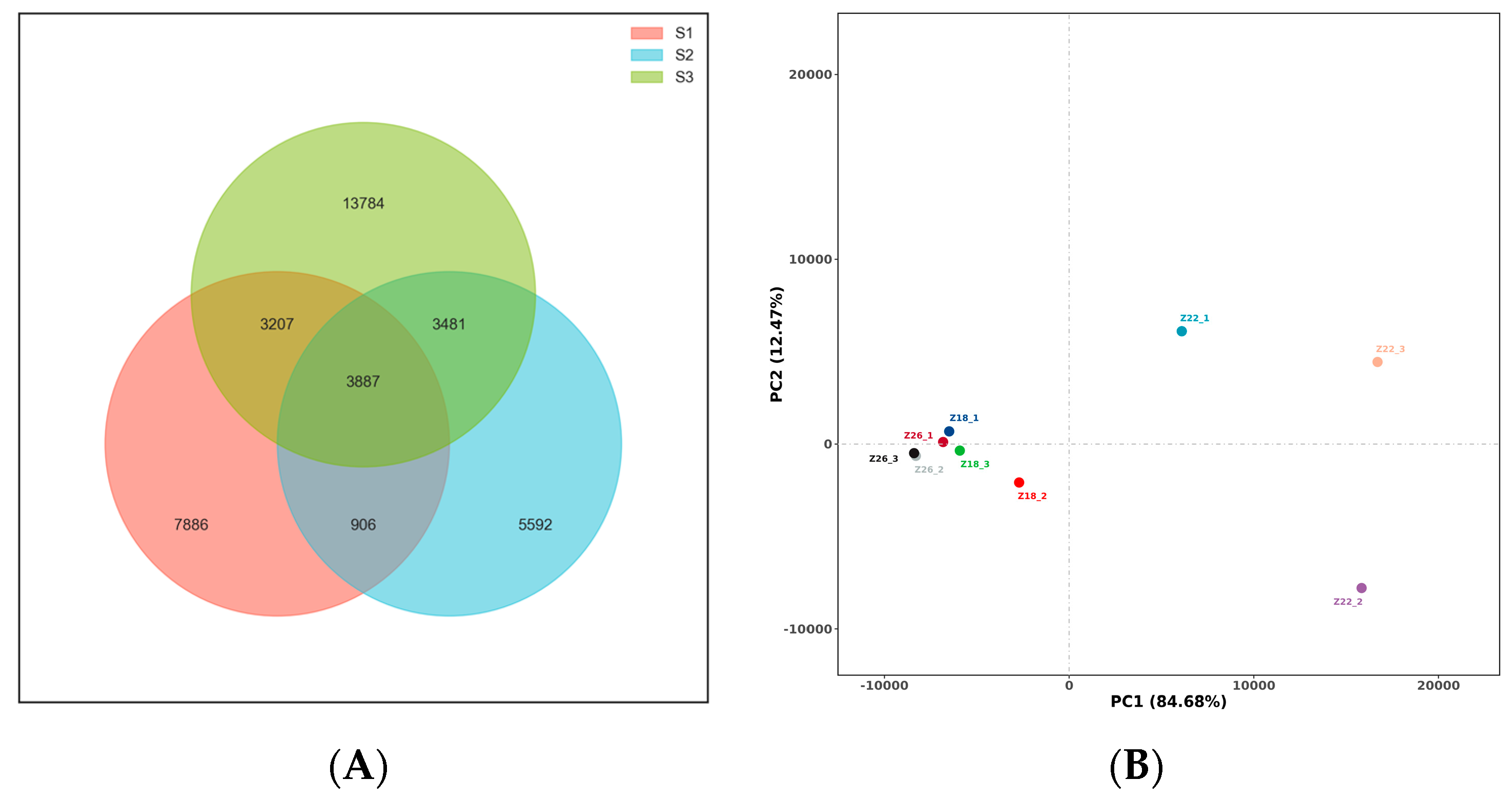
3.1.1. Three-Sample OTU Analysis and Correlation

In the study, a total of 54,111 operational taxonomic units (OTUs) were identified from the rhizosphere soil samples of three distinct groups (Figure 1A). Among these, 15,886 OTUs were associated with healthy pine trees, 13,866 OTUs with dead pine trees, and 24,359 OTUs with diseased pine trees. Notably, a shared pool of 3887 OTUs was detected across all three groups. The analysis of OTU abundance revealed that diseased pine trees exhibited the highest richness of OTUs, whereas dead pine trees displayed the lowest OTU richness. Principal component analysis (PCA) was conducted at the genus level to analyze the rhizosphere soil microbiota of the three sample groups (Figure 1B). The first two principal components accounted for 84.68% and 12.47% of the total variation, respectively. The results revealed discernible differences in the bacterial community structure among the healthy, diseased, and dead pine trees. Specifically, the dissimilarities between the healthy and diseased pine trees at the genus level were relatively minor, whereas the rhizosphere community structure of the dead pine trees exhibited significant dissimilarities compared to the other two groups.

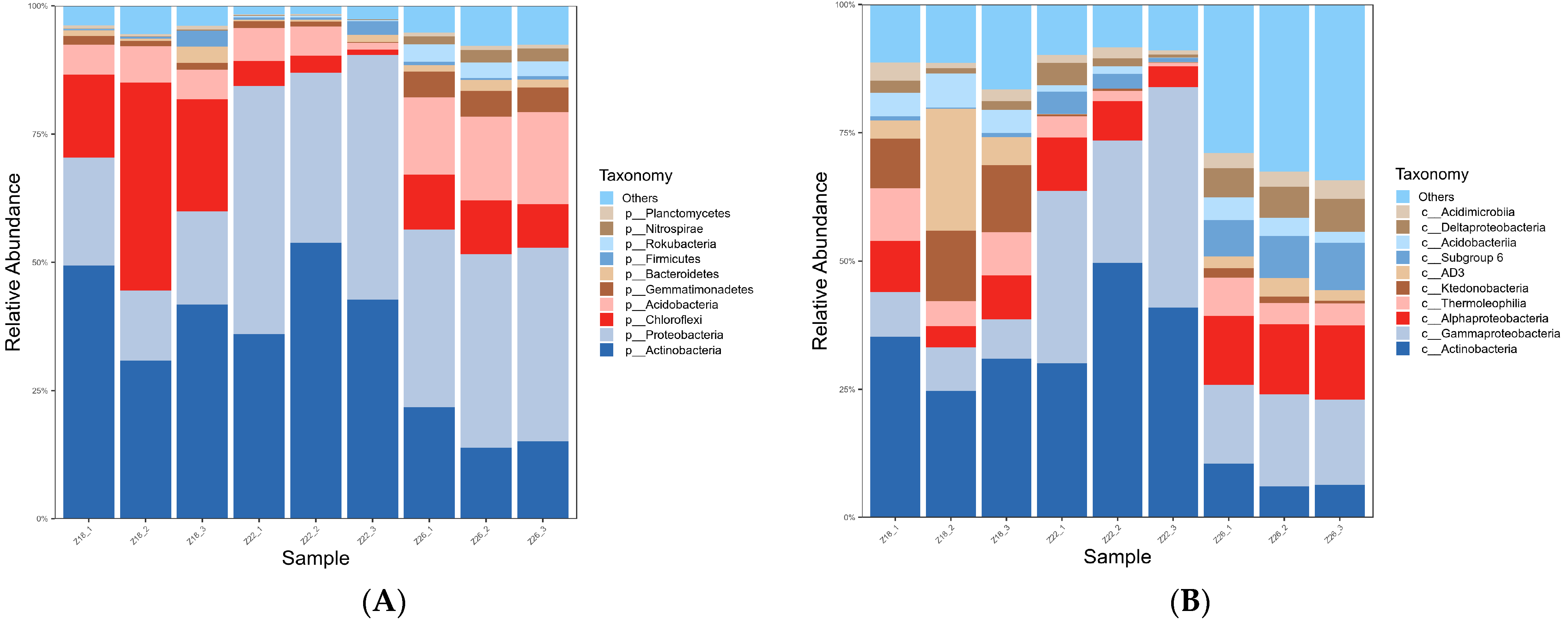
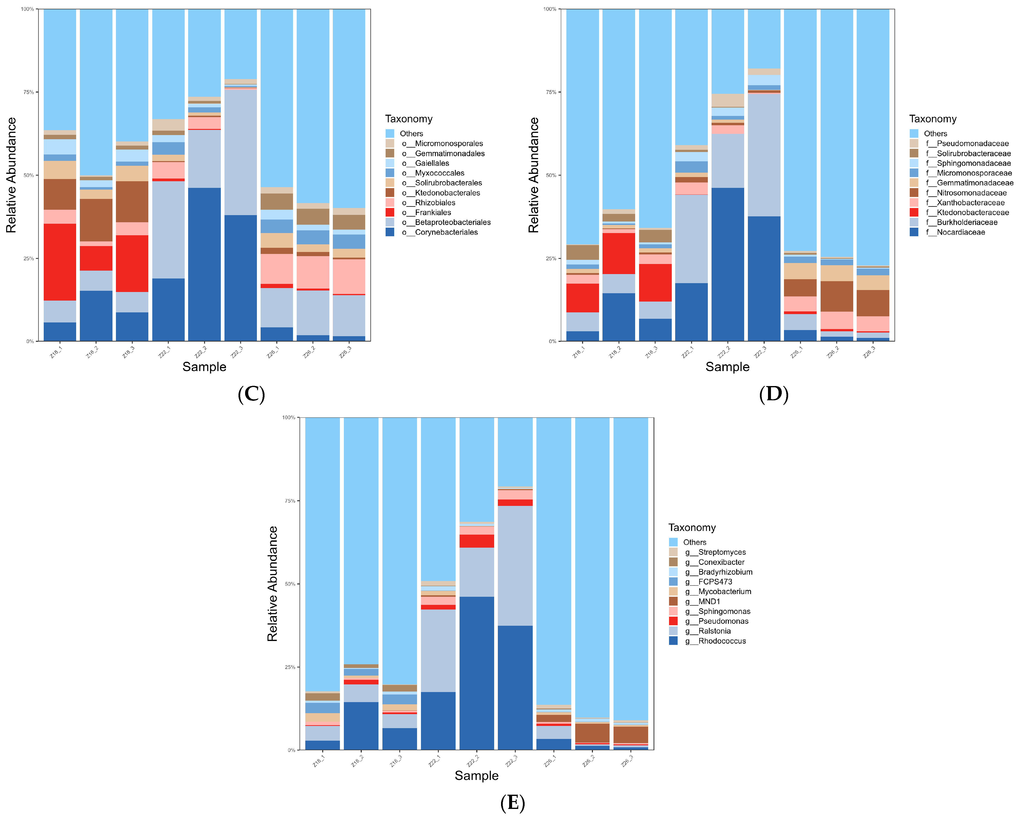
3.1.2. Analysis of Rhizosphere Microbial Species Composition in Three Sample Groups

In order to analyze the microbial community structures at different taxonomic levels, we conducted a comparative analysis by selecting the top 10 most abundant species based on species annotation. The results, presented in (Figure 2), provide insights into the composition of these communities. At the phylum level (Figure 2A), Proteobacteria, Actinobacteria, Acidobacteria, and Chloroflexi are the major phyla. In healthy rhizosphere soil of pine trees, Actinobacteria and Chloroflexi are the most abundant, with Chloroflexi being more abundant than other Chloroflexi groups. In the rhizosphere soil of dead pine trees, Actinobacteria and Proteobacteria are the most abundant, with Actinobacteria being more abundant than other Actinobacteria groups. At the class level (Figure 2B), Gammaproteobacteria, Acidobacteria, Alphaproteobacteria, and Ktedonobacteria are the dominant classes in the rhizosphere soil microbiota. Among them, Gammaproteobacteria, Acidobacteria, Alphaproteobacteria, and Ktedonobacteria show relatively high abundance. In the rhizosphere soil of healthy pine trees, Actinobacteria is relatively more abundant. In the rhizosphere soil of dead pine trees, Actinobacteria and Gammaproteobacteria have the highest abundance among the three bacterial groups. In the rhizosphere soil of diseased pine trees, Gammaproteobacteria and Alphaproteobacteria show relatively high abundance. At the order level (Figure 2C), Frankiaceae has a relatively high abundance in the rhizosphere soil of healthy pine trees. Meanwhile, Bacillariophyceae has the highest abundance in the rhizosphere soil of dead pine trees. At the family level (Figure 2D), Nocardioidaceae, Burkholderiaceae, Ktedonobacteraceae, and Xanthobacteraceae exhibited relatively high abundances in all three soil groups under investigation. Among them, Ktedonobacteraceae displayed the highest abundance in the rhizosphere soil of healthy pine trees, while Nocardiaceae and Burkholderiaceae showed the highest abundance in the rhizosphere soil of diseased pine trees. At the genus level (Figure 2E), the prominent bacterial genera identified were Rhodococcus, Ralstonia, Acidibacter, Burkholderia, and Bradyrhizobium. In the rhizosphere soil of healthy pine trees, Rhodococcus and Ralstonia were relatively more abundant.

3.2. Isolation and Screening of Threadkilling Bacteria

After isolating 620 bacterial strains from the soil samples, a 24 h nematocidal activity assay was conducted to screen for bacteria with nematocidal activity. Among the tested strains, 35 strains exhibited nematocidal activity. These strains were further categorized based on their source, with 20 strains isolated from healthy Pinus massoniana rhizosphere soil, 5 strains from diseased Pinus massoniana rhizosphere soil, and 10 strains from dead Pinus massoniana rhizosphere soil. A secondary screening was performed on the selected 35 strains to identify those with a corrected mortality rate exceeding 80% after 24 h (Table 1). The strains were identified as primarily Bacillus subtilis, Burkholderia cepacia, Acidobacterium, and Pseudomonas (Table 2). Among the isolated bacterial strains, namely 13-4, W45, and W49, their nematode-killing activity was found to be the strongest after a 24 h assay. The corrected mortality rates for these strains were determined as 92.23%, 90.85%, and 93.62%. To visually represent the nematode-killing effects, a schematic diagram illustrating the results is provided (Figure 3).

3.3. Determination of Nematocidal (Nematicidal) Activity of Fermentation Filtrate and Bacterial Suspension of 3 Bacterial Strains

Inorderto investigate the impact of fermentation filtrate and bacterial suspension on thenematocidal activity of four selected strains, aseries of experiments was conducted One milliliter (1 mL) of fermentation filtrate and 1 mL of bacterial suspension obtained from each of the three highly efficient nematode-killing strains were mixed with 1 mL of pine wood nematode solution using the soaking method. The mixtures were then subjected to incubation at 25 °C for 24 and 48 h. The resulting nematocidal activity was quantified by calculating the corrected mortality rate of the pine wood nematodes (Table 3). The findings suggest that the nematode-killing activity of the bacterial cell suspension of strain 13-4 surpasses that of the fermentation filtrate, as evidenced by the higher corrected mortality rate of 98.07% at 48 h for the former, compared to 95.74% for the latter. Moreover, the nematode-killing activity of the fermentation filtrate of strains W45 and W49 exhibited a significant increase compared to their respective bacterial cell suspensions. At 48 h, strain W45 demonstrated a corrected mortality rate of 94.84% for the fermentation filtrate, whereas the bacterial cell suspension exhibited a slightly lower rate of 93.42%. In the case of strain W49, the fermentation filtrate displayed a notable corrected mortality rate of 97.68% at 48 h, while the bacterial cell suspension exhibited a comparatively lower rate of 88.61% during the same time period.
Upon performing a two-fold and four-fold dilution of the bacterial cell suspensions and fermentation broth, it was observed that the nematode-killing activity of the fermentation broth surpassed that of the bacterial cell suspensions. Furthermore, when subjected to a four-fold dilution, significant differences in the corrected mortality rates were evident between the W13-4, W49, and W45 strains, regardless of whether it was the bacterial cell suspension or the fermentation broth. This distinction was particularly pronounced after a 48-h incubation period(Table 4).

3.4. Molecular Biological Identification of Three Bacterial Strains

The full-length sequences of the 16S rDNA gene fragments amplified from strains 13-4, W45, and W49 were obtained through sequencing and submitted to GenBank. The accession numbers assigned to these sequences are OR478150, OR478102, and OR478079, respectively. Subsequently, the 16S rDNA gene sequences of strains 13-4, W45, and W49 were aligned, and a phylogenetic analysis based on the 16S rDNA gene was conducted. The phylogenetic analysis revealed that strain 13-4 clustered together with Lysinibacillus capsici, forming a distinct subclade (Figure 4A) with a branch support value of 94.00%. Strain W45 was found to be closely related to Bacillus paramycoides, showing a shared branch (Figure 4B) with a branch support value of 96.00%; and strain 49 clustered with Delftia tsuruhatensisin one unit as shown in (Figure 4C) with 100.00% branching support.

3.5. Effects of Three Strains on PAL Content of Pinus Massoniana

Phenylalanine ammonia-lyase (PAL) is an essential defensive enzyme in plants, involved not only in the phenylpropanoid metabolic pathway but also in the synthesis of lignin and other plant secondary metabolites. During environmental stress or external stimuli, plants activate their defense systems, increasing PAL activity. Thus, PAL activity serves as a physiological indicator of plant stress resistance and is considered one of the key markers for evaluating plant responses to adverse conditions. The results, presented in (Figure 5A), demonstrate that within 96 h of inoculation, all treatment groups exhibited significantly higher PAL activity compared to the control group. In the 13-4F treatment, PAL activity exhibited a sustained increase, reaching its peak at 24 h, measuring 429.957 U/g min−1, followed by a subsequent decline and stabilization at 72 h. A similar trend was observed in the W45F treatment, where PAL activity initially increased, reaching its highest level at 12 h, measuring 391.63 U/g min−1, and then stabilized at 48 h. Under the W49F treatment, PAL activity showed an initial increase, followed by a decrease until 12 h, and then resumed an upward trend, reaching its maximum activity at 24 h, measuring 298.30 U/g min−1. In contrast, the control group exhibited no significant changes in PAL enzyme activity. Moreover, the application of different bacterial cell suspensions 13-4J, W45J, and W49J also induced significant variations in PAL activity within Pinus massoniana tissues (Figure 5C). The 13-4J treatment resulted in the highest PAL activity at 12 h, measuring 334.97 U/g min−1, followed by a gradual decline. Similarly, the W45J treatment exhibited the highest PAL activity at 12 h, measuring 453.29 U/g min−1, while the W49J treatment reached its maximum activity at 72 h, measuring 249.98 U/gmin−1. Notably, the control group displayed a slight upward trend in PAL activity, peaking at 72 h and measuring 151.65 U/g min−1. These findings provide compelling evidence that strains 13-4, W45, and W49 significantly induce PAL enzyme activity in Pinus massoniana tissues.
After 96 h of bacterial treatment and subsequent inoculation with pine wood nematodes (PWN), substantial changes in the activity of phenylalanine ammonia-lyase (PAL) were observed in Pinus massoniana. Under the fermentation broth treatment (Figure 5B), only the PWN-inoculated with CK2 exhibited PAL activity that initially increased and subsequently decreased, consistently remaining lower than the control group (CK). Overall, PAL activity in all PWN-inoculated groups demonstrated an initial increase followed by a decline, while the PAL activity in the CK2 group inoculated with sterile water remained relatively stable. Specifically, in the 13-4F treatment, PAL activity in Chinese red pine displayed an initial increase followed by a decline, reaching its peak at 14 days, measuring 478.29 U/g min−1. In the W45F treatment, PAL activity peaked at 3 days, measuring 788.25 U/g min−1, and exhibited stable fluctuations thereafter. In the W49F treatment, PAL activity showed an overall increasing trend followed by a decline, reaching its maximum at 14 days, measuring 818.25 U/g min−1. The PAL activity in the CK group demonstrated an initial increase followed by a decline, with the highest activity observed at 21 days, measuring 463.29 U/g min−1. Throughout the 28 day period, PAL activity in all treatment groups remained lower than that of the CK group.
Under the bacterial cell suspension treatment (Figure 5D), the 13-4J treatment revealed two peaks in PAL activity at 7 days, measuring 788.25 U/g min−1 and 28 days, measuring 933.79 U/g min−1, respectively. In the W45J treatment, PAL activity displayed an initial increase followed by a decline, with the highest activity observed at 14 days, measuring 804.92 U/g min−1. Similarly, the W49J treatment exhibited two peaks in PAL activity at 3 days, measuring 166.65 U/g min−1, and 21 days, measuring 518.28 U/g min−1, respectively. The CK and CK2 groups demonstrated relatively stable changes, with all treatment groups surpassing the control group within the first 14 days. These findings provide compelling evidence that bacterial treatments induce a significant induction of PAL enzyme activity in Pinus massoniana tissues under both fermentation broth and bacterial cell suspension treatments.

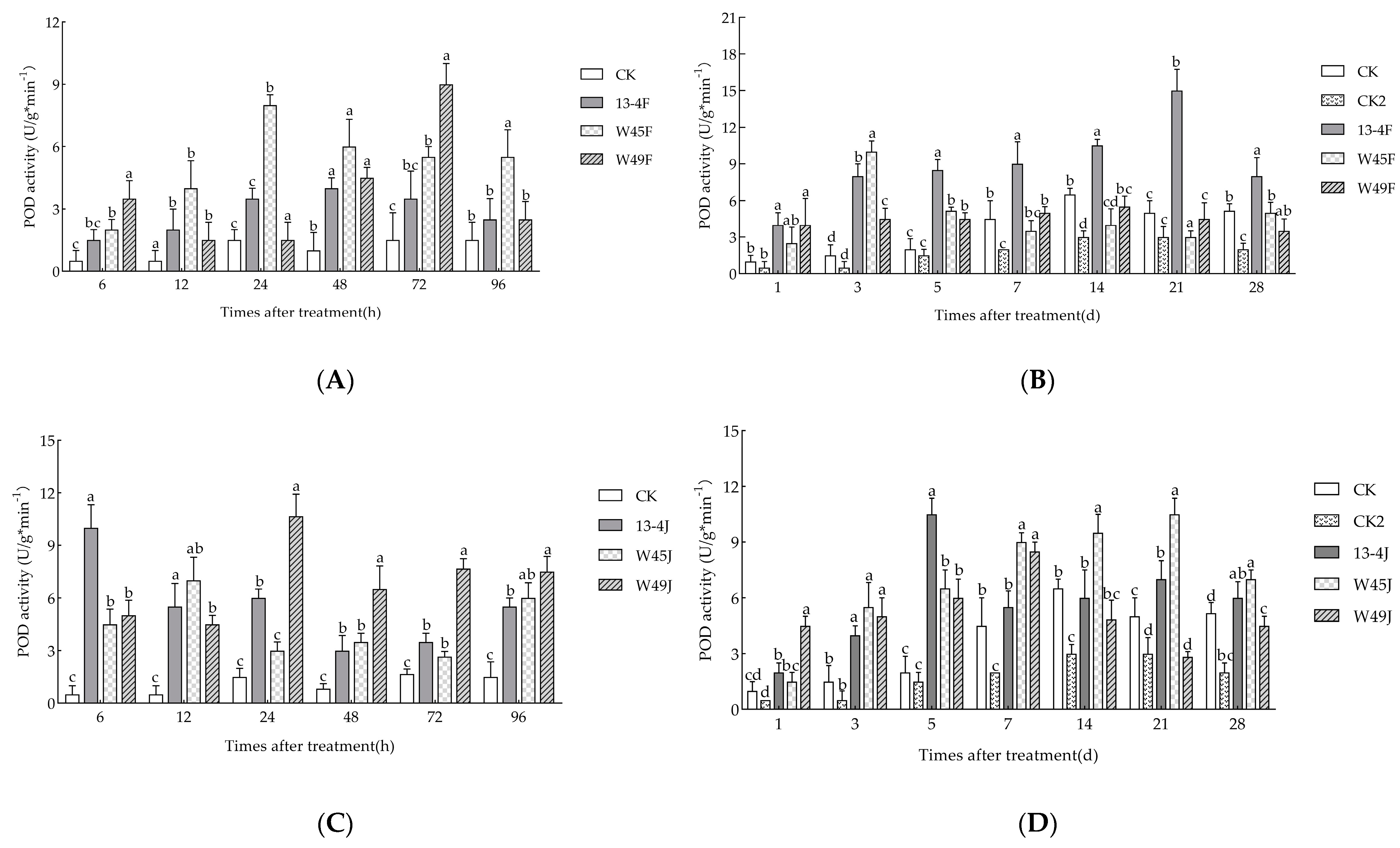
3.6. Effects of Three Strains on POD Content of Pinus massoniana

Peroxidase (POD) enzyme plays a crucial role in maintaining the hydrogen peroxide (H2O2) balance within plant organisms. Under the treatment with fermentation broth from different bacterial strains, significant changes were observed in the peroxidase (POD) activity within 96 h of inoculation in Pinus massoniana (Figure 6A). The enzyme activity in all treatment groups surpassed that of the control group. Specifically, in the 13-4F and W45F treatments, the POD activity exhibited an initial increase followed by a decline. The 13-4F treatment demonstrated the highest POD activity at 48 h, reaching 4 U/g min−1. Likewise, the W45F treatment reached its peak at 24 h, measuring 8 U/g min−1. Conversely, the W49F treatment displayed a decline followed by an increase in POD activity, with the highest level observed at 72 h, measuring 9 U/g min−1. In contrast, the control group showed no significant changes in POD enzyme activity. Similarly, under the treatment of bacterial cell suspension from different bacterial strains (Figure 6C), the POD activity in Pinus massoniana also exhibited notable variations. The 13-4J treatment reached its maximum enzyme activity at 6 h, measuring 10 U/g min−1, which subsequently declined and displayed an upward trend at 48 h. In the W45J treatment, the highest POD activity was recorded at 12 h, measuring 7 U/g min−1. Furthermore, the W49J treatment exhibited the highest activity at 24 h, measuring 10.67 U/g min−1. In contrast, the control group did not display any significant changes. These findings clearly indicate the remarkable inducing effect of bacterial strains 13-4, W45, and W49 on the POD activity in Pinus massoniana tissues.
After 96 h of bacterial inoculation, significant fluctuations in peroxidase (POD) activity were observed in Pinus massoniana upon subsequent inoculation with pine wood nematodes. Under fermentation broth treatment (Figure 6B), both the treatment and control groups exhibited an initial increase followed by a decline in POD activity. However, the POD activity in Chinese red pine treated with different bacterial strains’ fermentation broth consistently remained higher than the control group without bacterial inoculation. Specifically, in the 13-4F treatment, the POD activity displayed an initial increase followed by a decline, reaching its peak at 21 days with a value of 15 U/g min−1. Similarly, in the W45F treatment, the highest POD activity was observed at 3 days, measuring 10 U/g min−1, after which it gradually decreased. The W49F treatment showed an overall stable trend with an initial increase followed by a decrease in POD activity. However, from 14 days onwards, the control group’s (CK) POD activity exceeded that of all treatment groups except for 13-4F. At this time point, the CK group exhibited a POD activity of 6.5 U/g min−1, which persisted until 28 days.
Under bacterial cell suspension treatment (Figure 6D), the 13-4J treatment attained its highest enzyme activity at 5 days, measuring 10.5 U/g min−1, followed by a gradual decline until the 7th day. In the W45J treatment, POD activity initially increased and then decreased, reaching its peak at 21 days with a value of 10.5 U/g min−1. Similarly, the W49J treatment exhibited an upward trend followed by a decline, with the highest activity observed at 7 days, measuring 8.5 U/g min−1. In contrast, both the CK and CK2 treatments displayed an overall increasing trend followed by a decrease in POD activity. The highest levels were observed at 14 days, measuring 6.5 U/g min−1 and 3 U/g min−1, respectively. These findings clearly demonstrate the significant inducing effect of bacterial strains on POD activity in Pinus massoniana tissues under bacterial cell suspension treatment.

3.7. Screening and Control Effect of Pinus massoniana against Pine Wood Nematode Disease Induced by Linicide Strain

Within 96 h of inoculation, the changes in two defense enzymes were examined (Table 5) to preliminarily identify bacterial strains with strong inducible resistance capabilities. The strains 13-4 fermentation filtrate, 45 fermentation filtrate, and bacterial cell suspension were found to exhibit promising inducible resistance properties. After 28 days of pine wood nematode inoculation, the control effect was assessed (Figure 7). Ultimately, strain 13-4 was selected as a potent dual-resistant bacterial strain, possessing both pine wood nematode insecticidal activity and the ability to induce resistance against these nematodes.

4. Discussion

Microorganisms play a vital role in the pathogenesis of Pine Wilt Disease (PWD) [20,25]. It has now been shown that in the inter-root soils of pine trees infected with pine nematode disease and healthy pine trees in the natural state, putative microflora is the dominant microflora [26,27,28]; the abundance of microbial bacteria is lowest in the inter-root soils of P. massoniana; and there is a strong positive correlation between the abundance of acidic bacteria in the soils and the soil nitrogen cycle [29]. In our study, actinobacteria constituted a significant proportion of the rhizosphere soil, exhibiting distinct characteristics. Additionally, we observed a higher abundance of Acidobacteria in the rhizosphere soil of healthy Pinus massoniana compared to that of diseased counterparts. This disparity in Acidobacteria abundance may influence soil nitrogen cycling, contributing to a certain level of resistance in Pinus massoniana against pine wilt disease.
Studies have shown that microbial biocontrol mechanisms include (1) ecological niche competition [30], (2) inhibiting and killing pathogens directly or indirectly by antagonising substances produced by microbial metabolic processes [31], (3) secreting a range of hydrolytic enzymes, such as proteases, chitinases, and cellulases [32], (4) inducing resistance in plants (ISR and SAR) [33], and (5) transferring the pathogens from the plant body to the plant via another way of killing the pathogen by antiparasitism of a parasitic organism [34].
Research has indicated that specific bacteria associated with the pine wood nematode (B. xylophilus) have been reported to play an important role in the pathogenesis of PWD [35,36,37]. For example, some microorganisms of the genera Bacillus and Serratia have nematicidal activity through a parasitic mechanism or by producing toxic compounds [38,39]. Jeong et al. reported the inhibitory activity of lichen-associated Bacillus spp. against pine wood nematodes, suggesting their potential as agents for inducing plant resistance through the modulation of plant hormones [40]. Yuan et al. found that Bacillus cereus could not only affect the survival and fecundity of pine nematodes but also reduce the extent of pine nematode infestation in Sargasso pine [41].
It is noteworthy that the genus Lysinibacillus, which belongs to the family Bacillaceae, exhibits inhibitory effects on pine wood nematode (B. xylophilus) and also influences plant resistance and growth. Some bacteria within this genus or their secondary metabolites have been found to possess nematocidal activity, along with the ability to impact plant growth and resistance. Jennifer Jähne et al. discovered that the genome of Lysinibacillus capsici contains genes that promote plant growth [42]. Zhao Yong et al. demonstrated that Lysinibacillus capsici produces volatile organic compounds that regulate the growth of Arabidopsis thaliana [43]. Furthermore, previous studies have indicated that Lysinibacillus capsici possesses chitinase, which inhibits root-knot nematodes [44]. In our experiments, nine of the 13 strains of nematicidal bacteria screened and identified were Bacillus; one strain of Lysinibacillus capsici, one strain of Lelliottia jeotgali, one strain of Pseudomonas geniculata and one strain of Delftia tsuruhatensis. In addition, we conducted a greenhouse pot experiment to further investigate the biocontrol efficacy of three selected strains, namely 13-4 Lysinibacillus capsici, W45 Bacillus paramycoides, and W49 Delftia tsuruhatensis, which exhibited high nematocidal activity. The fermented broth and cell suspensions of these strains were utilized to evaluate their effectiveness against pine wilt disease caused by Bursaphelenchus xylophilus. Our results revealed that all three strains exhibited substantial inhibitory effects on the development of pine wilt disease in Pinus massoniana. Notably, the strain 13-4 Lysinibacillus capsici demonstrated significant regulatory effects on pine wood nematodes externally and internally in P. massoniana. These findings highlight the potential of the Lysinibacillus capsici 13-4 strain and its metabolites as promising microbial biocontrol agents for managing pine wilt disease.
Upon invasion by pathogens or microorganisms, plants elicit various defense responses [45]. Systemic acquired resistance (SAR) and induced systemic resistance (ISR) are two crucial defense mechanisms employed by plants in response to pathogen attacks. ISR is predominantly mediated through the jasmonic acid (JA) and ethylene (ET) signaling pathways, while SAR is typically governed by the salicylic acid (SA) signaling pathway [46]. In rice samples treated with the B51 strain, elevated activities of peroxidase (POD) and phenylalanine ammonia-lyase (PAL) were detected. Additionally, the expression levels of genes associated with the JA/ET signal transduction pathway were also enhanced [47]. In a study conducted by Wang et al., the treatment of loquat fruits with B. cereus AR156 resulted in augmented activities of defense-related enzymes, including PAL and POD. This treatment also led to an increased accumulation of hydrogen peroxide (H2O2), thereby inducing host defense responses and reducing the incidence of anthracnose rot [48]. Furthermore, the treatment of solanaceous plants with Trichoderma spp. demonstrated notable inhibition of pathogenic fungal mycelial growth and a significant increase in POD activity [49]. Jamal Qazi Mohammad Sajid et al.’s research revealed that Bacillus solubilis not only synthesizes plant hormones such as indole-3-acetic acid and gibberellic acid but also triggers plant defense responses against pathogens, enabling plants to sustain growth under adverse conditions [50]. Similarly, in our study, treatment with strains 13-4 enhanced the activities of defense enzymes, including POD and PAL, in Pinus massoniana and exhibited prolonged effects.
The issues proposed for further research include (1) conducting transcriptomic analysis using RNA sequencing to elucidate the signal transduction pathways initiated by strains 13-4 upon treatment. (2) Isolating and identifying additional functional metabolites produced by strains 13-4, particularly focusing on their potential to induce systemic resistance in the hosts.

5. Conclusions

In this study, the results of high-throughput sequencing showed that the differences in inter-root microorganisms between healthy and diseased ponytail pines were relatively small, while the root community structure of dead Masson pine was significantly different from the other two groups. The community structure of each group also varied at different levels, and there were more inter-root microorganisms in healthy ponytail pines than in diseased Masson pine. We isolated three strains, namely 13-4, W45, and W49, that exhiited effective nematode-killing properties. Strain 13-4 was identified as Lysinibacillus capsici, strain W45 as Bacillus paramycoides, and strain W49 as Delftia tsuruhatensis. Through a pot experiment, we found that the suspension and fermentation filtrate of these three strains induced various defense reactions in Pinus massoniana against pine wood nematodes, including increased activities of phenylalanine ammonia-lyase (PAL) and peroxidase (POD) enzymes. Consequently, the incidence of pine wood nematode disease was significantly reduced. Our findings highlight the strong nematode-killing effects of Lysinibacillus capsici, Bacillus paramycoides, and Delftia tsuruhatensis. Notably, the fermentation filtrate of strain 13-4 exhibited excellent nematode-killing efficacy and demonstrated the ability to induce resistance. Therefore, this strain holds promise as an effective biological control agent (BCA) for managing pine wood nematode disease.

Author Contributions

Conceptualization, M.L., Y.W., J.Z., G.Z. and J.L.; conducted the experiments and analyzed the data M.L., Y.W. and J.Z.; wrote the manuscript M.L., Y.W., J.Z., G.Z. and J.L.; supervision, G.Z. and J.L.; project administration, G.Z. and J.L.; funding acquisition, J.L. All authors have read and agreed to the published version of the manuscript.

Funding

National Key R&D Program of the 14th Five-Year Plan (No. 2021YFD1400904): Research on disaster mechanism and sustainable prevention and control technology of pine wood nematode.

Data Availability Statement

All relevant data are within this paper.

Conflicts of Interest

The authors declare no conflict of interest. The funders had no role of the study; in the collection, analyses, or interpretation of data; in the writing of the manuscript; or in the decision to publish the results.

References

  1. Nickle, W.R.; Golden, A.M.; Mamiya, Y.; Wergin, W. On the taxonomy and morphology of the pine wood nematode, Bursaphelenchus xylophilus (Steiner &Buhrer 1934) Nickle 1970. J. Nematol. 1981, 13, 385–392. [Google Scholar] [PubMed]
  2. Mota, M.M.; Braasch, H.; Bravo, M.A.; Penas, A.C.; Burgermeister, W.; Metge, K.; Sousa, E. First report of Bursaphelenchus xylophilus in Portugal and in Europe. Nematology 1999, 1, 727–734. [Google Scholar] [CrossRef]
  3. Zamora, P.; Rodríguez, V.; Renedo, F.; Sanz, A.V.; Domínguez, J.C.; Pérez-Escolar, G.; Miranda, J.; Alvarez, B.; Gonzalez-Casas, A.; Mayor, E.; et al. First report of Bursaphelenchus xylophilus causing pine wilt disease on Pinus radiata in Spain. Plant Dis. 2015, 99, 1449. [Google Scholar] [CrossRef]
  4. Futai, K. Pine wood nematode, Bursaphelenchus xylophilus. Annu. Rev. Phytopathol. 2013, 51, 61–83. [Google Scholar] [CrossRef] [PubMed]
  5. Kim, B.N.; Kim, J.H.; Ahn, J.Y.; Kim, S.; Cho, B.K.; Kim, Y.H.; Min, J. A short review of the pinewood nematode, Bursaphelenchus xylophilus. Toxicol. Environ. Health Sci. 2020, 12, 297–304. [Google Scholar] [CrossRef]
  6. Chan, S.Y.; Liu, S.Y.; Seng, Z.; Chua, S.L. Biofilm matrix disrupts nematode motility and predatory behavior. ISME J. 2021, 15, 260–269. [Google Scholar] [CrossRef]
  7. Cheng, C.; Qin, J.; Wu, C.; Lei, M.; Wang, Y.; Zhang, L. Suppressing a plant-parasitic nematode with fungivorous behavior by fungal transformation of a Bt cry gene. Microb. Cell Factories 2018, 17, 116. [Google Scholar] [CrossRef]
  8. Jansen-Girgan, C.; Claassens, S.; Fourie, H. In vitro evaluations to determine the effect of Bacillus firmus strains on the motility of Meloidogyne incognita second-stage juveniles. Trop. Plant Pathol. 2016, 41, 320–324. [Google Scholar] [CrossRef]
  9. Paiva, G.; Proença, D.N.; Francisco, R.; Verissimo, P.; Santos, S.S.; Fonseca, L.; Morais, P.V. Nematicidal bacteria associated to pinewood nematode produce extracellular proteases. PLoS ONE 2013, 8, e79705. [Google Scholar] [CrossRef]
  10. Wang, Y.; Mei, L.; Wu, J.; Zhang, L. Detection and characterisation of a Bacillus thuringiensis crystal protein with nematicidal activity against the pinewood nematode Bursaphelenchus xylophilus. Biocontrol Sci. Technol. 2012, 22, 1143–1153. [Google Scholar] [CrossRef]
  11. Yu, J.; Du, G.; Li, R.; Li, L.; Li, Z.; Zhou, C.; Guo, D. Nematicidal activities of bacterial volatiles and components from two marine bacteria, Pseudoalteromonas marina strain H-42 and Vibrio atlanticus strain S-16, against the pine wood nematode, Bursaphelenchus xylophilus. Nematology 2015, 17, 1011–1025. [Google Scholar] [CrossRef]
  12. Wu, J.; Yin, Y.; Tan, J.; Hao, D. A preliminary study on resistance of Pinus massoniana induced by Bacillus cereus NJSZ-13 strain to Bursaphelenchus xylophilus. J. Nanjing For. Univ. (Nat. Sci. Ed.) 2022, 46, 4. [Google Scholar] [CrossRef]
  13. Han, G.; Mannaa, M.; Kim, N.; Jeon, H.W.; Jung, H.; Lee, H.H.; Kim, J.; Park, J.; Park, A.R.; Kim, J.C.; et al. Response of Pine Rhizosphere Microbiota to Foliar Treatment with Resistance-Inducing Bacteria against Pine Wilt Disease. Microorganisms 2021, 9, 688. [Google Scholar] [CrossRef]
  14. Zhang, W.; Zhao, L.; Zhou, J.; Yu, H.; Zhang, C.; Lv, Y.; Sun, J. Enhancement of oxidative stress contributes to increased pathogenicity of the invasive pine wood nematode. Philos. Trans. R. Soc. B 2019, 374, 20180323. [Google Scholar] [CrossRef]
  15. Li, Y.; Wang, X.; Liu, Z.; Zhang, X. Research advance of pathogenic mechanism of pine wood nematode. For. Pest. Dis. 2022, 41, 11–20. [Google Scholar]
  16. Cao, Y.; Wang, L.; Wang, X.; Fan, J. Pathogenicity of Bursaphelenchus xylophilus to Larix olgensis Seedlings. Sci. Silv. Sin. 2020, 56, 108–115. [Google Scholar] [CrossRef]
  17. Zhang, W.; Li, Y.; Zhon, L.; Shen, D.; Zhu, L.; Fan, B. Inhibition of pine wood nematode Bursaphelenchus xylophilus by rhizobacterium Bacillus velezensis FZB42. Acta Microbiol. Sin. 2021, 61, 1287–1298. [Google Scholar] [CrossRef]
  18. Wang, J.; Jiang, X.; Luan, Q.; Feng, J.; Wang, J.; Liu, M. Effects of Bursaphelenchus xylophilus Infestation on Physiological Indexes of Larix kaempferi. J. Southwest. For. Univ. 2023, 43, 6. [Google Scholar]
  19. Ren, W.; Ren, G.; Teng, Y.; Li, Z.; Li, L. Time-dependent effect of grapheneon the structure, abundance, and function of the soil bacterial community. J. Hazard. Mater. 2015, 297, 286–294. [Google Scholar] [CrossRef]
  20. Han, Z.M.; Hong, Y.D.; Zhao, B.G. A study on pathogenicity of bacteria carried by pine wood nematodes. J. Phytopathol. 2003, 151, 683–689. [Google Scholar] [CrossRef]
  21. Liu, M.J.; Hwang, B.S.; Jin, C.Z.; Li, W.J.; Park, D.J.; Seo, S.T.; Kim, C.J. Screening, isolation and evaluation of a nematicidal compound from actinomycetes against the pine wood nematode, Bursaphelenchus xylophilus. Pest Manag. Sci. 2018, 75, 1585–1593. [Google Scholar] [CrossRef]
  22. Elamary, R.; Salem, W. Optimizing and purifying extracellular amylase from soil bacteria to inhibit clinical biofilm-forming bacteria. PeerJ 2020, 8, e10288. [Google Scholar] [CrossRef]
  23. Maehara, N.; Futai, K. Effect of fungal interactions on the numbers of the pinewood nematode, Bursaphelenchus xylophilus (Nematoda: Aphelenchoididae), carried by the Japanese pine sawyer, Monochamus alternatus (Coleoptera: Cerambycidae). Fundam. Appl. Nematol. 1997, 20, 611–617. [Google Scholar] [CrossRef]
  24. Xiong, Y.; Xia, F.; Huang, Z.; Liu, L.; Weng, X.; Wu, S. Isolation and Identification of Bacillus sp. Strain BRC-XYT1 and its Toxicity to Monochamus alternatus Hope. Curr. Biotechnol. 2016, 6, 5. [Google Scholar] [CrossRef]
  25. Vicente, C.S.; Ikuyo, Y.; Mota, M.; Hasegawa, K. Pinewood nematode-associated bacteria contribute to oxidative stress resistance of Bursaphelenchus xylophilus. BMC Microbiol. 2013, 13, 299. [Google Scholar] [CrossRef]
  26. Fukuda, K. Physiological process of the symptom development and resistance mechanism in pine wilt disease. J. For. Res. 1997, 2, 171–181. [Google Scholar] [CrossRef]
  27. Mabuhay, J.A.; Nakagoshi, N. Response of soil microbial communities to changes in a forest ecosystem brought about by pine wilt disease. Landsc. Ecol. Eng. 2012, 8, 189–196. [Google Scholar] [CrossRef]
  28. Liu, Y.; Ponpandian, L.N.; Kim, H.; Jeon, J.; Hwang, B.S.; Lee, S.K.; Park, S.-C.; Bae, H. Distribution and diversity of bacterial endophytes from four Pinus species and their efficacy as biocontrol agents for devastating pine wood nematodes. Sci. Rep. 2019, 9, 12461. [Google Scholar] [CrossRef]
  29. Zhang, Y.; Ding, K.; Yrjälä, K.; Liu, H.; Tong, Z.; Zhang, J. Introduction of broadleaf species into monospecific Cunninghamia lanceolata plantations changed the soil Acidobacteria subgroups composition and nitrogen-cycling gene abundances. Plant Soil 2021, 467, 29–46. [Google Scholar] [CrossRef]
  30. Pérez-García, A.; Romero, D.; De Vicente, A. Plant protection and growth stimulation by microorganisms: Biotechnological applications of Bacilli in agriculture. Curr. Opin. Biotechnol. 2011, 22, 187–193. [Google Scholar] [CrossRef]
  31. Hu, J.; Wei, Z.; Friman, V.P.; Gu, S.H.; Wang, X.F.; Eisenhauer, N.; Yang, T.J.; Ma, J.; Shen, Q.R.; Xu, Y.C.; et al. Probiotic diversity enhances rhizosphere microbiome function and plant disease suppression. MBio 2016, 7, 10–1128. [Google Scholar] [CrossRef]
  32. Leclère, V.; Béchet, M.; Adam, A.; Guez, J.S.; Wathelet, B.; Ongena, M.; Thonat, P.; Gancel, F.; Cholletimbeert, M.; Jacques, P. Mycosubtilin overproduction by Bacillus subtilis BBG100 enhances the organism’s antagonistic and biocontrol activities. Appl. Environ. Microbiol. 2005, 71, 4577–4584. [Google Scholar] [CrossRef]
  33. Han, L.; Zhang, G.; Miao, G.; Zhang, X.; Feng, J. Streptomyces kanasensis sp. nov., an antiviral glycoprotein producing actinomycete isolated from forest soil around kanas lake of China. Curr. Microbiol. 2015, 71, 627–631. [Google Scholar] [CrossRef]
  34. Ballhausen, M.B.; Vandamme, P.; De Boer, W. Trait differentiation within the fungus-feeding (mycophagous) bacterial genus Collimonas. PLoS ONE 2016, 11, e0157552. [Google Scholar] [CrossRef]
  35. Oku, H.; Shiraishi, T.; Ouchi, S.; Kurozumi, S.; Ohta, H. Pine wilt toxin, the metabolite of a bacterium associated with a nematode. Naturwissenschaften 1980, 67, 198–199. [Google Scholar] [CrossRef]
  36. Le Dang, Q.; Son, S.W.; Cheon, H.M.; Choi, G.J.; Choi, Y.H.; Jang, K.S.; Kim, J.C. Pyochelin isolated from Burkholderia arboris KRICT1 carried by pine wood nematodes exhibits phytotoxicity in pine callus. Nematology 2011, 13, 521–528. [Google Scholar] [CrossRef]
  37. Zhao, B.G.; Wang, H.L.; Han, S.F.; Han, Z.M. Distribution and pathogenicity of bacteria species carried by Bursaphelenchus xylophilus in China. Nematology 2003, 5, 899–906. [Google Scholar] [CrossRef]
  38. Wang, Z.; Zhang, Y.; Wang, C.; Wang, Y.; Sung, C. Esteya vermicola controls the pinewood nematode, Bursaphelenchus xylophilus, in pine seedlings. J. Nematol. 2017, 49, 86. [Google Scholar] [CrossRef]
  39. Maehara, N. Reduction of Bursaphelenchus xylophilus (Nematoda: Parasitaphelenchidae) population by inoculating Trichoderma spp. into pine wilt-killed trees. Biol. Control 2008, 44, 61–66. [Google Scholar] [CrossRef]
  40. Jeong, M.H.; Yang, S.Y.; Lee, Y.S.; Ahn, Y.S.; Kim, K.Y. Selection and characterization of bacillus licheniformis mh48 for the biocontrol of pine wood nematode (Bursaphelenchus xylophilus). J. Korean For. Soc. 2015, 104, 512–518. [Google Scholar] [CrossRef]
  41. Yuan, Y.; Yan, Z.; Chen, Y.; Ye, J.; Tan, J. Effects of Bacillus cereus on Survival, Fecundity, and Host Adaptability of Pine Wood Nematode. Diversity 2023, 15, 566. [Google Scholar] [CrossRef]
  42. Jähne, J.; Le Thi, T.T.; Blumenscheit, C.; Schneider, A.; Pham, T.L.; Le Thi, P.T.; Blom, J.; Vater, J.; Schweder, T.; Lasch, P.; et al. Novel plant-associated Brevibacillus and Lysinibacillus genomospecies harbor a rich biosynthetic potential of antimicrobial compounds. Microorganisms 2023, 11, 168. [Google Scholar] [CrossRef]
  43. Zhao, D.; Jiao, J.; Du, B.; Liu, K.; Wang, C.; Ding, Y. Volatile organic compounds from Lysinibacillus macroides regulating the seedling growth of Arabidopsis thaliana. Physiol. Mol. Biol. Plants 2022, 28, 1997–2009. [Google Scholar] [CrossRef] [PubMed]
  44. Lee, Y.S.; Anees, M.; Park, Y.S.; Kim, S.B.; Jung, W.J.; Kim, K.Y. Purification and properties of a Meloidogyne-antagonistic chitinase from Lysobacter capsici YS1215. Nematology 2014, 16, 63–72. [Google Scholar] [CrossRef]
  45. Durrant, W.E.; Dong, X. Systemic acquired resistance. Annu. Rev. Phytopathol. 2004, 42, 185–209. [Google Scholar] [CrossRef] [PubMed]
  46. Kusajima, M.; Shima, S.; Fujita, M.; Minamisawa, K.; Che, F.-S.; Yamakawa, H.; Nakashita, H. Involvement of ethylene signalingin Azospirillum sp. B510-induced disease resistance in rice. Biosci. Biotechnol. Biochem. 2018, 82, 1522–1526. [Google Scholar] [CrossRef]
  47. Khare, E.; Mishra, J.; Arora, N.K. Multifaceted Interactions Between Endophytes and Plant: Developments and Prospects. Front. Microbiol. 2018, 9, 2732. [Google Scholar] [CrossRef]
  48. Wang, X.; Wang, L.; Wang, J.; Jin, P.; Liu, H.; Zheng, Y. Bacillus cereus AR156-Induced Resistance to Colletotrichum acutatum Is Associated with Priming of Defense Responses in Loquat Fruit. PLoS ONE 2014, 9, e112494. [Google Scholar] [CrossRef]
  49. Khalifa, E.A.; Khalifa, R.K.M.; Haggag, H.E. Effect of Trichoderma species on damping off diseases incidence, some plant enzymes activity and nutritional status of bean plants. Am. J. Sci. 2010, 6, 10–57. [Google Scholar] [CrossRef]
  50. Jamal, Q.M.S.; Ahmad, V. Lysinibacilli: A biological factories intended for bio-insecticidal, bio-control, and bioremediation activities. J. Fungi 2022, 8, 1288. [Google Scholar] [CrossRef]
Figure 1. Rhizosphere bacterial communities in pine trees with different disease severity levels. (A) Venn Diagram. S1 represents soil samples from healthy pine trees, S2 represents soil samples from dead pine trees, and S3 represents soil samples from diseased pine trees. (B) Principal Component Analysis. Z18 represents samples from healthy pine trees, Z22 represents samples from dead pine trees, and Z26 represents samples from diseased pine trees.
Forests 14 02230 g001
Figure 2. Analysis of inter-root microbial composition of Masson pine with different disease levels. (A) Phylum level; (B) Class level; (C) Taxonomic level; (D) Order level; (E) Genus level; Z18 healthy pine trees, Z22 dead pine tree, Z26 diseased pine trees.
Forests 14 02230 g002aForests 14 02230 g002b
Figure 3. (A) Morphology of pine wood nematode in normal state. (B) Morphology of pine wood nematode in treated dead state.
Forests 14 02230 g003
Figure 4. Phylogenetic tree of isolated strains: (A) strain13-4 16S rDNA phylogenetic tree; (B) strain W45 16S rDNA phylogenetic tree; (C) strain W49 16S rDNA phylogenetic tree. The red triangles are labelled to make it easier to see the strains.
Forests 14 02230 g004
Figure 5. Variations in PAL activity within Pinus massoniana under different treatments with three bacterial strains. (A) Application of fermentation filtrate for the initial four days; (B) application of fermentation filtrate after nematode inoculation; (C) application of bacterial suspension for the first four days; (D) application of bacterial suspension after nematode inoculation. CK(sterile water); CK2 (sterile water with pine wood nematodes). Error bars represent standard deviation. Different lowercase letters indicate significant differences between treatments (p < 0.05).
Forests 14 02230 g005
Figure 6. Variations in POD activity within Pinus massoniana under different treatments with three bacterial strains. (A) Application of fermentation filtrate for the initial four days; (B) application of fermentation filtrate after nematode inoculation; (C) application of bacterial suspension for the first four days; (D) application of bacterial suspension after nematode inoculation. CK (sterile water); CK2 (sterile water with pine wood nematodes). Error bars represent standard deviation. Different lowercase letters indicate significant differences between treatments (p < 0.05).
Forests 14 02230 g006
Figure 7. Incidence of Pinus massoniana under different treatments of strains. (A) the bacterial suspension treatment group; (B) the fermentation filtrate treatment group.
Forests 14 02230 g007
Table 1. The 13 selected root-associated bacterial strains were subjected to a 24 h nematocidal activity assay as part of the secondary screening process.
Strain Number24 h Mortality %Adjusted Mortality %
13-384.67 ± 0.73 e83.64 ± 0.78 e
W7388.53 ± 1.54 cde87.77 ± 1.64 cde
W4591.42 ± 0.76 abc90.85 ± 0.81 abc
13-492.72 ± 0.93 ab92.23 ± 0.99 ab
W388.93 ± 1.19 bcd88.20 ± 1.27 bcd
W285.5 ± 1.04 de84.53 ± 1.11 de
W6090.18 ± 1.18 abc89.52 ± 1.26 abc
W4994.02 ± 0.87 a93.62 ± 0.93 a
W6188.49 ± 1.17 cde87.73 ± 1.24 cde
W7689.36 ± 1.14 bcd88.65 ± 1.21 bcd
W7788.47 ± 1.02 cde87.70 ± 1.85 cde
W8085.58 ± 0.70 de84.62 ± 1.09 de
W8488.93 ± 0.50 bcd88.19 ± 0.75 bcd
Different lowercase letters in the same column indicate significant differences between treatments (p < 0.05).
Table 2. Isolation and screening of 13 strains for molecular biological characterisation.
Strain NumberLatin Name
13-3Bacillus cereus
W73Bacillus siamensis
W45Bacillus paramycoides
13-4Lysinibacillus capsici
W3Bacillus siamensis
W2Bacillus zanthoxyli
W60Lelliottia jeotgali
W49Delftia tsuruhatensis
W61Bacillus siamensis
W76Lysinibacillus
W77Pseudomonas geniculata
W80Bacillus siamensis
W84Bacillus siamensis
Table 3. Stability determination of fermentation filtrate and bacterial suspension of three strains.
TreatmentStrain
Number
Dilution
Times
24 h
Mortality %
Adjusted
Mortality %
48 h Mortality %Adjusted
Mortality %
CK 01.00 8.00
Fermentation filtrate13-40 90 ± 0.64 b89.93 ± 1.11 b96.33 ± 0.75 ab95.74 ± 0.87 ab
W450 87.1 ± 0.67 b87.84 ± 1.17 b95.56 ± 0.83 ab94.84 ± 0.97 ab
W490 94.74 ± 0.41 a94.63 ± 0.71 a98 ± 0.26 a97.68 ± 0.30 a
Bacterial suspension13-40 81.98 ± 1.19 c83.22 ± 2.08 c98.33 ± 0.91 a98.068 ± 1.05 a
W450 71.51 ± 0.91 d70.36 ± 1.58 d94.33 ± 1.74 b93.42 ± 2.02 b
W490 80.11 ± 1.40 c80.54 ± 2.45 c90.19 ± 0.81 c88.61 ± 0.94 c
Different lowercase letters in the same column indicate significant differences between treatments (p < 0.05).
Table 4. Determination of nematoidal activity of fermentation filtrate and bacterial suspension of three strains.
TreatmentStrain NumberDilution
Times
24 h
Mortality %
Adjusted
Mortality %
48 h Mortality %Adjusted
Mortality %
CK 01.00 8.00
Fermentation filtrate13-4281.04 ± 1.15 a79.77 ± 1.23 a95.26 ± 0.67 ab91.02 ± 0.77 ab
475.84 ± 1.04 abc74.23 ± 1.11 abc88.5 ± 0.76 bbc86.65 ± 0.89 abc
W45270 ± 1.47 d68 ± 1.57 d84.58 ± 1.85 cd82.1 ± 2.15 cd
442.78 ± 1.47 ef38.96 ± 1.57 ef81.64 ± 1.08 d78.68 ± 1.25 d
W49280 ± 1.15 ab78.67 ± 1.23 ab94.01 ± 0.75 a93.04 ± 0.87 a
470.33 ± 1.57 d68.36 ± 1.68 d83.88 ± 1.25 d81.27 ± 1.46 d
Bacterial suspension13-4275.73 ± 1.34 bc74.12 ± 1.43 bc84.86 ± 1.94 cd82.41 ± 2.26 cd
435.94 ± 1.31 g31.67 ± 1.40 g67.04 ± 1.62 ef61.72 ± 1.88 ef
W45272.43 ± 2.41 cd70.59 ± 2.57 cd80.54 ± 1.42 d77.4 ± 1.65 d
445.65 ± 1.55 e42.03 ± 1.65 e64.81 ± 1.44 f59.13 ± 1.68 f
W49267.78 ± 1.49 d65.63 ± 1.59 d82.94 ± 0.93 d80.192 ± 1.08 d
440.01 ± 1.21 fg36.01 ± 1.29 fg70.83 ± 1.11 e66.12 ± 1.28 e
Different lowercase letters in the same column indicate significant differences between treatments (p < 0.05).
Table 5. Changes of two defence enzymes in 96 h under different treatments.
13-4FW45FW49F13-4JW45JW49J
POD++-+++
PAL++-+++
Note: “+” means the effect of induction is obvious, and “-” means the effect of induction is not obvious.
Disclaimer/Publisher’s Note: The statements, opinions and data contained in all publications are solely those of the individual author(s) and contributor(s) and not of MDPI and/or the editor(s). MDPI and/or the editor(s) disclaim responsibility for any injury to people or property resulting from any ideas, methods, instructions or products referred to in the content.

