Article

Expression and Functional Analysis of the PaPIP1-2 Gene during Dormancy and Germination Periods of Kernel-Using Apricot (Prunus armeniaca L.)

1
State Key Laboratory of Tree Genetics and Breeding, Experimental Center of Forestry in North China, National Permanent Scientific Research Base for Warm Temperate Zone Forestry of Jiulong Mountain in Beijing, Chinese Academy of Forestry, Beijing 100091, China
2
Jiangsu Key Laboratory for the Research and Utilization of Plant Resources, Institute of Botany, Jiangsu Province and Chinese Academy of Sciences (Nanjing Botany Garden Mem. Sun Yat-Sen), Nanjing 210014, China
*
Authors to whom correspondence should be addressed.
Forests 2023, 14(12), 2306; https://doi.org/10.3390/f14122306
Submission received: 31 October 2023 / Revised: 10 November 2023 / Accepted: 14 November 2023 / Published: 24 November 2023
(This article belongs to the Section Genetics and Molecular Biology)

Abstract

:
Aquaporins play a crucial role in helping water molecules move across cell membranes. While some studies have examined the role of AQPs in model plants like Arabidopsis, their impact on the ability of non-model plants to withstand environmental stress is largely unknown. In this study, we have explored the functions of the PaPIP1-2 gene, which encodes a protein called PIP, in apricot kernels (Prunus armeniaca L.). Our findings reveal how the PaPIP1-2 gene behaves during both dormancy and sprouting phases. Using a network analysis, we identified its interaction with six genes related to cold resistance. The mRNA levels of PaAQP genes, which co-express with cold resistance genes, remain consistent throughout different stages of P. armeniaca flower bud development, including physiological dormancy (PD), ecological dormancy (ED), sprouting period (SP), and germination stage (GS). Furthermore, our investigation of the location of the GFP-tagged PaPIP1-2 protein showed that it is mainly found in the cell membrane. Yeast strains with overexpressed PaPIP1-2 exhibited improved cold resistance and higher protein content. Similarly, when we overexpressed PaPIP1-2 in Arabidopsis, it enhanced the growth of these transgenic plants under cold stress. This improvement was associated with reduced levels of MDA (malondialdehyde); decreased ion leakage; increased proline accumulation; superoxide dismutase (SOD) activity; and the expression of cold resistance genes like AtPUB26, AtBTF3L, AtEBF1-1, and AtRAV1, compared with the wild-type plants. In summary, our results highlight the role of the P. armeniaca PaPIP1-2 gene in enhancing cold resistance and its importance in the dormancy and germination stages.

1. Introduction

Aquaporins (AQPs) are crucial proteins that facilitate the movement of water molecules across cell membranes, as well as the transport of small solutes such as H2O2, silicic acid, boric acid, and glycerol [1,2]. These proteins play a vital role in various aspects of plant biology, including growth, development, and their ability to withstand environmental stresses like drought, cold, and salt [3]. Plant AQPs fall into five distinct subgroups: (1) nodulin26-like (NIPs), (2) plasma membrane-related (PIPs), (3) small basic (SIPs), (4) tonoplast intrinsic (TIPs), and (5) uncategorized X (XIPs) [4]. The PIP subgroup further divides into the PIP1 and PIP2 categories. PIP1 members have a long N-terminus and short C-terminal tails, while PIP2 variants have a short N-terminus and a longer C-terminus [5].
The structural features of AQPs show considerable conservation, including six transmembrane domains connected by five loops, known as LA-LE loops. Asn-Pro-Ala (NPA) motifs found in loop E (LE) and loop B (LB) play a critical role in transport selectivity [6,7]. Another vital structural element is the ar/R selectivity filter, formed by residues from LE (LE1 and LE2), TM2 (H2), and TM5 (H5), which determines substrate specificity [3,8]. The positions P1-P5 within Froger’s framework consist of five conserved amino acids that distinguish between AQPs conducting water and those transporting glycerol (aquaglyceroporins, GLPs) [9]. The conservation of the aromatic/arginine (ar/R) selectivity, NPA motifs, and Froger’s positions is essential for the proper functioning of AQPs.
Previous research has highlighted the significant roles of PIP subfamily members in adapting to cold temperatures and developing freezing tolerance. For example, during cold stress, specific upregulation of Arabidopsis PIP1;4 and PIP2;5 genes was observed, and increased PIP2;5 protein levels were confirmed through immunoblotting, emphasizing their importance in cold adaptation [10]. The overexpression of MusaPIP1-2 in transgenic banana plants improved their resistance to both cold and drought conditions [11]. Similarly, transgenic tobacco plants overexpressing the wheat TaAQP7 (PIP2) gene exhibited enhanced cold tolerance compared with non-transgenic plants [12]. Additionally, the overexpression of the aquaporin gene EsPIP1;4 from Eutrema salsugineum in Arabidopsis enhanced abiotic stress tolerance [5]. Despite a growing body of evidence supporting the roles of PIP subfamily genes in plant responses to environmental stresses like cold temperatures, our understanding of the specific functions of individual PIP members in P. armeniaca remains limited. Moreover, the mRNA expression patterns of PIP subfamily genes during different stages of dormancy and germination in P. armeniaca flowers have not been thoroughly investigated. Among the PIP subfamily genes, the apricot counterpart of the Arabidopsis gene displayed distinct expression patterns during the dormancy and sprouting phases in P. armeniaca. These variations in expression strongly suggest the potential involvement and significant contribution of PaPIP1-2 in regulating dormancy and germination stages in flower buds.
Apricot (Prunus armeniaca L., also known as Prunus armeniaca L. × Prunus sibirica L.) is classified among kernel-using fruits and shares close kinship with plums and peaches. In the northern regions of China, apricot is highly regarded for its ecological and economic significance. It holds a prominent position as a globally significant stone fruit species, celebrated for its rich content of antioxidants like vitamins A, E, C, and beta-carotenes. Furthermore, it finds extensive use in the production of protein-based beverages and woody oils. Notably, spring frost is a leading cause of reduced apricot (P. armeniaca L.) yields in China. Therefore, it is imperative to gain insights into the mechanisms of frost protection, to identify key genes governing flower bud dormancy and resistance to low temperatures, and to unravel the regulatory processes governing dormancy and the subsequent sprouting period (SP).
Buds that do not undergo bud burst even under favorable environmental conditions are said to be in a state of physiological dormancy (PD) or endodormancy. Buds unable to burst due to insufficient temperatures required for flower bud germination are in a state of ecological dormancy (ED). When environmental conditions become conducive, the flower buds progress initially into the sprouting period (SP), followed by the germination stage (GS), leading to subsequent flower development.
In this study, we investigated the mRNA profiles of PaPIP1-2 during the dormancy and sprouting phases of P. armeniaca. To assess its impact on growth and antioxidant enzymes, we conducted experiments involving the overexpression of PaPIP1-2 in yeast and transgenic A. thaliana. Examination of the PaPIP1-2 co-expression network unveiled its influence on genes associated with dormancy, germination, and adaptation to cold conditions. Our findings suggest that the PaPIP1-2 gene plays a pivotal role in response to flower bud dormancy and germination, enhancing resilience against various forms of stress, including the challenges posed by low-temperature stress.

2. Materials and Methods

2.1. Recognition of PIP Subfamily Components in P. armeniaca by Phylogenetic Analyses

The amino acid sequences of PIP subfamily components were obtained from the unpublished genome database of P. armeniaca L. To compare the PaPIP1-2 sequence with other PIP members in P. armeniaca (Longwangmao), the ClustalW program, accessible at http://www.ebi.ac.uk/clustalw/ (accessed on 18 April 2023) was used for sequence alignment. Subsequently, the phylogenetic tree was designed using the Molecular Evolutionary Genetics Analysis (MEGA) program v7.0, with the reliability of the tree assessed through the application of the maximum likelihood technique with 1000 bootstrap resamplings [13].

2.2. Analysis of Structural Features of Putative PaPIPs

The molecular weight (Mw), isoelectric point (pI), and grand average of hydropathy (GRAVY) numbers were acquired through ProtParam procedures, available at http://web.expasy.org/protparam/ (accessed on 18 April 2023), and the ExPASy database maintained by the Swiss Institute of Bioinformatics [14]. To analyze the transmembrane helical domains (TMHs), the TMHMM Server 2.0, accessible via http://www.cbs.dtu.dk/services/TMHMM/ [15] (accessed on 18 April 2023), was utilized. For determining the protein subcellular location, two tools were employed: PlantmPLoc, available at http://www.csbio.sjtu.edu.cn/bioinf/plant-multi/ [16] (accessed on 18 April 2023), and WoLF PSORT, accessible at http://www.genscript.com/wolf-psort.html [17] (accessed on 18 April 2023). Functional prediction was carried out by examining the various sequence alignment outcomes generated by ClustalW, where the presence and arrangement of two NPA motifs, specifically the ar/R filter with LE1, LE2, H2, and H5, as well as Froger’s positions with P1–P5, were assessed.

2.3. Subcellular Localization Analysis of PaPIP1-2 in A. thaliana Protoplasts

The GFP DNA sequence was amplified by PCR, and subsequently, it was subcloned into the pBI221 vector (Clontech, Takara Bio USA, Inc., Mountain View, CA, USA) (USA). The fusion construct was created by linking the coding regions of PaPIP1-2 cDNA to the 5′ end of the GFP DNA. Two vectors were generated: a GFP-only control vector and a PaPIP1-2-GFP construct. To introduce these vectors into protoplasts of Arabidopsis thaliana, a procedure involving polyethylene glycol (PEG) was employed [18]. To confirm the plasma membrane localization, the transfected protoplasts were stained with the FM4-64 dye, which specifically targets the membrane [19]. Subsequently, the fluorescence of GFP (excitation: 488 nm, argon laser; emission: 505–525 nm) and FM4-64 (excitation: 551 nm, helium–neon laser; emission: 498–588 nm) present in the cellular sample was examined using a confocal microscope, specifically the TCS SP microscope acquired from confocal scanning microscopy (TCS SP8, Leica, Germany).

2.4. cDNA-Library Preparation and Illumina Sequencing for Transcriptome Analysis

To explore the mRNA profiles of the PaPIP1-2 gene in the ‘Youyi’ variety of P. armeniaca at different stages of dormancy and sprouting, RNA sequencing (RNA-seq) was conducted on flower buds. RNA samples were collected from all stages of floral buds from 2 Youyi specimens. Two ‘Youyi’ specimens, raised under identical conditions, provided the samples [20]. Evaluation during the PD or ED phase ensured that flower buds did not burst under external force. In contrast, during the SP or GS phase, flower buds had the potential to burst under applied pressure. In the SP phase, the flower buds lacked sepals, with concealed petals. Conversely, during the GS phase, while scales remained concealed, the petals became visible. For the cDNA-library preparation and sequencing purpose, the Plant RNA Extraction Kit from Autolab (Beijing, China) was utilized following the manufacturer’s instructions. The quantitative and qualitative assessment of each RNA sample was conducted using a NanoDrop 2000TM from Thermo Scientific (Wilmington, MA, USA) and gel electrophoresis.
Subsequently, 10 mg of total RNA was employed to isolate poly(A) mRNA using magnetic oligo (dT) beads, which was then fragmented using fragmentation buffer (Ambion, Austin, TX, USA). The synthesis of the first-strand cDNA was achieved using random hexamer primers. Subsequently, the second-strand cDNA was synthesized using DNA polymerase I from New England Biolabs, along with RNase H from Invitrogen, dNTPs, and buffer. Small DNA fragments were purified using the QIAquick PCR Purification Kit from Qiagen (Valencia, CA, USA), followed by end repair and poly(A) incorporation before ligation to sequencing adapters. Fragments of appropriate sizes, ranging from 350 to 450 bp, were purified through agarose gel electrophoresis and subjected to PCR amplification. A total of 6 cDNA libraries (comprising SP, ED, and GS stages from 2 plants) were subjected to sequencing using the Illumina HiSeq™ 2000 system. The sequenced material of P. armeniaca has been archived in the NCBI Sequence Read Archive (SRA) under the code SRS1042411. Data analysis involved examining and assessing heatmaps and hierarchical clusters using HemI 1.0 (accessible at http://hemi.biocuckoo.org/down.php) (accessed on 25 April 2023), with measurements based on fragments per kilobase of transcript per million mapped reads (FPKM). High-quality, uncontaminated reads were aligned to the reference genome to estimate fragments per kilobase of transcript per million fragments mapped (FPKM).

2.5. Gene Expression Analysis and qRT–PCR

Real-time PCR (qRT-PCR) was employed to quantify mRNA expression profiles of PaPIP1-2 and the 6 co-expressed genes in both the stems and flower buds of three P. armeniaca specimens, which consistently grew throughout the PD, ED, SP, and GS stages. Total RNA extraction from stems and flower buds utilized the RNeasy Plant Mini Kit from Qiagen (Valencia, CA, USA), and reverse transcription was performed using the Superscript II reverse transcriptase from Invitrogen (Grand Island, NY, USA), following kit protocols. Quantitative PCR primers were designed using the Primer Premier 5.0 program from Premier Biosoft Int. (Palo Alto, CA, USA) (see Table S1 for primer details). Gene transcript levels were quantified using the ABI Prism 7500 sequence detector from Applied Biosystems (Foster City, CA, USA) and the SYBR® Premix Ex TaqTM Kit from TaKaRa (Tokyo, Japan). PaElf (elongation factor-1α) served as the internal reference in quantitative PCR. Each biological replicate underwent three technical replicates during qRT-PCR analysis, and data analysis involved using the 2−ΔΔCT method for relative quantification [21].

2.6. Co-Expression Network Construction and Analysis

Co-expression data for PaPIP1-2 were retrieved from RNA-Seq data available in the SRA records with accession value SRS1042411. To build a genome-wide co-expression network for P. armeniaca, we utilized RNA-Seq data from floral buds of 2 P. armeniaca specimens. Genes exhibiting Pearson’s correlation values exceeding 0.876 were selected, resulting in the identification of 50 genes as co-expressed partners of PaPIP1-2. From this pool of 50 genes, 6 genes associated with low-temperature stress were specifically chosen to construct a co-expression network involving PaPIP1-2. The co-expression network was visualized using the Cytoscape program, accessible at http://www.cytoscape.org/ (accessed on 27 April 2023).

2.7. Yeast Transformation and Cold-Condition Handling

The coding DNA sequences (CDSs) corresponding to PaPIP1-2 were integrated into the yeast vector pGAPZA, generating the recombinant vector pGAPZA::PaPIP1-2. This recombinant vector was introduced into the GS115 yeast strain, and positive clones were selected and confirmed via PCR analysis. For initiating low-temperature treatment, both solid and liquid Simmons Citrate-Ura (SC-U) media were utilized, following the protocol outlined by Liu et al. in their 2015 publication [22]. Briefly, yeast strains containing either the empty vector (pGAPZA vector) or the PaPIP1-2 gene (considered as the positive condition) were cultured with agitation (200 rpm) at 30 °C for a full day to induce the expression of the PaPIP1-2 protein. Subsequently, a 1 mL aliquot of the liquid yeast culture, containing either the pGAPZA::PaPIP1-2 recombinant vector or the pGAPZA empty vector, was exposed to a cold treatment (−20 °C) for 24 h at an optical density of 600 (OD600) equal to 1.0. The yeast culture was then serially diluted (1:10, 1:100, 1:1000, and 1:10,000), and 4 µL of both the original and diluted yeast cultures were spotted onto solid SC-U medium containing galactose (2%). After a 48 h incubation period at 30 °C, the growth of transformed cells was observed and recorded. Furthermore, a 1 mL aliquot of the yeast culture, containing either the pGAPZA:PaPIP1-2 recombinant vector or the pGAPZA empty vector, at an OD600 value = 1.0, was transferred into 10 mL of liquid SC-U medium supplemented with galactose (2%). After incubation at −20 °C with shaking (200 rpm) at 30 °C for an entire day, the OD600 of the transformed cells was measured. A control group that did not undergo low-temperature stress was included for comparison.

2.8. Plant Transformants and Low-Temperature Resistance Assessment

We conducted cloning and insertion of the coding DNA sequence (CDS) of PaPIP1-2 into a pBI121 vector designed for plant expression (Table S2). Subsequently, the recombinant plasmids pBI121::PaPIP1-2 were transferred into Agrobacterium tumefaciens strain LBA4404. For the genetic transformation of Arabidopsis, the inflorescence infection method was employed [23]. To investigate the effects of low-temperature stress on sprout development, we initiated seed germination under standard growth conditions and then transferred mature plants to growth chambers with temperature cycles of 22 °C for sixteen hours, followed by 16 °C for sixteen hours, and finally 4 °C for eight h. After a treatment duration of 10 days, we assessed the activity of superoxide dismutase (SOD) and the levels of malondialdehyde (MDA) and proline in both the transgenic Arabidopsis and wild-type specimens. These assessments were carried out following established protocols [24,25]. Each sample consisted of three distinct plants, and experiments related to physiological indices were performed in triplicate.

2.9. Statistical Assessments

The obtained results, including gene transcription, physiological measurements, OD600 values, and others, underwent one-way analysis of variance (ANOVA) followed by Tukey’s post hoc test. Each statistical test was performed three times. In the figures, lowercase letters were used to indicate statistical significance between two groups, as determined by one-way ANOVA (p < 0.05). Statistical analyses were conducted using SPSS v16.0 (SPSS, Inc., Chicago, IL, USA).

3. Results

3.1. PaPIP1-2 Showed High Expression in the Dormancy and Sprouting Periods of P. armeniaca

In a previous study, we conducted transcriptome sequencing to examine the expression patterns of PaAQP during the dormancy and sprouting phases of P. armeniaca. Notably, PaPIP1-2 consistently displayed elevated expression levels across various stages, including ED, SP, and GS, compared with most PaAQPs (Figure S1). The FPKM values from the measurement data of two replicates in the ED stage were 197.1 and 196.8, peaking at 347.3 and 380.9 during the SP period. In the GS period, the FPKM values slightly decreased to 217.3 and 200.7 (Figure 1a). The results obtained from qRT-PCR confirmed that the expression pattern of PaPIP1-2 matched the variations in transcript levels detected through an RNA-seq analysis during the dormancy and germination periods of flower buds (Figure 1b). Additionally, the expression of PaPIP1-2 in the stems exhibited similar trends to those observed in the flower buds across all three phases analyzed (ED, SP, and GS) in P. armeniaca.

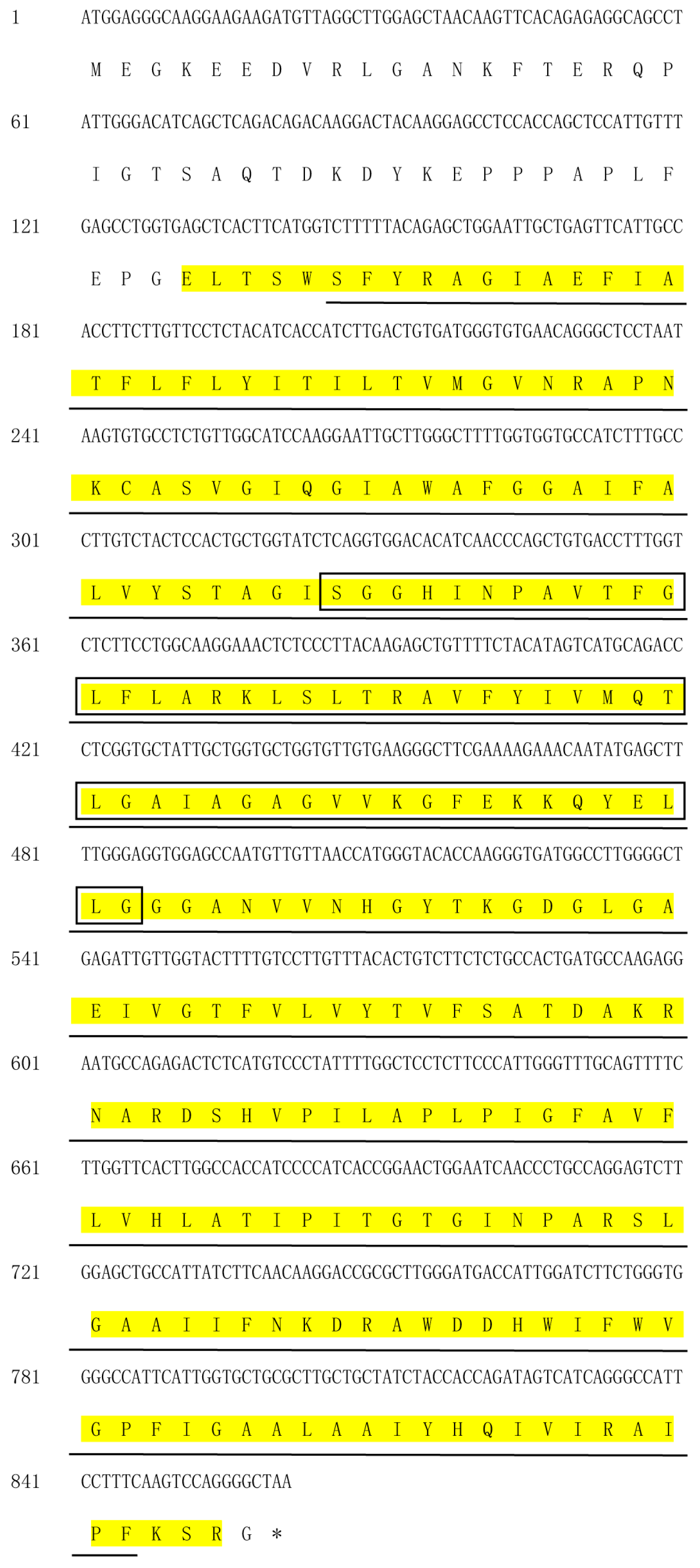
3.2. Isolation and Characterization of PaPIP1-2

An 861-bp cDNA sequence was isolated from the floral bud of P. armeniaca (Figure 2). This sequence exhibited an 86.6% similarity to the open reading frame (ORF) of P. armeniaca PaPIP1-1 and was designated as PaPIP1-2. An analysis of evolutionary relationships and sequence comparison revealed that the cDNA sequence of PaPIP1-2 shares 70.8%–86.6% similarity with the cDNA sequences of PIPs from P. armeniaca (Figure S2, Table S3). PaPIP1-2 encodes a protein comprising 286 amino acids (Figure 2). The estimated molecular mass of this protein is 30.66 kilodaltons (kD), and it has an isoelectric point (pI) of 9.25. The protein possesses a GRAVY score of 0.338 and contains six transmembrane helices (TMHs). A characteristic NPA motif, found in most PIP subfamily members, was detected in PaPIP1-2. Additionally, PaPIP1-2 exhibited conserved residues in the ar/R filter, including phenylalanine (F) at position H2, histidine (H) at H5, threonine (T) at LE1, and arginine (R) at LE2. Furthermore, Q/E-S-A-F-W residues were observed at Froger positions, a common feature in almost all PIP subfamily members of P. armeniaca (Figure S3).
A structural analysis conducted at http://smart.embl-heidelberg.de/ (accessed on 28 April 2023) predicted the presence of typical protein domains in the PaPIP1-2 protein (Figure 2). PaPIP1-2 encompasses a conserved MIP domain between residues 44 and 273, forming transmembrane channels responsible for the transport of small carbohydrates (e.g., glycerol), CO2, NH3, water, and possibly ions via an energy-independent mechanism (E = 1.7 × 10−81, Table S4). Additionally, a transmembrane organic solute transport domain is situated between residues 49 and 282, known in plants for its capacity to transport brassinosteroid-like compounds and to act as a regulator of cell death (E = 363,762.75, Table S4). The Crp-type HTH domain spanning residues 109 to 162, approximately 70–75 amino acids long with a winged structure, functions as a DNA-binding domain and is commonly found in transcriptional modulators of the crp-fnr division. This domain participates in the regulation of enzymes related to nitrogen fixation, respiration, aromatic ring degradation, and various forms of photosynthesis (E = 1562.48, Table S4). These findings suggest that the PaPIP1-2 protein may be involved in critical biological processes, including signal transduction, transcriptional activation, and material transport, among others.

3.3. Subcellular Localization of PaPIP1-2 Protein

The prediction of PaPIP1-2 protein’s subcellular localization was performed using Plant-mPLoc and WoLF PSORT. Both predictions consistently indicated that PaPIP1-2 localizes to the plasma membrane, aligning with the typical localization pattern of most members of the PaPIP subgroup [26]. To further investigate the subcellular localization of the PaPIP1-2 gene in plant cells, we fused the PaPIP1-2 gene with the EGFP (Enhanced Green Fluorescent Protein) vector under the control of the CaMV35S promoter. Transient expression analyses of localization were conducted using A. thaliana leaf mesophyll cells. The green fluorescence of the PBI221-PaPIP1-2-EGFP recombinant protein was specifically observed in the plasma membrane (Figure 3), confirming the previously predicted subcellular localization. In contrast, A. thaliana cells expressing GFP alone exhibited green fluorescence throughout the entire cell (Figure 3). These results further corroborate the plasma membrane localization of PaPIP1-2 in plant cells.

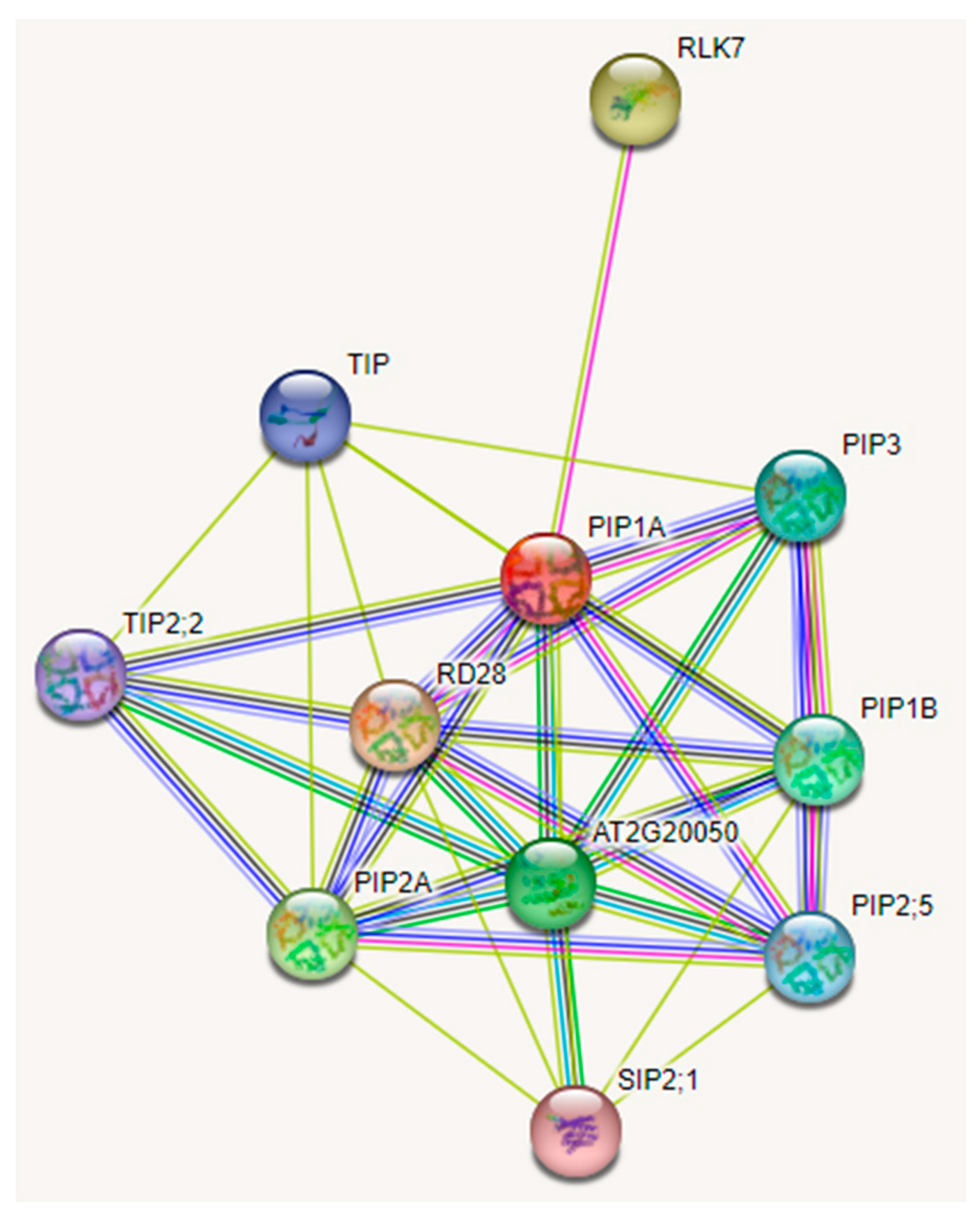
3.4. Protein–Protein Interaction Network of PaPIP1-2

To gain deeper insights into the function of PaPIP1-2 and its interactions with other proteins, a protein–protein interaction network was constructed. This network allowed us to analyze potential functional partners of PaPIP1-2. Several PIP and TIP subfamily proteins interacted with PaPIP1-2, including PIP2A, PIP1B, PIP3, PIP2;5, TIP, and TIP2;2. Notably, PIP and TIP proteins have been identified in P. armeniaca, and PaPIP1-3 and PaTIP1-1 overexpressing (OE) Arabidopsis, where they enhance cold tolerance to cold stress [26]. Furthermore, the AT2G20050 protein, exhibiting homology to protein phosphatase 2C, has been observed to undergo downregulation in Arabidopsis, reinforcing freezing resistance within transgenic plants [27]. Moreover, the leucine-rich receptor-like protein kinase family protein RLK7 is involved in the control of germination speed, with its homologous gene participating in cold resistance [28]. These potential protein interactions may indicate that PaPIP1-2 collaborates with genes related to germination and cold tolerance, playing a crucial role in flower bud dormancy and cold resistance (Figure 4).

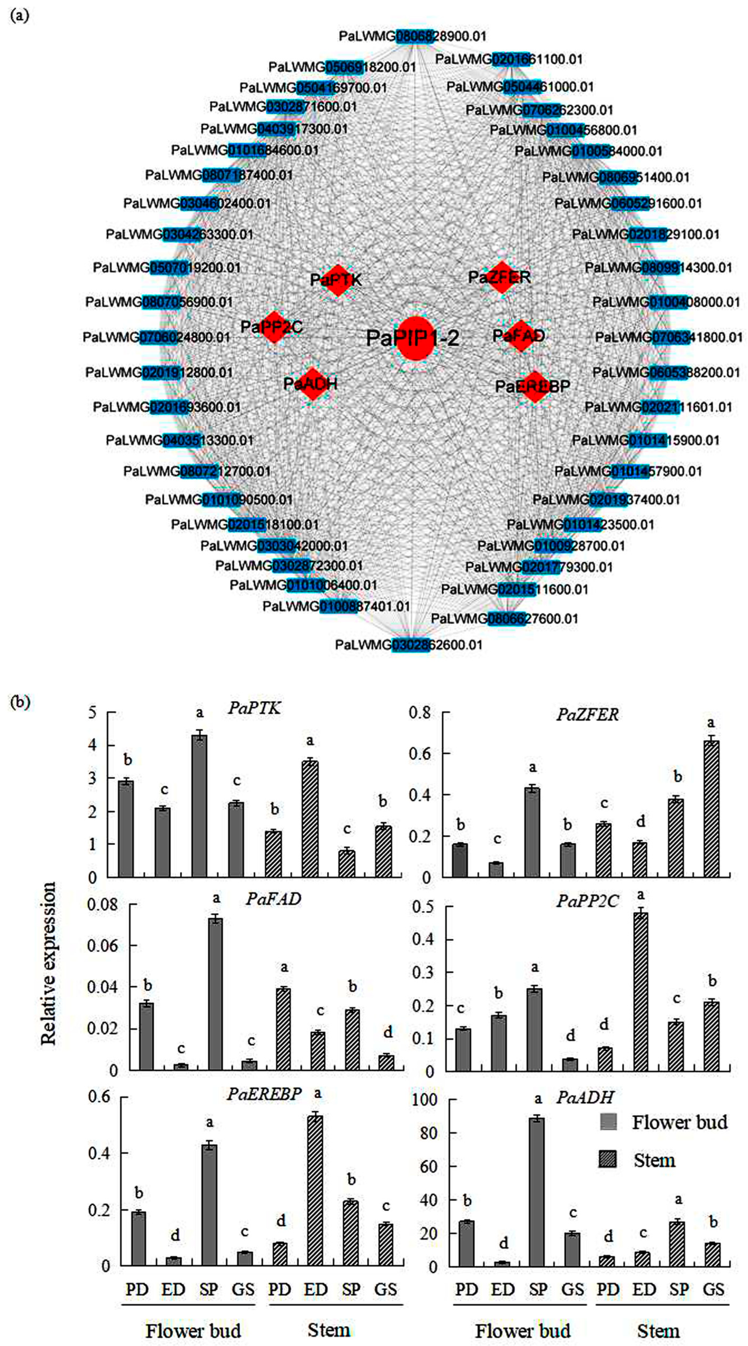
3.5. PaPIP1-2 Co-Expression Network in P. armeniaca

To elucidate the role of PaPIP1-2 in the dormancy and germination periods of P. armeniaca, a co-expression network was constructed (Figure 5a). This network revealed 50 genes strongly associated with PaPIP1-2 (co-expression coefficient > 0.995) (Table S5). Among these genes, six (12%) were related to cold tolerance (Table S6) [29,30,31,32]. Specifically, these six genes were protein tyrosine kinase (PaPTK), zinc finger protein (PaZFER), fatty acid desaturase family gene (PaFAD), protein phosphatase 2C (PaPP2C), ethylene-responsive element-binding protein (PaEREBP), and alcohol dehydrogenase protein (PaADH), displaying notable co-expression with PaPIP1-2 across various tissues. This suggests the potential involvement of these six proteins in conferring resilience against low-temperature stress to P. armeniaca.
To further analyze the network regulated by key PaPIP1-2, we examined the expression levels of the six genes associated with cold resistance within the co-expression network of PaPIP1-2. These genes were evaluated in the ED, PD, GS, and SP stages of flower buds and stems (Figure 5b). Notably, only PaPP2C exhibited conspicuously consistent expression profiles alongside PaPIP1-2 throughout all four scrutinized phases. Additional gene pairs, namely PaPTK and PaPIP1-2, PaFAD and PaPIP1-2, PaEREBP and PaPIP1-2, PaADH and PaPIP1-2, as well as PaZFER and PaPIP1-2, showcased largely similar expression trends in tandem with PaPIP1-2 across all four examined phases, with the exception being that the expressions of PaPTK, PaFAD, PaEREBP, PaADH, and PaZFER genes had declined during the ED phase. These findings suggest a coordinated relationship between PaPIP1-2 and these cold resistance-associated genes during different developmental stages of flower buds and stems. The analysis of gene expression patterns has proven valuable in identifying co-expressed genes and understanding their functional interactions.

3.6. The PaPIP1-2 Gene Conferred Cold Resistance to Yeast

To investigate the impact of PaPIP1-2 expression on low-temperature tolerance, an experiment was conducted using yeast strains transformed with either the pGAPZA empty vector, the pGAPZA::PaPIP1-2 (Figure S4), the pGAPZA::PaPIP1-1, or the pGAPZA::PaPIP2-3 recombinant expression vectors. These transformed yeast strains were subjected to low-temperature treatment by incubating them in SC-U medium in both solid and liquid forms, supplemented with 2% galactose. Yeast cells carrying the pGAPZA::PaPIP1-2 recombinant expression vector on solid SC-U media supplemented with sorbitol (2 M) after exposure to −20 °C showed significantly enhanced growth compared with cells transformed with the control empty vector.
Furthermore, the yeast colonies carrying the PaPIP1-2 gene exhibited more favorable growth compared with those carrying the empty vector strains (Figure 6a). In the 2 M liquid SC-U sorbitol medium, the average OD value for the three strains with empty vectors was 2.33. In contrast, the yeast strains with PaPIP1-2 displayed an average OD value of 2.55. After the application of low-temperature stress, the average OD values were observed as 1.99 and 2.54 for the empty vector strains and the PaPIP1-2 strains, respectively (Figure 6b). These results clearly demonstrate that the presence of the PaPIP1-2 gene positively enhances the cold resistance of yeast cells.

3.7. PaPIP1-2 Was Transformed in A. thaliana and Conferred Low-Temperature Tolerance

To investigate the specific role of the PaPIP1-2 gene in response to low-temperature stress, PaPIP1-2 was individually introduced into A. thaliana plants. These transgenic Arabidopsis plants overexpressing PaPIP1-2, along with the wild-type Arabidopsis plants, were subjected to various temperature treatments within growth chambers. Following a 10-day treatment period, noticeable distinctions emerged between the two groups. The wild-type plants displayed yellow leaves, signifying their susceptibility to low-temperature stress, whereas the PaPIP1-2-overexpressing (OE) Arabidopsis plants exhibited enhanced growth and resilience to low-temperature stress (Figure 7a). To validate the efficacy of PaPIP1-2 overexpression, qRT-PCR was conducted on two independent PaPIP1-2 transgenic lines (PIPOE-1 and PIPOE-2) after exposure to low cold stress. Both lines displayed elevated gene expression levels compared with untreated transgenic lines, confirming successful PaPIP1-2 overexpression (Figure 7b).
The activities of SOD, the primary ROS antioxidant enzyme in plants, were further evaluated in PaPIP1-2-OE plants. The PIPOE-1 and PIPOE-2 lines exhibited markedly higher SOD activity than the wild-type line (Figure 7c). The cold resistance physiological characteristics of the PaPIP1-2-transformed plants were further analyzed by assessing the MDA (malondialdehyde) and proline contents following cold stress. The MDA content was higher in the leaves of cold-stressed wild-type plants compared with PaPIP1-2 transgenic plants, indicating more significant plasma membrane damage in the wild-type plants (Figure 7e). Conversely, the proline content was significantly higher in the PaPIP1-2-transformed plants than in the wild-type plants under low-temperature stress, indicating the adaptability of PaPIP1-2 transgenic plants to the low-temperature environment (Figure 7d). This transgenic study underscores the association between the PaPIP1-2 gene and cold resistance in plants, demonstrating that improved PaPIP1-2 expression in transgenic A. thaliana could enhance the physiological activity of antioxidant enzymes and the low-temperature resistance of these plants.

4. Discussion

AQPs exhibit functional diversity and are widely distributed among various plant species. Among them, the plasma membrane intrinsic protein (PIP) subgroups play vital roles in ion transport, seed germination, root growth, flowering promotion, and abiotic stress tolerance, such as tolerance to salt, cold, or drought [5,15]. The transport activity and substrate selection of AQPs are influenced by NPA motifs, Froger’s positions, and the ar/R selectivity filter. In PaPIP proteins, typical Q/E-S-A-F-W residues at Froger’s positions, NPA motifs, and F-H-T-R residues in the ar/R selectivity filter are highly conserved, similar to PIP subfamilies in other plants like H. brasiliensis [33] and chickpea (C. arietinum L.) [34]. These typical characteristics are believed to play crucial roles in plant photosynthesis, the regulation of root and leaf hydraulics, and the facilitation of CO2 diffusion [35]. Based on these previous findings, it can be inferred that the homologous PIPs of P. armeniaca likely share similar functions in controlling vegetal development and responding to environmental stresses. These functions may involve participation in photosynthetic processes, plant hydraulics, as well as the transport of water and solutes.
Plant-mPLoc and WoLF PSORT predictions concurred that the PaPIP1-2 protein is located in the plasma membrane. This prediction aligns with experimental evidence from other plant species like flax, banana, and others [2,36]. However, Chaumont et al. (2000) [37] reported that the expression of the maize PIP gene ZmPIP1-2 can be localized both in the plasma membrane and the endoplasmic reticulum. In our experiment, the green fluorescence of PBI121-PaPIP1-2-GFP was observed exclusively in the plasma membrane of A. thaliana cells. The membrane localization of the PaPIP1-2 protein is consistent with the expression pattern of PIP genes in other plant species, such as chickpea and castor bean [34,38].
AQP genes are expressed across multiple plant organs, and their expression patterns in specific tissues or stages of development are closely associated with distinct developmental stages [39]. The expression profiles of PaPIP1-2 in diverse tissues (flower buds and stems) and under different stress conditions provide valuable insights into their molecular functions. Through qRT-PCR experiments, it was observed that PaPIP1-2 was expressed in all examined tissues and dormancy periods. However, there were significant variations in gene expression levels among different tissues (Figure 5), indicating potential functional differences of PaPIP1-2 across these tissues. Particularly, during the sprouting (SP) period, the mRNA levels of PaPIP1-2 were found to be high, suggesting its involvement in morphological growth recovery rather than the release of (endo)dormancy. Additionally, PaPIP1-2 showed higher expression in stems during the PD period compared with other dormancy and sprouting periods, suggesting its role in improving the substances and the energy loading capacity required for bud progress and germination.
In the co-expression network of PaPIP1-2, comparable expression profiles were observed among several gene pairs across the dormancy and sprouting phases. Examples of such gene pairs include PaPTK and PaPIP1-2, PaPP2C and PaPIP1-2, PaEREBP and PaPIP1-2, and PaZFER and PaPIP1-2. These conspicuously analogous expression patterns suggest a potential collaboration between PaPIP1-2 and pivotal genes linked to cold stress response, floral bud dormancy, and bud germination within P. armeniaca. For instance, in a study by Zhu et al. (2021) [32], 68 AP2/ERF genes were detected in dormant Chinese cherry floral buds, and several PpcAP2/ERF transcription factors, homologous to PaEREBP, were found to be involved in the transition of flower bud dormancy. Moreover, a single zinc-finger protein, sharing homology with the gene PaZFER, has been identified as a mediator of seed dormancy controlled by ABA in Arabidopsis [40]. Additionally, Kim et al. (2013) [41] demonstrated that PP2C activates gibberellic acid signaling, inhibits ABA signaling, and promotes the release of seed dormancy in Arabidopsis.
AQPs play a crucial role in facilitating water movement among cells, enhancing their ability to regulate bud dormancy in response to abiotic stressors by improving water transport [42]. Notably, a previous study demonstrated that transgenic bananas overexpressing MusaPIP1-2 displayed enhanced tolerance to low-temperature environments [11]. Similarly, in A. thaliana, the genes AtPIP2-5 and AtPIP2-6 are classified as cold-induced genes, meaning they are upregulated in response to low-temperature stress [43]. The overexpression of AtPIP1-4 or AtPIP2-5 in this species has been shown to confer improved tolerance to cold conditions compared with wild-type plants [44]. During our investigation, we observed elevated expression levels of PaPIP1-2 during the sprouting period (SP) in P. armeniaca flower buds. Remarkably, when subjected to low-temperature stress, yeast cells expressing PaPIP1-2 displayed significantly improved growth compared with cells transformed with the empty vector on SG-U medium, as shown in Figure 6. Additionally, our findings revealed that transgenic Arabidopsis plants overexpressing PaPIP1-2 exhibited enhanced tolerance to low temperatures. This improvement was attributed to a reduction in malondialdehyde (MDA) content and increased activities of superoxide dismutase (SOD) and cold resistance genes AtPUB26, AtBTF3L, AtEBF1-1, and AtRAV1 [45,46,47,48], as depicted in Figure 7. These results strongly suggest that PaPIP1-2 plays a significant role as a gene associated with low-temperature resistance. Its involvement in maintaining normal growth and conferring resistance under low-temperature conditions appears to be of considerable importance.

5. Conclusions

This study has significantly advanced our understanding of the functional characteristics of the PaPIP1-2 gene, a member of the PIP subgroup within the P. armeniaca AQP gene family. The expression of PaPIP1-2 exhibits dynamic changes during the dormancy and sprouting periods of flower buds, indicating its differential regulation in response to these developmental stages. Subcellular localization experiments confirmed that PaPIP1-2 is specifically present in the plasma membrane, which aligns with the typical localization pattern of PIP proteins in various plant species. Furthermore, a protein–protein interaction prediction analysis revealed the interactions between PaPIP1-2 and several PIP and TIP subfamily proteins, suggesting its potential involvement in various physiological processes. The co-expression network analysis demonstrated consistent co-expression between PaPIP1-2 and six low-temperature resistance genes. These genes displayed stable expression profiles across different developmental stages of P. armeniaca flower buds, suggesting their coordinated roles in response to cold stress and bud development. Experimental evidence from yeast and Arabidopsis studies indicated that PaPIP1-2 enhances resistance to low-temperature stress. The overexpression of PaPIP1-2 in Arabidopsis led to increased activities of antioxidative enzymes and improved tolerance to low temperatures in the transgenic plants. This research has identified a promising candidate gene closely associated with cold tolerance and the dormancy and sprouting phases in P. armeniaca flower buds. These findings provide valuable insights into the molecular mechanisms underlying cold tolerance and flower bud development, with potential applications in agriculture and horticulture for the controlled manipulation of flowering periods and the mitigation of frost-induced damage.

Supplementary Materials

The following supporting information can be downloaded at https://www.mdpi.com/article/10.3390/f14122306/s1, Table S1: Primers used in the qRT-PCR analysis of the PaPIP1-2 gene, and cold resistance genes co-expressed with PaPIP1-2 gene; Table S2: CaMV 35S promoter sequence of pBI121 vector; Table S3: Complete coding sequences and corresponding amino acid sequences of the PIP subfamily of AQP genes of P. armeniaca; Table S4: Structural components of PaPIP1-2 protein; Table S5: Predicted functional partners of PaPIP1-2; Table S6: Genes related to cold resistance in the PaPIP1-2 co-expression network [27,29,30,31,32,49]; Figure S1: Expression profiles of PaPIP1-2 and other PaAQPs genes at different developmental stages of flower buds; Figure S2: Phylogenetic relationship analysis of the PIP subfamily of Aquaporin (AQP) genes within the genomes of Prunus armeniaca (apricot); Figure S3: Protein sequence alignment of PaPIPs (Plasma membrane Intrinsic Proteins) identified in P. armeniaca; Figure S4: Vector map of pGAPZA.

Author Contributions

S.L. and Y.X. conceived and designed the research. S.L., F.L. and F.W. performed all the experiments. S.W. and H.G. contributed reagents/materials/analysis tools. G.Z. and H.Y. analyzed the data. S.L. wrote the paper. All authors have read and agreed to the published version of the manuscript.

Funding

This work was supported by the Fundamental Research Funds of CAF (CAFYBB2018MA003) and the National Natural Science Foundation of China (31770705 and 31400570).

Data Availability Statement

All relevant data can be found within the manuscript and its uploaded Supplementary Materials.

Conflicts of Interest

The authors declare no conflict of interest.

References

  1. Maurel, C.; Santoni, V.; Luu, D.T.; Wudick, M.M.; Verdoucq, L. The cellular dynamics of plant aquaporin expression and functions. Curr. Opin. Plant Biol. 2009, 12, 690–698. [Google Scholar] [CrossRef]
  2. Maurel, C.; Verdoucq, L.; Luu, D.T.; Santoni, V. Plant aquaporins: Membrane channels with multiple integrated functions. Annu. Rev. Plant Biol. 2008, 59, 595–624. [Google Scholar] [CrossRef]
  3. Mitani-Ueno, N.; Yamaji, N.; Zhao, F.J.; Ma, J.F. The aromatic/arginine selectivity filter of NIP aquaporins plays a critical role in substrate selectivity for silicon, boron, and arsenic. J. Exp. Bot. 2011, 62, 4391–4398. [Google Scholar] [CrossRef]
  4. Anderberg, H.I.; Kjellbom, P.; Johanson, U. Annotation of Selaginella moellendorffii Major Intrinsic Proteins and the Evolution of the Protein Family in Terrestrial Plants. Front. Plant Sci. 2012, 3, 33. [Google Scholar] [CrossRef]
  5. Yang, X.; Li, J.; Ji, C.; Wei, Z.; Zhao, T.; Pang, Q. Overexpression of an aquaporin gene EsPIP1;4 enhances abiotic stress tolerance and promotes flowering in Arabidopsis thaliana. Plant Physiol. Biochem. 2022, 193, 25–35. [Google Scholar] [CrossRef]
  6. Kayum, M.A.; Park, J.I.; Nath, U.K.; Biswas, M.K.; Kim, H.T.; Nou, I.S. Genome-wide expression profiling of aquaporin genes confer responses to abiotic and biotic stresses in Brassica rapa. BMC Plant Biol. 2017, 17, 23. [Google Scholar] [CrossRef]
  7. Bansal, A.; Sankararamakrishnan, R. Homology modeling of major intrinsic proteins in rice, maize and Arabidopsis: Comparative analysis of transmembrane helix association and aromatic/arginine selectivity filters. BMC Struct. Biol. 2007, 7, 27. [Google Scholar] [CrossRef]
  8. Hove, R.M.; Bhave, M. Plant aquaporins with non-aqua functions: Deciphering the signature sequences. Plant Mol. Biol. 2011, 75, 413–430. [Google Scholar] [CrossRef]
  9. Froger, A.; Tallur, B.; Thomas, D.; Delamarche, C. Prediction of functional residues in water channels and related proteins. Protein Sci. 1998, 7, 1458–1468. [Google Scholar] [CrossRef]
  10. Rahman, A.; Kawamura, Y.; Maeshima, M.; Rahman, A.; Uemura, M. Plasma Membrane Aquaporin Members PIPs Act in Concert to Regulate Cold Acclimation and Freezing Tolerance Responses in Arabidopsis thaliana. Plant Cell Physiol. 2020, 61, 787–802. [Google Scholar] [CrossRef]
  11. Shekhawat, U.K.S.; Ganapathi, T.R. MusaWRKY71 Overexpression in Banana Plants Leads to Altered Abiotic and Biotic Stress Responses. PLoS ONE 2013, 8, e75506. [Google Scholar] [CrossRef]
  12. Zhou, S.; Hu, W.; Deng, X.; Ma, Z.; Chen, L.; Huang, C.; Wang, C.; Wang, J.; He, Y.; Yang, G.; et al. Overexpression of the wheat aquaporin gene, TaAQP7, enhances drought tolerance in transgenic tobacco. PLoS ONE 2012, 7, e52439. [Google Scholar] [CrossRef]
  13. Qu, J.; Xu, S.; Zhang, Z.; Chen, G.; Zhong, Y.; Liu, L.; Zhang, R.; Xue, J.; Guo, D. Evolutionary, structural and expression analysis of core genes involved in starch synthesis. Sci. Rep. 2018, 8, 12736. [Google Scholar] [CrossRef]
  14. Gasteiger, E.; Gattiker, A.; Hoogland, C.; Ivanyi, I.; Appel, R.D.; Bairoch, A. ExPASy: The proteomics server for in-depth protein knowledge and analysis. Nucleic Acids Res. 2003, 31, 3784–3788. [Google Scholar] [CrossRef]
  15. Kong, W.; Yang, S.; Wang, Y.; Bendahmane, M.; Fu, X. Genome-wide identification and characterization of aquaporin gene family in Beta vulgaris. PeerJ 2017, 5, e3747. [Google Scholar] [CrossRef]
  16. Chou, K.-C.; Shen, H.-B. Plant-mPLoc: A Top-Down Strategy to Augment the Power for Predicting Plant Protein Subcellular Localization. PLoS ONE 2010, 5, e11335. [Google Scholar] [CrossRef]
  17. Horton, P.; Park, K.-J.; Obayashi, T.; Fujita, N.; Harada, H.; Adams-Collier, C.J.; Nakai, K. WoLF PSORT: Protein localization predictor. Nucleic Acids Res. 2007, 35, W585–W587. [Google Scholar] [CrossRef]
  18. Sheen, J. Signal Transduction in Maize and Arabidopsis Mesophyll Protoplasts. Plant Physiol. 2001, 127, 1466–1475. [Google Scholar] [CrossRef]
  19. Nelson, B.K.; Cai, X.; Nebenführ, A. A multicolored set of in vivo organelle markers for co-localization studies in Arabidopsis and other plants. Plant J. 2007, 51, 1126–1136. [Google Scholar] [CrossRef]
  20. Song, C.; Yin, M.; Jiang, Z.; Li, H.; Wuyun, T.; Song, J. Relationship between flower bud differentiation and change of endogenous hormone of ‘Youyi’ during dormancy period. J. Northwest A F Univ. Nat. Sci. Ed. 2017, 45, 170–184. [Google Scholar]
  21. Livak, K.J.; Schmittgen, T.D. Analysis of relative gene expression data using real-time quantitative PCR and the 2(-Delta Delta C(T)) Method. Methods 2001, 25, 402–408. [Google Scholar] [CrossRef]
  22. Liu, Y.; Xie, L.; Liang, X.; Zhang, S. CpLEA5, the Late Embryogenesis Abundant protein gene from Chimonanthus praecox, possesses low temperature and osmotic resistances in Prokaryote and Eukaryotes. Int. J. Mol. Sci. 2015, 16, 26978–26990. [Google Scholar] [CrossRef]
  23. Clough, S.J.; Bent, A.F. Floral dip: A simplified method for Agrobacterium-mediated transformation of Arabidopsis thaliana. Plant J. 1998, 16, 735–743. [Google Scholar] [CrossRef]
  24. Wang, L.; Xu, C.; Wang, C.; Wang, Y. Characterization of a eukaryotic translation initiation factor 5A homolog from Tamarix androssowii involved in plant abiotic stress tolerance. BMC Plant Biol. 2012, 12, 118. [Google Scholar] [CrossRef]
  25. Bates, L.S.; Waldren, R.P.; Teare, I.D. Rapid determination of free proline for water-stress studies. Plant Soil. 1973, 39, 205–207. [Google Scholar] [CrossRef]
  26. Li, S.; Wang, L.; Zhang, Y.; Zhu, G.; Zhu, X.; Xia, Y.; Li, J.; Gao, X.; Wang, S.; Zhang, J.; et al. Genome-Wide identification and function of aquaporin genes during dormancy and sprouting periods of Kernel-Using Apricot (Prunus armeniaca L.). Front. Plant Sci. 2021, 12, 690040. [Google Scholar] [CrossRef]
  27. Tähtiharju, S.; Palva, T. Antisense inhibition of protein phosphatase 2C accelerates cold acclimation in Arabidopsis thaliana. Plant J. 2001, 26, 461–470. [Google Scholar] [CrossRef]
  28. Chen, X.; Ding, Y.; Yang, Y.; Song, C.; Wang, B.; Yang, S.; Guo, Y.; Gong, Z. Protein kinases in plant responses to drought, salt, and cold stress. J. Integr. Plant Biol. 2021, 63, 53–78. [Google Scholar] [CrossRef]
  29. Jung, Y.J.; Lee, I.H.; Nou, I.S.; Lee, K.D.; Rashotte, A.M.; Kang, K.K. BrRZFP1 a Brassica rapa C3HC4-type RING zinc finger protein involved in cold, salt and dehydration stress. Plant Biol. 2013, 15, 274–283. [Google Scholar] [CrossRef]
  30. Jingu, D.; Iino, M.; Kawasaki, J.; Urano, E.; Kusakari, S.; Hayashi, Y.; Matozaki, T.; Ohnishi, H. Protein tyrosine phosphatase Shp2 positively regulates cold stress-induced tyrosine phosphorylation of SIRPα in neurons. Biochem. Biophys. Res. Commun. 2021, 569, 72–78. [Google Scholar] [CrossRef]
  31. Khodakovskaya, M.; McAvoy, R.; Peters, J.; Wu, H.; Li, Y. Enhanced cold tolerance in transgenic tobacco expressing a chloroplast omega-3 fatty acid desaturase gene under the control of a cold-inducible promoter. Planta 2006, 223, 1090–1100. [Google Scholar] [CrossRef]
  32. Zhu, Y.; Liu, X.; Gao, Y.; Li, K.; Guo, W. Transcriptome-based identification of AP2/ERF family genes and their cold-regulated expression during the dormancy phase transition of Chinese cherry flower buds. Sci. Hortic. 2021, 275, 109666. [Google Scholar] [CrossRef]
  33. Murata, K.; Mitsuoka, K.; Hirai, T.; Walz, T.; Agre, P.; Heymann, J.B.; Engel, A.; Fujiyoshi, Y. Structural determinants of water permeation through aquaporin-1. Nature 2000, 407, 599–605. [Google Scholar] [CrossRef]
  34. Deokar, A.A.; Tar’an, B. Genome-Wide Analysis of the Aquaporin Gene Family in Chickpea (Cicer arietinum L.). Front. Plant Sci. 2016, 7, 1802. [Google Scholar] [CrossRef]
  35. Gupta, A.B.; Sankararamakrishnan, R. Genome-wide analysis of major intrinsic proteins in the tree plant Populus trichocarpa: Characterization of XIP subfamily of aquaporins from evolutionary perspective. BMC Plant Biol. 2009, 9, 134. [Google Scholar] [CrossRef]
  36. Shivaraj, S.M.; Deshmukh, R.K.; Rai, R.; Bélanger, R.; Agrawal, P.K.; Dash, P.K. Genome-wide identification, characterization, and expression profile of aquaporin gene family in flax (Linum usitatissimum). Sci. Rep. 2017, 7, 46137. [Google Scholar] [CrossRef]
  37. Chaumont, F.; Barrieu, F.; Jung, R.; Chrispeels, M.J. Plasma membrane intrinsic proteins from maize cluster in two sequence subgroups with differential aquaporin activity. Plant Physiol. 2000, 122, 1025–1034. [Google Scholar] [CrossRef]
  38. Zou, Z.; Gong, J.; Huang, Q.; Mo, Y.; Yang, L.; Xie, G. Gene structures, evolution, classification and expression profiles of the Aquaporin gene family in Castor Bean (Ricinus communis L.). PLoS ONE 2015, 10, e0141022. [Google Scholar] [CrossRef]
  39. Di Giorgio, J.A.; Bienert, G.P.; Ayub, N.D.; Yaneff, A.; Barberini, M.L.; Mecchia, M.A.; Amodeo, G.; Soto, G.C.; Muschietti, J.P. Pollen-specific Aquaporins NIP4;1 and NIP4;2 are required for pollen development and pollination in Arabidopsis thaliana. Plant Cell 2016, 28, 1053–1077. [Google Scholar] [CrossRef]
  40. He, Y.; Gan, S. A novel zinc-finger protein with a proline-rich domain mediates ABA-regulated seed dormancy in Arabidopsis. Plant Mol. Biol. 2004, 54, 1–9. [Google Scholar] [CrossRef]
  41. Kim, W.; Lee, Y.; Park, J.; Lee, N.; Choi, G. HONSU, a protein phosphatase 2C, regulates seed dormancy by inhibiting ABA signaling in Arabidopsis. Plant Cell Physiol. 2013, 54, 555–572. [Google Scholar] [CrossRef]
  42. Yooyongwech, S.; Horigane, A.K.; Yoshida, M.; Yamaguchi, M.; Sekozawa, Y.; Sugaya, S.; Gemma, H. Changes in aquaporin gene expression and magnetic resonance imaging of water status in peach tree flower buds during dormancy. Physiol. Plant. 2008, 134, 522–533. [Google Scholar] [CrossRef]
  43. Alexandersson, E.; Fraysse, L.; Sjövall-Larsen, S.; Gustavsson, S.; Fellert, M.; Karlsson, M.; Johanson, U.; Kjellbom, P. Whole gene family expression and drought stress regulation of aquaporins. Plant Mol. Biol. 2005, 59, 469–484. [Google Scholar] [CrossRef]
  44. Afzal, Z.; Howton, T.C.; Sun, Y.; Mukhtar, M.S. The Roles of Aquaporins in Plant Stress Responses. J. Dev. Biol. 2016, 4, 9. [Google Scholar] [CrossRef]
  45. Ding, Y.; Jia, Y.; Shi, Y.; Zhang, X.; Song, C.; Gong, Z.; Yang, S. OST1-mediated BTF3L phosphorylation positively regulates CBFs during plant cold responses. EMBO J. 2018, 37, e98228. [Google Scholar] [CrossRef]
  46. Jiang, B.; Shi, Y.; Zhang, X.; Xin, X.; Qi, L.; Guo, H.; Li, J.; Yang, S. PIF3 is a negative regulator of the CBF pathway and freezing tolerance in Arabidopsis. Proc. Natl. Acad. Sci. USA 2017, 114, E6695–E6702. [Google Scholar] [CrossRef]
  47. Wang, X.; Ding, Y.; Li, Z.; Shi, Y.; Wang, J.; Hua, J.; Gong, Z.; Zhou, J.M.; Yang, S. PUB25 and PUB26 promote plant freezing tolerance by degrading the cold signaling negative regulator MYB15. Dev. Cell 2019, 51, 222–235. [Google Scholar] [CrossRef]
  48. Ren, C.; Huayang, L.; Wang, Z.; Dai, Z.; Lecourieux, F.; Kuang, F.; Xin, H.; Li, S.; Liang, Z. Characterization of chromatin accessibility and gene expression upon cold stress reveals the transcription factor RAV1 functions in cold response in Vitis amurensis. Plant Cell Physiol. 2021, 62, 1615–1629. [Google Scholar] [CrossRef]
  49. Dolferus, R.; Jacobs, M.; Dennis, P. Differential interactions of promoter elements in stress responses of the Arabidopsis Adh gene. Plant Physiol. 1994, 105, 1075–1087. [Google Scholar] [CrossRef]
Figure 1. (a) Average FPKM values of the PaPIP1-2 gene during the ED, SP, and GS periods of P. armeniaca. The average FPKM value of the PaPIP1-2 gene is based on RNA-seq data. (b) Transcript levels of PaPIP1-2 in flower buds and stems during the PD, ED, SP, and GS periods of P. armeniaca. Different lowercase letters (a, b, c, d) on the bars indicate significant differences at p < 0.05, determined by ANOVA.
Forests 14 02306 g001
Figure 2. Analysis of the PaPIP1-2 sequence, including the open reading frame and deduced amino acid sequences. The Solute_trans_a domain of PaPIP1-2 is indicated by a thin black underline, and the SCOP domain d1j4na_ is highlighted in yellow. The HTH_CRP motif is enclosed in a box for clear identification. * denotes protein termination translation.
Forests 14 02306 g002
Figure 3. Subcellular localization of the PaPIP1-2 protein in the plasma membrane. Bright-field images (Bright) are provided. Nuclei are counterstained with 4′,6-diamidino-2-phenylindole (DAPI), a nuclear marker, as shown in the DAPI images (blue). The plasma membrane is specifically targeted using FM4-64FX dye, and its images are provided. The Merge images represent the overlap of GFP (green) and FM4-64FX (red) fluorescence, highlighting the co-localization of the PaPIP1-2::GFP fusion protein and the plasma membrane-specific dye. Row 1 displays A. thaliana protoplasts expressing GFP alone, serving as a control group. Row 2 exhibits protoplasts expressing the PaPIP1-2::GFP fusion protein with FM4-64 dye, enabling the visualization of PaPIP1-2 localization in the plasma membrane. The scale bars in the images represent a length of 5 μm.
Forests 14 02306 g003
Figure 4. Predicted interaction network of PaPIP1-2 proteins based on interactions observed among their orthologs in Arabidopsis thaliana. The network was generated using data from the STRING database. In the figure, red circles represent the queried protein, and other circles in the network represent interacting proteins. Annotations for the predicted interacting proteins were obtained from the UniProtKB database (https://www.uniprot.org/ (accessed on 29 April 2023)). The thickness of the lines connecting the circles in the network indicates the strength of data support for the interactions.
Forests 14 02306 g004
Figure 5. (a) Co-expression network of PaPIP1-2 and its associated genes. The network includes 50 genes co-expressed with PaPIP1-2, with 6 genes involved in cold resistance represented by red nodes. The central red node is PaPIP1-2. (b) Transcript levels of the 6 cold resistance genes in flower buds and stems during the PD, ED, SP, and GS stages of P. armeniaca. Expression levels are normalized to PaElf (elongation factor-1α). Each bar represents the mean ± SD of three independent experiments. Different lowercase letters on the bars indicate significant differences at p < 0.05, determined by ANOVA.
Forests 14 02306 g005
Figure 6. Impact of PaPIP1-2 overexpression on cold acclimation and tolerance in yeast. (a) Growth of GS115 yeast strain after transformation with different constructs. Yeast strains were transformed with the empty vector (pGAPZA), pGAPZA containing PaPIP1-2, or pGAPZA alone (control). (b) OD600 values of yeast transformants under cold stress conditions. The data are means ± SDs of three replicates. “n.s.” indicates no significant difference between the two sample groups, while different letters indicate significant differences at p < 0.05 as determined by one-way ANOVA.
Forests 14 02306 g006
Figure 7. Images of PaPIP1-2-OE and wild-type Arabidopsis plants grown in growth chambers at different temperatures: 22 °C, 16 °C, and 16 °C for 16 h followed by 4 °C for 8 h (a). (b) Expression levels of PaPIP1-2 quantified using qRT-PCR in two distinct PaPIP1-2 transgenic lines (PIP1-2 OE-1 and PIP1-2 OE-2). (c) Superoxide dismutase (SOD) activity after 10 days of treatment. (d) Proline content after 10 days of treatment. (e) Malondialdehyde (MDA) content after 10 days of treatment. Expression levels of cold resistance genes including (f) AtPUB26, (g) AtBTF3L, (h) AtEBF1, and (i) AtRAV1. Abbreviations used are PIP1-2 OE-1, PIP1-2 OE-2, and PaPIP1-2-OE transgenic lines. Actin was used as a control gene for normalization. Error bars represent standard errors (SEs) from three independent repeats. “n.s.” indicates no significant difference between the two groups of samples, while different letters represent significant differences at p < 0.05, determined by one-way ANOVA.
Forests 14 02306 g007
Disclaimer/Publisher’s Note: The statements, opinions and data contained in all publications are solely those of the individual author(s) and contributor(s) and not of MDPI and/or the editor(s). MDPI and/or the editor(s) disclaim responsibility for any injury to people or property resulting from any ideas, methods, instructions or products referred to in the content.

