Article

Alternaria alternata and Alternaria koreana, the Causal Agents of Leaf Spot in Celtis sinensis and Their Sensitivity to Fungicides

1
College of Forestry, Nanjing Forestry University, Nanjing 210037, China
2
Co-Innovation Center for Sustainable Forestry in Southern China, Nanjing Forestry University, Nanjing 210037, China
*
Author to whom correspondence should be addressed.
Forests 2023, 14(12), 2389; https://doi.org/10.3390/f14122389
Submission received: 3 November 2023 / Revised: 29 November 2023 / Accepted: 4 December 2023 / Published: 7 December 2023
(This article belongs to the Section Forest Health)

Abstract

:
Celtis sinensis is a highly versatile species that is commonly cultivated in the southern regions of China. In June 2022, leaf spot disease was detected in C. sinensis in Nanjing, Jiangsu, China. Based on morphological characteristics, three isolates were determined to be of the Alternaria species. A phylogenetic analysis of combined ITS, GAPDH, TEF1-α, RPB2, and Alt a 1 sequences identified the three isolates we obtained as Alternaria alternata and A. koreana. We determined the pathogenicity of A. alternata and A. koreana in C. sinensis leaves using inoculation through in vivo experiments. Symptoms of inoculation onset in indoor pots were in accordance with those observed in the open country. A. alternata and A. koreana can grow at 15–35 °C, with the best growth occurring at 25 °C. The findings from fungicide susceptibility experiments demonstrated that A. alternata and A. koreana were the most sensitive to prochloraz, which could offer an effective approach for future prevention and control measures against A. alternata and A. koreana. This study provides the initial step toward further research on A. alternata and A. koreana as pathogens of C. sinensis and provides the stage for future control strategy development.

1. Introduction

Celtis sinensis is an important member of the Ulmaceae family [1] that is native to China, Japan, and Korea [2]. Mainly concentrated in central and eastern China, with straight trunks and beautiful tree shapes, it is a common street and landscaping tree species and a valuable resistant tree species that can adsorb toxic gases, such as sulfur dioxide and chlorine, and has a certain degree of adsorption capacity for dust [3]. C. sinensis demonstrates remarkable resilience, as it is able to withstand both dry conditions and moist, infertile soils. These trees possess strong adaptability to diverse environmental factors. Moreover, the branches, leaves, roots, and bark of C. sinensis hold important medicinal value and can be utilized in the treatment of burns and urticaria [4]. In addition, it has a wide range of industrial uses. The roots and branches can be used to make artificial cotton and furniture, and the fruit can also be used as raw materials for lubricant production [5]. However, in Xuanwu District, Nanjing, Jiangsu Province, a large number of cases of leaf spot and leaf curling occurred in C. sinensis, which seriously affected its ornamental value and damaged the ecological environment.
In August 2023, leaf blotch on Celtis julianae caused by A. arborescens and A. italica was discovered in Nanjing [6]. Alternaria is a widely distributed group of fungi in the natural environment and is also economically important as a dematiaceous fungus [7]. More than 95% of these species are plant parthenogenetic parasites. They can grow and multiply in low-temperature and humid environments, causing a variety of plant diseases, as well as fruit rot, which severely jeopardizes the growth of crops and plants and causes enormous economic losses while destroying natural ecosystems globally [8]. In addition, the spores of Alternaria can also release allergens [9], triggering diseases such as asthma [10] and posing a major threat to human health. The pathogenic fungi of Alternaria establish their population by colonizing the surface of seeds with mycelium or conidia [11] on diseased plant residues and in the soil. These serve as primary infection sources for the following year. The conidia of the fungi are spread and dispersed by air currents, leading to secondary infections, thus perpetuating the cycle of transmission and spread of disease.
At present, the most direct and effective method for the control of diseases caused by Alternaria is chemical control [12]. Fungicides, such as dithiocarbamates, triazoles, strobilurins, iprodione, and copper fungicides, are used in most areas for disease control [13]. Copper-based fungicides are widely used due to their long residual effectiveness and resistance to washing [14]. Methoxyacrylate, dimethomorph, and mancozeb fungicides have also been registered for the control of brown spot disease in countries such as the United States, Israel, and Spain [15]. Due to the difficulty of mixing copper-based formulations with other pesticides and the single-site action of methoxyacrylate and dimethomorph fungicides, resistant populations can quickly emerge in the field, leading to decreased or ineffective efficacy of these fungicides. Recently, nicotinamide fungicides, such as cyazofamid, have been registered in the United States for the control of this disease [16]. These fungicides can be used for preseed disinfection, as well as direct application to the soil, effectively providing preventive protection [17].
This study reveals the characteristics of Alternaria related to leaf spot in C. sinensis in Xuanwu District, Nanjing, Jiangsu Province, China. This disease has caused significant damage to the local ecosystem. This study was conducted using the following methodologies: to determine the causative agent of brown spot disease in C. sinensis using Koch’s postulates; to identify the pathogen through molecular biological and morphological identification methods; and to screen fungicides for high sensitivity to pathogen mycelium through culture-based plate phenotype experiments.

2. Materials and Methods

2.1. Sampling and Fungal Isolations

In June 2022, the disease was observed on the leaves of C. sinensis on the campus of Nanjing Forestry University, Nanjing City, Jiangsu Province (119°46′43″ E, 32°02′38″ N), and 20 diseased leaves were collected as samples from 5 infected C. sinensis plants. The diseased leaves were wiped with absorbent paper and sterile water to remove surface dust and impurities. One hundred small tissues (3 × 3 mm2) were cut from lesion margins and surface-sterilized in 75% ethanol for 30 s, followed by 5% NaClO for 90 s, rinsed in sterile water 3 times, dried on sterilized filter paper, and plated onto potato dextrose agar (PDA) for 4 days at 25 °C [18]. On the third day, fungal hyphae emerging from leaf tissue were shifted to fresh PDA [19]. The isolated strains were preliminarily classified according to their morphological characteristics and ITS sequence alignment for subsequent experiments.

2.2. Pathogenicity Tests

Healthy C. sinensis and seedlings were obtained from the laboratory of Nanjing Forestry University, and the seedlings had heights of approximately 80 cm. Before the pathogenicity experiment, the leaf surface was wiped twice with 75% alcohol and sprayed with sterile water three times. The water stains on the surface of the leaves were wiped dry with absorbent paper. The colony morphology of 100 fungal samples was assessed, and they were divided into different groups. Isolates 11 and 13 (A. alternata) and isolate 12 (A. koreana) from the group with the highest frequency of occurrence were selected for pathogenicity testing and the inoculation of leaf blades and seedlings, respectively [20]. Five detached leaves were inoculated with each isolate, wounds were created on both sides of the face of the leaf veins with a sterile needle, and a 6 mm mycelium block was placed face down on each puncture wound and removed after 24 h [21]. In addition, five leaves were inoculated with sterilized PDA agar blocks as a control. The samples were cultured for four days under environmental conditions of a temperature of 25 °C, 70–80% humidity, and 24 h of light exposure per day. Nine healthy C. sinensis seedlings were selected from each plant, 3 leaves from each a plant were inoculated with the spores of one of the 3 isolates by puncturing the leaf with a sterile needle as a wound suspension (106 conidia/mL), and the other three plantlets were inoculated with sterile water as a control group. The plantlets were covered with plastic bags and sprayed with water to maintain a high level of humidity.

2.3. Morphological Identification and Biological Characteristics

Three isolates (11, 12, and 13) were cultured on PDA medium at 25 °C for 7 days in a constant temperature incubator. Morphological identification was based on colony appearance and conidia characteristics. The morphology of conidia was determined using a Zeiss Axio Imager A2m microscope (Carl Zeiss, Jena, Germany) (n = 30).
Three isolates (11, 12, and 13) were chosen for the purpose of conducting morphological observations and biological characterization. Isolates were cultured on potato dextrose agar medium (PDA), Czapek dox agar medium (CDA), Richard medium (Richard), corn meal agar medium (CMA), oatmeal agar medium (OA), and potato saccharose agar medium (PSA) for 7 days at 25 °C, and the appearance and coloration of the colonies were documented.
In order to ascertain the isolates’ optimal growth temperature, mycelial plugs with a diameter of 6 mm were positioned on freshly prepared PDA medium, which had a diameter of 90 mm, and incubated at 5 °C, 15 °C, 20 °C, 25 °C, 30 °C, and 35 °C. The colony growth diameter was measured and recorded daily.

2.4. DNA Extraction, PCR Amplification, and Sequencing

The isolated fungi were cultured on PDA medium for 7 days, and the mature mycelium was scraped off using a sterilized surgical blade. DNA extraction was performed using the CTAB method [22]. All DNA extracts were preserved at a temperature of −20 °C for future utilization.
Polymerase chain reaction was performed on ITS [23], TEF1-α [24], GAPDH [25], RPB2 [26], and Alt a 1 [27] genes, which were sequenced using primers ITS1/4, EF1-728F/EF1-986R, GPD1/GPD2, RPB2-5F2/RPB2-7C, and Alt-for/Alt-rev, respectively. PCR was performed in a 25 µL reaction mixture containing 21 μL of Tmix, 1 μL of DNA template, 1 μL of Taq DNA polymerase, 1 μL of forward primer, and 1 μL of reverse primer (Table 1). The PCR products were sequenced at Sangon Biotech Co. Ltd. (Nanjing, China).

2.5. Phylogenetic Analyses

The phylogenetic tree was constructed using sequences from the fungal cultures obtained in the course of this research and sequences associated with Alternaria sp. that are available in GenBank (Table 2); Alternaria alternantherae was used as the outgroup. Sequence alignment analysis using MAFFT version 7 (https://mafft.cbrc.jp/alignment/software/, accessed on 1 January 2023) [28] was performed to trim sequences to ensure a high degree of sequence alignment [29]. Phylogenetic analysis was conducted through the concatenation of five loci (ITS, TEF1-α, GAPDH, RPB2, and Alt a 1) [30]. Phylogenetic tree construction was conducted based on a combination of sequence maximum likelihood (ML) and Bayesian inference (BI). The ML analysis used the GTR + F + I + G4 model, and branch stability was determined using 1000 bootstrap replicates. BI analysis used the GTR + I + G + F model, including two parallel runs of 2,000,000 generations. The resulting trees were plotted using FigTree v.1.4.3 (http://tree.bio.ed.ac.uk/software/figtree/, accessed on 1 January 2023).

2.6. Evaluation of Fungicides against A. alternata (Isolates 11, 13) and A. koreana (Isolate 12)

Fungicides were kindly provided by Mrs. Tingting Dai of Nanjing Forestry University as 96% prochloraz [31], 97% myclobutanil [32], 97% tebuconazole [33], and 98% pyraclostrobin [34]. Each fungicide was prepared as a stock solution, which was then diluted to different concentrations. The solutions were added to sterilized PDA medium at a rate of 2% to create agar plates containing the respective concentrations of the fungicide. PDA plates without the use of any fungicide were used as controls. A fungal plug was extracted from the colony’s edge using a 6 mm punch and relocated to the center of the medium, and each treatment was replicated 3 times. After incubation at 25 °C for 7 days, the diameter of the fungal mycelium was measured. The EC50 value was calculated using IBM SPSS Statistics 26.

3. Results

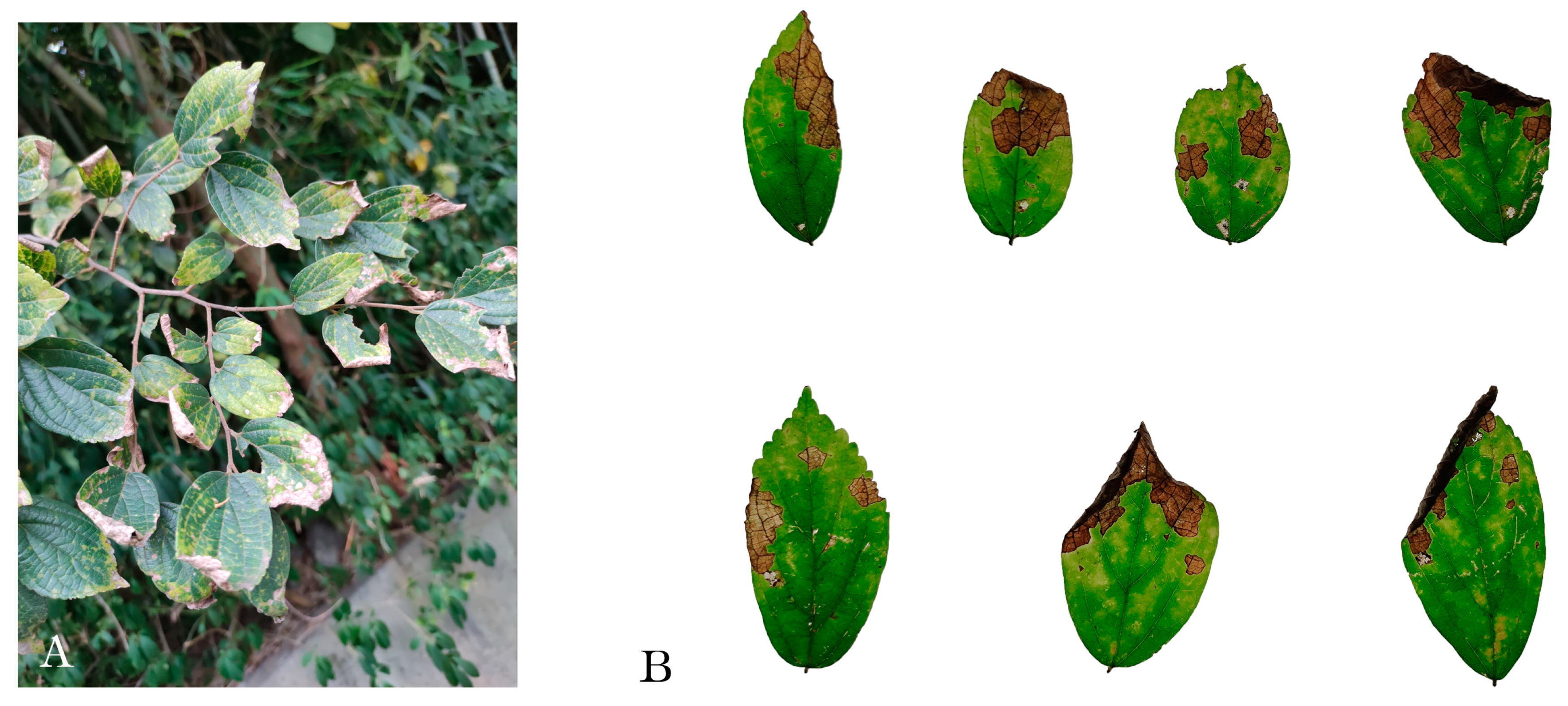
3.1. Field Observations and Fungal Isolation

The survey results indicated that nearly 50% of C. sinensis in Xuanwu District, Nanjing, have shown signs of disease. Most of the spots were produced on the apex of the leaves (Figure 1A), and the spots were brown with darker margins and round to irregular in shape (Figure 1B), on which dark brown chains of conidia were visible. Over time, the spots gradually expanded, and the leaves became dark brown, dry, curled, and necrotic and eventually fell off. In total, 90 fungal colonies developed from the tissue pieces and were grouped into three types according to their colony characteristics and RPB2 sequencing results, with frequencies of 84.4% (76 of 90), 10% (9 of 90), and 5.5% (5 of 90). Three isolates from the highest frequency were selected for purification. According to the ITS sequences, three fungi belonged to Alternaria and were used in further research.

3.2. Pathogenicity Test

Three representative isolates (11, 12, and 13) were inoculated on healthy leaves, and black spots were observed on the leaves after 4 days (Figure 2). The induced symptoms matched those observed in the wild. Conversely, the control group remained healthy with no symptoms of disease. Observing the size of the lesions, those caused by isolates 11 and 13 had smaller affected areas. In contrast, the lesions caused by isolate 12 exhibited a larger affected area. Strains isolated from artificially diseased leaves exhibit colony similarities to the originally isolated strains. DNA was extracted and subjected to PCR using the primer ITS1/4, and the obtained sequences were similar to the original sequences. The symptoms observed on seedlings inoculated in a controlled environment closely resembled those observed in natural field conditions, thus meeting the criteria of Koch’s postulates. Therefore, A. alternata and A. koreana are the pathogens of leaf spot disease in C. sinensis.

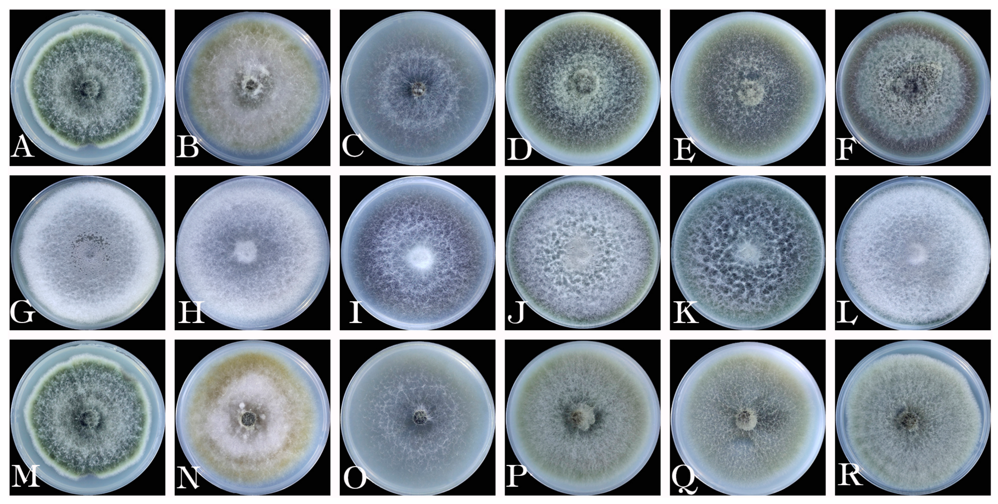
3.3. Morphological Characteristics

Three representative isolates were grown on PDA medium and hatched for 7 days at 25 °C, and their morphological characteristics were recorded based on visual observations (Figure 3). The colonies of isolates 11 and 13 on PDA were initially gray and became grayish brown over time. Conidiophores produced numerous conidia in long chains, mostly unbranched. Conidia were obclavate and dark brown, with two to five transverse and zero to two longitudinal or oblique septa and measured 10.1 to 35.9 × 7.6 to 24.3 μm (n = 30). The colony of isolate 12 on PDA was grayish green. Hyaline (young) or brown (old) hyphae, with septa and smooth walls, were slightly constricted at the septa and branched. Conidiophores produced numerous conidia in long chains. These conidia were globose to ellipsoidal and 13.5 to 34.5 × 7.0 to 14.1 μm (n = 30). The morphological characteristics of the three isolates matched those of Alternaria spp. The three isolates showed different morphological characteristics on different media.

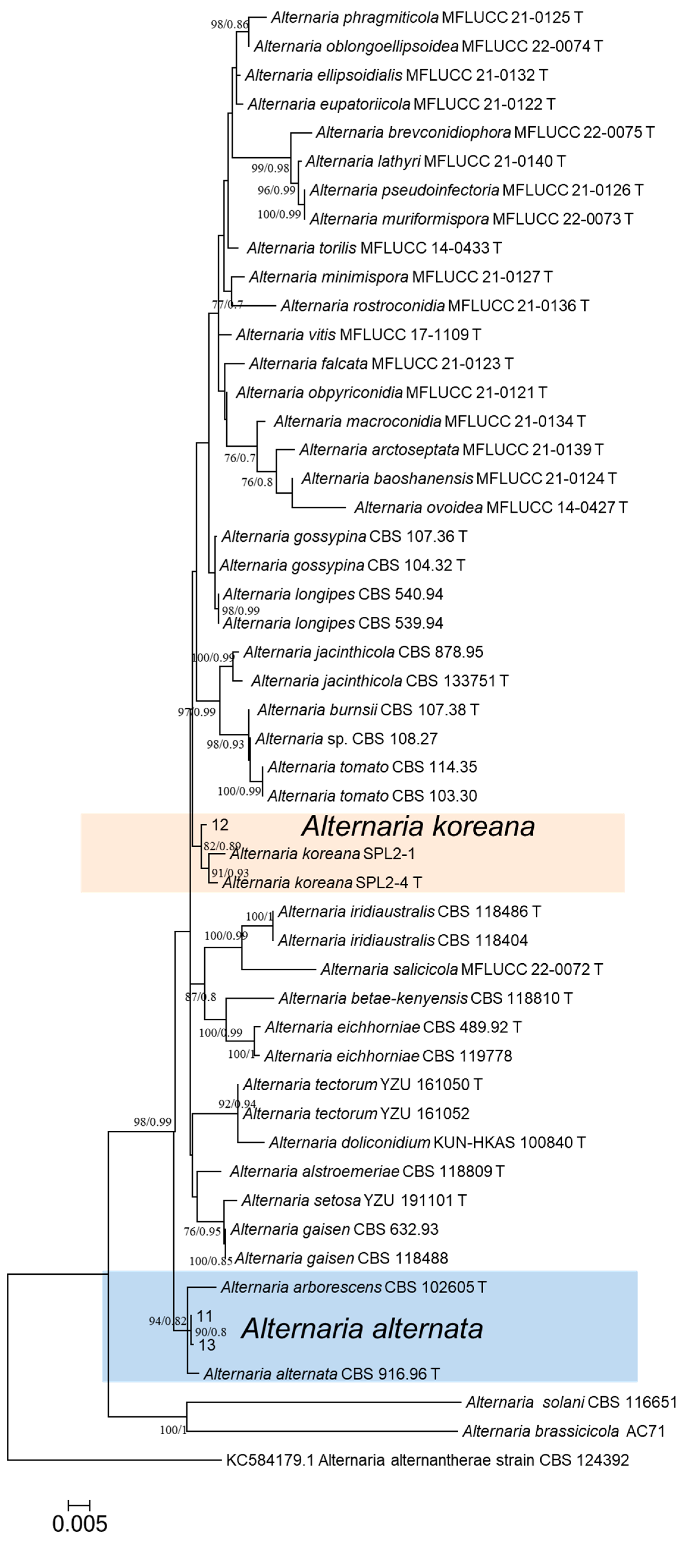
3.4. Multigene Phylogenetic Analysis

BLAST results showed that the ITS, GAPDH, TEF1-α, RPB2, and Alt a 1 sequences of isolate 12 were highly similar (≥98%) to those of A. koreana (culture ex-type SPL2-1), while the sequences of isolates 11 and 13 were highly similar (≥99%) to those of A. alternata (culture representative CBS 918.96). The sequences of genes/region ITS, GAPDH, TEF1-α, RPB2, and Alt a 1 from the three isolates (11, 12, and 13) were uploaded to NCBI, and the registration numbers are shown in Table 2. Phylogenetic analyses using concatenated sequences placed isolate 12 in the clade of A. koreana and isolates 11 and 13 in the clade of A. alternata. Based on the phylogeny and morphology, isolate 12 was identified as A. koreana, and isolates 11 and 13 were identified as A. alternata (Figure 4).

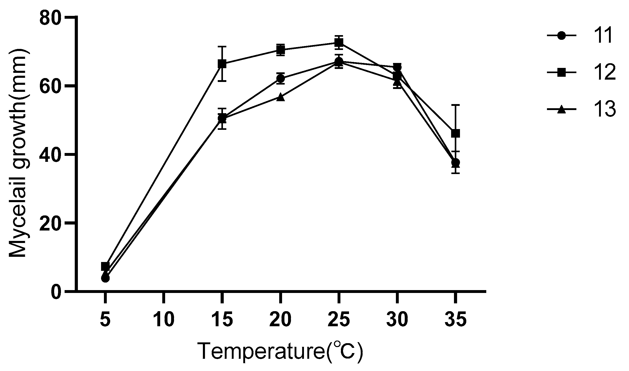
3.5. Biological Characteristics

The colonies of isolates 11 and 13 on CDA appeared as yellowish colonies with flattened colonies (Figure 5B,N). On Richard’s medium, the colony appeared grayish white, with very few hyphae and weak growth (Figure 5C,O). The colony of isolate 11 was dark green with dense mycelium on CMA and OA media, and on PSA media, the colony was dark gray in the middle with a brown ring around it and abundant mycelium (Figure 5D,E). The colony of isolate 13 was light green and radial on CMA, OA, and PSA media (Figure 5P–R). The colony of isolate 12 on CDA was almost identical to the colony morphology on PDA and PSA, with grayish white colonies and dense hyphae (Figure 5G,H,L). It showed a brownish color on Richard, with reticulated colonies and fewer hyphae (Figure 5I), a whitish green color on CMA, with loosely packed colonies (Figure 5J), and a dark green color on PSA medium, with fuzzy colonies (Figure 5K).
Isolates 11, 12, and 13 were cultured between 5 and 35 °C for 7 days, and the colony diameter reached a maximum and grew most vigorously at 25 °C, but at 5 °C, the mycelium hardly grew, which shows that 25 °C is the optimum culture temperature for isolates 11, 12, and 13 (Figure 6).

3.6. Susceptibility of Alternaria Isolates to Fungicides

The three isolates showed similar biological responses to the four fungicides (Figure 7). All four fungicides showed significant growth inhibition of the isolates on PDA media. Prochloraz had a lower EC50 on mycelial growth than the other three fungicides and showed the best inhibitory effect on isolates 11, 12, and 13 (Table 3). Pyraclostrobin had the highest EC50 of isolate 13 and the weakest inhibitory effect but still had a strong inhibitory effect on isolates 11 and 12. Based on the EC50 values, myclobutanil inhibited isolates 11 and 13 almost equally but more strongly inhibited isolate 12. Isolate 12 was the most sensitive to tebuconazole compared with 11 and 13, and isolate 13 was the least sensitive. The results indicate that prochloraz was the most effective fungicide against Alternaria spp. in this study.

4. Discussion

Celtis sinensis is a common deciduous tree that not only has value in food and medicine but also serves a crucial function in cityscape greening and the economy. However, leaf spot disease dilapidates the visual impression of leaves and severely alters the ornamental value of C. sinensis. This disease also leads to leaf damage and interferes with photosynthesis, which prevents C. sinensis from fully utilizing sunlight for nutrient synthesis. This results in the slow growth of the plant and weakening of the leaves, which in turn affects the health and growth of the whole tree and reduces the quality of the timber [35], resulting in an economic loss to the timber industry and affecting the ecological function of C. sinensis. Timely identification of the pathogen responsible for C. sinensis leaf spot is essential. By combining morphological identification, molecular analysis, and phylogenetic research [36], A. alternata and A. koreana were identified as the causal agents of C. sinensis leaf spot in China. To the best of our knowledge, this is the first report of A. alternata and A. koreana causing leaf spot in C. sinensis.
The majority of pathogenic species within the Alternaria genus are the primary cause of the most detrimental plant diseases, commonly referred to as “leaf spot” or “leaf blight”. These diseases are characterized by the development of circular or irregular necrotic lesions on the surface of the leaf, accompanied by a distinct concentric ring-like pattern at the center of the lesions. It is difficult to classify species of Alternaria based on spore morphology characteristics. This challenge arises because a considerable number of small-spore Alternaria species, commonly referred to as A. alternata, exhibit closely similar morphological characteristics [37]. Currently, there is very little research available on A. koreana species. The spore size observed in this study closely resembles the findings reported by Romain et al. [38]. However, there is actually a large amount of research available on Alternaria spp., and considerable variation in spore sizes has been reported. The spore sizes observed in this report are notably disparate from those explored by Ramirez et al. [39], but they are comparable to the description by Sun et al. [40]. Because its morphological characteristics are susceptible to environmental conditions and the presence of many uncontrollable factors, it is highly prone to variations [41].
Currently, the classification of Alternaria species primarily relies on a combination of morphological characteristics and multigene phylogenetic analysis methods. Lawrence et al. [42], Woudenberg et al. [43], Grum-Grzhimaylo et al. [44], and Ghafri et al. [45] used multigene analyses to reconstruct phylogenetic relationships within the genus Alternaria, resulting in the division of the genus into 28 clades, each represented by a type specimen. Phylogenetic analysis of Alternaria using multiple nucleotide sequences, such as ITS, mtLSU, endoPG, TUB, mtSSU, ATP, EF-1α, gpd, Alt a 1, CAL, CHS, ACT, OPA2-1, IGS, HIS, TMA22, PGS1, and REV3, is often employed because these genes play a role in the identification of the genus Alternaria, as well as similar interspecifics. Alternaria sect. Alternaria contains most small-spored Alternaria species with concatenated conidia, including important plant, human, and postharvest pathogens [46]. Alternaria sect. Alternaria consists of only 11 phylogenetic species and one species complex [42].
Temperature is typically regarded as the primary environmental factor influencing plant diseases [47]. In this study, the most favorable growth temperature for the representative isolates was 25 °C, mycelial growth was stagnant at 5 °C, and growth was weaker at 15 °C, 20 °C, 30 °C, and 35 °C than at 25 °C. This aligns with the findings of prior studies regarding the ideal temperature for the growth of A. alternata [48]. Therefore, it is advisable to implement timely pathogen control measures before the optimal growth temperature is reached. Leaf spot in C. sinensis tends to occur in March and April, with outbreaks peaking in May and June and continuing through October. During this period, the pathogen thrives in suitable environments and produces highly infectious conidia that infect the plant host.
Evaluating the biological characteristics of pathogens is of utmost significance in preventing and controlling plant disease outbreaks, as it can lay the groundwork for scientifically informed disease prevention and control measures. It is important to detect the presence of pathogens as early as possible to effectively prevent and control plant diseases [49]. Leaf spot in C. sinensis caused by Alternaria not only affects the health and appearance of plants but also has an impact on their economic value. Fungal leaf spot disease usually spreads under wet conditions and can spread to other healthy C. sinensis or plants through wind, raindrops, insects, or artificial means, thus spreading the disease further and causing the occurrence of leaf spot disease on large areas of C. sinensis, as well as the epidemic occurrence of other plant diseases, which disrupts the ecological balance. Therefore, the negative impacts of C. sinensis leaf spot on agriculture, forestry, urbanization, ecosystems, and the economy are manifold, and measures must be taken to prevent and manage this plant disease. It is obligatory to retard the flourishing of fungal mycelium, thus further controlling the spread and transmission of the disease to prevent greater losses. The temperature experiment conducted in this study conclusively demonstrates the importance of determining the timing of fungicide application before the pathogen reaches its optimal growth temperature.
Different fungicides exert their fungicidal effects through various mechanisms based on their type, chemical structure, etc. [50]. Prochloraz inhibits sterol synthesis in fungal cell membranes, leading to cell wall rupture and cell death. It also interferes with fungal DNA and protein synthesis, resulting in fungicidal effects. In 2009, researchers conducted in vitro susceptibility tests using six fungicides against the pathogen Alternaria solani. The results showed that prochloraz, with EC50 values ranging from 0.03 to 0.11 µg/mL, was effective in inhibiting the growth of the pathogen [51]. This result is consistent with the results of the present study, which showed that prochloraz has the best inhibitory effect on A. alternata and A. koreana, causing leaf spot in C. sinensis. However, by comparing the EC50 values, the sensitivity of prochloraz to A. alternata and A. koreana are much less than that of A. solani, and the inhibitory effect is still unsatisfactory. In 2023, the EC50 value of fludioxonil for 43 A. alternata was only 0.089 ± 0.020 in Wang et al.’s study [52], whereas the EC50 value of prochloraz for A. alternata in this study was 2.95 ± 0.31, which clearly shows that fludioxonil requires a smaller dosage to achieve the same inhibitory effect. Fewer fungicides were selected in this study, and the selection of efficient, environmentally friendly, and economical fungicides must be further explored.

5. Conclusions

In this study, we isolated fungal pathogens and performed pathogenicity tests. We identified three isolates (11–13) responsible for causing leaf spot in C. sinensis through multilocus phylogenetic analyses involving ITS, GAPDH, TEF1-α, RPB2, and Alt a 1 loci, as well as physical attributes. Three isolates were identified as A. alternata and A. koreana. The pathogen’s susceptibility to the four fungicides was ascertained through observational experiments conducted on culture medium plates. To the best of our knowledge, this is the first report that A. alternata and A. koreana cause leaf spot in C. sinensis on a global scale. This revelation will yield valuable insights for future investigations focusing on the prevention and therapeutic policies of this recently evolved disease.

Author Contributions

Q.W., X.Z., Y.W. and Y.Z. designed the study and were involved in writing the paper; Q.W. and Y.Z. were responsible for sample collections; Q.W. was responsible for pathogenicity tests; Q.W., X.Z., and Y.W. were involved in morphological identification; Q.W., X.Z. and Y.W. were involved in phylogenetic analyses; Q.W., X.Z., Y.W. and Y.Z. contributed to planning and editing of the paper. All authors have read and agreed to the published version of the manuscript.

Funding

This research received no external funding.

Data Availability Statement

All data generated or analyzed during this study are included in this article.

Acknowledgments

The authors would like to thank those who provided assistance and advice for this study.

Conflicts of Interest

The authors declare no conflict of interest.

References

  1. Fan, J.; Chen, Y.; Luo, M.; Liang, Z.; Nong, X. The chloroplast genome characteristics, comparative genomics and gene resource mining of Celtis sinensis (Persoon, 1805). Mitochondrial DNA Part B 2022, 7, 698–704. [Google Scholar] [CrossRef] [PubMed]
  2. Lee, H.B.; Lee, K.H.; Nguyen, T.T.T. First Report of Powdery Mildew Caused by Erysiphe michikoae on Celtis sinensis in Korea. Plant Dis. 2020, 104, 984–985. [Google Scholar] [CrossRef]
  3. Guozheng, W.; Yunyan, Z.; Jiehan, Z.; Mengyuan, Z.; Yimin, H.; Yongjing, T.; Jingbo, Z.; Zhongsheng, W. Characterization of the first complete chloroplast genome sequence of Celtis sinensis (Cannabaceae) and its phylogenetic implications. Mitochondrial DNA Part B 2019, 4, 4091–4093. [Google Scholar]
  4. Huizheng, W.; Shilong, W.; Yubin, L. First Report of Botryosphaeria dothidea Causing Leaf Spot and Wilt on Celtis sinensis in China. Plant Dis. 2021, 105, 217. [Google Scholar]
  5. Liang, L.; Li, H.; Zhou, L.F.; Chen, F.M. Lasiodiplodia pseudotheobromae causes stem canker of Chinese hackberry in China. J. For. Res. 2020, 31, 2571–2580. [Google Scholar] [CrossRef]
  6. Liao, Y.C.Z.; Cao, Y.J.; Wan, Y.; Li, H.; Li, D.W.; Zhu, L.H. Alternaria arborescens and A. italica Causing Leaf Blotch on Celtis julianae in China. Plants 2023, 12, 3113. [Google Scholar] [CrossRef]
  7. Woudenberg, J.H.C.; Groenewald, J.Z.; Binder, M.; Crous, P.W. Alternaria redefined. Stud. Mycol. 2013, 75, 171–212. [Google Scholar] [CrossRef]
  8. Qin, Q.M.; Fan, Y.Y.; Jia, Q.L.; Duan, S.S.; Liu, F.J.; Jia, B.X.; Wang, G.Q.; Guo, W.H.; Wang, C. The Potential of Alternaria Toxins Production by A. alternata in Processing Tomatoes. Toxins 2022, 14, 827. [Google Scholar] [CrossRef]
  9. Mitakakis, T.Z.; Barnes, C.; Tovey, E.R. Spore germination increases allergen release from Alternaria. J. Allergy Clin. Immunol. 2001, 107, 388–390. [Google Scholar] [CrossRef]
  10. Bush, R.K.; Prochnau, J.J. Alternaria-induced asthma. J. Allergy Clin. Immunol. 2004, 113, 227–234. [Google Scholar] [CrossRef]
  11. Pochon, S.; Terrasson, E.; Guillemette, T.; Iacomi-Vasilescu, B.; Georgeault, S.; Juchaux, M.; Berruyer, R.; Debeaujon, I.; Simoneau, P.; Campion, C. The Arabidopsis thaliana-Alternaria brassicicola pathosystem: A model interaction for investigating seed transmission of necrotrophic fungi. Plant Methods 2012, 8, 16. [Google Scholar] [CrossRef]
  12. Baez-Flores, M.E.; Troncoso-Rojas, R.; Osuna, M.A.I.; Dominguez, M.R.; Pryor, B.; Tiznado-Hernandez, M.E. Differentially expressed cDNAs in Alternaria alternata treated with 2-propenyl isothiocyanate. Microbiol. Res. 2011, 166, 566–577. [Google Scholar] [CrossRef]
  13. Timmer, L.W.; Peever, T.L.; Solel, Z.; Akimitsu, K. Alternaria diseases of citrus—Novel pathosystems. Phytopathol. Mediterr. 2003, 42, 1000–1014. [Google Scholar]
  14. Vicent, A.; Badal, J.; Asensi, M.J.; Sanz, N.; Armengol, J.; Garcia-Jimenez, J. Laboratory evaluation of citrus cultivars susceptibility and influence of fruit size on Fortune mandarin to infection by Alternaria alternata pv. citri. Eur. J. Plant Pathol. 2004, 110, 245–251. [Google Scholar] [CrossRef]
  15. Vega, B.; Dewdney, M.M. Distribution of Qol Resistance in Populations of Tangerine-Infecting Alternaria alternata in Florida. Plant Dis. 2014, 98, 67–76. [Google Scholar] [CrossRef] [PubMed]
  16. Vega, B.; Dewdney, M.M. Sensitivity of Alternaria alternata from Citrus to Boscalid and Polymorphism in Iron-Sulfur and in Anchored Membrane Subunits of Succinate Dehydrogenase. Plant Dis. 2015, 99, 231–239. [Google Scholar] [CrossRef]
  17. Song, J.H.; Wang, Y.F.; Yin, W.X.; Huang, J.B.; Luo, C.X. Effect of Chemical Seed Treatment on Rice False Smut Control in Field. Plant Dis. 2021, 105, 3218–3223. [Google Scholar] [CrossRef] [PubMed]
  18. Ju, Y.; Si, Y.Z.; Li, D.W.; Xu, W.; Sun, J.W.; Zhu, L.H. First Report of Leaf Blotch of Salix babylonica Caused by Botryosphaeria dothidea in China. Plant Dis. 2021, 105, 224. [Google Scholar] [CrossRef] [PubMed]
  19. Zhu, L.H.; Xu, W.; Huang, L.; Ye, J.R.; Li, D.W. Pathogenicity and Biological Characteristics of Septotinia populiperda Causing Leaf Blotch of Willow. Plant Dis. 2022, 106, 1262–1270. [Google Scholar] [CrossRef]
  20. Liao, Y.C.Z.; Wan, Y.; Si, Y.Z.; Geng, J.X.; Li, D.W.; Zhu, L.H. Leaf spot of Ligustrum japonicum caused by Diaporthe eres newly reported in China. Crop Prot. 2023, 164, 106139. [Google Scholar] [CrossRef]
  21. Si, Y.Z.; Sun, J.W.; Wan, Y.; Chen, Y.N.; He, J.; Li, W.Z.; Li, D.W.; Zhu, L.H. Neofusicoccum cryptomeriae sp. nov. and N. parvum Cause Stem Basal Canker of Cryptomeria japonica in China. J. Fungi 2023, 9, 404. [Google Scholar] [CrossRef]
  22. He, J.; Sun, M.-L.; Li, D.-W.; Zhu, L.-H.; Ye, J.-R.; Huang, L. A real-time PCR for detection of pathogens of anthracnose on Chinese fir using TaqMan probe targeting ApMat gene. Pest Manag. Sci. 2022, 79, 980–988. [Google Scholar] [CrossRef]
  23. White, T.J.; Bruns, T.D.; Lee, S.B.; Taylor, J.W. Amplification and direct sequencing of fungal ribosomal RNA genes for phylogenetics. In PCR Protocols: A Guide to Methods and Applications; University of New Mexico: Albuquerque, NM, USA, 1990; Volume 18, pp. 315–322. [Google Scholar]
  24. Carbone, I.; Kohn, L. A method for designing primer sets for speciation studies in filamentous ascomycetes. Mycologia 1999, 91, 553–556. [Google Scholar] [CrossRef]
  25. Berbee, M.L.; Pirseyedi, M.; Hubbard, S. Cochliobolus phylogenetics and the origin of known, highly virulent pathogens, inferred from ITS and glyceraldehydes-3-phosphate dehydrogenase gene sequences. Mycologia 1999, 91, 964–977. [Google Scholar] [CrossRef]
  26. Liu, Y.J.; Whelen, S.; Hall, B.D. Phylogenetic relationships among ascomycetes: Evidence from an RNA polymerse II subunit. Mol. Biol. Evol. 1999, 16, 1799–1808. [Google Scholar] [CrossRef] [PubMed]
  27. Hong, S.G.; Cramer, R.A.; Lawrence, C.B.; Pryor, B.M. Alt a 1 allergen homologs from Alternaria and related taxa: Analysis of phylogenetic content and secondary structure. Fungal Genet. Biol. 2005, 42, 119–129. [Google Scholar] [CrossRef] [PubMed]
  28. Katoh, K.; Standley, D.M. MAFFT multiple sequence alignment software version 7: Improvements in performance and usability. Mol. Biol. Evol. 2013, 30, 772–780. [Google Scholar] [CrossRef]
  29. Zhang, D.; Gao, F.; Jakovlic, I.; Zou, H.; Zhang, J.; Li, W.X.; Wang, G.T. PhyloSuite: An integrated and scalable desktop platform for streamlined molecular sequence data management and evolutionary phylogenetics studies. Mol. Ecol. Resour. 2020, 20, 348–355. [Google Scholar] [CrossRef] [PubMed]
  30. Li, J.-F.; Jiang, H.-B.; Jeewon, R.; Hongsanan, S.; Bhat, D.J.; Tang, S.-M.; Lumyong, S.; Mortimer, P.E.; Xu, J.-C.; Camporesi, E.; et al. Alternaria: Update on species limits, evolution, multi-locus phylogeny, and classification. Stud. Fungi 2023, 8, 1–61. [Google Scholar] [CrossRef]
  31. Zhang, Y.; Zhang, B.; Luo, C.; Fu, Y.; Zhu, F. Fungicidal Actions and Resistance Mechanisms of Prochloraz to Penicillium digitatum. Plant Dis. 2021, 105, 408–415. [Google Scholar] [CrossRef]
  32. Ypema, H.L.; Ypema, M.; Gubler, W.D. Sensitivity of Uncinula necator to Benomyl, Triadimefon, Myclobutanil, and Fenarimol in California. Plant Dis. 1997, 81, 293–297. [Google Scholar] [CrossRef] [PubMed]
  33. Gama, A.B.; Baggio, J.S.; Rebello, C.S.; Lourenco, S.d.A.; Gasparoto, M.C.d.G.; da Silva Junior, G.J.; Peres, N.A.; Amoriml, L. Sensitivity of Colletotrichum acutatum Isolates from Citrus to Carbendazim, Difenoconazole, Tebuconazole, and Trifloxystrobin. Plant Dis. 2020, 104, 1621–1628. [Google Scholar] [CrossRef]
  34. Han, S.; Lu, J.; Gao, J.; Cheng, J.; Xu, W.; Tao, L.; Zhang, Y. Pyraclostrobin induced AMPK/mTOR pathways mediated autophagy in RAW264.7 macrophages. J. Environ. Sci. Health Part B 2021, 56, 793–800. [Google Scholar] [CrossRef] [PubMed]
  35. Zhang, M.J.; Zheng, X.R.; Li, H.; Chen, F.M. Alternaria alternata, the Causal Agent of a New Needle Blight Disease on Pinus bungeana. J. Fungi 2023, 9, 71. [Google Scholar] [CrossRef]
  36. Meng, J.X.; Zhang, X.Y.; Han, X.S.; Fan, B. Application and Development of Biocontrol Agents in China. Pathogens 2022, 11, 1120. [Google Scholar] [CrossRef] [PubMed]
  37. Zhu, X.Q.; Xiao, C.L. Phylogenetic, Morphological, and Pathogenic Characterization of Alternaria Species Associated with Fruit Rot of Blueberry in California. Phytopathology 2015, 105, 1555–1567. [Google Scholar] [CrossRef]
  38. Romain, B.B.N.D.; Hassan, O.; Kim, J.S.; Chang, T. Alternaria koreana sp. nov., a new pathogen isolated from leaf spot of ovate-leaf Atractylodes in South Korea. Mol. Biol. Rep. 2022, 49, 413–420. [Google Scholar] [CrossRef] [PubMed]
  39. Ramirez-Villacis, D.; Barriga-Medina, N.; Llerena-Llerena, S.; Pazmino-Guevara, C.; Leon-Reyes, A. First Report of Alternaria alternata Causing Leaf Spot on Broccoli in Ecuador. Plant Dis. 2023, 107, 2866. [Google Scholar] [CrossRef]
  40. Sun, H.F.; Jiang, X.; Li, Z.; Wang, H.; Yan, Y.; Yang, H. First report of Alternaria alternata causing leaf blight on Trollius chinensis in China. Plant Dis. 2023, 107, 2533. [Google Scholar] [CrossRef]
  41. Pryor, B.M.; Michailides, T.J. Morphological, pathogenic, and molecular characterization of Alternaria isolates associated with Alternaria late blight of pistachio. Phytopathology 2002, 92, 406–416. [Google Scholar] [CrossRef]
  42. Lawrence, D.P.; Gannibal, P.B.; Peever, T.L.; Pryor, B.M. The sections of Alternaria: Formalizing species-group concepts. Mycologia 2013, 105, 530–546. [Google Scholar] [CrossRef]
  43. Woudenberg, J.H.C.; Truter, M.; Groenewald, J.Z.; Crous, P.W. Large-spored Alternaria pathogens in section Porri disentangled. Stud. Mycol. 2014, 79, 1–47. [Google Scholar] [CrossRef] [PubMed]
  44. Grum-Grzhimaylo, A.A.; Georgieva, M.L.; Bondarenko, S.A.; Debets, A.J.M.; Bilanenko, E.N. On the diversity of fungi from soda soils. Fungal Divers. 2016, 76, 27–74. [Google Scholar] [CrossRef]
  45. Ghafri, A.; Maharachchikumbura, S.S.N.; Hyde, K.D.; Al-Saady, N.; Al-Sadi, A.M. A new section and a new species of Alternaria encountered from Oman. Phytotaxa 2019, 405, 279–289. [Google Scholar] [CrossRef]
  46. Woudenberg, J.H.C.; Seidl, M.F.; Groenewald, J.Z.; de Vries, M.; Stielow, J.B.; Thomma, B.P.H.J.; Crous, P.W. Alternaria section Alternaria: Species, formae speciales or pathotypes? Stud. Mycol. 2015, 82, 1–21. [Google Scholar] [CrossRef] [PubMed]
  47. Giner, M.M.; Garcia, J.S.C.; Camacho, C.N. Airborne Alternaria spores in SE Spain (1993–98)—Occurrence patterns, relationship with weather variables and prediction models. Grana 2001, 40, 111–118. [Google Scholar] [CrossRef]
  48. Cabral, L.D.; Rodriguez, A.; Delgado, J.; Patriarca, A. Understanding the effect of postharvest tomato temperatures on two toxigenic Alternaria spp. strains: Growth, mycotoxins and cell-wall integrity-related gene expression. J. Sci. Food Agric. 2019, 99, 6689–6695. [Google Scholar] [CrossRef]
  49. Cao, X.M.; Li, Y.L.; Fan, J.L.; Zhao, Y.J.; Borriss, R.; Fan, B. Two Lysine Sites That Can Be Malonylated Are Important for LuxS Regulatory Roles in Bacillus velezensis. Microorganisms 2021, 9, 1338. [Google Scholar] [CrossRef]
  50. Gisi, U.; Sierotzki, H. Fungicide modes of action and resistance in downy mildews. Eur. J. Plant Pathol. 2008, 122, 157–167. [Google Scholar] [CrossRef]
  51. Stepanovic, M.; Vuksa, P.; Potocnik, I.; Milijasevic, S.; Rekanovic, E.; Todorovic, B. Integrated Management of Viral Sensitivity of Alternaria solani Isolates to Some Fungicides. In Proceedings of the II International Symposium on Tomato Diseases, Kusadasi, Turkey, 8–12 October 2007; pp. 377–380. [Google Scholar]
  52. Wang, F.; Saito, S.; Xiao, C.L. Fungicide Resistance of Alternaria alternata and A. arborescens Isolates from Mandarin Fruit and Its Influence on Control of Postharvest Alternaria Rot. Plant Dis. 2023, 107, 1538–1543. [Google Scholar] [CrossRef]
Figure 1. Symptoms of infected leaves in C. sinensis in the wild. (A) Diseased leaves naturally. (B) Closeup view of diseased leaves.
Forests 14 02389 g001
Figure 2. Pathogenicity of fungal isolates. (A) Signs on leaves of controls treated with PDA after 4 days. (BD) Signs on leaves inoculated with mycelium blocks of A. alternata (isolates 11, 13) and A. koreana (isolate 12) after 4 days. (E) Signs on leaves of controls treated with sterile water after 5 days. (FH) Signs on leaves of controls handled with sterile water after 5 days. Signs on leaves inoculated with 10 µL of conidial suspension (106 conidia/mL) of A. alternata (isolates 11, 13) and A. koreana (isolate 12) after 5 days.
Forests 14 02389 g002
Figure 3. Morphological characteristics of fungal isolates 11 and 13 (A. alternata) and isolate 12 (A. koreana). (A) Seven-day-old front and back view colony of 11 on PDA. (B,C) Conidial chain and conidia of isolate 11, Bars: (B)= 100 μm, (C) = 10 μm. (D) Seven-day-old front and back view colony of 12 on PDA. (E,F) Conidial chain and conidia of isolate 12, Bars: (E) = 100 μm, (F) = 10 μm. (G) Seven-day-old front and back view colony of 13 on PDA. (H,I) Conidial chain and conidia of isolate 13, Bars: (H) = 100 μm, (I) = 10 μm.
Forests 14 02389 g003
Figure 4. Phylogenetic relationship of 11, 12, and 13 and related taxa derived from concatenated sequences of the ITS, GAPDH, TEF1-α, RPB2, and Alt a 1 genes using the maximum likelihood algorithm and Bayesian analysis. Bootstrap values > 75% (1000 replications) and Bayesian posterior probability (PP ≥ 0.90) are shown at the nodes (ML/PP). Alternaria alternantherae (CBS 124392) was used as an outgroup. Bar = 0.002 substitutions per nucleotide position. T indicates ex-types.
Forests 14 02389 g004
Figure 5. Colony formation of isolates 11 and 13 (A. alternata) and isolate 12 (A. koreana) isolated from Celtis sinensis hatched for 7 days on 6 media at 25 °C. (AF). Colony morphology of isolate 11 cultured on PDA, CDA, Richard, CMA, OA, and PSA media for 7 days. (GL). Colony morphology of isolate 12 cultured on PDA, CDA, Richard, CMA, OA, and PSA media for 7 days. (MR). Colony morphology of isolate 13 cultured on PDA, CDA, Richard, CMA, OA, and PSA media for 7 days.
Forests 14 02389 g005
Figure 6. The impact of temperature on the growth of colony diameters in isolates 11 and 13 (A. alternata) and isolate 12 (A. koreana) after 7 days of culture on PDA media.
Forests 14 02389 g006
Figure 7. The suppression effect on isolates 11 and 13 (A. alternata) and isolate 12 (A. koreana) on plates caused by various doses of four diverse fungicides (prochloraz, myclobutanil, tebuconazole, and pyraclostrobin) based on fresh PDA for 7 days.
Forests 14 02389 g007
Table 1. Primers used for PCR amplification in the molecular identification of three isolates (11–13).
GenePrimerSequence (5′-3′)PCR Amplification Cycle Parameters:
Internal transcribed spacer
(ITS)
ITS1
ITS4
TCCGTAGGTGAACCTGCGG
TCCTCCGCTTATTGATATGC
95 °C: 3 min, (95 °C: 30 s, 55 °C: 30 s, 72 °C: 35 s) × 32 cycles, 72 °C: 8 min
Elongation factor 1-alpha
(TEF1-α)
EF1-728F
EF1-986R
CATCGAGAAGTTCGAGAAGG
TACTTGAAGGAACCCTTACC
95 °C: 3 min, (95 °C: 30 s, 59 °C: 30 s, 72 °C: 20 s) × 32 cycles, 72 °C: 8 min
Glycerol-3-phosphate
dehydrogenase (GAPDH)
GPD1
GPD2
CAACGGCTTCGGTCGCATTG
GCCAAGCAGTTG GTTGT
94 °C: 3 min, (94 °C: 30 s, 59.5 °C: 30 s, 72 °C: 40 s) × 32 cycles, 72 °C: 8 min
RNA polymerase second-largest subunit (RPB2)RPB2-5F2
RPB2-7cR
GGGGWGAYCAGAAGAAGGC
CCCATRGCTTGTYYRCCCAT
94 °C: 3 min, (94 °C: 30 s, 53 °C: 30 s, 72 °C: 50 s) × 32 cycles, 72 °C: 8 min
Alternaria major allergen gene
(Alt a 1)
Alt-al-for
Alt-al-rev
ATGCAGTTCACCACCATCGC
ACGAGGGTGAYGTAGGCGTC
95 °C: 3 min, (95 °C: 30 s, 60.5 °C: 30 s, 72 °C: 30 s) × 32 cycles, 72 °C: 8 min
Table 2. The isolates of Alternaria species used in this study for phylogenetic analysis.
SpeciesIsolateAccession Numbers
ITSGAPDHTEF1-αRPB2Alt a 1
Alternaria alstroemeriaeCBS 118809 TKP124297KP124154KP125072KP124765KP123845
Alternaria sp.CBS 108.27KC584236KC584162KC584727KC584468-
A. alternantheraeCBS 124392KC584179KC584096KC584633KC584374KP123846
A. alternataCBS 916.96 TAF347031AY278808KC584634KC584375AY563301
A. arborescensCBS 102605 TAY347033AY278810KC584636KC584377AY563303
A. alternata11OP476716OP609771OP609768OP604538OP609775
A. alternata13OP476718OP609773OP609770OP604540OP609776
A. arctoseptataMFLUCC
21-0139 T
-OK2367020K236608OK236655OK236755
A. baoshanensisMFLUCC
21-0124 T
MZ622003OK236706OK236613OK236659OK236760
A. betae-kenyensisCBS 118810 TKP124419KP124270KP125197KP124888KP123966
A. breviconidiophoraMFLUCC
22-0075 T
MZ621997OK236698OK236604OK236651-
A. burnsiiCBS 107.38 TKP124420JQ646305KP125198KP124889KP123967
A. brassicicolaAC71LC440588LC482009LC480214LC476794LC481621
A. doliconidiumKUN-HKAS
100840 T
NR158361----
A. ellipsoidialisMFLUCC
21-0132 T
MZ621989OK236690OK236596OK236643OK236743
A. eupatoriicolaMFLUCC
21-0122 T
MZ621982OK236683OK236589OK236636OK236736
A. eichhorniaeCBS 489.92 TKC146356KP124276KP125204KP124895KP123973
A. eichhorniaeCBS 119778KP124426KP124277KP125205KP124896KP123973
A. falcataMFLUCC
21-0123 T
MZ621992OK236693OK236599OK236646OK236746
A. gaisenCBS 632.93KC584197KC584116KC584658KC584399KP123974
A. gaisenCBS 118488KP124427KP124278KP125206KP124897KP123975
A. gossypinaCBS 104.32 TKP124430JQ646312KP125209KP124900JQ646395
A. gossypinaCBS 107.36 TKP124431JQ646310KP125210KP124901JQ646393
A. iridiaustralisCBS 118404KP124434KP124283KP125213KP124904KP123980
A. iridiaustralisCBS 118486 TKP124435KP124284KP125214KP124905KP123981
A. jacinthicolaCBS 878.95KP124437KP124286KP125216KP124907KP123983
A. jacinthicolaCBS 133751 TKP124438KP124287KP125217KP124908KP123984
A. koreanaSPL2-1
(KACC49833) T
LC621613LC621647LC621715LC621681LC631831
A. koreanaSPL2-4LC621615LC621649LC621717LC621683LC631832
A. koreana12OP476717OP609772OP609769OP604539OP609774
A. lathyriMFLUCC
21-0140 T
MZ621974OK236675OK236581OK236628OK236728
A. longipesCBS 539.94KP124441KP124290KP125220KP124911KP123987
A. longipesCBS 540.94AY278835AY278811KC584667KC584409AY563304
A. macroconidiaMFLUCC
21-0134 T
MZ622001OK236704OK236610OK236657OK236757
A. minimisporaMFLUCC
21-0127 T
MZ621980OK236681OK236587OK236634OK236734
A. muriformisporaMFLUCC
22-0073 T
MZ621976OK236677OK236583OK236630OK236730
A. ovoideaMFLUCC
14-0427 T
MZ622005OK236708OK236614OK236661OK236761
A. oblongoellipsoideaMFLUCC
22-0074 T
MZ621967OK236668OK236574OK236621OK236721
A. obpyriconidiaMFLUCC
21-0121 T
MZ621978OK236680OK236585OOK236633OK236732
A. phragmiticolaMFLUCC
21-0125 T
MZ621994OK236696OK236602OK236649OK236749
A. pseudoinfectoriaMFLUCC
21-0126 T
MZ621984OK236685OK236591OK236638OK236738
A. rostroconidiaMFLUCC
21-0136 T
MZ621969OK236670OK236576OK236623OK236723
A. salicicolaMFLUCC
22-0072 T
MZ621999OK236700OK236606OK236653OK236753
A. setosaYZU 191101 TOP341770OP352306OP374459OP352294OP293717
A. solaniCBS 103.30KC584217KC584139KC584688KC584430GQ180097
A. tectorumYZU 161050 TOP341728OP352303OP374456OP352291OP293714
A. tectorumYZU 161052OP341817OP352304OP374457OP352292OP293715
A. tomatoCBS 103.30KP124445KP124294KP125224KP124915KP123991
A. tomatoCBS 114.35KP124446KP124295KP125225KP124916KP123992
A. torilisMFLUCC
14-0433 T
MZ621988OK236688OK236594OK236641OK236741
A. vitisMFLUCC
17-1109 T
MG764007----
T ex-type isolate; bolded are the isolates for this study.
Table 3. Concentration at 50% of maximum effect (EC50 values) of isolates. (Data = mean ± standard error).
Fungicide EC50 Values (µg/mL)
111213
Prochloraz2.95 ± 0.31 d3.96 ± 0.4 c4.05 ± 0.42 d
Myclobutanil84.41 ± 3.33 a55.14 ± 2.34 a87.89 ± 3.73 c
Tebuconazole35.32 ± 5.98 c13.96 ± 2.52 c143.17 ± 8.93 b
Pyraclostrobin50.53 ± 2.05 b34.5 ± 7.66 b422.26 ± 27.69 a
Isolates 11 and 13 were A. alternata; isolate 12 was A. koreana. Data were analyzed with SPSS Statistics 19.0 by one-way ANOVA, and means were compared using Duncan’s test at a significance level of p = 0.05.
Disclaimer/Publisher’s Note: The statements, opinions and data contained in all publications are solely those of the individual author(s) and contributor(s) and not of MDPI and/or the editor(s). MDPI and/or the editor(s) disclaim responsibility for any injury to people or property resulting from any ideas, methods, instructions or products referred to in the content.

