Article

Function of Tryptophan 2,3-Dioxygenase in Monochamus alternatus Hope Revealed by RNA Interference

by
Liang-Jing Sheng
1,2,†,
Xiao-Qian Weng
1,2,†,
Ming-Qing Weng
1,2,†,
Ya-Jie Guo
1,3,
Rebeca Carballar-Lejarazú
4,
Fei-Ping Zhang
1,2,* and
Song-Qing Wu
1,2,*
1
College of Forestry, Fujian Agriculture and Forestry University, Fuzhou 350002, China
2
Key Laboratory of Integrated Pest Management in Ecological Forests, Fujian Agriculture and Forestry University, Fuzhou 350002, China
3
Laboratory of Forest Symbiology, Asian Research Center for Bioresource and Environmental Sciences, Graduate School of Agricultural and Life Sciences, The University of Tokyo, Tokyo 188-0002, Japan
4
Department of Microbiology & Molecular Genetics, University of California Irvine, Irvine, CA 92697, USA
*
Authors to whom correspondence should be addressed.
These authors contributed equally to this work.
Forests 2023, 14(2), 215; https://doi.org/10.3390/f14020215
Submission received: 25 November 2022 / Revised: 18 January 2023 / Accepted: 20 January 2023 / Published: 23 January 2023
(This article belongs to the Section Forest Ecophysiology and Biology)

Abstract

:
Monochamus alternatus Hope (Coleoptera: Cerambycidae), an invasive beetle that has caused billions of dollars of economic losses, is a serious pest of Pinus massoniana in many Asian countries. An efficient RNAi system is helpful for functional genomics research on M. alternatus. In this study, a tryptophan 2,3-dioxygenase (TDO) related to the ommochrome synthesis in insects was identified. Using RNAi technology, the M. alternatus TDO gene was silenced by injecting dsRNA into pupae, and individuals were analyzed by phenotype and expression of the TDO gene by RT-qPCR. The results show that TDO is expressed in different developmental stages of M. alternatus, having its peak expression during the prepupal stage. White-eye phenotypes were observed in the pupal and adult stages after dsRNA injection, and a significant 81% decrease in TDO mRNA levels 48 h after injection was determined by RT-qPCR. This gene can be used as a genetic marker and is an important discovery for future genetic engineering tools to control M. alternatus populations.

1. Introduction

Being the most important vector for pine wilt disease (PWD), the Japanese pine sawyer beetle, Monochamus alternatus Hope (Coleoptera: Cerambycidae), has critically threatened the pine woodland ecosystem and affected ecological safety in China [1]. In China, the direct and oblique financial loss brought on by way of the pine wood nematode (PWN) Bursaphelenchus xylophilus (Steiner and Buhrer) Nickle has already passed RMB 27.5 billion [2]. Throughout the process of PWN transmission, adults find new pine trees to replenish nutrition and lay eggs, which leads to PWN getting into the wound generated by adult activity [3,4]. Thus, an urgent and effective way to protect the rest of the pine forest resources is by controlling the population of the vector of PWN [3,5]. The current efforts to eradicate PWN are focused on controlling M. alternatus populations through the application of chemical or biological insecticides. However, both strategies impose some limitations and have been not successful in eradicating PWN. Although biological control has some advantages for the control of M. alternatus, including long persistence, lower threat to the environment, and high specialty in the targeting of the organism, its use has not had the desired impact in controlling the disease.
New technologies based on genetic engineering have been developed to modify or suppress insect populations [6,7,8]. Currently, the genetic toolkit for several insects includes assembled and annotated genome sequences [6], efficient methods for germline transformation (transposons, phiC31 integrase-mediated) [9,10,11], a variety of genetically marked strains [12], genotype/phenotype analysis using RNA interference [13], and most recently, methods for and use of CRISPR-based genome editing [14,15,16]. At present, RNAi technology is widely used to elucidate gene function and link phenotype with gene function and has been used to control pests such as Lepidoptera, Diptera, and Coleoptera [17,18,19,20]. However, for M. alternatus, some of these tools are still not available, and to develop a “proof of principle” model for this, Coleoptera phenotypic genes play an important role as they are usually the target to assess and improve genome editing capacity [21,22].
Eye pigmentation genes have been used in other insect systems for genetic manipulation due to the facility of determining viable mutant phenotypes [23]. However, insect eye color is regulated by complex pigmentation pathways that can vary among species [24,25]. In general, ommochromes are an important source of visible eye pigments and are the major products of a series of tryptophan degradations in various insects [24]. Tryptophan is degraded by tryptophan 2,3-dioxygenase (TDO; encoded by the vermilion gene), which is required for the synthesis of brown eye pigment in Drosophila [26]. The brown pigment is derived biosynthetically from tryptophan by a series of steps, including the intermediates N-formylkynurenine, kynurenine, and 3-hydroxykynurenine [27,28]. The vermilion gene has been used in germline transformation vectors as the selectable marker gene in Drosophila melanogaster [29].
Here, we identified the vermilion gene (TDO) in M. alternatus and showed that the silencing of the gene results in an eye-color change phenotype in pupae and adults. This work contributes to expanding the genetic tools in M. alternatus by providing a visible marker to develop new genetic control tools for this pest.

2. Materials and Methods

2.1. Experimental Insects

Monochamus alternatus adults were collected from Minhou County, Fuzhou City, Fujian Province, China, in the summer of 2021. Sexually mature adults were reared as usual at 27 ± 1 °C under a 14:10 h light/dark photoperiod and 70%–80% relative humidity with fresh branches of Pinus massoniana. Wood for laying eggs was collected from the wild, and larvae were raised on an artificial diet until pupation.

2.2. Identification of the TDO Gene in M. alternatus

The TDO gene was identified from the genome of M. alternatus. The conserved domain and Blast analysis was performed by using the CD-Search and BLAST (https://www.ncbi.nlm.nih.gov/ (accessed on 23 Nov 2021)), respectively. The TDO protein sequences of different insect species were downloaded from the NCBI website (https://www.ncbi.nlm.nih.gov/ (accessed on 23 Nov 2021)). Protein sequences were aligned by MEGA, and homology analysis was performed by using the GeneDoc software [30]. The sequences from the transmembrane helix residues were predicted by using the SMART website (http://smart.embl-heidelberg.de/ (accessed on 14 Oct 2022)).

2.3. Isolation of M. alternatus RNA and Synthesis of dsRNA Molecules

Total RNA was isolated from M. alternatus pupa or the adult heads by homogenizing the tissue in 1 mL Trizol [31]. Then, 50–100 mg homogenate was immediately transferred to a 1.5 mL Eppendorf tube and allowed to sit for 5 min at room temperature. Then, 200 µL chloroform was added, mixed manually for 15 s, and incubated for 2 min at room temperature, followed by centrifugation at 12,000 rpm at 4 °C for 5 min. The upper water phase was transferred to a new Eppendorf tube, followed by isopropyl alcohol precipitation and a subsequent wash with pre-cooled 75% ethanol. Centrifugation was performed at 7500 rpm at 4 °C for 5 min and the supernatant was discarded. The RNA pellet was resuspended in RNase-free H2O.
cDNA was made by reverse transcription of extracted RNA using 1 µg total RNA to synthesize first-strand cDNA in a Yeasen Hifair® II 1st Strand cDNA Synthesis Kit (Yeasen, Shanghai, China) according to the manufacturer’s instructions. To obtain a cDNA fragment containing T7 RNA polymerase promoter, we performed PCR with 1 µg cDNA as a template using TDO-F and TDO-R (Table 1). As a negative control, dsRNA from the green florescent protein (GFP) was used. PCR products were purified with a FastPure Gel DNA Extraction Mini Kit (Vazyme, Nanjing, China), and we used 0.5 µg as a template for in vitro transcription using a T7 RNAi Transcription Kit (Vazyme, Nanjing, China) to generate sense and antisense RNA in the same tube. The synthesized dsRNAs were purified with magnetic beads, the concentration of the products was determined, and the products were stored at −80 °C until use.

2.4. RNAi in M. alternatus in the Pupal Stage

New pupae were disinfected with 75% alcohol and dried with tissue. The sterilized pupae were placed in Petri dishes on ice for 2 min before being injected with a syringe sterilized with ethanol and ddH2O. dsGFP and ddH2O were used as negative controls. Approximately 1 µg of each dsRNA was injected into the pronotum of M. alternatus. A group of 10 new pupae were injected as a replicate, and injections were repeated three times. The heads were removed at 24, 48, and 96 h post injection and used for qRT-PCR analysis to determine the TDO expression levels.

2.5. Real-Time Quantitative PCR (RT-qPCR)

Gene expression analysis was performed with three independent biological replicates. The expression levels from five heads of M. alternatus were detected 24 h, 48 h, and 96 h after injection. RNA extraction and cDNA synthesis was performed as described in Section 2.3. Briefly, 2 µL from each cDNA sample was mixed with 10 µL 2× ChamQ Universal SYBR qPCR Master Mix, 0.4 µL of each primer (10 µM) (Table 1), and 7.2 µL RNase-free water. RT-qPCR reactions were performed with ChamQ Universal SYBR qPCR Master Mix (Vazyme, Nanjing, China) in a QuantStudio™ 6 Flex Real-Time PCR System.

2.6. Statistical Analysis

RT-qPCR results are presented as means ± SD of three independent biological replicates, and the relative expression of non-injected and dsTDO was determined using the QuantStudio™ 6 Flex Real-Time PCR System (Life Technologies) with 2−ΔΔCt methodology. Relative expression was analyzed by the Tukey’s test for significant differences between the average values using GraphPad Prism 8.0.2 for Windows. p < 0.05 was used as the level of statistical significance.

3. Results

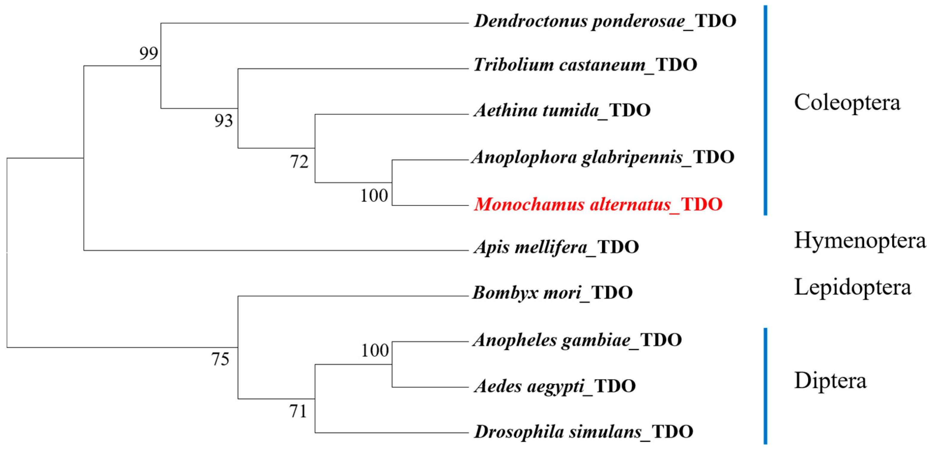
3.1. Identification of the TDO Gene and Homology Analysis

TDO plays an important role in the kynurenine pathway and is used in the synthesis of ommochrome, which is common to all insects [14,22,32]. To identify the TDO gene in M. alternatus, a total of nine protein sequences from annotated TDO protein sequences of Coleoptera, Hymenoptera, Lepidoptera, and Diptera from the National Center for Biotechnology Information reference sequence database were analyzed. A neighbor-joining tree was constructed to examine the phylogenetic relationships of the M. alternatus sequences with homologous proteins from representative species across four insect orders (Coleoptera, Lepidoptera, Hymenoptera, and Diptera; see Figure 1).
TDO is located on chromosome 5 of M. alternatus (MaTDO). Only one domain of the TDO gene translated to amino acid sequence was identified by conserved region TDO, amino acid region 9–355 (Figure 2A and Figure S1). In addition, nucleotide and protein sequences for of M. alternatus TDO are also shown (Figure S1). According to the tree and the protein alignment of Coleoptera, MaTDO is 94% identical to Anoplophora glabripennis TDO, 85.3% identical to Aethina tumida TDO isoform X1, and 83.6% identical to Tribolium castaneum TDO (Figure 2B). All sequences possess a single Trp_dioxygenase domain and do not contain a transmembrane helix structure.

3.2. TDO Silencing and Its Phenotypic Impact in M. alternatus

The phenotypes observed in M. alternatus following the injection with 1 µg target dsTDOs are shown in Figure 3. All insects injected with the vermilion dsRNA survived and presented a change in eye color, exhibiting a distinct lack of pigment in comparison with the control treatments. All injected individuals emerged normally after 10 days. Pupae injected with the dsTDO had a light brown compound eye compared to the black eye color from the pupae injected with ddH2O and dsGFP. Therefore, we have reason to believe that the dose of dsGFP does not affect the eye phenotype in this study. Pupae injected on day 8 showed no accumulation of pigment in the central part of the compound eye, and the small eye in the compound eye was obvious and appeared light brown on day 10. In contrast, the small eye in the compound eye of pupae from the control group on day 8 and day 10 could not be observed due to the large accumulation of ommochrome pigments. After eclosion, the difference in the eyes between injected dsTDO and injected ddH2O was more pronounced.

3.3. Gene Expression of TDO during Different Developmental Stages of M. alternatus

Although TDO was detected in different developmental stages of M. alternatus, the transcript expression detected from the prepupal stage was significantly higher than in older pupa (eyes and mouth are tanned in the pupal stage) and adult stages (Figure 4A). The transcription level was high in the prepupal stage and decreased to a certain extent, but not significantly, in the pupal stages (p < 0.05, one-way ANOVA followed by Tukey’s test). Expression of TDO showed a rapid downward trend in old pupal stages and continued throughout the adult stage to almost undetectable levels.
To confirm the functional roles of TDO in M. alternatus, we injected dsRNA derived from TDO transcripts into the pronotum of first-day pupae. ddH2O and dsGFP were used as negative controls. The expression of TDO in the experimental and control groups were calculated at 24, 48, and 96 h after injection (p < 0.05, two-way ANOVA followed by Tukey’s test). There was no significant difference in the expression between ddH2O and dsGFP, indicating that GFP injection had no effect on the silencing of the vermilion gene. Each dsRNA injection caused a significant decrease in the target mRNA level at 48 h but had little effect on the transcript level of the reference gene. As shown in Figure 4B, pupae injected with dsTDO exhibited a significant decrease (81%) in TDO mRNA levels 48 h after injection relative to controls. Analyses of pupae 96 h after injection showed a nonsignificant decrease of approximately 53% in TDO transcript levels compared to controls. In addition, mean mRNA levels in insects injected with TDO were lower 48 h after injection than 96 h after injection.

4. Discussion

Insect genetic engineering has contributed to great advances in insect biology and is a powerful tool for pest control. One of the components for insect transgenesis is the availability of having recessive marker genes that produce visible but viable phenotypes that can be used to screen and select the modified organisms [33,34]. Marker genes associated with eye color are commonly used because their mutation results in a change of eye color that is easily distinguished from wild-type individuals [34]. Detectable eye color markers have contributed to huge advancements in Drosophila transformations and also in mosquitoes (Anopheles gambiae) and some Coleopterans, such as T. castaneum [26,35,36]. In this study, we silenced the tryptophan 2,3-dioxygenase gene using dsRNA injection into first-day pupae. Using dsRNA for gene silencing in the pupal stage is rarely reported in Coleoptera. In D. melanogaster, both pteridine and ommochrome are required to determine the red-brown eye color of wild-type individuals [26,37]. Here, we obtained a reduction in the eye pigmentation rather than the white eye of M. alternatus, which is possibly due to the pteridine pathway being absent or not playing an important role in eye pigmentation or that the silencing did not reach 100%. Pteridine pigments proved in Hemiptera that white is involved in both ommochrome and pteridine pigment pathways in Nezara viridula [38]. In Coleoptera, it has been shown that the Tribolium eye lacks pteridine pigments, while the tryptophan-to-ommochrome pathway causes white eyes in beetles [36]. Therefore, it is possible that silencing efficiency affects the eyes phenotype of M. alternatus.
TDO transcripts were detected at the lowest levels during the old pupal stage compared to all other pupal stages and were almost undetectable in adults. The maximum expression level of TDO during the new pupal stage coincides with the beginning of eye pigmentation and subsequently declines to a level close to the detection limit in newly emerged adults. This may be because the gene is fully expressed in late larvae or early pupae, and phenotypic traits are expressed in old pupae and adults. In Carausius morosus, tryptophan 2,3-dioxygenase-specific activity increases during the feeding period of the sixth instar larvae and drops during the premolt stage, while the activity at 15–20 days after emergence was much higher than that at the sixth instar larvae [32]. The results of RT-qPCR in Henosepilachna vigintioctopunctata revealed that white transcripts were detectable in all stages from egg to adult, whereas the white 2 transcript has lower transcript levels in old pupae and adults but is relatively significant in larvae [39]. Transcript levels are generally higher in the transformation stages of development, presumably with the highest levels detected in the ultimate larvae and early pupae.
TDO gene silencing is feasible in the pupal stage before the appearance of eye pigment, as shown by our experiments. TDO is one of the key enzymes in the formation of eye pigment. In ommochrome biogenesis, tryptophans are mostly brown, the pivotal enzyme in the eye pigment pathway, and pigment granules are produced in a series of steps [40] (Figure 5). TDO, an enzyme found in insects used in the formation of eye pigments, is essential for normal ommochrome deposition [41]. In Drosophila, vermilion is the selectable marker gene in a germ line transformation vector [26]. The white gene plays an essential role in tryptophan, which was supported as a marker for genetic transformation in N. viridula [38]. TDO was applied to transformation markers, and CRISPR/Cas9 technology with this gene also succeeded in T. castaneum [14]. Eye color genes (white, scarlet, brown, and Ok) were knocked-out in the germline and somatic cells of the cotton Helicoverpa armigera and also by CRISPR/Cas9 editing [21]. Injections of dsRNA encoding TDO into pupal M. alternatus resulted in a visible light brown eye phenotype by reducing the amount of eye pigments, which means this gene can be used as a genetic transformation marker.
In summary, we identified the TDO gene the ortholog to the vermillion genes and showed that silencing of this gene results in a visible eye color, which makes this gene a good candidate to be used as a marker in transgenesis. The future plan is to lay the groundwork to extend CRISPR-based genome editing to M. alternatus so that irreversible gene transcriptional inhibition can passed on to the offspring population. CRISPR/Cas9 technology was used in T. castaneum to knockout TDO, showing that gene editing can be successful in Coleopterans [14]. Gene editing is a powerful strategy for pest control [42,43,44], but many genetic tools have to be developed for the future control of pine wood nematode disease.

5. Conclusions

Research on RNAi-based gene silencing has been attempted in many species. However, cutting-edge studies are relatively preliminary, and studies on RNAi in M. alternatus are rare. In this study, a TDO gene was identified, and it was determined by RNAi-based gene silencing that it is involved in eye pigment synthesis in M. alternatus. Moreover, it was demonstrated that pupal injection and gene silencing are feasible, resulting in the decrease in TDO transcript expression and resulting in a white eye phenotype in the treated individuals. This study describes the first genetic marker in M. alternatus that results in a phenotypic change and that can be used as a marker for future transgenesis strategies to genetically engineer this pest.

Supplementary Materials

The following supporting information can be downloaded at: https://www.mdpi.com/article/10.3390/f14020215/s1, Figure S1: TDO nucleotide and protein sequences of M. alternatus TDO. Nucleic acids translation initiation starts from the methionine in the open reading frame. The highly conserved region, Trp_dioxygenase, is highlighted. The asterisk indicates translation stop codons.

Author Contributions

Conceptualization, S.-Q.W. and F.-P.Z.; methodology, L.-J.S. and X.-Q.W.; data curation, L.-J.S., X.-Q.W. and M.-Q.W.; Project administration, F.-P.Z.; Resources, S.-Q.W.; Supervision, Y.-J.G.; formal analysis, L.-J.S. and M.-Q.W.; writing—original draft preparation, L.-J.S.; writing—review and editing, R.C.-L., X.-Q.W. and M.-Q.W. All authors have read and agreed to the published version of the manuscript.

Funding

This research was funded by the National Key R & D Program of China (grant number 2021YFD1400900); the National Major Emergency Science and Technology Program of China (grant number ZD202001); the National Natural Science Foundation of China (grant numbers U1905201 and 32171805); the Forestry Key Program of Science and Technology in Fujian Province (grant number 2021FKJ03); the Natural Science Foundation of Fujian Province, China (grant number 2021J01056); the Forestry Programs of Science and Technology in Fujian Province (grant number Mincaizhi (2020) 601); the Science and Technology Program of Fujian Province (grant number 2018N5002); the Forestry Science Research Project of Fujian Forestry Department (grant number Minlinke (2017) 03); the Forestry Peak Discipline Construction Project of Fujian Agriculture and Forestry University (grant number 72202200205); the Forest Science Peak Project of College of Forestry, Fujian Agriculture and Forestry University (grant number 71201800720); and the Undergraduate Training Program for Innovation and Entrepreneurship of China (grant numbers 202210389029, X202210389174, and X202210389176).

Institutional Review Board Statement

Not applicable.

Informed Consent Statement

Not applicable.

Data Availability Statement

The data presented in this study are available in the article and Supplementary Materials.

Conflicts of Interest

The authors declare no conflict of interest.

References

  1. Li, M.; Li, H.; Sheng, R.C.; Sun, H.; Sun, S.H.; Chen, F.M. The first record of Monochamus saltuarius (Coleoptera; Cerambycidae) as vector of Bursaphelenchus xylophilus and its new potential hosts in China. Insects 2020, 11, 636. [Google Scholar] [CrossRef]
  2. Shi, J.; Luo, Y.; Wu, H.; Kari, H.; Liang, L. Impact of the invasion by Bursaphelenchus xylophilus on forest growth and related growth models of Pinus massoniana population. Acta Ecol. Sin. 2008, 28, 3193–3204. [Google Scholar]
  3. Ryss, A.Y.; Kulinich, O.A.; Sutherland, J.R. Pine wilt disease: A short review of worldwide research. For. Stud. China 2011, 13, 132–138. [Google Scholar] [CrossRef]
  4. Kim, H.M.; Choi, I.S.; Lee, S.; Hwang, I.M.; Chun, H.H.; Wi, S.G.; Kim, J.C.; Shin, T.Y.; Kim, J.C.; Kim, J.S.; et al. Advanced strategy to produce insecticidal destruxins from lignocellulosic biomass Miscanthus. Biotechnol. Biofuels 2019, 12, 1–10. [Google Scholar] [CrossRef] [PubMed]
  5. Zhang, F.; Merchant, A.; Zhao, Z.; Zhang, Y.; Zhang, J.; Zhang, Q.; Wang, Q.; Zhou, X.; Li, X. Characterization of MaltOBP1, a minus-C odorant-binding protein, from the Japanese pine sawyer beetle, Monochamus alternatus Hope (Coleoptera: Cerambycidae). Front. Physiol. 2020, 11, 212. [Google Scholar] [CrossRef] [Green Version]
  6. Tittiger, C. Functional genomics and insect chemical ecology. J. Chem. Ecol. 2004, 30, 2335–2358. [Google Scholar] [CrossRef]
  7. Phelps, T.J.; Palumbo, A.V.; Beliaev, A.S. Metabolomics and microarrays for improved understanding of phenotypic characteristics controlled by both genomics and environmental constraints. Curr. Opin. Biotechnol. 2002, 13, 20–24. [Google Scholar] [CrossRef]
  8. Li, Y.; Wang, X.; Hou, Y.; Zhou, X.; Chen, Q.; Guo, C.; Xia, Q.; Zhang, Y.; Zhao, P. Integrative proteomics and metabolomics analysis of insect larva brain: Novel insights into the molecular mechanism of insect wandering behavior. J. Proteome Res. 2016, 15, 193–204. [Google Scholar] [CrossRef]
  9. Bachmann, A.; Knust, E. The Use of P-Element Transposons to Generate Transgenic Flies. Methods Mol. Biol. 2008, 420, 61–77. [Google Scholar]
  10. Labbe, G.M.; Nimmo, D.D.; Alphey, L. piggybac- and PhiC31-mediated genetic transformation of the Asian tiger mosquito, Aedes albopictus (Skuse). PLoS Negl. Trop. Dis. 2010, 4, e788. [Google Scholar] [CrossRef] [Green Version]
  11. Pondeville, E.; Puchot, N.; Meredith, J.M.; Lynd, A.; Vernick, K.D.; Lycett, G.J.; Eggleston, P.; Bourgouin, C. Efficient ΦC31 integrase–mediated site-specific germline transformation of Anopheles gambiae. Nat. Protoc. 2014, 9, 1698–1712. [Google Scholar] [CrossRef]
  12. Chu, F.; Klobasa, W.; Wu, P.; Pinzi, S.; Grubbs, N.; Gorski, S.; Cardoza, Y.; Lorenzen, M.D. Germline transformation of the western corn rootworm, Diabrotica virgifera virgifera. Insect Mol. Biol. 2017, 26, 440–452. [Google Scholar] [CrossRef] [PubMed]
  13. Quan, G.X.; Kanda, T.; Tamura, T. Induction of the white egg 3 mutant phenotype by injection of the double-stranded RNA of the silkworm white gene. Insect Mol. Biol. 2002, 11, 217–222. [Google Scholar] [CrossRef] [PubMed]
  14. Adrianos, S.; Lorenzen, M.; Oppert, B. Metabolic pathway interruption: CRISPR/Cas9-mediated knockout of tryptophan 2,3-dioxygenase in Tribolium castaneum. J. Insect Physiol. 2018, 107, 104–109. [Google Scholar] [CrossRef] [PubMed]
  15. Kistler, K.E.; Vosshall, L.B.; Matthews, B.J. Genome engineering with CRISPR-Cas9 in the mosquito Aedes aegypti. Cell Rep. 2015, 11, 51–60. [Google Scholar] [CrossRef] [Green Version]
  16. Zhao, Q.; Ma, D.; Huang, Y.; He, W.; Li, Y.; Vasseur, L.; You, M. Genome-wide investigation of transcription factors provides insights into transcriptional regulation in Plutella xylostella. Mol. Genet. Genomics 2018, 293, 435–449. [Google Scholar] [CrossRef] [PubMed]
  17. Kumar, M.; Gupta, G.P.; Rajam, M.V. Silencing of acetylcholinesterase gene of Helicoverpa armigera by siRNA affects larval growth and its life cycle. J. Insect Physiol. 2009, 55, 273–278. [Google Scholar] [CrossRef]
  18. Fishilevich, E.; Vélez, A.M.; Storer, N.P.; Li, H.; Bowling, A.J.; Rangasamy, M.; Worden, S.E.; Narva, K.E.; Siegfried, B.D. RNAi as a management tool for the western corn rootworm, Diabrotica virgifera virgifera. Pest Manag. Sci. 2016, 72, 1652–1663. [Google Scholar] [CrossRef] [Green Version]
  19. Spit, J.; Philips, A.; Wynant, N.; Santos, D.; Plaetinck, G.; Broeck, J.V. Knockdown of nuclease activity in the gut enhances RNAi efficiency in the Colorado potato beetle, Leptinotarsa decemlineata, but not in the desert locust, Schistocerca gregaria. Insect Biochem. Mol. Biol. 2017, 81, 103–116. [Google Scholar] [CrossRef]
  20. Camargo, R.A.; Barbosa, G.O.; Possignolo, I.P.; Peres, L.E.; Lam, E.; Lima, J.E.; Figueira, A.; Marques-Souza, H. RNA interference as a gene silencing tool to control Tuta absoluta in tomato (Solanum lycopersicum). PeerJ 2016, 4, e2673. [Google Scholar] [CrossRef] [Green Version]
  21. Khan, S.A.; Reichelt, M.; Heckel, D.G. Functional analysis of the ABCs of eye color in Helicoverpa armigera with CRISPR/Cas9-induced mutations. Sci. Rep. 2017, 7, 40025. [Google Scholar] [CrossRef] [Green Version]
  22. Paglino, A.; Lombardo, F.; Arca, B.; Rizzi, M.; Rossi, F. Purification and biochemical characterization of a recombinant Anopheles gambiae tryptophan 2,3-dioxygenase expressed in Escherichia coli. Insect Biochem. Mol. Biol. 2008, 38, 871–876. [Google Scholar] [CrossRef] [PubMed]
  23. Quan, G.X.; Kim, I.; Komoto, N.; Sezutsu, H.; Ote, M.; Shimada, T.; Kanda, T.; Mita, K.; Kobayashi, M.; Tamura, T. Characterization of the kynurenine 3-monooxygenase gene corresponding to the white egg 1 mutant in the silkworm Bombyx mori. Mol. Genet. Genom. 2002, 267, 1–9. [Google Scholar] [CrossRef] [PubMed]
  24. Linzen, B. The Tryptophan → Ommochrome Pathway in Insects. In Advances in Insect Physiology; Treherne, J.E., Berridge, M.J., Wigglesworth, V.B., Eds.; Academic Press: Cambridge, MA, USA, 1974; pp. 117–246. [Google Scholar]
  25. Dustmann, J.H. Pigment studies on several eye-colour mutants of the honey bee, Apis mellifera. Nature 1968, 219, 950–952. [Google Scholar] [CrossRef] [PubMed]
  26. Fridell, Y.-W.C.; Searles, L.L. Vermilion as a small selectable marker gene for Drosophila transformation. Nucleic Acids Res. 1991, 19, 5082. [Google Scholar] [CrossRef] [Green Version]
  27. Bottino-Rojas, V.; Ferreira-Almeida, I.; Nunes, R.D.; Feng, X.; Pham, T.B.; Kelsey, A.; Carballar-Lejarazu, R.; Gantz, V.; Oliveira, P.L.; James, A.A. Beyond the eye: Kynurenine pathway impairment causes midgut homeostasis dysfunction and survival and reproductive costs in blood-feeding mosquitoes. Insect Biochem. Mol. Biol. 2022, 142, 103720. [Google Scholar] [CrossRef] [PubMed]
  28. Yamamoto, M.; Howells, A.J.; Ryall, R.L. The ommochrome biosynthetic pathway in Drosophila melanogaster: The head particulate phenoxazinone synthase and the developmental onset of xanthommatin synthesis. Biochem. Genet. 1976, 14, 1077. [Google Scholar] [CrossRef]
  29. Klemenz, R.; Weber, U.; Gehring, W.J. The white gene as a marker in a new P-element vector for gene transfer in Drosophila. Nucleic Acids Res. 1987, 15, 3947–3959. [Google Scholar] [CrossRef] [Green Version]
  30. Nicholas, K.B.; Nicholas, H.B.J.; Deerfield, D.W.I. GeneDoc: Analysis and visualization of genetic variation. Embnew News 1997, 4, 1–6. [Google Scholar]
  31. Corthell, J.T. DNA and RNA extraction protocols, In Basic Molecular Protocols in Neuroscience: Tips, Tricks, and Pitfalls; Academic Press: Cambridge, MA, USA, 2014; pp. 11–19. [Google Scholar]
  32. Stratakis, E.; Schartau, W. Tryptophan 2,3-dioxygenase in the development of the stick insect, Carausius morosus Br. J. Comp. Physiol. B 1979, 132, 351–355. [Google Scholar] [CrossRef]
  33. Handler, A.M. A current perspective on insect gene transformation. Insect Biochem. Mol. Biol. 2001, 31, 111–128. [Google Scholar] [CrossRef] [PubMed]
  34. Horn, C.; Schmid, B.G.M.; Pogoda, F.S.; Wimmer, E.A. Fluorescent transformation markers for insect transgenesis. Insect Biochem. Mol. Biol. 2002, 32, 1221–1235. [Google Scholar] [CrossRef] [PubMed]
  35. Besansky, N.J.; Fahey, G.T. Utility of white gene in estimating phylogenetic relationships among mosquitoes (Diptera: Culicidae). Moleciular Biol. Evol. 1997, 14, 442–454. [Google Scholar] [CrossRef] [PubMed]
  36. Lorenzen, M.D.; Brown, S.J.; Denell, R.E.; Beeman, R.W. Cloning and characterization of the Tribolium castaneum eye-color genes encoding tryptophan oxygenase and kynurenine 3-monooxygenase. Genetics 2002, 160, 225–234. [Google Scholar] [CrossRef]
  37. Sullivan, D.T.; Bell, L.A.; Paton, D.R.; Sullivan, M.C. Purine transport by malpighian tubules of pteridine-deficient eye color mutants of Drosophila melanogaster. Biochem. Genet. 1979, 17, 565–573. [Google Scholar] [CrossRef]
  38. Souza, D.; Christensen, S.A.; Wu, K.; Buss, L.; Kleckner, K.; Darrisaw, C.; Shirk, P.D.; Siegfried, B.D. RNAi-induced knockdown of white gene in the southern green stink bug (Nezara viridula L.). Sci. Rep. 2022, 12, 10396. [Google Scholar] [CrossRef]
  39. Xu, P.; Ze, L.J.; Kang, W.N.; Wu, J.J.; Jin, L.; Anjum, A.A.; Li, G.Q. Functional divergence of white genes in Henosepilachna vigintioctopunctata revealed by RNA interference. Insect Mol. Biol. 2020, 29, 466–476. [Google Scholar]
  40. Osanai-Futahashi, M.; Tatematsu, K.I.; Yamamoto, K.; Narukawa, J.; Uchino, K.; Kayukawa, T.; Shinoda, T.; Banno, Y.; Tamura, T.; Sezutsu, H. Identification of the Bombyx red egg gene reveals involvement of a novel transporter family gene in late steps of the insect ommochrome biosynthesis pathway. J. Biol. Chem. 2012, 287, 17706–17714. [Google Scholar] [CrossRef] [Green Version]
  41. Beard, C.B.; Benedict, M.Q.; Primus, J.P.; Finnerty, V.; Collins, F.H. Eye pigments in wild-type and eye-color mutant strains of the African malaria vector Anopheles gambiae. J. Hered. 1995, 86, 375–380. [Google Scholar] [CrossRef]
  42. Grubbs, N.; Haas, S.; Beeman, R.W.; Lorenzen, M.D. The ABCs of eye color in Tribolium castaneum: Orthologs of the Drosophila white, scarlet, and brown Genes. Genetics 2015, 199, 749–759. [Google Scholar] [CrossRef] [Green Version]
  43. Powell, M.E.; Bradish, H.M.; Gatehouse, J.A.; Fitches, E.C. Systemic RNAi in the small hive beetle Aethina tumida Murray (Coleoptera: Nitidulidae), a serious pest of the European honey bee Apis mellifera. Pest Manage. Sci. 2017, 73, 53–63. [Google Scholar] [CrossRef] [PubMed] [Green Version]
  44. Moreira-Pinto, C.E.; Coelho, R.R.; Leite, A.G.B.; Silveira, D.A.; de Souza, D.A.; Lopes, R.B.; Macedo, L.L.P.; Silva, M.C.M.; Ribeiro, T.P.; Morgante, C.V.; et al. Increasing Anthonomus grandis susceptibility to Metarhizium anisopliae through RNAi-induced AgraRelish knockdown: A perspective to combine biocontrol and biotechnology. Pest Manag. Sci. 2021, 77, 4054–4063. [Google Scholar] [CrossRef] [PubMed]
Figure 1. Phylogenetic analysis of tryptophan 2,3-dioxygenase (TDO) derived from chromosome 5 of Monochamus alternatus. Other TDO proteins are from four different coleopteran species, Dendroctonus ponderosae (XP_019756918.1), Tribolium castaneum (NP_001034499.1), Aethina tumida (XP_019868462.1), and Anoplophora glabripennis (XP_018573815.1); a hymenopteran, Apis mellifera (XP_006569049.1); a lepidopteran, Bombyx mori (XP_004922659.1); and three dipterans, Anopheles gambiae (AAC27663.1), Aedes aegypti (XP_001656460.1), and Drosophila simulans (AAA81532.1). We constructed the tree using the neighbor-joining method based on full-length protein sequence alignments. Bootstrap analyses of 1000 replications were performed, and bootstrap values > 50% are shown on the tree.
Forests 14 00215 g001
Figure 2. Conserved domains of the tryptophan 2,3-dioxygenase (TDO) protein in Monochamus alternatus and comparison to the domains of other insects. (A) Trp_dioxygenase domain of the TDO protein. (B) Multi alignment of TDO protein sequences in Tribolium castaneum (NP_001034499), Anoplophora glabripennis (Asian long-horned beetle; XP_018573815), Nicrophorus vespilloides (Boreal carrion beetle; XP_017783763), Aethina tumida (small hive beetle; XP_019868462), and Dendroctonus ponderosae (mountain pine beetle; XP_019756918). Residues that are completely conserved are in sapphire blue. Identical residues are in cyan. Similar residues are in gray, and different residues are in white. The TDO domain is indicated by the red line above the sequence.
Forests 14 00215 g002
Figure 3. Monochamus alternatus phenotype after injection with dsRNAs. (AC) Control pupae injected with 1 µL ddH2O. (DF) Treatment pupae injected with 1 µg dsGFP. (GI) Treatment pupae injected with 1 µg dsTDO. From left to right: 8, 10, and 12 days after injection.
Forests 14 00215 g003
Figure 4. Relative expression of TDO in Monochamus alternatus at different stages of development (A) and 24, 48, and 96 h after the injection of 1 µg TDO dsRNA (B). Insects were microinjected with ddH2O (control), dsGFP, or dsTDO. Different letters indicate statistical differences in two-way ANOVA and Tukey’s test (p < 0.05). Error bars indicate the SD of three independent biological replicates.
Forests 14 00215 g004
Figure 5. Model of the ommochrome biosynthesis pathway in beetle pigmentation.
Forests 14 00215 g005
Table 1. List of primer sequences.
PrimerNameSequence (5′-3′)
Primers for dsRNA
synthesis
TDO-FTAATACGACTCACTATAGGGAGATGGGGGAAATACCAACGAGC
TDO-RTAATACGACTCACTATAGGGAGACTTCTCATGGCGGAGGTGAG
GFP-FTAATACGACTCACTATAGGGATGAGTAAAGGAGAAGAACT
GFP-RTAATACGACTCACTATAGGGTTTGTATAGTTCATCCATGC
Primers for RT-qPCRActin-FAGCCGGTTTCGCCGGTGATGAC
Actin-RCACTTCATGATGGAGTTGTAGAC
TDO-qPCR-FTCAGCGATGGTTGGAGAGGA
TDO-qPCR-RTCTCCGTCTTTTCGTCGTCA
Note: Underlined nucleotides correspond to the T7 RNA polymerase promoter sequence.
Disclaimer/Publisher’s Note: The statements, opinions and data contained in all publications are solely those of the individual author(s) and contributor(s) and not of MDPI and/or the editor(s). MDPI and/or the editor(s) disclaim responsibility for any injury to people or property resulting from any ideas, methods, instructions or products referred to in the content.

