Communication

Identification of Structural Differentiation and Differentially Expressed Genes between Sulcus and Culm of Phyllostachys violascens cv. Viridisulcata

1
State Key Laboratory of Subtropical Silviculture, Zhejiang A & F University, Hangzhou 311300, China
2
Guangdong Key Laboratory for Innovative Development and Utilization of Forest Plant Germplasm, College of Forestry and Landscape Architecture, South China Agricultural University, Guangzhou 510642, China
3
State Key Laboratory for Conservation and Utilisation of Subtropical Agro-Bioresources, South China Agricultural University, Guangzhou 510642, China
*
Author to whom correspondence should be addressed.
These authors contributed equally to this work.
Forests 2023, 14(6), 1073; https://doi.org/10.3390/f14061073
Submission received: 3 April 2023 / Revised: 11 May 2023 / Accepted: 22 May 2023 / Published: 23 May 2023
(This article belongs to the Special Issue Genetic Regulation of Growth and Development of Woody Plants)

Abstract

:
Bamboo is one of the essential ornamental plants that is widely used as a decorative landscape element in gardens. Phyllostachys violascens cv. Viridisulcata has a unique internode color phenotype with yellow culm and green sulcus, but their structural and development differences remain unknown. In the current study, we analyzed the histological analysis of internode cross-sections through SEM and microscopy. These results revealed that the vascular bundles distributed in the culm were organized in oblique rows and multiple lines. In contrast, the vascular bundles’ distribution in the sulcus was much more random. The distribution density, maximum length, and maximum width of vascular bundles were also differentiated between the sulcus and the culm. Further, the cell wall thickness of fiber cells in the culm was more than 30% thicker than the sulcus. The FT-IR analysis identified that the culm and sulcus had similar structural properties. The total lignin content measurement revealed that lignin accumulated more in the sulcus than in the culm. Additionally, we identified the lignin biosynthesis pathway genes, Pv4CL and PvC4H, which were differentially expressed between the culm and sulcus through transcriptomic data and qPCR analyses. In conclusion, our results identified that the vascular bundles’ structure differed between the culm and sulcus, and Pv4CL and PvC4H genes might play an essential role in their development.

1. Introduction

Bamboos (Bambusoideae subfamily) are evergreen perennial monocarpic plants belonging to the Poaceae family and are primarily distributed in tropical, subtropical, and temperate regions between 46° N and 47° S [1]. China is home to ~40% of the total bamboo species worldwide, known as the Kingdom of Bamboo. Bamboo is designated as the “green gold of the forest” because of its extensive use in animal diets, human food, fuel, building materials, furniture, paper production, musical instruments, and also as an ornamental plant [2,3]. Bamboo international trade reached 72.1 billion US dollars globally in 2019, and one-fifth of the global supply of bamboo originates from China (https://studycli.org/chinese-culture/chinese-bamboo/ (accessed on 27 April 2023)) [2,4]. The total cultivated area of bamboo is ~31.5 million hectares worldwide, and China accounts for 19% of the world’s cultivated bamboo area [5].
Several studies have identified that plants with various colors and shapes may benefit health care, rehabilitation, and emotional regulation [6,7]. Bamboo has a long history of garden landscape application, especially in Asia, due to its high aesthetic value and ornamental interest [8]. The important ornamental features of bamboo are mainly internode color, internode stripes, leaf stripes, internode variants, etc. [8]. The bamboo internode has two parts: the culm and sulcus. The bamboo sulcus is the groove along the length of the internode and appears on alternating sides of the culm, while variation in the structural properties between them might affect the processing of the bamboo products [9,10]. Further, internode color variation and differences between the culm and sulcus naturally exist in bamboo species. For example, Phyllostachys violascens cv. Viridisulcata [3], Phyllostachys vivax cv. Aureocaulis [11] and P. edulis cv. Tao Kiang [12] has a yellow culm and green sulcus. By contrast, P. pubescens cv. Luteosulcata [10] and P. violascens cv. Notata [13] has a yellow sulcus on the green culm. Plant color characterization can be associated with biochromes, which absorb or reflect different wavelengths. These pigments reflect the poorly absorbed color back to our eyes. The pigments that are responsible for this coloration can be classified into four main categories: betalains (betaxanthin, betacyanin), carotenoids (xanthophylls, carotenes), chlorophylls, and flavonoids (anthocyanins, chalcones, flavonols, flavones) [14].
In a previous study, we identified a stable color variation in the culm and sulcus of P. violascens cv. Viridisulcata was likely due to two flavonoids, prunin and rhoifolin [3]. However, the structural and development features between the sulcus and culm have not been investigated. In the current study, we conducted histological and Fourier transform infrared spectroscopy (FT-IR) analyses between the sulcus and the culm of P. violascens cv. Viridisulcata. The FT-IR results showed that the sulcus and culm had similar material properties, but vascular bundles’ density and lignin content in the sulcus were higher than in the culm. Here, we also analyzed transcriptomic data between the sulcus and culm to reveal the sulcus and culm developmental mechanism in bamboo. We identified two genes, 4-Coumarate-CoA ligase (Pv4CL) and Cinnamate-4-hydroxylase (PvC4H), and encoded the most important enzymes to enhance lignin biosynthesis, which could be involved in bamboo sulcus development. Our results suggest that Pv4CL and PvC4H may play an important role in bamboo internode development. We hope our study may exploit possible regulatory genes for future genetic breeding and help the bamboo industry better utilize bamboo resources.

2. Materials and Methods

2.1. Plant Materials

P. violascens cv. Viridisulcata was cultivated at the bamboo garden of Zhejiang Agriculture and Forestry University, Zhejiang Province, China (30°15′ N, 119°43′ E). The planting place has four distinct seasons with abundant rainfall, which belongs to the mid-subtropical monsoon climate zone. We selected six young bamboos for sampling with uniform sizes at a height of 3 m in the bamboo garden. The samples were collected from the longest internode of each bamboo on 24 April 2019.

2.2. Scanning Electron Microscopy

Scanning electron microscopy (SEM) samples were prepared according to the method described by Xia et al. [11]. Bamboo sulcus and culm were cut into 2.0 × 2.0 mm2 pieces using a sharp blade by hand and were maintained in the water before scanning. A scanning electron microscope TM4000 (HITACHI, Ibaraki, Japan) was used in our experiments. The calibrated microscope was collected at 50× with a 15 kV acceleration potential. The scale was 1.00 mm.

2.3. Light Microscopy and Cell Wall Thickness Measurement

Bamboo internode cross-sections were stained by phloroglucinol-HCl (2%) and toluidine blue O (0.05%) for 2 min. The microscopy scale was 60 μm. Photographs of the cross-sections were used for vascular bundle characteristic measurements. The distribution of vascular bundles was measured on the 1 mm (radial) × 1.8 mm (tangential) area from the photographs. The area, maximum length, and maximum width of single vascular bundles were measured within each photograph according to the method described by Kanzawa et al. [15].

2.4. FT-IR and Lignin Content Analysis

All the samples were ground into powder after total freeze-drying. The FT-IR was conducted with a Bruker Vertex 70 spectrometer (Bruker, Germany) using a KBr disc containing 1% (w/w) of finely ground samples [16]. Further, to measure the lignin content, sample powders were incubated with 4% diluted sulfuric acid at 121 °C for 60 min to hydrolyze completely. The hydrolysate was filtered to determine the acid-soluble lignin, and the residue was measured for acid-insoluble lignin. Determination and calculation methods were followed as per Qin et al. [17].

2.5. Transcriptome Analysis

The transcriptomic data published earlier by our lab were used in this study (GSE157799) [3]. Here, we analyzed the previous transcriptomic data in order to analyze the relative genes in this study. Further, we annotated unigenes through BLASTX with a threshold value of E ≤ 10−5 against different databases such as the NCBI Non-Redundant, Swiss-Prot/UniProt, Eukaryotic Orthologous Group (KOG), Kyoto Encyclopedia of Genes and Genomes (KEGG) and Gene Ontology (GO) databases. Moreover, differentially expressed genes were identified through the DESeq2 R package [18]. The FPKM values of the genes were extracted and used to develop the heatmap by TBtools [19].

2.6. Quantitative Real-Time PCR

The expression profiles of the key genes relating to lignin biosynthesis were validated using qRT-PCR [20]. The total RNA was isolated from the culm and sulcus using the Trizol reagent (TaKaRa, Beijing, China). cDNA was prepared by a PrimeScript RT reagent kit with gDNA Eraser (TaKaRa, Shiga, Japan) using the manufacturer’s instructions. The qRT-PCR amplification was conducted in a real-time instrument (BioRad, Hercules, CA, USA) using the 2XNovoStart SYBR qPCR SuperMix Plus (Novoprotein, Suzhou, China). Data were analyzed by the 2−ΔΔt method described by Schmittgen and Livak [21], and the reference gene was actin expression [22,23]. The details of the primers used in this study are given in Supplementary Table S1.

3. Results

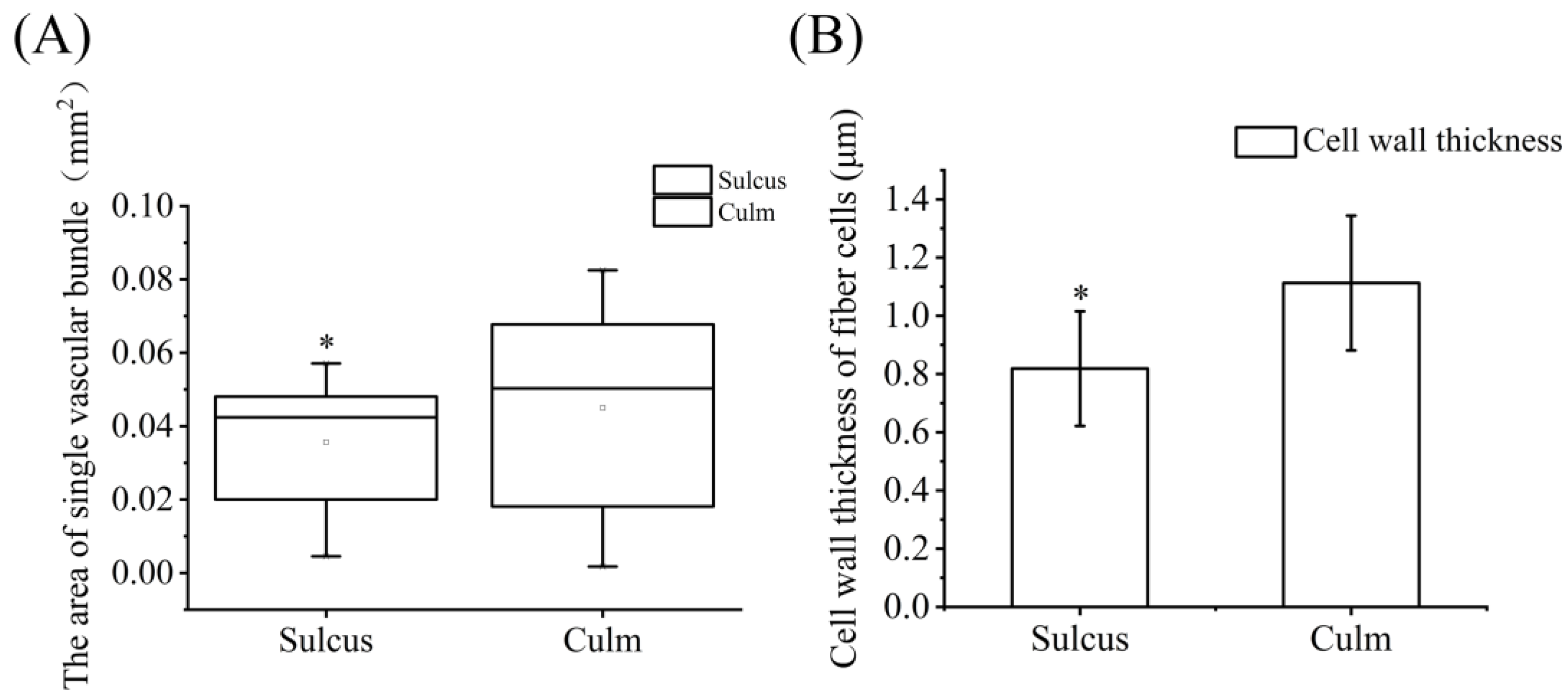
3.1. Histological Analysis of Internode Cross-Sections

To identify the structural differentiation between the culm and sulcus of P. violascens, we investigated these tissues through scanning electron microscopy (SEM) and cross-section staining. From Figure 1A,B, we can observe that the vascular bundles near the inner periphery were larger, while those adjacent to the outer periphery were smaller. Further, the vascular bundles distributed in the culm were organized in oblique rows and multiple lines (Figure 1B). By contrast, the vascular bundles’ distribution in the sulcus was much more random (Figure 1A). Further, the distribution density of vascular bundles in the sulcus and culm was 13.33 and 11.56/mm2, respectively (Table 1). The structural properties of vascular bundles, such as the maximum length of vascular bundles in the sulcus and culm, were approximately 0.28 and 0.38 mm (an increase in the size of 35.7% in the culm), respectively. The maximum width of vascular bundles, in the sulcus and culm was 0.29 and 0.32 mm (an increase in the size of 10.35% in the culm), respectively (Table 1). The vascular bundle’s length and width were relative to the vascular bundle’s area. Therefore, the culm average vascular bundles’ area also increased 26.2% more than in the sulcus (Figure 2A). Moreover, we also measured the cell wall thickness of bamboo fiber cells, and the result showed that the cell wall thickness in the culm was approximately 30% thicker than in the sulcus (Figure 2B). These observations indicated that the fiber cells in the culm might be more mature than that in the sulcus.
Further, to observe the lignin accumulation in the bamboo sulcus and culm, the cross-sections of the sulcus and culm were stained with phloroglucinol-HCl. The results showed that lignin accumulated around the vascular bundles. Further, the small vascular bundles in the outer sulcus surface were higher in number than in the culm (Figure 3A,B). The vascular bundles are spatial and more organized in the culm than the sulcus in the outer periphery sections (Figure 3A,B). At the same time, the inner sulcus periphery sections also contained a smaller and larger number of vascular bundles than the culm (Figure 3E,F). These structural observations of phloroglucinol-HCl stained sections were also confirmed with the culm and sulcus sections stained with toluidine blue O (Figure 3C,D,G,H). Further, the distribution density and size of vascular bundles in Figure 3 were also consistent with the SEM observations.

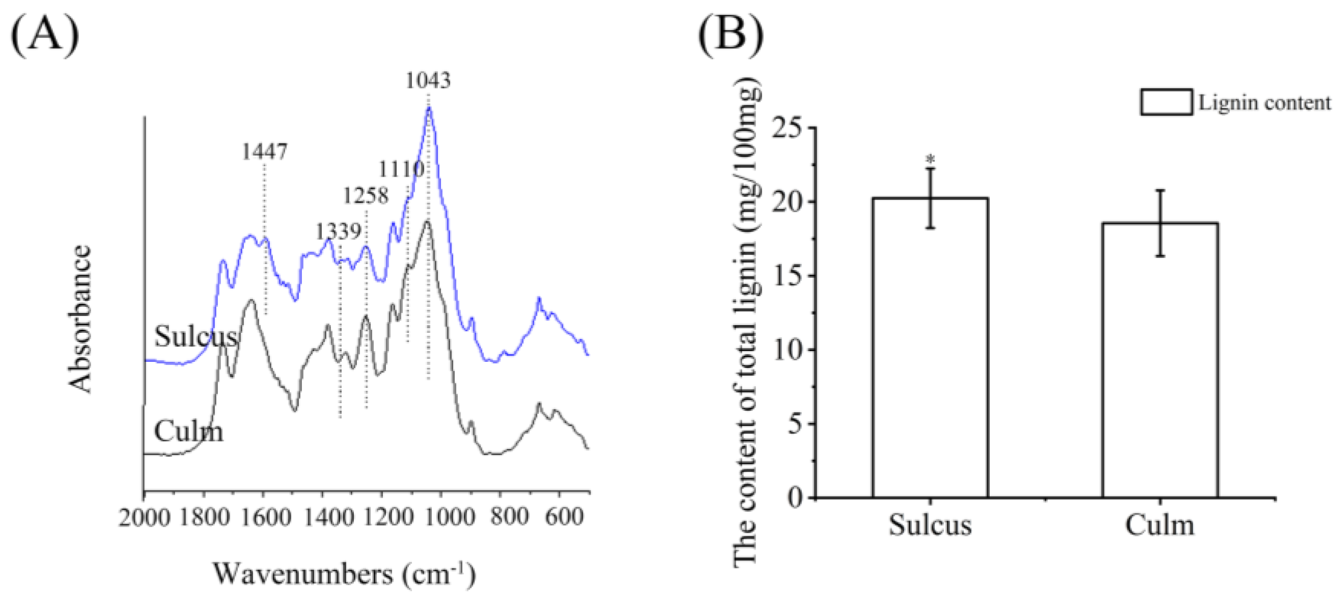
3.2. FT-IR Analysis and Lignin Content Measurement

In the current study, to analyze the differences in structural components such as lignin, cellulose, and hemicellulose between the culm and sulcus of P. violascens, FT-IR spectroscopy was used (Figure 4A). The FT-IR spectra results identified that the bamboo sulcus had basic structural similarities to the culm. Although the sulcus and culm had similar peaks, the absorbance of the sulcus was higher than the culm. Further, the absorption band that occurred at 1447 cm−1 was derived from the CH2 deformation vibration of lignin and xylan [24]. However, the signal at 1447 cm−1 in the culm was very weak. This meant that the sulcus and culm might differ in CH2 deformation vibrations in lignin and xylan. The peak value at 1339 cm−1 showed the CH2 bending vibration of lignin [25]. The peak value at 1258 cm−1 was caused by the stretching in the phenol–ether bonds of the lignin. By contrast, the most prominent peak at 1043 cm−1 was due to the C-O stretching of hemicellulose and cellulose [26]. The C-O stretching and ring asymmetric valence vibration at the 1110 cm−1 band belonged to lignin [27]. Further, we also analyzed the total lignin content (Figure 4B). The lignin content in the sulcus was approximately 20.24 ± 2.10 mg per 100 mg, while the lignin content in the culm was approximately 18.55 ± 2.22 mg per 100 mg. This result showed that the accumulation of lignin in the sulcus increased by 9%, which was consistent with the higher absorbance of the sulcus in the FT-IR spectra.

3.3. The Lignin Biosynthesis Genes Differentially Expressed between Sulcus and Culm

The lignin polymer is an essential component in the plant’s secondary cell wall, especially in wood and bark [28]. Lignin is the basal component that provides mechanical strength to plant tissues [29]. Altering the expression of lignin biosynthesis genes also affects the plant phenotype [30,31]. Therefore, in the current study, to identify the possible regulatory networks during internode development, we analyzed transcriptome data between the sulcus and the culm of P. violascens [3]. We identified many transcription factors, such as PvMYB108 (Cluster-4806.17597) and PvWRKY33 (Cluster-8360.0), which are differentially expressed genes (DEGs) that belong to various biological processes through GO analysis. The KEGG pathway enrichment results showed the precursor pathway to lignin biosynthesis, i.e., phenylpropanoid biosynthesis, which was enriched significantly among the two tissues. Here, we identified two genes directly involved in the lignin biosynthesis pathway that were highly expressed in the culm compared to the sulcus (Figure 5). These genes were Pv4CL (Cluster-4806.43335) and PvC4H (Cluster-4806.38638), and their fold change values were 1.77 and 2.61 (Supplementary Table S2). Their expressions were also quantified by the qRT-PCR (Figure 5), and the results were consistent with the transcriptomic data. The other lignin biosynthesis genes did not show different expression levels between the sulcus and culm, such as Phenylalanine ammonia lyase1-3 (PvPAL1-3) (Cluster-4806.23860, Cluster-4806.23845, and Cluster-4806.23685), Cinnamyl-alcohol dehydrogenase (PvCAD) (Cluster-4806.9308), Cinnamoyl-CoA reductase (PvCCR) (Cluster-4806.5663), p-Coumarate 3-hydroxylase (PvC3H) (Cluster-4806.17937), Caffeoyl-CoA O-methyltransferase (PvCCoAOMT) (Cluster-4806.30654), Catechol-O-methyltransferase (PvCOMT) (Cluster-4806.23962), Ferulate-5-hydroxylase (PvF5H) (Cluster-4806.19849) and Shikimate hydroxycinnamoyltransferase (PvHCT) (Cluster-4806.19849). However, these structural genes were highly expressed in both tissues, as confirmed by transcriptome and qPCR analysis. Therefore, we believe that Pv4CL and PvC4H might play the most critical roles in the lignin biosynthesis of the culm and sulcus.

4. Discussion

4.1. Phenotype Analysis

The bamboo internode contains two particular tissues: the culm and sulcus. The sulcus is usually a groove along the length of the internode and appears on alternating sides of the culm [9]. To identify the structural differences between the sulcus and culm, we conducted a histological analysis and lignin content measurement. Our results identified that vascular bundles near the inner periphery were larger in both the culm and sulcus, while those adjacent to the outer periphery were smaller. These results were consistent with the earlier results, but the earlier reports identified that smaller vascular bundles were very dense compared to larger ones [32,33]. These results might be because we used a 3 m bamboo for SEM analysis. The increase in the number of smaller vascular bundles might be due to an increase in the age of the bamboo culm. Further, the vascular bundles distributed in the culm were organized in oblique rows and multiple lines (Figure 1B and Figure 2). These results were also consistent with earlier studies [32,33]. By contrast, our results identified that vascular bundles in the sulcus were in a more random arrangement. Because of the smaller vascular bundles in the sulcus, the arrangement of the vascular bundles might be affected.

4.2. Bamboo Culm and Sulcus Composition Analysis

Similar to other species, bamboo’s major structural components also contain lignin, cellulose, and hemicellulose [34]. In the current study, the FT-IR spectra analysis showed that the sulcus and culm had similar basic structural features (Figure 4A). The similarities in the FT-IR features indicated that the basic components between the sulcus and culm were identical. The previous reports identified 900–1200 cm−1 bands attributed to the C-O stretching of bamboo [35,36]. The current study also found two peaks, 1043 cm−1 and 1110 cm−1, in this range between the sulcus and the culm. Further, an absorbance at 1447 cm−1 corresponding to lignin and xylan was also found in various bamboo species [34]. Although a peak was observed at 1447 cm−1 in the sulcus, the current study identified a weak signal in the culm, contrary to previous findings. Moreover, although the cell wall thickness of fiber cells in the culm was thicker (Figure 3B), the distribution density of the vascular bundle in the sulcus was much more than that in the culm. Therefore, there may be a greater number of fiber cells in the sulcus, which could explain why lignin content is higher in the sulcus than in the culm. Here, we conjectured that the compact density of the vascular bundle might lead to the development of the sulcus and also to higher lignin content.

4.3. Genes May Contribute to Lignin Biosynthesis in Bamboo Internodes

Currently, transcriptome technologies help to greatly unmask the hidden facts behind phenotypes in plants [37]. We identified two genes, Pv4CL and PvC4H, which were expressed higher in the culm by transcriptome and qPCR analysis between the sulcus and culm. Further, histological analysis and lignin content measurements revealed a higher distribution density of vascular bundles and lignin content in the sulcus. In Arabidopsis thaliana, At4CL1 and At4CL2 were primarily responsible for controlling the branching of the growing lignin molecule [38]. The ref3 mutant, which had mutations in the C4H gene, had reduced lignin deposition and an altered lignin monomer content [39]. In Oryza sativa, Os4CL1, Os4CL3, Os4CL4, and Os4CL5 genes were all responses to lignin biosynthesis, plant growth, and other morphological changes [40]. It was also reported that lignin synthesis in rice could be reduced by C4H and 4CL using RNAi [41]. In Populus tremuloides, Pt4CL1 was also associated with lignin biosynthesis when developing the xylem of woody stems [42]. Therefore, our results show that Pv4CL and PvC4H might play an essential role in lignin biosynthesis during sulcus and culm development. However, vascular bundles in the sulcus were much smaller and experienced abnormal growth. These results may relate to the lower expression levels of Pv4CL and PvC4H in the sulcus.
Plant variation is one of the most essential traits of ornamental plants. Recently, variations in the plant have been in great demand, and studying the underlying mechanism for future breeding is of great importance. As an ornamental bamboo in China, P. violascens cv. Viridisulcata displays a colorful internode. In this study, we tried to discover the structural differences and development between the sulcus and the culm. We found that the vascular bundles’ distribution density and size differentiated between the sulcus and the culm. Many enzymes in relation to lignin biosynthesis may promote lignin biosynthesis in the bamboo internode. Here, lignin accumulated more in the sulcus than in the culm. Therefore, the low expression of Pv4CL and PvC4H genes might lead to a smaller size, with a higher density of vascular bundles and higher lignin content in the sulcus. During the bamboo internode development, the culm with the sulcus become two different tissues and appear alternately on the bamboo internode. Moreover, bamboo products have entered all aspects and become indispensable to our life [43,44]. Xie et al. (2016) found that different vascular bundle shapes and sizes from different bamboo species could affect the physical and mechanical properties of fiber bundle-reinforced composites [45]. As a result of the mixing of sulcus and culm, bamboo sulcus with a high lignin content and abnormal vascular bundles may affect the quality of bamboo products. In the future, the bamboo internode may be divided into culms and sulcus for better product consistency in bamboo processing and utilization.

5. Conclusions

The bamboo internode contains a sulcus and culm, but the structural differences and development between the sulcus and culm remain unknown. In this study, we further conducted histological, composition, and gene expression analyses to explore the underlying developmental mechanism between the sulcus and the culm. Through histological analysis, we identified that vascular bundles in the sulcus were much smaller and had abnormal growth. Furthermore, vascular bundles in the sulcus were in a random arrangement, but the distribution density of vascular bundles in the sulcus was higher. The higher density of vascular bundles may relate to the higher lignin content in the sulcus. FT-IR analysis showed that sulcus and culm had similar basic structural features and also pointed out that they were different tissues. By transcriptome and qPCR analysis, the expression level of Pv4CL and PvC4H genes might affect the development of sulcus and culm. Our study may help discover the underlying mechanism between the sulcus and culm in bamboo and also pave the foundation for the genetic breeding of bamboo forests.

Supplementary Materials

The following supporting information can be downloaded at: https://www.mdpi.com/article/10.3390/f14061073/s1, Table S1: Primer sequences used in qRT-PCR experiments; Table S2: The expression information of relative genes involved flavonoid biosynthesis.

Author Contributions

X.-C.L., N.V., D.H. and A.-M.W. designed the experiments and reviewed the manuscript. H.-T.W. performed the experiments and wrote the original draft. H.-W.L. and J.-L.P. performed the sample sections. All authors have read and agreed to the published version of the manuscript.

Funding

This research work was supported by the National Key R&D Program of China (2021YFD2200503) and the Research Fund for International Young Scientists by the National Natural Science Foundation of China (32150410354).

Data Availability Statement

The transcriptomic data in this study can be found here: National Center for Biotechnology Information (NCBI) BioProject database under accession number GSE157799.

Conflicts of Interest

The authors declare no conflict of interest.

References

  1. Pei, J.L.; Wang, Y.; Zhuo, J.; Gao, H.B.; Vasupalli, N.; Hou, D.; Lin, X. Complete Chloroplast Genome Features of Dendrocalamusfarinosus and Its Comparison and Evolutionary Analysis with Other Bambusoideae Species. Genes 2022, 13, 1519. [Google Scholar] [CrossRef] [PubMed]
  2. Vasupalli, N.; Hou, D.; Singh, R.M.; Wei, H.T.; Zou, L.H.; Yrjälä, K.; Wu, A.; Lin, X. Homo-and hetero-dimers of CAD enzymes regulate lignification and abiotic stress response in Moso Bamboo. Int. J. Mol. Sci. 2021, 22, 12917. [Google Scholar] [CrossRef] [PubMed]
  3. Wei, H.T.; Hou, D.; Ashraf, M.F.; Lu, H.W.; Zhuo, J.; Pei, J.L.; Qian, Q.X. Metabolic Profiling and Transcriptome Analysis Reveal the Key Role of Flavonoids in Internode Coloration of Phyllostachys violascens cv. Viridisulcata. Front. Plant Sci. 2022, 12, 3413. [Google Scholar] [CrossRef]
  4. Ye, S.; Chen, G.; Kohnen, M.V.; Wang, W.; Cai, C.; Ding, W.S.; Wu, C.; Gu, L.; Zheng, Y.; Ma, X.; et al. Robust CRISPR/Cas9 mediated genome editing and its application in manipulating plant height in the first generation of hexaploid Ma bamboo (Dendrocalamus latiflorus Munro). Plant Biotechnol. J. 2020, 18, 1501–1503. [Google Scholar] [CrossRef] [PubMed]
  5. Fang, W.; Gui, R.Y.; Ma, L.F.; Jin, A.W.; Lin, X.C.; Yu, X.J.; Ma, L.J.; Qian, J. Chinese Economic Bamboo; China Science Publishing & Media: Beijing, China, 2015; p. 402. [Google Scholar]
  6. Jin, H.Y.; Zhi, W.Z. Design of Household Multi-Function Vacuum Cleaner. Adv. Mater. Res. 2014, 945, 266–269. [Google Scholar] [CrossRef]
  7. Song, C.; Igarashi, M.; Ikei, H.; Miyazaki, Y. Physiological effects of viewing fresh red roses. Complement. Ther. Med. 2017, 35, 78–84. [Google Scholar] [CrossRef]
  8. Zheng, J.M.; Tian, M.W.K.; Jiang, D.H.; Li, M.; Ye, J.; Chen, L.Y.; He, T.; Zheng, Y. Which ornamental features of bamboo plants will attract the people most? Urban For. Urban Green. 2021, 61, 127101. [Google Scholar] [CrossRef]
  9. Lin, X.C.; Ruan, X.S.; Lou, Y.F.; Guo, X.Q.; Fang, W. Genetic similarity among cultivars of Phyllostachys pubescens. Plant Syst. Evol. 2009, 277, 67–73. [Google Scholar] [CrossRef]
  10. Lin, X.C.; Lou, Y.F.; Zhang, Y.Z.; Yuan, X.L.; He, J.C.; Fang, W. Identification of Genetic Diversity Among Cultivars of Phyllostachys violascens Using ISSR, SRAP and AFLP Markers. Bot. Rev. 2011, 77, 223–232. [Google Scholar] [CrossRef]
  11. Xia, X.; Gui, R.; Yang, H.; Fu, Y.; Wei, F.; Zhou, M. Identification of genes involved in color variation of bamboo culms by suppression subtractive hybridization. Plant Physiol. Biochem. 2015, 97, 156–164. [Google Scholar] [CrossRef]
  12. Jin, Y.C.; Yuan, K. Studies on the functional components and bioactivity and the relativity of bamboo shoots and shells. Adv. Mater. Res. 2011, 108, 314–319. [Google Scholar] [CrossRef]
  13. Cai, K.; Zhu, L.; Zhang, K.; Li, L.; Zhao, Z.; Zeng, W.; Lin, X. Development and characterization of EST-SSR markers from RNA-Seq data in Phyllostachys violascens. Front. Plant Sci. 2019, 10, 50. [Google Scholar] [CrossRef] [PubMed]
  14. Młodzińska, E. Survey of plant pigments: Molecular and environmental determinants of plant colors. Acta Biol. Crac. Ser. Bot. 2009, 51, 7–16. [Google Scholar] [CrossRef]
  15. Kanzawa, E.; Aoyagi, S.; Nakano, T. Vascular bundle shape in cross-section and relaxation properties of Moso bamboo (Phyllostachys pubescens). Mater. Sci. Eng. C 2011, 31, 1050–1054. [Google Scholar] [CrossRef]
  16. Wang, K.L.; Wang, B.; Hu, R.; Zhao, X.; Li, H.; Zhou, G.; Song, L.; Wu, A.M. Characterization of hemicelluloses in Phyllostachys edulis (moso bamboo) culm during xylogenesis. Carbohydr. Polym. 2019, 221, 127–136. [Google Scholar] [CrossRef]
  17. Qin, W.; Yin, Q.; Chen, J.; Zhao, X.; Yue, F.; He, J.; Yang, L.; Liu, L.; Zeng, Q.; Lu, F.; et al. The Class II KNOX transcription factors KNAT3 and KNAT7 synergistically regulate monolignol biosynthesis in Arabidopsis. J. Exp. Bot. 2020, 71, 5469–5483. [Google Scholar] [CrossRef]
  18. Love, M.I.; Huber, W.; Anders, S. Moderated estimation of fold change and dispersion for RNA-seq data with DESeq2. Genome Biol. 2014, 15, 550. [Google Scholar] [CrossRef]
  19. Chen, C.J.; Chen, H.; Zhang, Y.; Thomas, H.R.; Frank, M.H.; He, Y.H.; Xia, R. TBtools: An Integrative Toolkit Developed for Interactive Analyses of Big Biological Data. Mol. Plant 2020, 13, 1194–1202. [Google Scholar] [CrossRef]
  20. Hou, D.; Zhao, Z.Y.; Hu, Q.T.; Li, L.; Vasupalli, N.; Zhuo, J.; Zeng, W.; Wu, A.; Lin, X. PeSNAC-1 a NAC transcription factor from moso bamboo (Phyllostachys edulis) confers tolerance to salinity and drought stress in transgenic rice. Tree Physiol. 2020, 40, 1792–1806. [Google Scholar] [CrossRef]
  21. Schmittgen, T.D.; Livak, K.J. Analyzing real-time PCR data by thecomparative CT method. Nat. Protoc. 2008, 3, 1101–1108. [Google Scholar] [CrossRef]
  22. Fan, C.; Ma, J.; Guo, Q.; Li, X.; Wang, H.; Lu, M. Selection of reference genes for quantitative real-time PCR in bamboo (Phyllostachys edulis). PLoS ONE 2013, 8, e56573. [Google Scholar] [CrossRef]
  23. Hou, D.; Li, L.; Ma, T.F.; Pei, J.L.; Zhao, Z.Y.; Lu, M.Z.; Wu, A.; Lin, X. The SOC1-like gene BoMADS50 is associated with the flowering of Bambusa oldhamii. Hortic. Res. 2021, 8, 133. [Google Scholar] [CrossRef] [PubMed]
  24. Schwanninger, M.; Rodrigues, J.C.; Pereira, H.; Hinterstoisser, B. Effects of short-time vibratory ball milling on the shape of FT-IR spectra of wood and cellulose. Vib. Spectrosc. 2004, 36, 23–40. [Google Scholar] [CrossRef]
  25. Åkerholm, M.; Salmén, L. The Oriented Structure of Lignin and its Viscoelastic Properties Studied by Static and Dynamic FT-IR Spectroscopy. Holzforschung 2003, 57, 459–465. [Google Scholar] [CrossRef]
  26. Kačuráková, M.; Smith, A.C.; Gidley, M.J.; Wilson, R.H. Molecular interactions in bacterial cellulose composites studied by 1D FT-IR and dynamic 2D FT-IR spectroscopy. Carbohydr. Res. 2002, 337, 1145–1153. [Google Scholar] [CrossRef]
  27. Cheng, S.; Huang, A.; Wang, S.; Zhang, Q. Effect of different heat treatment temperatures on the chemical composition and structure of Chinese fir wood. BioResources 2016, 11, 4006–4016. [Google Scholar] [CrossRef]
  28. Legay, S.; Sivadon, P.; Blervacq, A.; Pavy, N.; Baghdady, A.; Tremblay, L.; Levasseur, C.; Ladouce, N.; Lapierre, C.; Séguin, A.; et al. EgMYB1, an R2R3 MYB transcription factor from eucalyptus negatively regulates secondary cell wall formation in Arabidopsis and poplar. New Phytol. 2021, 188, 774–786. [Google Scholar] [CrossRef] [PubMed]
  29. Kamran, M.; Cui, W.; Ahmad, I.; Meng, X.; Zhang, X.; Su, W.; Chen, J.; Ahmad, S.; Fahad, S.; Han, Q.; et al. Effect of paclobutrazol, a potential growth regulator on stalk mechanical strength, lignin accumulation and its relation with lodging resistance of maize. Plant Growth Regul. 2018, 84, 317–332. [Google Scholar] [CrossRef]
  30. Yoon, J.; Choi, H.; An, G. Roles of lignin biosynthesis and regulatory genes in plant development. J. Integr. Plant Biol. 2015, 57, 902–912. [Google Scholar] [CrossRef]
  31. Zhao, Q.; Dixon, R.A. Transcriptional networks for lignin biosynthesis: More complex than we thought? Trends Plant Sci. 2011, 16, 227–233. [Google Scholar] [CrossRef]
  32. Shang, L.L.; Sun, Z.J.; Liu, X.; Jiang, Z.H. A novel method for measuring mechanical properties of vascular bundles in moso bamboo. J. Wood Sci. 2015, 61, 562–568. [Google Scholar] [CrossRef]
  33. Li, J.; Xu, H.C.; Yu, Y.; Chen, H.; Yi, W.K.; Wang, H.K. Intelligent analysis technology of bamboo structure. Part I: The variability of vascular bundles and fiber sheath area. Ind. Crops Prod. 2021, 174, 114163. [Google Scholar] [CrossRef]
  34. Mohd, T.B.M.; Bhat, I.U.H.; Mohmod, A.L.; Aditiawati, P.; Abdul Khalil, H.P.S. Thermal and FT-IR Characterization of Gigantochloa levis and Gigantochloa scortechinii Bamboo, a Naturally Occurring Polymeric Composite. J. Polym. Environ. 2013, 21, 534–544. [Google Scholar] [CrossRef]
  35. Sun, B.; Huang, A.; Wang, Y.; Liu, J. Natural bamboo (Neosinocalamus affinis Keng) fiber identification using FT-IR and 2D-IR correlation spectroscopy. J. Nat. Fibers 2015, 12, 796907. [Google Scholar] [CrossRef]
  36. Cai, Q.; Fan, Z.; Chen, J.; Guo, W.; Ma, F.; Sun, S.; Hu, L.; Zhou, Q. Dissolving process of bamboo powder analyzed by FT-IR spectroscopy. J. Mol. Struct. 2018, 1171, 639–643. [Google Scholar] [CrossRef]
  37. Ashraf, M.F.; Hou, D.; Hussain, Q.; Imran, M.; Pei, J.; Ali, M.; Shehzad, A.; Anwar, M.; Noman, A.; Waseem, M.; et al. Entailing the Next-Generation Sequencing and Metabolome for Sustainable Agriculture by Improving Plant Tolerance. Int. J. Mol. Sci. 2022, 23, 651. [Google Scholar] [CrossRef]
  38. Soltani, B.M.; Ehlting, J.; Douglas, C.J. Genetic analysis and epigenetic silencing of At4CL1 and At4Cl2 expression in transgenic Arabidopsis. Biotechnol. J. 2006, 1, 1124–1136. [Google Scholar] [CrossRef]
  39. Schilmiller, A.L.; Stout, J.; Weng, J.K.; Humphreys, J.; Ruegger, M.O.; Chapple, C. Mutations in the cinnamate 4-hydroxylase gene impact metabolism, growth and development in Arabidopsis. Plant J. 2009, 60, 771–782. [Google Scholar] [CrossRef]
  40. Gui, J.; Shen, J.; Li, L. Functional characterization of evolutionarily divergent 4-coumarate: Coenzyme a ligases in rice. Plant Physiol. 2011, 157, 574–586. [Google Scholar] [CrossRef]
  41. Xia, G.S. Repression of Lignin Synthesis in Rice by C4H and 4CL Using RNAi. Int. J. Biosci. Biochem. Bioinform. 2013, 3, 226–228. [Google Scholar] [CrossRef]
  42. Hu, W.J.; Kawaoka, A.; Tsai, C.J.; Lung, J.; Osakabe, K.; Ebinuma, H.; Chiang, V.L. Compartmentalized expression of two structurally and functionally distinct 4-coumarate:CoA ligase genes in aspen (Populus tremuloides). Proc. Natl. Acad. Sci. USA 1998, 95, 5407–5541. [Google Scholar] [CrossRef] [PubMed]
  43. Taskhiri, M.S.; Jeswani, H.; Geldermann, J.; Azapagic, A. Optimizing cascaded utilization of wood resources considering economic and environmental aspects. Comput. Chem. Eng. 2019, 124, 302–316. [Google Scholar] [CrossRef]
  44. Peng, Z.; Lu, Y.; Li, L.; Zhao, Q.; Feng, Q.; Gao, Z.; Lu, H.; Hu, T.; Yao, N.; Liu, K.; et al. The draft genome of the fast-growing non-timber forest species moso bamboo (Phyllostachys heterocycla). Nat. Genet. 2013, 45, 456–461. [Google Scholar] [CrossRef]
  45. Xie, J.L.; Qi, J.Q.; Hu, T.X.; Hoop, C.F.D.; Hse, C.Y.; Shupe, T.F. Effect of fabricated density and bamboo species on physical–mechanical properties of bamboo fiber bundle reinforced composites. J. Mater. Sci. 2016, 51, 7480–7490. [Google Scholar] [CrossRef]
Figure 1. Scanning electron microscopy observation of P. violascens cv. Viridisulcata. The internode consists of the sulcus (A) and culm (B) in P. violascens.
Forests 14 01073 g001
Figure 2. Measurement of vascular bundle area and cell wall thickness of fiber cells. The area of the single vascular bundle (A) and cell wall thickness of fiber cells. The small square represents the average number. (B) were measured between the sulcus and culm. Significant differences were determined by t-test (* p < 0.05).
Forests 14 01073 g002
Figure 3. Phloroglucinol-HCl and toluidine blue O staining of cross sections between sulcus and culm. The sulcus and culm were divided into an outer- and inner-side to observe the whole thickness of the bamboo internode. (A,B,E,F) were stained by phloroglucinol-HCl, and (C,D,G,H) were stained by toluidine blue O. The scale was 60 μm.
Forests 14 01073 g003
Figure 4. The sulcus and culm FT–IR analysis and total lignin content measurement. (A) FT−IR spectrum of sulcus and culm. The blue spectrum indicates the sulcus, and the black spectrum indicates the culm (B) Lignin content measurement between the sulcus and the culm. Significant differences were determined by t-test (* p < 0.05).
Forests 14 01073 g004
Figure 5. Total genes relative to the lignin biosynthesis pathway in this study. Lignin biosynthesis pathway; the red color genes represent differentially expressed genes between the sulcus and culm. The enzymes involved in the lignin biosynthesis pathway: PAL, phenylalanine ammonia lyase; 4CL, 4-coumarate-CoA ligase; C3H, p-coumarate 3-hydroxylase; C4H, cinnamate-4-hydroxylase; F5H, Ferulate-5-hydroxylase; COMT, Catechol-O-methyltransferase; HCT, 4-coumaroyl-CoA:shikimate O-(hydroxycinnamoyl) transferase; C3′H, p-Coumaroyl Quinate/Shikimate 3′-Hydroxylase; CAD, cinnamyl-alcohol dehydrogenase; CCoAOMT, Caffeoyl-CoA O-methyltransferase; CCR, cinnamoyl-CoA reductase; LAC, laccase; PRX, peroxidase. Relative expression of genes at the transcription level by qRT-PCR.
Forests 14 01073 g005
Table 1. The vascular bundle density and structural parameters of sulcus and culm.
TissueThe Distribution Density of Vascular Bundles
(per·mm2)
Vascular Bundle’s Structural Parameters
Maximum Length (mm)Maximum Width (mm)
sulcus13.33 ± 0.351 a0.28 ± 0.02 a0.29 ± 0.01 a
culm11.56 ± 0.426 b0.38 ± 0.01 b0.32 ± 0.02 b
Significant differences were determined by t-test (p < 0.05). Values are the means ± SD for six technical replicates. The significance is indicated by lowercase letters a and b.
Disclaimer/Publisher’s Note: The statements, opinions and data contained in all publications are solely those of the individual author(s) and contributor(s) and not of MDPI and/or the editor(s). MDPI and/or the editor(s) disclaim responsibility for any injury to people or property resulting from any ideas, methods, instructions or products referred to in the content.

