Article

Cinnamyl Alcohol Dehydrogenase Gene Regulates Bursaphelenchus xylophilus Reproduction and Development

College of Life Science, Qingdao University, Qingdao 266071, China
*
Authors to whom correspondence should be addressed.
Forests 2023, 14(7), 1379; https://doi.org/10.3390/f14071379
Submission received: 5 June 2023 / Revised: 28 June 2023 / Accepted: 29 June 2023 / Published: 5 July 2023
(This article belongs to the Section Forest Health)

Abstract

:
Pine wilt disease (PWD) caused by the pine wood nematode (PWN), Bursaphelenchus xylophilus, is a globally distributed destructive disease of pine forest. To study the PWD pathogenic mechanism, the cinnamyl alcohol dehydrogenase gene (BxCAD-1) from B. xylophilus was selected. The BxCAD-1 gene was amplified by PCR from the cDNA library of B. xylophilus and cloned into the expression vector pET-15b to construct the recombinant vector pET-15b-BxCAD-1. The recombinant cinnamyl alcohol dehydrogenase protein was purified by Ni-NTA affinity chromatography from Escherichia coli BL21 (DE3) harboring pET-15b-BxCAD-1 induced by IPTG. The effects of pH, temperature, metal ions and substrates on the activity of BxCAD-1 were determined, showing the highest catalytic activity at pH 8.0 and 40 °C with cinnamyl alcohol as substrate and Zn2+ as an activator. To elucidate the functions of BxCAD-1 in B. xylophilus, the expression of the gene was down-regulated by RNA interference. Results showed that the movement, feeding, reproduction, spawning rate, hatching rate, lifespan, infectivity and sensitivity to ethanol decreased compared with negative controls. RNA interference also affected the development of B. xylophilus from the larval stage to the adult stage. In situ hybridization showed that the gene was expressed in the digestive tract of male and female adults. This study revealed a promising target for PWD control.

1. Introduction

Pine wilt disease (PWD) is a devastating disease leading to the rapid death of pine trees and the large-scale destruction of pine forests. It is characterized by rapid transmission, a short onset cycle and difficult control [1]. The disease is caused by the pine wood nematode (PWN) (Bursaphelenchus xylophilus) through insect vectors such as Monochamus alternatus and other cerambycid beetles [2]. Although the pine trees have no visible symptoms at the initial stage of infection, PWD causes a decrease in resin secretion, lipid peroxidation and exudation of cell contents. As the disease progresses, the pine needles turn yellowish brown or reddish brown, wilt and droop until the trees wither and die [3,4,5]. Pine trees in North America, where PWD originated, are not severely affected [6,7]. In Asia, the PWN was first discovered in Nagasaki on Kyushu Island in Japan in 1905 and then rapidly spread to other countries in East Asia and Europe, resulting in huge losses to the local ecological environment and economy [8,9,10].
Pine wilt disease is related to complex interactions among B. xylophilus, insect vectors and host pines. The PWN pathogenic mechanism has not been fully understood [11]. The enzyme hypothesis suggests that B. xylophilus invades the pine tree by secreting a large number of enzymes such as cellulase, lipase, glycoside hydrolase and peptidase, which attack the cell wall and membrane of the parenchyma tissue of the pine tree, resulting in the leakage of oil and the obstruction of water transmission [12,13,14,15,16]. The cavitation hypothesis proposes that tracheid cavitation occurs in the xylem at the early stage of infection before the symptoms of PWD appear, which is caused by the production of hydrophobic terpenes in B. xylophilus infected pine trees and blocks water transport [17,18,19,20]. The toxin hypothesis suggests that the pathogenicity of B. xylophilus alone is not strong or even unable to cause disease in pine trees, but the symbiosis with toxin-producing bacteria can lead to the death of pine trees [21,22,23,24].
Cinnamyl alcohol dehydrogenase (CAD) (EC1.1.1.195) was first found in soybean [25]. This enzyme exists in plants and participates in the final step of cell wall synthesis, containing a highly conserved NAD(P)H/NAD(P)+ binding domain, which is also dependent on the activation of Zn2+ [26,27]. Studies have shown that CAD is involved in lignin synthesis in Artemisia annua [28], Oryza sativa [29], Arabidopsis [30], Triticum aestivum [31] and Populus tomentosa [32]. In Saccharomyces cerevisiae, CADs may be involved in fusel synthesis, lignin decomposition and NADP(H) dynamic balance [33,34]. In Sclerotinia sclerotiorum, CAD regulates its development through the reactive oxygen species (ROS) pathway [35]. Moreover, CAD is involved in the lipid biosynthesis of cell membranes in Mycobacterium bovis BCG [36], and it can disproportionate benzaldehyde to produce benzyl alcohol and benzoic acid in Helicobacter pylori [37].
RNA interference (RNAi) is an effective method to study the biological function of genes. It utilizes homologous double-stranded RNA (dsRNA) to silence gene function at the post-transcriptional level, resulting in the degradation of the target mRNA sequence [38,39,40,41]. Xue et al. [42] found three cathepsin L-like cysteine proteinase (CPL) genes, Bx-Cpls-1, Bx-Cpls-2 and Bx-Cpls-3 of B. xylophilus. Silencing Bx-Cpl gene can reduce the feeding energy, reproductive ability and pathogenicity of B. xylophilus. Qiu et al. [43] and Qiu et al. [44] inhibited the expression of cytochrome P450 gene, CYP-33C9 and pectin lyase gene, Bxpel1 in B. xylophilus by RNAi technology, respectively, and effectively slowed B. xylophilus migration. Hu et al. [45] found that the expression level of BxSapB1, an effector factor of B. xylophilus, was upregulated in the highly virulent strain. Silencing this gene significantly reduced B. xylophilus virulence.
4-Methylpyrazole is a competitive inhibitor of alcohol dehydrogenase [46,47,48]. In a previous study on the transcriptome of B. xylophilus treated with 4-Methylpyrazole, an up-regulated gene BxCAD-1 coding cinnamyl alcohol dehydrogenase was identified [49]. At present, there are few studies on cinnamyl alcohol dehydrogenase in animals, and cinnamyl alcohol dehydrogenase has never been reported in B. xylophilus. The data from this study will support the understanding of BxCAD-1 in the reproduction and development of B. xylophilus, which may be an important pathogenic factor of B. xylophilus and provide a new molecular target for the exploration of nematicides and control of PWD. If it is universal, it can provide some data support for the control of other nematodes.

2. Materials and Methods

2.1. Materials

Bursaphelenchus xylophilus were collected from infected Pinus thunbergii by the Baermann funnel method [1] from Muping District, Yantai City, Shandong Province, China. Botrytis cinerea was kept in our laboratory. The products and manufacturers used in this experiment are listed in Table 1.
Mature cones of P. thunbergii on the campus of Qingdao University were collected, and the seeds were soaked overnight and placed in the seedling substrate. The seedlings were cultured at 25 °C in greenhouse for about 30 days to obtain P. thunbergii seedlings.

2.2. Isolation and Culture of B. xylophilus

B. xylophilus extracted from infected P. thunbergii were inoculated in B. cinerea cultures on Potato Dextrose Agar (PDA) medium at 25 °C in the dark for 9 days [50]. B. xylophilus were collected from B. cinerea cultures by the Baermann funnel method.

2.3. Construction of cDNA Library of B. xylophilus

Approximately one thousand PWNs (mixed developmental stages) were used to extract the total RNA following the instruction of the RNA Easy Fast Tissue/Cell Kit. The construction of cDNA library was performed using the PrimeScriptTM II First Strand cDNA Synthesis Kit following the manufacturer’s instructions.

2.4. Cloning of BxCAD-1 Gene

A pair of specific primers, CAD-F and CAD-R (Table 2), were generated from the gene sequence of BxCAD-1 gene and designed using Primer Premier 5.0 software (Premier, Canada). PCR amplification was carried out with the cDNA library as the template. The amplification program included one cycle at 94 °C for 5 min, 35 cycles at 94 °C for 40 s, 56 °C for 42 s and 72 °C for 90 s respectively, followed by one cycle at 72 °C for 10 min. The PCR amplification products were analyzed by 1% agarose gel electrophoresis, and the target gene was recovered by Extraction Mini Kit. The BxCAD-1 gene was cloned into vector pEASY-T1 to obtain pEASY-T1-BxCAD-1, which was amplified in E. coli DH5α and sequenced at Sangon Biotech Co., Ltd. (Shanghai, China).

2.5. Biological Information Analysis of BxCAD-1 Gene

The domain (https://www.ebi.ac.uk/interpro/result/InterProScan/iprscan5-R20220307-030447-0221-57005514-p2m/) (accessed on 14 May 2021), signal peptide (https://services.healthtech.dtu.dk/service.php?SignalP-5.0) (accessed on 14 May 2021), transmembrane domain (https://services.healthtech.dtu.dk/service.php?TMHMM-2.0) (accessed on 14 May 2021) and three-dimensional structure (https://swissmodel.expasy.org/) (accessed on 14 May 2021) of the amino acid residue sequence encoded by BxCAD-1 gene were predicted and analyzed. The BxCAD-1 gene was entered into NCBI database for homology analysis. The DNAMAN software (6.0.3.99) was used for sequence alignment, and phylogenetic tree of amino acids was constructed by MEGA software (X-10) [51].

2.6. Construction of Engineered Bacteria

The recombinant cloning vector pEASY-T1-BxCAD-1 and expression vector pET-15b were double digested by restriction endonuclease NdeI, XhoI. The BxCAD-1 gene was recovered after agarose gel electrophoresis and cloned into the expression vector pET-15b by T4 DNA ligase, and the recombinant pET-15b-BxCAD-1 was transformed into E. coli BL21 (DE3) to construct the engineered bacteria [52].

2.7. Expression and Purification of Recombinant BxCAD-1

A single colony of engineered bacteria was cultured in 100 mL of Luria–Bertani (LB) medium with ampicillin (100 μg/mL) at 37 °C for 12 h, and 60 mL culture was transferred to 3 L of LB medium with ampicillin (100 μg/mL) for further incubation at 37 °C for 5 h. Next, IPTG was added into the culture to a final concentration of 1.0 mmol/L, and incubation lasted for another 5 h. The fermentation broth was centrifuged at 16,500× g for 40 min. The bacterial pellet was resuspended with 20 mL of binding buffer (10% glycerol, 0.5 mol/L NaCl, 20 mmol/L Tris-Cl pH 8.0, 5.0 mmol/L imidazole) and lyzed by intermittent sonication (sonication for 5 s and intermission for 15 s) on ice at 400 W for 2 h. The cell lysates were centrifuged at 4140× g for 15 min at 4 °C; inclusion bodies were collected and washed three times with 6 mL of washing buffer (0.5 mol/L NaCl, 2.0 mol/L Urea, 0.1 mmol/L PMSF, 0.5% TritonX-100, 20 mmol/L Tris-Cl pH 8.0) followed by centrifugation at 4140× g for 15 min at 4 °C. The final inclusion bodies were resuspended in 3 mL of denaturing buffer (0.5 mol/L NaCl, 8.0 mol/L Urea, 0.1 mmol/L phenylmethanesulfonyl fluoride, 20 mmol/L Tris-Cl pH 8.0). The suspension was serially diluted using refolding buffer (0.5 mol/L NaCl, 0.1 mmol/L phenylmethanesulfonyl fluoride, 20 mmol/L Tris-Cl pH 8.0) to reach a final urea concentration of less than 1.0 mol/L. Next, the refolded recombinant protein was purified by Ni-NTA affinity chromatography and identified by SDS-PAGE (12% gel) [53].

2.8. Determination of Physical and Chemical Properties of BxCAD-1

The reaction system for enzyme activity determination includes 100 mmol/L Tris-Cl (pH 8.0), 5 mmol/L NAD, 92.4 μg/mL enzyme solution, 100 mmol/L ethanol and 0.1 mmol/L Zn2+. The reaction was performed at 40 °C for 1 h, followed by the measurement of optical density at 340 nm (A340). One unit (U) of enzyme activity was defined as the amount of enzyme that increases by 0.001 at A340 per minute. The specific activity (U/mg) of the enzyme was defined as the units of enzyme activity per mg protein. The enzyme activity assay was carried out in three replications [53].
The effects of different temperatures (10 °C, 20 °C, 30 °C, 40 °C and 50 °C), pH (6.0, 7.0, 8.0, 9.0 and 10.0), metal ions (Zn2+, Fe2+, Ni2+, Mg2+ and K+), and substrates (methanol, ethanol, isopropanol, sorbitol, cinnamyl alcohol, D-pinitol and 2-methoxyphenol) on BxCAD-1 activity were determined.

2.9. RNA Interference for BxCAD-1 Gene

2.9.1. Synthesis of dsRNA

Three fragments of BxCAD-1 for RNA interference were selected, which included fragment 1 (1 to 430 bp of ORF, with a total length of 430 bp), fragment 2 (200 to 700 bp of ORF, with a total length of 500 bp) and fragment 3 (600 to 1000 bp of ORF, with a total length of 400 bp). PCR amplification was carried out with three pairs of specific primers: CAD-1-F and CAD-1-R, CAD-2-F and CAD-2-R, CAD-3-F and CAD-3-R, respectively (Table 2), and pET-15b-BxCAD-1 was used as the template. The amplifications of the three fragments were finished according to the program: one cycle at 94 °C for 5 min, 35 cycles at 94 °C for 40 s, 56 °C for 42 s, 72 °C for 40 s. The three fragments were further amplified using specific primers CAD-1T7-F, CAD-1T7-R; CAD-2T7-F, CAD-2T7-R and CAD-3T7-F, CAD-3T7-R (Table 2), respectively, to add T7 promoter to the up-stream of three fragments. A pair of specific primers GFP-F and GFP-R (Table 2) with T7 promoter were used for PCR amplification with pET-15b-GFP as template; the amplification conditions were as described above. DsRNA was synthesized by using the amplified product as template, and the synthesis method was referred to the instructions of the MEGAscriptTM RNAi Kit [53].

2.9.2. Efficiency Assessment of RNAi

QRT-PCR was used to verify the effect of BxCAD-1 silencing through RNAi on mRNA levels. Approximately 3000 B. xylophilus (mixed developmental stages) were soaked in 50 μL dsRNA solution (1.0 μg/μL) and cultured in the dark at 20 °C at 180 rpm for 72 h [53]. B. xylophilus soaked in distilled H2O and gfp dsRNA solution (1.0 μg/μL) were used as control. B. xylophilus were washed with sterile water 4 times, and the extraction of total RNA and construction of cDNA library was performed using the RNA Easy Fast Tissue/Cell Kit and HiScript III RT SuperMix for qPCR, respectively. QRT-PCR has used the ChanQ Universal SYBR qPCR Master Mix. The three fragments were further amplified using specific primers Q1F, Q1R; Q2F, Q2R and Q3F, Q3R (Table 2), respectively. The actin gene amplified was used as the internal control using specific primers CTF and CTR [51]. The PCR program was set as follows: 30 s at 95 °C, followed by 20 cycles of 5 s at 95 °C and 30 s at 58 °C. Each experiment was performed with three biological and three technical replicates.

2.9.3. Effect of RNAi on Movement

Approximately 3000 B. xylophilus (mixed developmental stages) were soaked in 50 μL dsRNA solution (1.0 μg/μL) and cultured in the dark at 20 °C at 180 rpm for 72 h, and all B. xylophilus used in this study were interfered in this way [53]. Fifty B. xylophilus were randomly selected for optical microscope observation. The head thrashing frequency in 1 min was monitored as an indicator of vitality [54]. B. xylophilus soaked in distilled H2O, gfp dsRNA solution (1.0 μg/μL) and 0.02 mmol/L 4-methylpyrazole solution were used as control.

2.9.4. Effect of RNAi on Feeding and Reproduction

Approximately 200 B. xylophilus with ratio of female to male of 1:1 were inoculated on PDA plates full of B. cinerea and cultured in the dark at 25 °C. The feeding situation was observed every day. After 9 days of culture, B. xylophilus were isolated from PDA plates by the Baermann funnel method and counted under an optical microscope [51]. B. xylophilus soaked in distilled H2O, gfp dsRNA solution (1.0 μg/μL) and 0.02 mmol/L 4-methylpyrazole solution were used as control. The experiment was repeated five times.

2.9.5. Effect of RNAi on Spawning Rate and Hatching Rate

One hundred microliters of B. xylophilus suspension (approximately 400 B. xylophilus with ratio of female to male of 1:1) were transferred into a 96-well plate in the dark at 25 °C for 12 h, and the number of eggs was counted under an optical microscope. Eggs were separated and incubated in the dark at 25 °C for 24 h for hatching. The number of total eggs at the beginning of assay and the remaining eggs after 24 h was recorded to calculate the number of hatched eggs and the hatching rate [51]. Bursaphelenchus xylophilus soaked in distilled H2O, gfp dsRNA solution (1.0 μg/μL) and 0.02 mmol/L 4-methylpyrazole solution were used as control, respectively. The experiment was repeated three times.

2.9.6. Effect of RNAi on Lifespan of PWN

One hundred microliters of B. xylophilus suspension (approximately 200 mixed developmental stages B. xylophilus) were transferred into a 96-well plate in the dark at 25 °C for 72 h; the number of surviving B. xylophilus were counted under an optical microscope and observed every 24 h [55]. Bursaphelenchus xylophilus soaked in distilled H2O, gfp dsRNA solution (1 μg/μL) and 0.02 mmol/L 4-methylpyrazole solution were used as control, respectively. The experiment was repeated three times.

2.9.7. Effect of RNAi on Infectivity

Approximately 200 B. xylophilus (mixed developmental stages) were inoculated into Pinus thunbergii seedlings. A small wound was made at the top of the seeding and B. xylophilus were inoculated into the wound [56]. The wilting of pine needles of the Pinus thurnbergii seedlings was observed every day [56]. And the seedling mortality was recorded at 0, 9 and 21 days, respectively. Inoculation of distilled water without B. xylophilus (negative control), B. xylophilus soaked in distilled H2O, gfp dsRNA solution (1 μg/μL) and 0.02 mmol/L 4-methylpyrazole solution were used as compared controls, respectively. The experiment was repeated eight times.

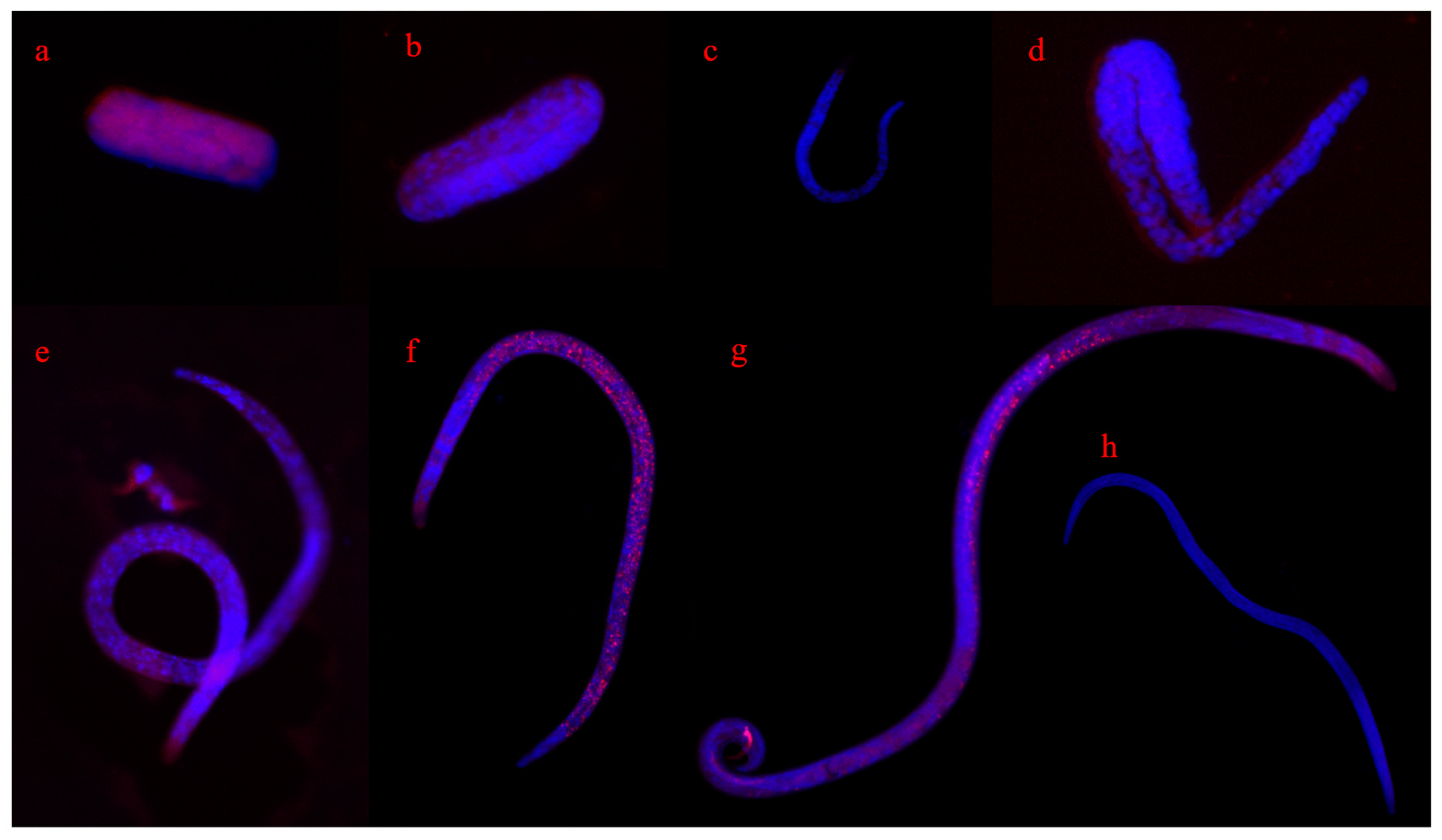
2.9.8. Effect of RNAi on Development

The B. xylophilus at J2 stages were collected according to the method described by Wang et al., Shinya et al. and Xue et al. with some modifications [42,51,57]. Approximately 10,000 B. xylophilus (mixed developmental stages) were inoculated in B. cinerea cultures on PDA medium at 25 °C in the dark for 9 h. The eggs were collected by the Baermann funnel method. The eggs were inoculated in B. cinerea cultures on PDA medium in the dark at 25 °C for 24 h. The J2 stage larvae were collected by Baermann funnel method. Approximately 3000 J2 stage larvae were soaked in dsRNA solution (1.0 μg/μL) as described above, and the development of the larvae was observed. B. xylophilus soaked in distilled H2O and gfp dsRNA (1.0 μg/μL) were used as control, respectively. The experiment was repeated three times.

2.9.9. Effect of RNAi on Sensitivity to Ethanol

One hundred microliters of B. xylophilus suspension (approximately 300 B. xylophilus with ratio of female to male of 1:1) were transferred into a 96-well plate, absolute ethanol was added to a final concentration of 1%, and nematodes were observed every 24 h for a total of 72 h. B. xylophilus soaked in distilled H2O and gfp dsRNA solution (1.0 μg/μL) were used as control, respectively. The experiment was repeated three times.

2.9.10. Effect of RNAi on the Reactive Oxygen Species and Activities of Antioxidant Enzymes

Approximately 3000 B. xylophilus (mixed developmental stages) were soaked in 50 μL dsRNA solution (1.0 μg/μL) and cultured in the dark at 20 °C at 180 rpm for 72 h [53]. The reactive oxygen species were detected following the instructions of the reactive oxygen species Assay Kit.
For superoxide dismutase (SOD) analysis, 2 mL reaction system contained 50 mmol/L PBS (pH 7.8), 13 mmol/L Met, 75 μmol/L NBT, 1 mmol/L EDTA, 20 μmol/L riboflavin and 100 μL enzyme solution. The added reaction system was placed under 4000 LX light source to react at 25 °C for 15 min, then the reaction was terminated immediately in the dark, and the absorbance was determined under 560 nm [58]. The amount of enzyme required when the inhibition rate reached 50% was defined as one unit of enzyme activity (U). The calculation formula of SOD activity was referred to in Formula (1) in Table 3. The experiment was repeated three times.
To detect peroxidase (POD), 2 mL reaction system contained 50 mmol/L PBS (pH 7.8), 0.005% H2O2, 0.001% guaiacol and 100 μL enzyme solution. The change of absorbance at 470 nm in 10 min was measured [58]. The value of A470 reduced by 0.01 per minute was defined as one unit of enzyme activity (U). The calculation formula of POD activity was referred to in Formula (2) in Table 3. To measure the activity of catalase (CAT), 2 mL reaction system contained 50 mmol/L PBS (pH 7.8), 0.001% H2O2, and 100 μL enzyme solution. The change of absorbance in 10 min was measured at 240 nm [58]. The value of A240 reduced by 0.01 per minute was defined as one unit of enzyme activity (U). The calculation formula of CAT activity is referred to in Table 3, Formula (3). The experiment was repeated three times.

2.10. Fluorescence In Situ Hybridization of BxCAD-1 Gene

Fluorescence in situ hybridization technology was performed to determine the spatial expression patterns of BxCAD-1 gene at different developmental stages of B. xylophilus. A red fluorescence-labeled probe (5′-CAACGATTTGACCGGGCTTCACATCCGCCTCCTTCAACGCCTTAT-3′) was generated from the cloned sequence of BxCAD-1 gene and designed using Primer Premier 5.0 software (Premier, Canada). Approximately three thousand B. xylophilus (mixed developmental stages) were fixed with 1.0 mL RNA-free paraformaldehyde fixative at 25 °C for 30 min. The method of in situ hybridization was referred to in the instruction of FISH in situ hybridization kit. The sense probe (5′-TTGTACTACACAAAAGTACTG-3′) was used as a negative control [59].

2.11. Analysis of KEGG Pathway of BxCAD-1 Gene

Analysis of the pathway involved in BxCAD-1 gene in the transcriptome of B. xylophilus treated with methylpyrazole was performed with KEGG ORTHOLOGY: K13953 (genome.jp).

2.12. Data Analysis

All of the experiments were performed at least three times. Data from repeated experiments were represented as means ± standard deviation (S.D.), and all statistical analyses were performed using SPSS 19.0 software (SPSS, Chicago, IL, USA). The independent sample t-test and one-way analysis of variance were used to assess differences between the groups. A p value of <0.05 was considered to denote statistical significance.

3. Results

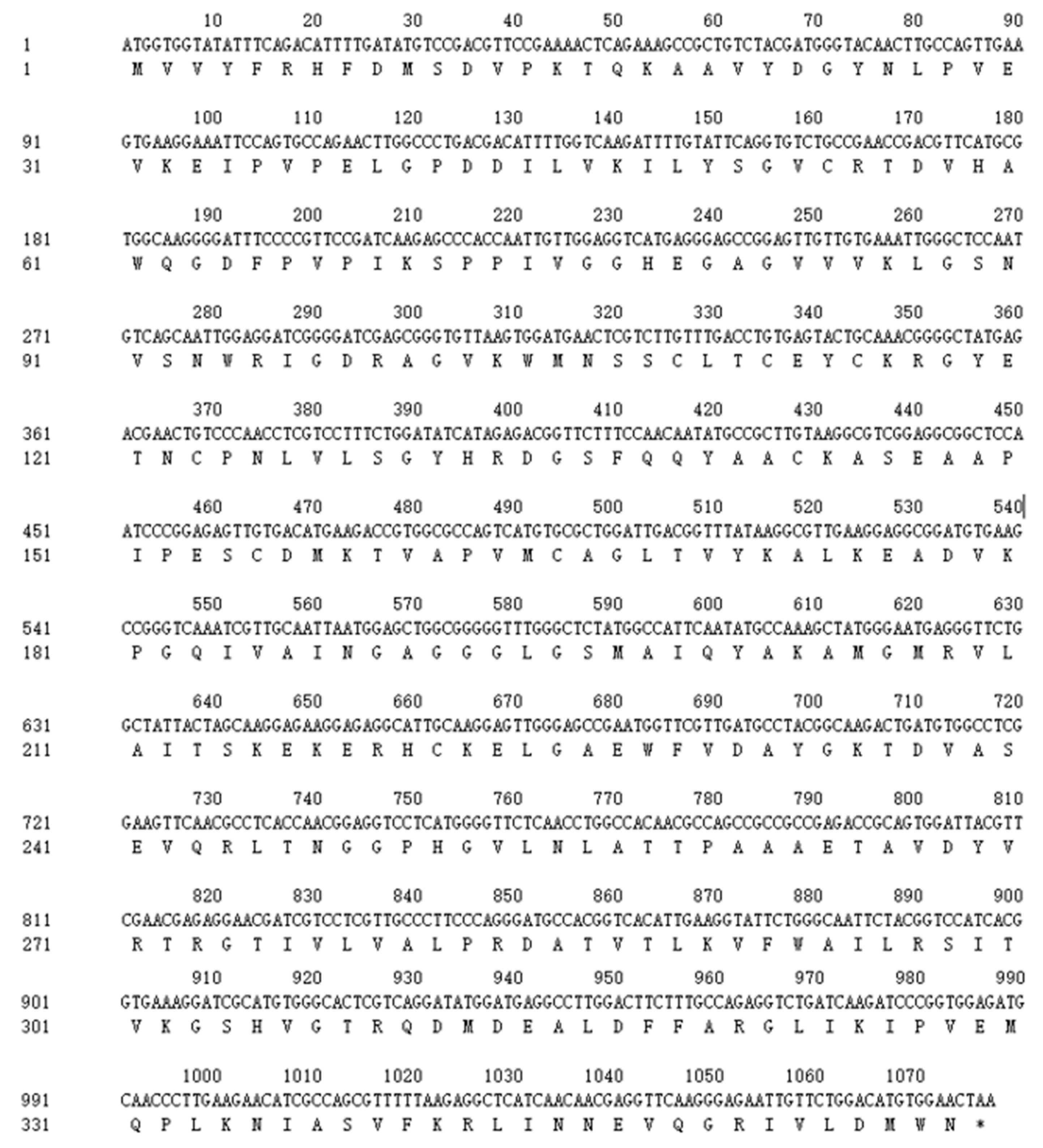
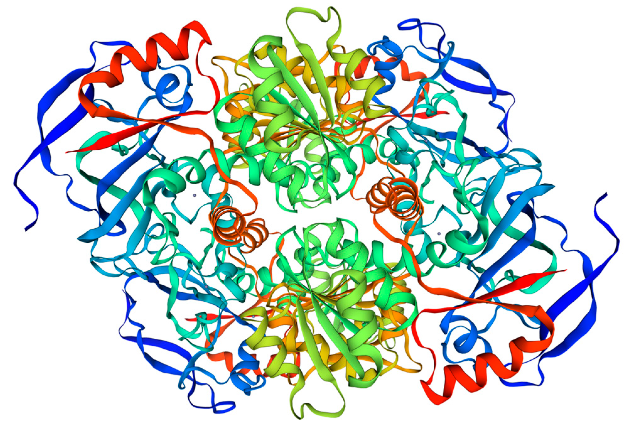
3.1. Sequence Analysis of BxCAD-1 Gene

The full-length ORF of BxCAD-1 gene is 1077 bp (GenBank accession no: ON540735), which encodes 358 amino acid residues (Figure 1) with a molecular mass of 38.93 kDa and a theoretical pI of 8.27. The major amino acids of BxCAD-1 protein are Val (11.2%), Ala (9.5%), Gly (8.9%), Leu (7.0%) and Lys (6.7%). The predicted structure includes one NAD binding domain (156–327 bp), two substrate binding domains (41–150 and 193–320 bp), one molecular chaperone binding domain (10–187 bp), and one Zn2+ binding domain (77–91 bp). Based on the analysis of a three-dimensional structure diagram, four polypeptide chains can form a highly symmetrical tetramer complex. One subunit can bind two Zn2+ and one NAD, with one Zn2+ participating in the catalytic reaction and the other stabilizing the structure (Figure 2). It is predicted that the protein has no signal peptide and transmembrane domain.
The alignment of amino acid sequences showed that the BxCAD-1 protein of B. xylophilus had the closest evolutionary relationship with cinnamyl alcohol dehydrogenase (GenBank accession no: CAD5217100.1) in B. xylophilus, with the highest homology of 98%. It also had strong homology of 96% with alcohol dehydrogenase (GenBank accession no: ARA71334.1) (Figure 3). The gene cloned in this study was named BxCAD-1.

3.2. Induced Expression and Purification of Recombinant BxCAD-1

The protein encoded by BxCAD-1 was over-expressed in E. coli BL21 (DE3) upon IPTG induction and appeared mainly in inclusion bodies. After the recombinant protein in inclusion bodies was resolved in urea solution and refolded by a series of dilutions, it was purified by affinity chromatography on nickel-binding resin. The recombinant protein was purified to homogeneity with a relative molecular weight of 38 kDa as analyzed by SDS-PAGE (Figure 4), which was consistent with that of cinnamyl alcohol dehydrogenase encoded by BxCAD-1.

3.3. Determination of Cinnamyl Alcohol Dehydrogenase Activity

The effects of pH, temperature, metal ions and substrates on the activity of cinnamyl alcohol dehydrogenase were determined. The recombinant cinnamyl alcohol dehydrogenase showed the highest catalytic activity at pH 8.0, and the optimal temperature for this enzyme was 40 °C. The Zn2+ achieved the highest enzyme activity compared to other metal ions (p < 0.05), and Fe2+, Mg2+ and Ni2+ had a similar activating effect, whereas K+ showed no effect on its activity (p > 0.05). The optimal substrate for this enzyme was cinnamyl alcohol, followed by sorbitol and ethanol (Figure 5).

3.4. RNAi of BxCAD-1

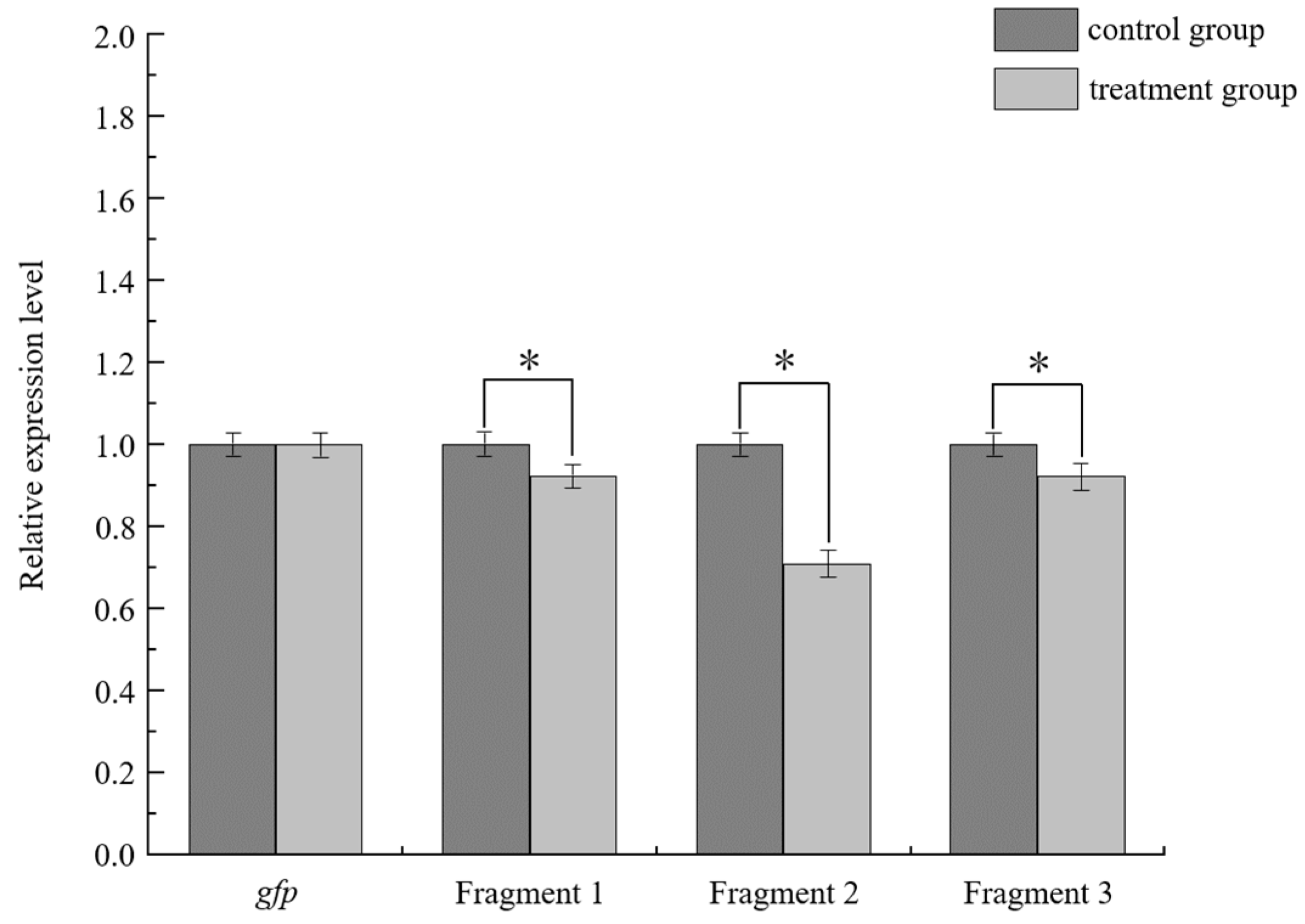
3.4.1. Efficiency Assessment of RNAi

The RNAi efficiency on the BxCAD-1 expression level was evaluated through qRT-PCR. The results showed that compared with the expression level of BxCAD-1 in the B. xylophilus soaked in sterilized water, that in the B. xylophilus soaked in fragment 1, 2 and 3 dsRNA solution decreased. Fragment 2 had the greatest impact on gene expression level. This result indicated that the expression of BxCAD-1 could be strongly inhibited by soaking the B. xylophilus in BxCAD-1 dsRNA. The gfp dsRNA, as one of the negative controls, demonstrated no significant effects on the expression level of BxCAD-1 compared with distilled water (Figure 6).
The markers labeled gfp, fragment 1, fragment 2 and fragment 3 were B. xylophilus treated with dsRNA of gfp, fragment 1, fragment 2 and fragment 3 of BxCAD-1, respectively.

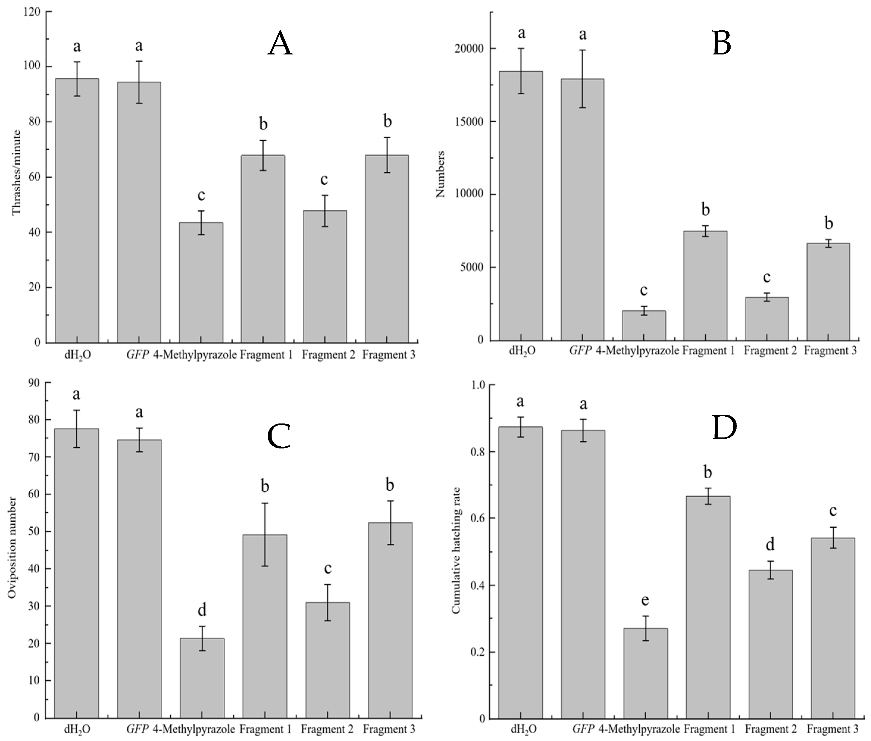
3.4.2. B. xylophilus Movement and Reproduction

The B. xylophilus in distilled H2O and dsRNA from gfp had the highest head thrashing frequency of 95.62 ± 6.26 and 94.34 ± 7.58, respectively (p > 0.05). The thrashes of nematodes treated with interfering fragments 1, 2 and 3 were 67.86 ± 5.40, 47.82 ± 5.71 and 67.96 ± 6.38, respectively. Notably, the inhibitory effect of fragment 2 was significantly higher than fragments 1 and 3. The thrashes of nematodes treated with 4-methylpyrazole were 43.54 ± 4.35, which was similar to the effect of fragment 2 (Figure 7A).
The number of B. xylophilus treated differently after 9 days of inoculation was recorded. The number of B. xylophilus in distilled H2O and dsRNA from gfp was the largest, with 18,450 ± 1556.44 and 17,916.67 ± 1973.50, respectively (p > 0.05). The number of PWN treated with interfering fragments 1, 2 and 3 were 7516.67 ± 371.56, 2983.33 ± 279.38 and 6650 ± 262.99, respectively, and the inhibitory effect of fragment 2 was the best. The PWN treated with 4-methylpyrazole was 2050 ± 298.61, which had a similar effect to fragment 2 (Figure 7B).

3.4.3. Spawning and Hatching Rate of B. xylophilus

The number of eggs for B. xylophilus in distilled H2O and dsRNA from gfp was 77.6 ± 5 and 74.6 ± 3.2, respectively (p > 0.05). The egg number for B. xylophilus treated with fragments 1, 2 and 3 were 49.2 ± 8.4, 31 ± 4.86 and 52.4 ± 5.85, respectively, and the inhibitory effect of fragment 2 was the best, which was significantly lower than that of fragments 1 and 3. The egg number for PWN treated with 4-methylpyrazole was 21.4 ± 3.26, which was significantly lower than that of fragment 2 (Figure 7C).
The hatching rate of eggs laid by B. xylophilus in distilled H2O and dsRNA from gfp were 87.37 ± 2.99% and 86.33 ± 3.31%, respectively, and 4-Methylpyrazole was 27.10 ± 3.61%, which is significantly lower than that of fragment 2 (Figure 7D).

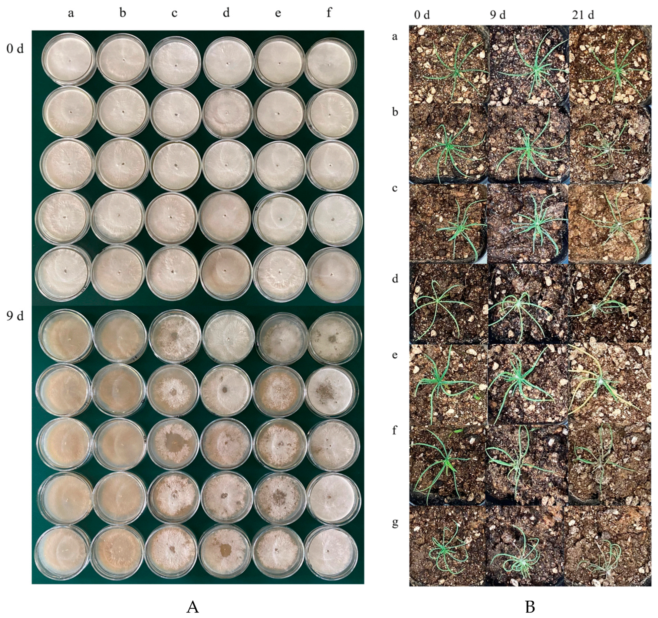
3.4.4. Feeding and Infectivity Ability of B. xylophilus

The feeding ability of B. xylophilus treated differently was recorded from day 1 to day 9 after B. xylophilus was inoculated on Botrytis cinerea cultures on PDA plates. The Botrytis cinerea inoculated with B. xylophilus in distilled H2O and dsRNA from gfp were depleted, approximately 1/2 and 1/4 of mycelia were ingested, and more than 3/4 mycelia was left for methylpyrazole-treated B. xylophilus on day 9. The feeding of fragment 1 and fragment 3 were similar, with a feeding area of 1/2, fragment 2 was 1/4, and 4-Methylpyrazole was the smallest, which was less than 1/4 (Figure 8A).
To investigate the infectivity of PWN treated differently, the seedlings of P. thunbergii were inoculated with different PWNs. The growth status of seedlings at 0, 9 and 21 days after infection were recorded, respectively. On the ninth day after infection, the seedlings treated with B. xylophilus in distilled H2O and dsRNA from gfp began to wither, and those in the negative control group, inoculated with B. xylophilus in dsRNA from BxCAD-1 and 4-methylpyrazole had no significant changes. On the twenty-first day after infection, all seedlings had completely withered and died, except those in the negative control group (Figure 8B).

3.4.5. Lifespan and Development of B. xylophilus

The survival rate of B. xylophilus per day was recorded. No significant difference was observed in the first three days, but the mortality of B. xylophilus treated with 4-methylpyrazole and dsRNA from three fragments of BxCAD-1 increased from the fourth day. Until the seventh day, the survival rate of PWN in distilled H2O, dsRNA from gfp and 4-methylpyrazole was 63 ± 4.64%, 61.33 ± 5.31% and 32 ± 4.19%, respectively, while the survival rate of PWN treated with interfering fragment 1, 2 and 3 was 46 ± 5.73%, 37.33 ± 3.09% and 42.33 ± 7.13%, respectively (p > 0.05) (Figure 9A).
After RNAi by dsRNA of fragment 2 of BxCAD-1, the proportion of larvae increased significantly, accounting for 28.33 ± 2.34%, while that in distilled H2O and dsRNA from gfp were 17.33 ± 2.05% and 17.17 ± 2.11%, respectively. The proportion of male PWN accounted for 4.67 ± 1.11%, and female PWN accounted for 66.33 ± 1.97%, whereas male PWN in distilled H2O and gfp were 9.5 ± 1.71% and 9.17 ± 1.34%, and female PWN were 72.67 ± 1.37% and 73.67 ± 1.97%, respectively (Figure 9B).

3.4.6. Sensitivity to Ethanol and Oviposition of B. xylophilus

The B. xylophilus in the distilled H2O and dsRNA from gfp control groups showed obvious clumping behavior 24 h after providing ethanol of 1%, and approximately one hundred B. xylophilus were cuddled together (Figure 9). After 72 h, the B. xylophilus scattered and began to lay eggs; the number of eggs laid for B. xylophilus in distilled H2O and dsRNA from gfp were 13.33 ± 1.25, 14.33 ± 1.24, respectively (p > 0.05) (Figure 9C). The B. xylophilus had no obvious clumping behavior after RNAi by dsRNA of fragment 2 of BxCAD-1, and the number of eggs laid after 72 h was only 3.33 ±1.25 (Figure 9C and Figure 10).

3.4.7. Reactive Oxygen Species and Activities of Antioxidant Enzymes

After RNAi by dsRNA of fragment 2 of BxCAD-1, the fluorescence value of reactive oxygen species of B. xylophilus was the highest, which was significantly different from the value of B. xylophilus in distilled H2O and dsRNA from gfp treatment groups (p < 0.05). (Table 4) (Figure 11). The activity of SOD, POD and CAT decreased to 43.72%, 79.89% and 58.74% of that of B. xylophilus in distilled H2O, which were also significantly lower (p < 0.05) (Figure 9D).

3.5. In Situ Hybridization

The in situ hybridization indicated that the BxCAD-1 gene was mainly expressed in the digestive tract of male and female adults. No obvious expression was observed in eggs and larvae at various stages (Figure 12).

3.6. Analysis of KEGG Pathway of BxCAD-1 Gene

The pathway that the BxCAD-1 gene is involved in includes glycolysis/gluconeogenesis, fatty acid degradation, tyrosine metabolism, pyruvate metabolism, chloroalkane and chloroalkene degradation, naphthalene degradation, retinol metabolism and metabolism of xenobiotics by cytochrome P450. Its function is mainly to participate in the catalytic reaction between various alcohols and aldehydes, such as ethanol and ethanol, retinol and retinal, 3-chloroallyl alcohol and 3-chloroallyl aldehyde, 1-naphthalenemethanol and 1-naphthaldehyde as well as trichloroethanol and chloral hydrate.

4. Discussion

Pine wilt disease causes severe damage to pine forests and the environment. Currently, the major preventive measures against PWD include cutting out disease trees, breeding PWN-resistant pine trees, managing vector insects and performing trunk injection of pesticides. However, the implementation of these procedures cannot eliminate PWD. Therefore, it is an economical and efficient method to find specific gene targets of PWN and design effective pesticides to control this disease.
As a pathogenic organism, PWN can activate the defense mechanism of an infected pine tree, including the production of toxic secondary metabolites, activation of defense genes, and cell structure changes [60,61,62,63]. In order to break through the defense system barrier of the host, PWN produces a variety of enzymes and secondary metabolites for reaction, such as peroxidase, aldehyde dehydrogenase, cellulase and toxic factor [64,65]. In this study, the cDNA coding cinnamyl alcohol dehydrogenase from pine wood nematode was cloned and overexpressed. The characteristics of the recombinant BxCAD-1 were studied, and the effects of RNAi of BxCAD-1 were investigated thoroughly. Additionally, the main expression pattern of BxCAD-1 was also investigated through in situ hybridization.
From the results of homology analysis, BxCAD-1 is most similar to the possible cinnamyl alcohol dehydrogenase and alcohol dehydrogenase in B. xylophilus, which is also confirmed by bioassay of BxCAD-1 with different substrates in this study. BxCAD-1 can catalyze seven substrates with cinnamyl alcohol as its optimal substrate, and show an optimal temperature of 40 °C, whereas the optimal temperature of alcohol dehydrogenase from PWN was reported to be 30 °C [53]. Considering the above factors, we assume BxCAD-1 is a cinnamyl alcohol dehydrogenase and speculate that BxCAD-1 and alcohol dehydrogenase are a group of isoenzymes, but their optimal reaction temperature and substrate are not the same. Therefore, further study of BxCAS-1 is warranted.
Cinnamyl alcohol dehydrogenase is mainly involved in the formation of lignin in plants but plays other roles in microorganisms [28,29,30,31,32,33,34,35,36,37]. According to the results of in situ hybridization, the BxCAD-1 gene is highly expressed in the digestive tract. The obvious fluoresce signal in the spicules of male adults might contribute to its tissue structure since in situ hybridization analysis of UGT440A1 expression patterns also indicated the aggregation of fluorescent probes in spicules [51]. Considering other phenomena caused by RNAi of BxCAD-1, we speculate that BxCAD-1 is mainly associated with the feeding and reproduction of pine wood nematode. The cytochrome P450 gene and pectin lyase gene of B. xylophilus can destroy plant cell tissues and help B. xylophilus to migrate. The slower migration rate of B. xylophilus can be observed by RNAi with gene expression [43,44]. Silencing of effector BxSapB1 can also reduce the pathogenicity of B. xylophilus [45]. Through RNAi of the BxCAD-1 gene, the motor ability, feeding, reproduction, infection, lifespan, spawning rate and hatching rate of B. xylophilus are all decreased. To further study the role of this gene in the development of B. xylophilus, we selected synchronous B. xylophilus to observe their development. After RNAi of BxCAD-1, the proportion of larvae increased, and the number of male adults decreased significantly. We speculate that the BxCAD-1 gene is related to the formation and function of spicules of male B. xylophilus, which further affects the transition from the larval stage to the adult stage.
Pine trees infected with PWD can produce a large amount of volatiles, including ethanol, the mechanism of which is still unknown [66]. A large number of studies have shown that ethanol can promote the reproduction and expansion of B. xylophilus in pine trees. There was a report that ethanol was produced and released in a large amount 10 d after the resin ceased to secrete in the PWN-infected pine trees. The number of PWN increased dramatically two weeks after infection [66]. Studies also showed that low concentrations of ethanol could promote the oviposition of B. xylophilus, whereas higher concentrations of ethanol could reduce the oviposition of B. xylophilus [67,68]. A low concentration of dihydroconiferylalcohol was also reported to cause serious pine wilt by promoting the reproduction of B. xylophilus [69]. By silencing the BxCAD-1 gene through RNAi, the sensitivity of B. xylophilus to ethanol decreased and B. xylophilus tended to lay fewer eggs, which indicated that BxCAD-1 participated in alcohol-induced regulation of reproduction. Additionally, we observed an interesting phenomenon that ethanol could promote the clumping behavior of PWNs. Pine wood nematodes began to clump after providing 1% ethanol for 1 h, and this aggregation with a large number might last for 3 d followed by redispersing and laying eggs, while B. xylophilus without ethanol did not appear to have clumping behavior. In contrast, PWNs of RNAi for BxCAD-1 did not show obvious massive aggregation behavior, and there was no significant change in behavior even after providing 1% ethanol. Small chemical signals, such as ascarosides with hydroxyls, have been reported to attract potential mates of B. xylophilus [70]. Considering that cinnamyl alcohol dehydrogenase could catalyze the reaction of various substrates containing hydroxyls such as D-pinitol, sorbitol and 2-methoxyphenol, we speculated that B. xylophilus might be stimulated to mate by hydroxyl-oxidated aldehyde group-containing substances catalyzed by BxCAD-1.
The increase of active oxygen species in PWN after RNAi of BxCAD-1 might be one of the reasons for the shortened lifespan of B. xylophilus. Combined with the analysis of the KEGG pathway, the BxCAD-1 gene could participate in the metabolic pathway of cytochrome P450 and the metabolism of reactive oxygen species. In addition, through KEGG pathway analysis, the BxCAD-1 gene is involved in a variety of energy metabolism pathways, which is consistent with the results of in situ hybridization.

5. Conclusions

In summary, BxCAD-1 coding cinnamyl alcohol dehydrogenase was found to participate in the reproduction and development of B. xylophilus. The movement ability, feeding ability, reproductive ability, spawning rate, hatching rate, lifespan, infectivity and sensitivity to ethanol could be suppressed, and reactive oxygen species could accumulate through down-regulation of BxCAD-1 expression, which makes BxCAD-1 a promising target for exploring pesticides to control B. xylophilus. Additional investigations are needed to elucidate the roles of BxCAD-1.

Author Contributions

Conceptualization, R.L. and G.D. (Guicai Du); formal analysis, G.D. (Guicai Du); funding acquisition, Q.G. and R.L.; investigation, C.W. and G.D. (Guosong Dong); methodology, Y.Z., G.D. (Guicai Du), G.D. (Guosong Dong), H.T. and R.L.; project administration, Q.G. and R.L.; resources, H.T. and G.D. (Guosong Dong); software, G.D. (Guosong Dong) and Q.G.; writing—original draft, G.D. (Guosong Dong); writing—review and editing, G.D. (Guosong Dong) and W.D. All authors have read and agreed to the published version of the manuscript.

Funding

This work was supported by the Project for Qingdao Science and Technology Plan, China (23-2-8-cspz-8-nsh) and Shandong Province Natural Science Foundation, China (ZR2020MC123).

Data Availability Statement

Research data are available on request from the corresponding authors.

Conflicts of Interest

The authors declare no conflict of interest.

References

  1. Zhao, B.G.; Futai, K.; Sutherland, J.R.; Takeuchi, Y. (Eds.) Pine Wilt Disease; Springer International Publishers: Tokyo, Japan, 2008; pp. 5–13. [Google Scholar]
  2. Wang, Y.; Chen, F.M.; Wang, L.C.; Zhou, L.F.; Song, J. Study of the departure of pine wood nematode, Bursaphelenchus xylophilus (Nematoda: Aphelenchoididae) from Monochamus alternatus (Coleoptera: Cerambycidae). J. Asia-Pac. Entomol. 2020, 23, 981–987. [Google Scholar] [CrossRef]
  3. Fukuda, K.; Hogetsu, T.; Suzuki, K. Cavitation and cytological changes in xylem of pine seedlings inoculated with virulent and avirulent isolates of Bursaphelenchus xylophilus and B. mucronatus. JJFS 1992, 74, 289–299. [Google Scholar]
  4. Ishida, K.; Hogetsu, T. Role of resin canals in the early stages of pine wilt disease caused by the pine wood nematode. Can. J. Bot. 1997, 75, 346–351. [Google Scholar] [CrossRef]
  5. Hara, N.; Futai, K. Histological changes in xylem parenchyma cells and the effects on tracheids of Japanese black pine inoculated with pine wood nematode, Bursaphelenchus xylophilus. J. Jpn. For. Soc. 2001, 83, 285–289. [Google Scholar]
  6. Ikegami, M.; Jenkins, T.A.R. Estimate global risks of a forest disease under current and future climates using species distribution model and simple thermal model—Pine Wilt disease as a model case. For. Ecol. Manag. 2018, 409, 343–352. [Google Scholar] [CrossRef]
  7. Yang, B.J.; He, Z.Y.; Wang, C.F. Survey of pine wood nematode disease abroad. For. Pest Dis. 1999, 5, 40–42. [Google Scholar]
  8. Mantas, V.; Fonseca, L.; Baltazar, E.; Canhoto, J.; Abrantes, I. Detection of tree decline (Pinus pinaster aiton) in european forests using sentinel-2 data. Remote. Sens. 2022, 14, 2028. [Google Scholar] [CrossRef]
  9. Ryss, A.Y.; Kulinich, O.A.; Sutherland, J.R. Pine wilt disease: A short review of worldwide research. For. Stud. China 2011, 13, 132–138. [Google Scholar] [CrossRef]
  10. Wu, W.B.; Zhang, Z.B.; Zheng, L.J.; Han, C.Y.; Wang, X.M.; Xu, J.; Wang, X.R. Research progress on the early monitoring of pine wilt disease using hyperspectral techniques. Sensors 2020, 20, 3729. [Google Scholar] [CrossRef]
  11. Futai, K. Pine wood nematode, Bursaphelenchus xylophilus. Annu. Rev. Phytopathol. 2013, 51, 61–83. [Google Scholar] [CrossRef] [Green Version]
  12. Zhang, Q.; Li, H.Y.; Bai, G.; Xiong, H.L.; Yang, W.B. Immunological Analysis of Bursaphelenchus xylophilus Pathogenous Cellulase. Sci. Silvae Sin. 2007, 43, 64–67. [Google Scholar]
  13. Kikuchi, T.; Jonesb, J.T.; Aikawa, T.; Kosaka, H.; Ogura, N. A family of glycosyl hydrolase family 45 cellulases from the pine wood nematode Bursaphelenchus xylophilus. FEBS Lett. 2004, 572, 201–205. [Google Scholar] [CrossRef] [PubMed] [Green Version]
  14. Cardoso, J.M.S.; Fonseca, L.; Abrantes, I. α-l-fucosidases from Bursaphelenchus xylophilus secretome—Molecular characterization and their possible role in breaking down plant cell walls. Forests 2020, 11, 265. [Google Scholar] [CrossRef] [Green Version]
  15. Shinya, R.; Kirino, H.; Morisaka, H.; Takeuchi-Kaneko, Y.; Futai, K.; Ueda, M. Comparative secretome and functional analyses reveal glycoside hydrolase family 30 and cysteine peptidase as virulence determinants in the pinewood nematode Bursaphelenchus xylophilus. Front. Plant Sci. 2021, 12, 640459. [Google Scholar] [CrossRef]
  16. Shinya, R.; Morisaka, H.; Kikuchi, T.; Takeuchi, Y.; Ueda, M.; Futai, K. Secretome analysis of the pine wood nematode Bursaphelenchus xylophilus reveals the tangled roots of parasitism and its potential for molecular mimicry. PLoS ONE 2013, 8, e67377. [Google Scholar] [CrossRef]
  17. Yazaki, K.; Takanashi, T.; Kanzaki, N.; Komatsu, M.; Levia, D.F.; Kabeya, D.; Tobita, H.; Kitao, M.; Ishida, A. Pine wilt disease causes cavitation around the resin canals and irrecoverable xylem conduit dysfunction. J. Exp. Bot. 2018, 69, 589–602. [Google Scholar] [CrossRef]
  18. Li, Y.X.; Ying, W.; Liu, Z.Y.; Wang, X.; Lu, Q.; Jia, X.Z.; Zhang, X.Y. Functional analysis of the venom allergen-like protein gene from pine wood nematode Bursaphelenchus xylophilus using a baculovirus expression system. Physiol. Mol. Plant. Pathol. 2015, 93, 58–66. [Google Scholar] [CrossRef]
  19. Hara, N.; Takeuchi, Y.; Futai, K. Cytological changes in ray parenchyma cells of seedlings of three pine species infected with the pine wilt disease. J. Nematol. 2006, 36, 23–32. [Google Scholar] [CrossRef] [Green Version]
  20. Umebayashi, T.; Fukuda, K.; Haishi, T.; Sotooka, R.; Zuhair, S.; Otsuki, K. The developmental process of xylem embolisms in pine wilt disease monitored by multipoint imaging using compact magnetic resonance imaging. Plant Physiol. 2011, 156, 943–951. [Google Scholar] [CrossRef] [Green Version]
  21. Wang, H.G.; Li, L.; Ju, Y.W.; Zhang, J.C.; Zhao, B.G. Pathological changes in ultrastructure of the Pinus thunbergii due to flagellin toxin. J. Nanjing For. Univ. (Nat. Sci. Ed.) 2018, 42, 137–144. [Google Scholar]
  22. Zhu, L.H.; Ye, J.R.; Negi, S.; Xu, X.L.; Wang, Z.L.; Ji, J.Y. Pathogenicity of aseptic Bursaphelenchus xylophilus. PLoS ONE 2012, 7, e38095. [Google Scholar] [CrossRef] [PubMed] [Green Version]
  23. Xue, Q.; Xiang, Y.; Wu, X.Q.; Li, M.J. Bacterial communities and virulence associated with pine wood nematode Bursaphelenchus xylophilus from different Pinus spp. Int. J. Mol. Sci. 2019, 20, 3342. [Google Scholar] [CrossRef] [Green Version]
  24. Han, Z.M.; Hong, Y.D.; Zhao, B.G. A study on pathogenicity of bacteria carried by pine wood nematodes. J. Phytopathol. 2010, 151, 683–689. [Google Scholar] [CrossRef]
  25. Ebel, J.; Grisebach, H. Reduction of cinnamic acids to cinnamyl alcohols with an enzyme preparation from cell suspension cultures of soybean (Glycine max). FEBS Lett. 1973, 30, 141–143. [Google Scholar] [CrossRef] [PubMed] [Green Version]
  26. Tang, R.; Zhang, X.Q.; Li, Y.H.; Xie, X.M. Cloning and in silico analysis of a cinnamyl alcohol dehydrogenase gene in Pennisetum purpureum. J. Genet. 2014, 93, 145–158. [Google Scholar] [CrossRef] [PubMed]
  27. Kasirajan, L.; Valiyaparambth, R.; Kubandiran, A.; Velu, J. Isolation, cloning and expression analysis of cinnamyl alcohol dehydrogenase (CAD) involved in phenylpropanoid pathway of Erianthus arundinaceus, a wild relative of sugarcane. 3 Biotech 2020, 10, 11. [Google Scholar] [CrossRef] [PubMed]
  28. Li, X.; Ma, D.M.; Chen, J.L.; Pu, G.B.; Ji, Y.P.; Lei, C.Y.; Du, Z.G.; Liu, B.Y.; Ye, H.C.; Wang, H. Biochemical characterization and identification of a cinnamyl alcohol dehydrogenase from Artemisia annua. Plant. Sci. 2012, 193–194, 85–95. [Google Scholar] [CrossRef]
  29. Park, H.L.; Kim, T.L.; Bhoo, S.H.; Lee, T.H.; Lee, S.W.; Cho, M.H. Biochemical characterization of the rice cinnamyl alcohol dehydrogenase gene family. Molecules 2018, 23, 2659. [Google Scholar] [CrossRef] [Green Version]
  30. Sibout, R.; Eudes, A.; Mouille, G.; Pollet, B.; Lapierre, C.; Jouanin, L.; Séguin, A. Cinnamyl alcohol dehydrogenase-C and -D are the primary genes involved in lignin biosynthesis in the floral stem of Arabidopsis. Plant Cell 2005, 17, 2059–2076. [Google Scholar] [CrossRef] [Green Version]
  31. Chen, C.; Chang, J.M.; Wang, S.; Lu, J.; Liu, Y.; Si, H.Q.; Sun, G.; Ma, C.X. Cloning, expression analysis and molecular marker development of cinnamyl alcohol dehydrogenase gene in common wheat. Protoplasma 2021, 17, 881–889. [Google Scholar] [CrossRef]
  32. Chao, N.; Liu, S.X.; Liu, B.M.; Li, N.; Jiang, X.N.; Gai, Y. Molecular cloning and functional analysis of nine cinnamyl alcohol dehydrogenase family members in Populus tomentosa. Planta 2014, 240, 1097–1112. [Google Scholar] [CrossRef] [PubMed]
  33. Larroy, C.; Pare´s, X.; Biosca, J.A. Characterization of a Saccharomyces cerevisiae NADP(H)-dependent alcohol dehydrogenase (ADHVⅡ), a member of the cinnamyl alcohol dehydrogenase family. Eur. J. Biochem. 2002, 269, 5738–5745. [Google Scholar] [CrossRef] [PubMed]
  34. Larroy, C.; Fernández, M.R.; González, E.; Parés, X.; Biosca, J.A. Properties and functional significance of Saccharomyces cerevisiae ADHVI. Chem. Biol. Interact. 2003, 143, 229–238. [Google Scholar] [CrossRef] [PubMed]
  35. Zhou, J.H.; Lin, Y.; Fu, Y.P.; Xie, J.T.; Jiang, D.H.; Cheng, J.S. A cinnamyl alcohol dehydrogenase required for sclerotial development in Sclerotinia sclerotiorum. Phytopathol. Res. 2020, 2, 13. [Google Scholar] [CrossRef]
  36. Wilkin, J.M.; Soetaert, K.; Ste´landre, M.; Buyssens, P.; Castillo, G.; Demoulin, V.; Bottu, G.; Laneelle, M.A.; Daffe, M.; Bruyn, J.D. Overexpression, purification and characterization of Mycobacterium bovis BCG alcohol dehydrogenase. Eur. J. Biochem. 1999, 262, 299–307. [Google Scholar] [CrossRef] [Green Version]
  37. Mee, B.; Kelleher, D.; Frias, J.; Malone, R.; Tipton, K.F.; Henehan, G.T.M.; Windle, H.J. Characterization of cinnamyl alcohol dehydrogenase of Helicobacter pylori. An aldehyde dismutating enzyme. FEBS J. 2010, 272, 1255–1264. [Google Scholar] [CrossRef]
  38. Fire, A.; Xu, S.Q.; Montgomery, M.K.; Kostas, S.A.; Driver, S.E.; Mello, C.C. Potent and specific genetic interference by double-stranded RNA in Caenorhabditis elegans. Nature 1998, 391, 806–811. [Google Scholar] [CrossRef]
  39. Chi-Ham, C.L.; Clark, K.L.; Bennett, A.B. The intellectual property landscape for gene suppression technologies in plants. Nat. Biotechnol. 2010, 28, 32–36. [Google Scholar] [CrossRef]
  40. Niu, J.H.; Jian, H.; Xu, J.M.; Guo, Y.D.; Liu, Q. RNAi technology extends its reach: Engineering plant resistance against harmful eukaryotes. Afr. J. Biotechnol. 2015, 9, 7573–7582. [Google Scholar]
  41. Wang, M.; Wang, D.; Zhang, X.; Wang, X.; Liu, W.; Hou, X.; Huang, X.Y.; Xie, B.Y.; Cheng, X.Y. Double-stranded RNA-mediated interference of dumpy genes in Bursaphelenchus xylophilus by feeding on filamentous fungal transformants. Int. J. Parasitol. 2016, 46, 351–360. [Google Scholar] [CrossRef]
  42. Xue, Q.; Wu, X.Q.; Zhang, W.J.; Deng, L.N.; Wu, M.M. Cathepsin L-like cysteine proteinase genes are associated with the development and pathogenicity of pine wood nematode, Bursaphelenchus xylophilus. Int. J. Mol. Sci. 2019, 20, 215. [Google Scholar] [CrossRef] [PubMed] [Green Version]
  43. Qiu, X.; Yang, L.; Ye, J.; Wang, W.; Zhao, T.; Hu, H.; Zhou, G. Silencing of cyp-33C9 gene affects the reproduction and pathogenicity of the pine wood nematode, Bursaphelenchus xylophilus. Int. J. Mol. Sci. 2019, 20, 4520. [Google Scholar] [CrossRef] [PubMed] [Green Version]
  44. Qiu, X.W.; Wu, X.Q.; Huang, L.; Ye, J.R. Influence of Bxpel1 gene silencing by dsRNA interference on the development and pathogenicity of the pine wood nematode, Bursaphelenchus xylophilus. Int. J. Mol. Sci. 2016, 17, 125. [Google Scholar] [CrossRef] [PubMed] [Green Version]
  45. Hu, L.J.; Wu, X.Q.; Li, H.Y.; Zhao, Q.; Wang, Y.C.; Ye, J.R. An effector, BxSapB1, induces cell death and contributes to virulence in the pine wood nematode Bursaphelenchus xylophilus. Mol. Plant Microbe Interact. 2019, 32, 452–463. [Google Scholar]
  46. Di, L.; Balesano, A.; Jordan, S.; Shi, S.M. The role of alcohol dehydrogenase in drug metabolism: Beyond ethanol oxidation. AAPS J. 2021, 23, 20. [Google Scholar] [CrossRef]
  47. Zhao, T.Y.; Han, Z.X.Y.; Zhang, J.M.; Ding, Y.; Chen, J.J.; Qiao, H.L.; Gao, N. Effect of ADHI on hepatic stellate cell activation and liver fibrosis in mice. Biochem. Biophys. Res. Commun. 2023, 651, 98–106. [Google Scholar]
  48. Villalobos-García, D.; Ayhllon-Osorio, C.A.; Hernández-Muñoz, R. The fructose-dependent acceleration of ethanol metabolism. Biochem. Pharmacol. 2021, 188, 114498. [Google Scholar] [CrossRef]
  49. Wang, L.S. Function Analysis of adh-1 of B. xylophilus. Ph.D. Thesis, Qingdao University, Qingdao, China, 2019. [Google Scholar]
  50. Guo, Q.Q.; Du, G.C.; Qi, H.T.; Zhang, Y.N.; Yue, T.Q.; Wang, J.C.; Li, R.G. A nematicidal tannin from Punica granatum L. rind and its physiological effect on pine wood nematode (Bursaphelenchus xylophilus). Pestic. Biochem. Physiol. 2017, 135, 64–68. [Google Scholar] [CrossRef]
  51. Wang, M.; Du, G.C.; Fang, J.N.; Wang, L.S.; Guo, Q.Q.; Zhang, T.T.; Li, R.G. UGT440A1 is associated with motility, reproduction, and pathogenicity of the plant-parasitic nematode Bursaphelenchus xylophilus. Front. Plant Sci. 2022, 13, 862594. [Google Scholar]
  52. Liu, G.H.; Feng, K.; Guo, D.S.; Li, R.G. Overexpression and activities of 1-Cys peroxiredoxin from Pseudomonas fluorescens GcM5-1A carried by pine wood nematode. Folia Microbiol. 2015, 60, 443–450. [Google Scholar]
  53. Wang, L.S.; Zhang, T.T.; Pan, Z.S.; Lin, L.L.; Dong, G.Q.; Wang, M.; Li, R.G. The alcohol dehydrogenase with a broad range of substrate specificity regulates vitality and reproduction of the plant-parasitic nematode Bursaphelenchus xylophilus. Parasitology 2018, 146, 497–505. [Google Scholar] [CrossRef] [PubMed]
  54. Tang, J.; Ma, R.Q.; Zhu, N.; Guo, K.; Guo, Y.; Bai, L.; Yu, H.; Hu, J.; Zhang, X. Bxy-fuca encoding alpha-L-fucosidase plays crucial roles in development and reproduction of the pathogenic pinewood nematode, Bursaphelenchus xylophilus. Pest. Manag. Sci. 2020, 76, 205–214. [Google Scholar] [CrossRef] [PubMed]
  55. Wang, B.; Ma, L.; Wang, F.; Wang, B.; Hao, X.; Xu, J.; Ma, Y. Low temperature extends the lifespan of Bursaphelenchus xylophilus through the cGMP pathway. Int. J. Mol. Sci. 2017, 18, 2320. [Google Scholar] [CrossRef] [Green Version]
  56. Sun, Y.X. Isolation and Identification of Nematicidal Bacteria from Rootzone Soils of Pinus thunbergii and Characterization of Derived Active Components against Bursaphelenchus xylophilus. Master’s Thesis, Qingdao University, Qingdao, China, 2022. [Google Scholar]
  57. Shinya, R.; Takeuchi, Y.; Futai, K. A technique for separating the developmental stages of the propagative form of the pine wood nematode, Bursaphelenchus xylophilus. Nematology 2009, 11, 305–307. [Google Scholar]
  58. Li, Z.Z.; Shen, H.J.; Jiang, Q.H.; Ji, B.Z. A study on the activities of endogenous enzymes of protective system in some insects. Acta Entomol. Sin. 1994, 37, 399–403. [Google Scholar]
  59. Kim, C.; Kim, J.; Kim, S.; Cook, D.E.; Evans, K.S.; Andersen, E.C.; Lee, J. Long-read sequencing reveals intra-species tolerance of substantial structural variations and new subtelomere formation in C. elegans. Genome Res. 2019, 29, 1023–1035. [Google Scholar] [CrossRef] [Green Version]
  60. Zhang, Y.; Du, G.; Guo, Q.; Dong, G.; Wang, M.; Zhang, T.; Li, R. Transcriptome sequencing and analysis of genes related to disease resistance in Pinus thunbergii. Forests 2023, 14, 650. [Google Scholar] [CrossRef]
  61. Wang, X.Y.; Wu, X.Q.; Wen, T.Y.; Feng, Y.Q.; Zhang, Y. Terpene production varies in Pinus thunbergii parl. with different levels of resistance, with potential effects on pinewood nematode behavior. Forests 2022, 13, 1140. [Google Scholar] [CrossRef]
  62. Hu, L.J.; Wu, X.Q.; Wen, T.Y.; Qiu, Y.J.; Rui, L.; Zhang, Y.; Ye, J.R. A Bursaphelenchus xylophilus effector, BxSCD3, suppresses plant defense and contributes to virulence. Int. J. Mol. Sci. 2022, 23, 6417. [Google Scholar] [CrossRef]
  63. Li, Y.; Hu, L.J.; Wu, X.Q.; Ye, J.R. A Bursaphelenchus xylophilus effector, Bx-FAR-1, suppresses plant defense and affects nematode infection of pine trees. Eur. J. Plant. Pathol. 2020, 157, 637–650. [Google Scholar] [CrossRef]
  64. Yan, D.H.; Yang, B.J. The enzymes in the secretions of pine wood nematode (Bursaphelenchus xylophilus). For. Res. 1997, 10, 265–269. [Google Scholar]
  65. Shinya, R.; Morisaka, H.; Takeuchi, Y.; Ueda, M.; Futai, K. Comparison of the surface coat proteins of the pine wood nematode appeared during host pine infection and in vitro culture by a proteomic approach. Phytopathology 2010, 100, 1289–1297. [Google Scholar] [CrossRef] [PubMed] [Green Version]
  66. Ikeda, T.; Oda, K. The occurrence of attractiveness for Monochamus alternatus Hope (Coleoptera: Cerambycidae) in nematode-infected pine trees. J. Jap. For. Soc. 1980, 62, 432–434. [Google Scholar]
  67. Shuto, Y.; Watanabe, H. Stimulating effect of ethanol on oviposition of the pine wood nematode, Bursaphelenchus xylophilus. Agr. Biol. Chem. 1988, 52, 2927–2928. [Google Scholar]
  68. Wang, M.; Wang, L.S.; Fang, J.N.; Du, G.C.; Zhang, T.T.; Li, R.G. Transcriptomic profiling of Bursaphelenchus xylophilus reveals differentially expressed genes in response to ethanol. Mol. Biochem. Parasitol. 2022, 248, 111460. [Google Scholar] [CrossRef]
  69. Oku, H. Role of phytotoxins in pine wilt diseases. J. Nematol. 1988, 20, 245–251. [Google Scholar]
  70. Gao, M.G.; Li, Y.X.; Zhang, W.; Wei, P.F.; Wang, X.; Feng, Y.Q.; Zhang, X.Y. Bx-daf-22 contributes to mate attraction in the gonochoristic nematode Bursaphelenchus xylophilus. Int. J. Mol. Sci. 2019, 20, 4316. [Google Scholar] [CrossRef] [Green Version]
Figure 1. Nucleotide and deduced amino acid sequence of BxCAD-1 from Bursaphelenchus xylophilus. The stop codon is indicated by an asterisk.
Forests 14 01379 g001
Figure 2. The predicted three-dimensional structure of BxCAD-1 coding protein of Bursaphelenchus xylophilus. Blue to red represents N-terminal to C-terminal.
Forests 14 01379 g002
Figure 3. Alignment of the amino acid sequence of BxCAD-1 protein with homologs from other species. The alignment compares with the amino acid sequences of BxCAD-1 proteins from Aphelenchoides besseyi (KAI6237526.1), Brettanomyces bruxellensis (EIF49617.1), Bursaphelenchus xylophilus (ARA71334.1), Bursaphelenchus xylophilus (CAD5217100.1), Candida auris (PSK74933.1), Candida maltose (EMG45683.1), Candida viswanathii (RCK65220.1), Kluyveromyces lactis (CAA45739.1), Ogataea parapolymorpha (AFH35136.1), Pichia membranifaciens (GAV27708.1), Saccharomyces arboricola (EJS42378.1), Saccharomyces bayanus (AAP51044.1), Saccharomyces cerevisiae (AJS96410.1) and Zygosaccharomyces mellis (GCE97244.1). Black, red and blue shadings indicate fully conserved, strongly conserved and weakly conserved amino acids, respectively.
Forests 14 01379 g003
Figure 4. SDS-PAGE analysis of recombinant BxCAD-1 protein expressed in E. coli BL21 (DE3). (M) standard protein marker; (a) total proteins of E. coli BL21 (DE3); (b) total proteins of engineered bacteria induced by IPTG; (c) purified recombinant BxCAD-1.
Forests 14 01379 g004
Figure 5. Determination of enzyme BxCAD-1 activity. (A) the effects of pH on enzyme BxCAD-1 activity; (B) the effects of temperatures on enzyme BxCAD-1 activity; (C) the effects of metal ions on enzyme BxCAD-1 activity; (D) the effects of substrates on enzyme BxCAD-1 activity. Different letters indicate significant differences among groups (p < 0.05).
Forests 14 01379 g005
Figure 6. Relative expression of BxCAD-1 after RNAi. Control group, Bursaphelenchus xylophilus treated with distilled water. Treatment group, B. xylophilus treated with dsRNA. (*: p < 0.05, the difference was statistically significant).
Forests 14 01379 g006
Figure 7. Effects of RNAi on movement (A), reproduction ability (B), spawning rate (C) and hatching rate (D) of Bursaphelenchus xylophilus. (A), The motor ability of B. xylophilus in each group is evaluated based on the number of head thrashing frequency in 1 min. (B), The reproduction ability of B. xylophilus in each group is represented by the overall number of B. xylophilus. (C), The spawning rate of B. xylophilus in each group was calculated by counting the number of eggs. (D), The hatching rate of B. xylophilus in each group is calculated by the number of hatched eggs/the number of total eggs. Different letters indicate significant differences among groups (p < 0.05). The markers labeled dH2O, GFP, 4-Methylpyrazole, fragment 1, fragment 2 and fragment 3 were B. xylophilus treated with distilled water, dsRNA of gfp, 4-Methylpyrazole, fragment 1, fragment 2 and fragment 3 of BxCAD-1, respectively.
Forests 14 01379 g007
Figure 8. Effects of RNAi of Bursaphelenchus xylophilus on feeding Botrytis cinerea on plates (A) and its infection ability to seedlings of P. thunbergii (B). (A): (a) distilled H2O group; (b) gfp group; (c) fragment 1 group; (d) fragment 2 group; (e) fragment 3 group; (f) 4-Methylpyrazole group. (B): (a) the negative control group; (b) fragment 1 group; (c) fragment 2 group; (d) fragment 3 group; (e) 4-Methylpyrazole group; (f) distilled H2O group; (g) gfp group.
Forests 14 01379 g008
Figure 9. Effects of RNAi of Bursaphelenchus xylophilus on its lifespan (A), development (B), oviposition (C) and activities of antioxidant enzymes (D). (A) Daily survival rate of B. xylophilus after RNAi. (B) The proportion of B. xylophilus in different stages and genders after RNAi. (C) The number of eggs laid by B. xylophilus three days after RNAi. (D) The activities of superoxide dismutase (SOD), peroxidase (POD) and catalase (CAT) of B. xylophilus after RNAi. Different letters and asterisks indicate significant differences among groups (p < 0.05). The markers labeled dH2O, GFP, 4-Methylpyrazole, fragment 1, fragment 2 and fragment 3 were B. xylophilus treated with distilled water, dsRNA of gfp, 4-Methylpyrazole, fragment 1, fragment 2 and fragment 3 of BxCAD-1, respectively.
Forests 14 01379 g009
Figure 10. Effect of RNAi on sensitivity of ethanol of Bursaphelenchus xylophilus. (a) B. xylophilus in distilled H2O group for 1 h; (b) B. xylophilus in distilled H2O group plus 1% ethanol for 1 h; (c) B. xylophilus in distilled H2O group for 24 h; (d) B. xylophilus in gfp group for 24 h; (e) B. xylophilus in RANi group for 24 h.
Forests 14 01379 g010
Figure 11. Fluorescence detection of reactive oxygen species of Bursaphelenchus xylophilus adults. (a) Distilled H2O group; (b) GFP group; (c) fragment 2 group; (d) negative control (treatment without adding probes); (e) positive control (treatment with reactive oxygen and probes).
Forests 14 01379 g011
Figure 12. In situ hybridization analysis of BxCAD-1 gene expression patterns at different developmental stages of Bursaphelenchus xylophilus. The blue fluorescence corresponded to nuclear staining, and the red fluorescence is the expression region of BxCAD-1 gene. (a), egg; (b), J1 stage larvae; (c), J2 stage larvae; (d), J3 stage larvae; (e), J4 stage larvae; (f), female adult; (g), male adult; (h), female adult in the control group.
Forests 14 01379 g012
Table 1. The products and manufacturers used in this experiment.
ProductsManufacturers
Escherichia coli DH5α, BL21 (DE3), vector pET-15bInvitrogen Co. (Carlsbad, CA, USA)
RNA Easy Fast Tissue/Cell KitTiangen Biotech (Beijing, China)
PrimeScriptTM II First Strand cDNA Synthesis KitTakara Biomedical Technology (Beijing, China)
pEASY®-T1 Cloning KitTransGen Biotech Co., Ltd. (Beijing, China)
T4 DNA ligase, restriction endonuclease NdeI, XhoINew England Biolabs (Beijing, China)
Extraction Mini Kit, HiScript III RT SuperMix for qPCR,
ChanQ Universal SYBR qPCR Master Mix
Vazyme Biotech Co., Ltd. (Nanjing, Jiangsu, China)
MEGAscriptTM RNAi KitThermo Fisher Scientific (Waltham, MA, USA)
Reactive oxygen species Assay KitNanjing Jiancheng Bioengineering Institute (Nanjing, Jiangsu, China)
RNA Free paraformaldehyde fixative, FISH in situ hybridization kitShanghai Gefan Biotechnology Co., Ltd. (Shanghai, China)
Table 2. PCR primers used in this study.
PrimerSequence (5′→3′)
CAD-FATGGTGGTATATTTCAGACATTTTG
CAD-RTTAGTTCCACATGTCCAGAAC
CAD-1-FATGGTGGTATATTTCAGACATTTTG
CAD-1-RTACAAGCGGCATATTGTTGG
CAD-2-FTCCGATCAAGAGCCCACC
CAD-2-RCGTAGGCATCAACGAACC
CAD-3-FTATGCCAAAGCTATGGGAATG
CAD-3-RTCAAGGGTTGCATCTCCAC
CAD-1T7-FTAATACGACTCACTATAGGGATGGTGGTATATTTCAGACATTTTG
CAD-1T7-RTAATACGACTCACTATAGGGTACAAGCGGCATATTGTTGG
CAD-2T7-FTAATACGACTCACTATAGGGTCCGATCAAGAGCCCACC
CAD-2T7-RTAATACGACTCACTATAGGGCGTAGGCATCAACGAACC
CAD-3T7-FTAATACGACTCACTATAGGGTATGCCAAAGCTATGGGAATG
CAD-3T7-RTAATACGACTCACTATAGGGTCAAGGGTTGCATCTCCAC
GFP-FTAATACGACTCACTATAGGGCAAAGATGACGGGAACTAC
GFP-RTAATACGACTCACTATAGGGGATAATGGTCTGCTAGTTG
Q1RCAGACATTTTGATATGTCCGACG
Q1FGGCAGACACCTGAATACAAAATC
Q2RGTACTGCAAACGGGGCTATG
Q2FCCTTCAACGCCTTATAAACCG
Q3RCCTACGGCAAGACTGATGTGG
Q3FCTTCAATGTGACCGTGGCATC
CTRCTGCTGAGCGTGAAATCGT
CTFGTTGTAGGTGGTCTCGTGGA
Table 3. Formulas used for calculating superoxide dismutase (SOD), peroxidase (POD) and catalase (CAT) activities of Bursaphelenchus xylophilus.
NameFormulaRemarks
Formula (1) S O D ( U / g p r o t ) = A c k A E V A c k 0.5 w v Ack: absorbance of sample
AE: absorbance of control
V: total volume of enzyme solution of the sample
w: total protein content of sample
v: volume of enzyme solution during measurement
Formula (2) P O D ( U / g p r o t ) = A 470 V 0.01 w v t ΔA470: difference in absorbance
V: total volume of enzyme solution of the sample
w: total protein content of sample
v: volume of enzyme solution during measurement
t: reaction time
Formula (3) C A T ( U / g p r o t ) = A 240 V 0.01 w v t ΔA240: difference in absorbance
V: total volume of enzyme solution of the sample
w: total protein content of sample
v: volume of enzyme solution during measurement
t: reaction time
Table 4. The fluorescence intensity of reactive oxygen species of Bursaphelenchus xylophilus after RNAi.
GroupFluorescence Intensity (OD)
distilled H2O22,935.33 ± 523.15
GFP28,191.33 ± 619.84
fragment 256,021.67 ± 1262.13
negative control1732.00 ± 10.20
positive control32,641.33 ± 479.17
Disclaimer/Publisher’s Note: The statements, opinions and data contained in all publications are solely those of the individual author(s) and contributor(s) and not of MDPI and/or the editor(s). MDPI and/or the editor(s) disclaim responsibility for any injury to people or property resulting from any ideas, methods, instructions or products referred to in the content.

