Communication

The Occurrence and Genetic Variability of Tea Plant Necrotic Ring Blotch Virus in Fujian Province, China

1
Fujian Key Laboratory of Plant Virology, Institute of Plant Virology, Fujian Agriculture and Forestry University, Fuzhou 350002, China
2
Fujian Key Laboratory for Technology Research of Inspection and Quarantine, Technology Center of Fuzhou Customs District, Fuzhou 350001, China
*
Authors to whom correspondence should be addressed.
Forests 2023, 14(9), 1755; https://doi.org/10.3390/f14091755
Submission received: 4 July 2023 / Revised: 29 August 2023 / Accepted: 29 August 2023 / Published: 30 August 2023
(This article belongs to the Section Forest Health)

Abstract

:
Tea plant necrotic ring blotch virus (TPNRBV) is a kitavirus that poses a serious threat to the cultivation of tea, one of the most economically important plants in many Asian countries. However, the occurrence and genetic variability of this virus remain poorly understood. This study investigated the occurrence of TPNRBV in Fujian Province, China, one of the largest tea producers in the country, and determined the complete genome of 10 new TPNRBV isolates. The results revealed that TPNRBV is widespread in Fujian. The nucleotide diversity values for the RNA1-4 segments of TPNRBV were found to be 0.027, 0.016, 0.027, and 0.014, respectively. Among the seven proteins of TPNRBV, P22 was the least conserved, while MP was the most conserved. The 5′ termini of the genomic segments of TPNRBV commonly possessed a sequence of 5′-AATTACGA-3′ (RNA1-3) or 5′-ATTAACGA-3′ (RNA4). Furthermore, the 3′ non-coding region of TPNRBV RNA4 exhibited significant length variation due to frequent deletion/insertion mutations. Recombination and purifying selection likely played important roles in shaping the genetic structure of TPNRBV. These findings provide a snapshot of the epidemiology and genetic structure of TPNRBV, offering valuable information for the development of targeted strategies to control and manage TPNRBV in tea cultivation.

1. Introduction

The tea plant (Camellia sinensis (L.) O. Kuntze) is a perennial shrub extensively cultivated across Asia, Africa, and Latin America for the production of tea, a popular non-alcoholic beverage enjoyed worldwide. In China, the tea industry holds significant economic importance, providing employment opportunities in both rural tea-growing regions and urban areas involved in processing, packaging, and distributing tea products. Chinese tea exports contribute to international trade and foster cultural exchanges. With tea cultivation spanning over 20 provinces, including Zhejiang, Jiangsu, Fujian, Hunan, Guangdong, and Anhui, China maintains its prominence as a major tea producer [1].
Tea plant necrotic ring blotch virus (TPNRBV) belongs to the Blunervirus genus within the recently established Kitaviridae family, members of which infect plants but are phylogenetically closer to arthropod viruses than to plant viruses [2]. Its genome comprises four single-stranded RNA segments, designated RNA1 to RNA4 [3]. RNA1, RNA2, and RNA4 are monocistronic. RNA1 encodes for a protein with methyltransferase and helicase domains (Mtr-Hel); RNA2 encodes the RNA-dependent RNA polymerase (RdRp); and the protein encoded by RNA4 is believed to function as a movement protein (MP). RNA3 has four open reading frames (ORFs) encoding four putative proteins, weighing 14 (P14), 29 (P29), 24 (P24), and 22 (P22) kDa, respectively, with their functions remaining unknown [4]. Whereas kitaviruses generally lack the ability for systemic movement within host plants, the genomic segments of TPNRBV have been detected in the upper uninoculated leaves of its host plants [3,5].
Tea plants infected by TPNRBV exhibit discoloration and necrotic ring blotches on their lower leaves and tend to experience reduced growth vigor. Since its initial discovery in 2018 by Hao et al. using deep sequencing in China’s Zhejiang Province [4], TPNRBV has been reported in Iran and Japan, indicating its wide distribution in Asia [6,7]. However, comprehensive surveys to assess the prevalence of TPNRBV have not been conducted. Moreover, only three TPNRBV isolates have had their (nearly) complete genomes determined. Owing to this, the genetic variability of TPNRBV is poorly understood. This lack of knowledge poses a considerable challenge in designing targeted strategies to control the virus effectively.
The objective of this study is to provide a snapshot of the occurrence and genetic variability of TPNRBV. To achieve this, we investigated the occurrence of TPNRBV in Fujian Province, China, one of the largest tea producers in the country. In addition, the complete genome of 10 representative TPNRBV isolates was determined.

2. Materials and Methods

2.1. Sample Collection

Surveys were carried out between 2019 and 2021. A total of 196 tea plants displaying symptoms suggestive of TPNRBV (Supplementary Figure S1) were selected for sampling. These plants were randomly chosen from three significant tea-growing regions in Fujian: Fuzhou, Quanzhou, and Wuyishan, respectively. From each tea plant, a single leaf sample was collected and promptly stored at −80 °C until further analysis.

2.2. TPNRBV Detection

To detect TPNRBV in the collected samples, RT-PCR was employed. In brief, total RNA was extracted from tea leaves using the CTAB method described by Li et al. [8]. The RNA was reverse transcribed using Oligo(dT)15 (Promega, Beijing, China), and the resulting cDNA was subjected to PCR amplification with a primer pair (TPNRBV-F: 5′-CCTTATGTCGACAGTTGCTAC-3′; TPNRBV-R: 5′-CTAAGTCATCCATATGTGTGG-3′) targeting a region of TPNRBV RNA3 (~315 bp). The PCR reaction was performed in a final volume of 25 µL, consisting of 12.5 µL of 2 × Go Taq Green Master Mix (Promega, Beijing, China), 1 µL of each primer (10 µM), 2 µL of cDNA, and 8.5 µL of DEPC-treated water. The PCR conditions were as follows: an initial denaturation step at 94 °C for 3 min, followed by 35 cycles of denaturation at 94 °C for 30 s, annealing at 54 °C for 45 s, and extension at 72 °C for 1 min. A final elongation step at 72 °C for 10 min was performed at the end of the PCR.

2.3. Full-Genome Determination

To determine the complete genome of TPNRBV, the coding regions of TPNRBV were amplified using 15 pairs of primers (Supplementary Table S1) designed based on conserved regions found in published TPNRBV sequences. The 5’- and 3’-terminal ends of TPNRBV were determined using the HiScript-TS 5’/3’ RACE Kit (Vazyme, Nanjing, China) for rapid amplification of cDNA ends (RACE). The PCR products were then ligated with the T5-Zero vector (TransGen, Beijing, China) and sequenced by Sangon Biotech Co., Ltd. using an ABI’s 3730 XL DNA Analyzer. The obtained sequences were assembled and analyzed using DNAMAN version 9 (Lynnon Biosoft, Quebec, QC, Canada). The complete genome sequences obtained in this study have been deposited in GenBank with accession numbers OQ948425–OQ948464 (Supplementary Table S2).

2.4. Sequence Analysis

To analyze the sequences, multiple sequence alignment was performed using the MUSCLE algorithm [9] implemented in MEGAX [10]. To visually compare the sequences, a color-coded pairwise identity matrix was generated using SDT 1.2 [11]. To assess the genetic variation of TPNRBV, haplotype and nucleotide diversity were computed using DnaSP 6.0 [12].
Maximum likelihood (ML) trees were reconstructed using IQ-TREE [13] implemented in PhyloSuite 1.23 [14], employing the best-fit model of nucleotide substitution selected through the Bayesian information criterion with ModelFinder [15]. For RNA1, RNA2, RNA3, and RNA4 of TPNRBV, the nucleotide substitution models TIM3 + F + Γ4, TIM2 + F + Γ4, HKY + F + Γ4, and K2P + Γ4 were applied to phylogenetic analysis, respectively. The reliability of the ML tree topology was assessed through 10,000 nonparametric ultrafast bootstrap replicates [16].
For recombination analysis, the aligned genomes of the TPNRBV were screened for potential crossover events using the RDP4 software. This analysis included methods such as GENECONV, BOOTSCAN, MaxChi, CHIMAERA, SISCAN, and 3SEQ [17]. Only recombination events predicted by at least four methods with a p-value < 10−6 were considered acceptable. The putative recombination points were confirmed using GARD (A Genetic Algorithm for Recombination Detection) [18].
To identify sites subject to positive selection in the TPNRBV genome, we used a codon-based site model implemented in the EasyCodeML package [19]. For the site models, three pairs of nested site-specific models (M3 vs. M0, M2a vs. M1a, and M8 vs. M7) were compared, and likelihood ratio tests (LRTs) were applied to assess a better fit of the models. When these tests yielded a significant result (p < 0.05), the Bayes empirical Bayes method was used to identify amino acid residues that potentially evolved under positive selection.

3. Results

3.1. Occurrence of TPNRBV in Three Tea-Growing Regions of Fujian

Using RT-PCR, the presence of TPNRBV was detected in 22 out of the 196 tea samples analyzed (Table 1). The positive rate varied significantly depending on the samples’ geographical origins. Specifically, among the 20 samples collected from Quanzhou, 16 were identified as TPNRBV-positive. In contrast, only 2 out of the 75 samples from Fuzhou and 4 out of the 101 samples from Wuyishan tested positive for TPNRBV. Due to the substantial variability in symptoms among the TPNRBV-positive samples, establishing a definitive connection between a specific symptom and TPNRBV infection remains challenging, if not impossible.

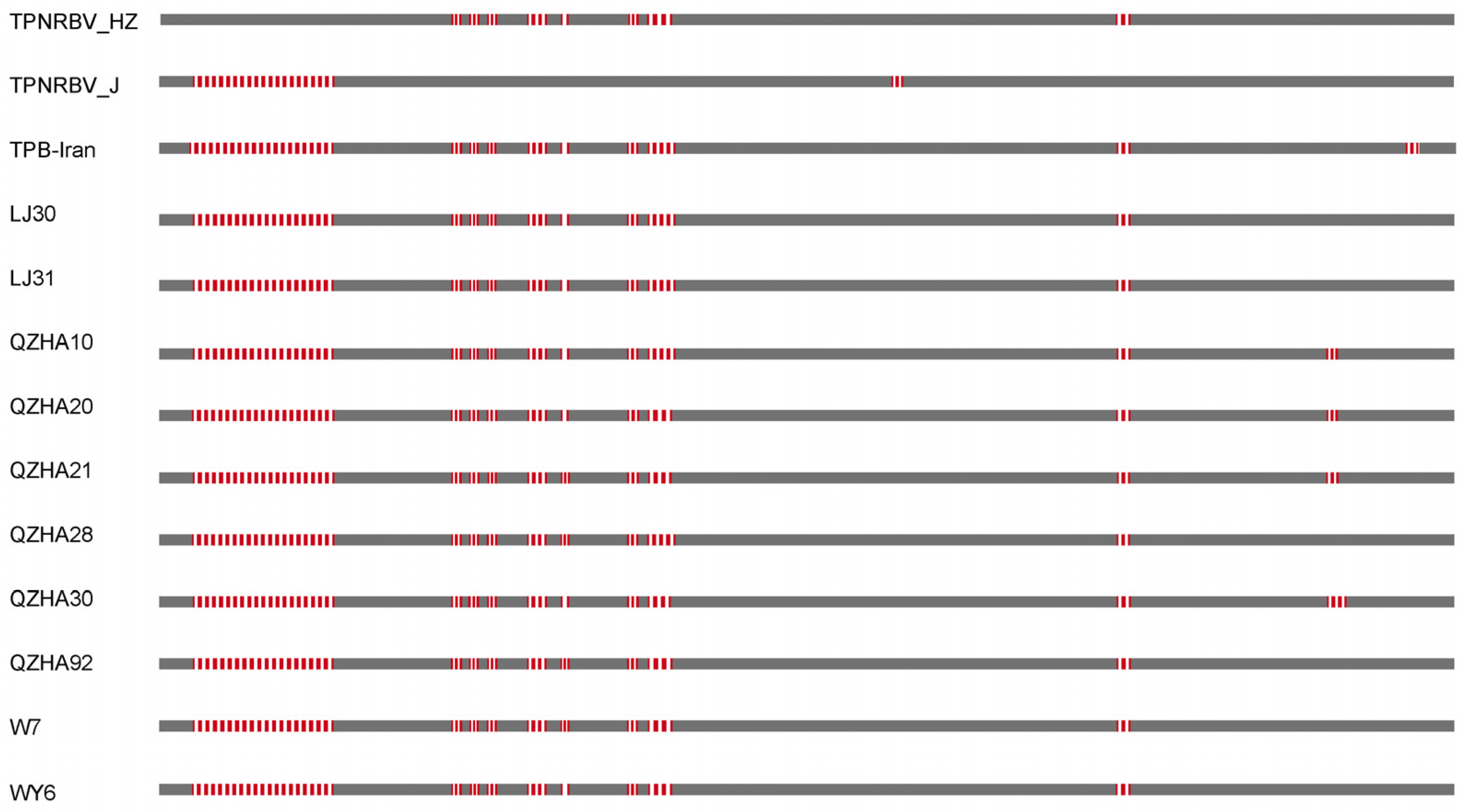
3.2. Genetic Variability of TPNRBV

To gain an understanding of the genetic variability of TPNRBV, the complete genome sequences of 10 TPNRBV isolates, including 2 from Fuzhou, 2 from Wuyishan, and 6 from Quanzhou, were determined. These 10 isolates were analyzed together with the 3 previously reported TPNRBV isolates: TPNRBV_HZ (from Hangzhou, Zhejiang Province of China), TPB-Iran (from Iran), and TPNRBV_J (from Japan) [4,6,7]. Since the reported sequences of TPB-Iran lack the 5’- and 3’-noncoding regions, our analysis focused on the coding sequences.
The aligned coding sequences of RNA1, RNA2, RNA3, and RNA4 were 5586, 3636, 2455, and 948 nucleotides in length, respectively (Table 2). All four segments exhibited a high haplotype diversity (Hd, Table 2). Statistical analysis revealed nucleotide diversity (Pi) values of 0.027, 0.016, 0.027, and 0.014 for RNA1, RNA2, RNA3, and RNA4, respectively (Table 2). The distribution of the nucleotide diversity in each segment was presented in Supplementary Figure S2. It is worth noting that the four ORFs of RNA3 showed Pi values of 0.019, 0.027, 0.019, and 0.027, respectively.
Pairwise comparisons revealed nucleotide sequence identities of 93.90%–100%, 94.10%–100%, 95.80%–100%, and 95.30%–100% for RNA1, RNA2, RNA3, and RNA4, respectively (Figure 1). For RNA1 and RNA2, the TPNRBV isolates determined in this study exhibited greater sequence identities with each other than with TPB-Iran and TPNRBV_HZ. For RNA3 and RNA4, however, similar observations were not made. Consistently, the TPNRBV isolates determined in this study tended to cluster together in phylogenetic trees inferred using the coding sequences of RNA1 and RNA2, but were assigned to separable branches in phylogenetic trees inferred using the coding sequences of RNA3 and RNA4 (Figure 1).
The aligned protein sequences of Mtr-Hel, RdRp, P14, P29, P24, P22, and MP were 1861, 1211, 123, 250, 182, 210, and 315 amino acids in length, respectively. Among these proteins, the least conserved was P22, with pairwise amino acid sequence identities ranging from 93.80% to 100%. Following this, P29 displayed amino acid sequence identities ranging from 94.00% to 100%. In contrast, the most conserved protein was MP, with amino acid sequences that were at least 98.00% identical (Supplementary Figure S3).

3.3. A Conserved Sequence Motif at the 5′ Termini of TPNRBV Genomic Segments

Maruyama et al. [9] identified a conserved sequence (5′-AATTACGA-3′) in the 5′ termini of TPNRBV_J RNA1-3, along with a variant of this sequence (5′-ATTAACGA-3′) in the 5′ termini of TPNRBV_J RNA4. Considering the significance of conserved terminal nucleotides in the replication and packaging of viruses with segmented genomes [20], we sought to determine if this feature is prevalent among TPNRBV isolates.
In TPNRBV_HZ, the sequence 5′-AATTACGA-3′ or 5′-ATTAACGA-3′ was absent in RNA2, RNA3, and RNA4. However, TPNRBV_HZ RNA1 contained the sequence 5′-AATTACGA-3′, albeit preceded by an additional four nucleotides (5′-TGGGAATTACGA-3′). Similarly, in QZHA21, one of the 10 isolates determined in this study, the sequence 5′-AATTACGA-3′, was absent in RNA3. For all other TPNRBV isolates, the sequence 5′-AATTACGA-3′ or 5′-ATTAACGA-3′ was readily detectable in all RNA segments, although most of these isolates had an additional G preceding the two conserved motifs (5′-GAATTACGA-3′ or 5′-GATTAACGA-3′). These observations suggest that the 5′-AATTACGA-3′ or 5′-ATTAACGA-3′ sequence is a common feature in the 5′ termini of the genomic segments of TPNRBV, and it is possible that the 5′ terminal nucleotides of certain segments of TPNRBV_HZ/QZHA21 were not captured during sequencing.
The aligned sequences of the 5′-noncoding regions of TPNRBV were 135, 134, 113, and 399 nucleotides in length for RNA1, RNA2, RNA3, and RNA4, respectively (Supplementary Figure S4). When the truncation of the 5′ termini of specific segments in TPNRBV_HZ and TPNRBV-QZHA21 were not considered, the pairwise nucleotide sequence identities in the 5′-noncoding regions of RNA1, RNA2, RNA3, and RNA4 ranged from 93.00% to 100%, 94.10% to 100%, 95.70% to 100%, and 95.80% to 100%, respectively. This indicates that the 5′-noncoding regions of TPNRBV are not more variable than its coding regions.

3.4. Extensive Deletion/Insertion Mutations in the 3′-Noncoding Regions of TPNRBV RNA4

The lengths of the 3′-noncoding regions of TPNRBV RNA4 vary significantly. For TPB-Iran, TPNRBV_HZ, and TPNRBV_J, the lengths are 823, 941, and 887 nucleotides, respectively. Among the 10 isolates determined in this study, the lengths range from 815 to 827 nucleotides. As shown in Figure 2, extensive deletion/insertion mutations are responsible for this observation. In an extreme case, a 110-nucleotide sequence found in TPNRBV_HZ is absent in all other TPNRBV isolates (Figure 2).
Extensive deletion/insertion mutations were also found in RNA3, but these mutations were concentrated in a small region located approximately 100 nucleotides upstream of the poly(A) tail. Every TPNRBV isolate exhibited at least one deletion/insertion in this region (Supplementary Figure S5). In contrast, extensive deletion/insertions were not found in RNA1 and RNA2.

3.5. The Role of Natural Selection and Recombination in Shaping the Genetic Structure of TPNRBV

To gain insights into the evolutionary processes underlying TPNRBV, the role of natural selection and recombination in shaping its genetic structure was investigated.
Recombination analyses conducted with the RDP4 suite revealed that the RNA2 of TPB-Iran exhibited recombination (RDP, p = 2.07 × 10⁻³; GENECONV, p = 2.60 × 10⁻⁴; Bootscan, p = 3.55 × 10⁻⁶; Maxchi, p = 6.02 × 10⁻¹⁰; Chimaera, p = 5.19 × 10⁻⁹; SiSscan, p = 6.61 × 10⁻⁶; and 3Seq, p = 6.41 × 10⁻¹¹). The RNA2 of TPNRBV_HZ was identified as its major parent, while the minor parent was an unknown TPNRBV isolate (Supplementary Figure S6A). This recombination event was further supported by GARD analysis (Supplementary Figure S6B).
Using EasyCodeML, purifying selection was detected at the majority of polymorphic sites in the ORFs of TPNRBV, as indicated by a dN/dS value significantly lower than one. However, one position in the ORF of RNA1 (position 1094, posterior probability > 0.95, Supplementary Table S3) was found to be under positive selection. Additionally, although not statistically supported, 19 and 22 codons were identified under positive selection in the ORFs of RNA1 and RNA2, respectively.

4. Discussion

TPNRBV is a recently discovered plant virus that infects tea plants. Although it is known to have a wide distribution across Asia, comprehensive regional-scale surveys to assess its prevalence have not been conducted. Additionally, the lack of knowledge about the genetic variability and diversity of TPNRBV not only hampers our understanding of its evolutionary dynamics, but also presents challenges in the development of accurate and reliable detection methods for this virus. In such a context, the occurrence of TPNRBV in Fujian Province, China was investigated and the complete genome of 10 new TPNRBV isolates was determined.
The results of our study revealed that TPNRBV is prevalent in Fujian, as it is present in all three major tea-growing regions of the province. However, there is considerable variation in TPNRBV prevalence among these regions. Several explanations can account for this observation [3]. For instance, the vector responsible for TPNRBV transmission may be a species of mites, as seen in other kitaviruses. It is possible that the abundance of TPNRBV-transmitting mites differs across various tea-growing regions. Alternatively, Fuzhou, Quanzhou, and Wuyishan may differ in terms of the tea cultivars they cultivate. It is plausible that different tea cultivars exhibit varying susceptibility to TPNRBV, as previously noted by Ren et al. [21].
TPNRBV possesses a genome that is relatively larger compared with most other plant viruses. Prior to this study, only three isolates of TPNRBV had complete or nearly complete genomes available [4,6,7]. Therefore, the sequencing of 10 new TPNRBV isolates presented here significantly contributes to our understanding of the virus’s population structure. Overall, our data reveal a low variability in TPNRBV, with nucleotide diversity values for its four RNA segments ranging from 0.014 to 0.027. Similar observations have been made for a few other kitaviruses, including citrus leprosis virus C (CiLV-C) [22,23], which is the most extensively studied kitavirus. The biological properties of these viruses, such as their frequent experience of transmission bottlenecks, have been suggested as an explanation for this observation [22,23]. Whereas transmission bottlenecks may indeed play a role in restricting the genetic diversity of TPNRBV, this study clearly demonstrates that other evolutionary forces, such as natural selection and recombination, also shape the virus’s population structure. Additionally, the data presented in Figure 1 indicate that reassortment may not be uncommon for TPNRBV. Moreover, the incongruence between the phylogeny and geographical origins of TPNRBV suggests frequent gene flow between different populations of this virus. One notable finding from this study aligns with the notion that the evolution of TPNRBV is complex: both the least conserved (P22) and the most conserved (MP) proteins of TPNRBV are encoded by RNA3 and RNA4, respectively, despite RNA4 not being the most variable genome fragment of TPNRBV.
In addition to its occurrence and genetic variability, this study reveals two important properties of TPNRBV. First, the motif 5′-AATTACGA-3′ or 5’-ATTAACGA-3′ may be shared by the four genomic segments of TPNRBV, as initially proposed by Maruyama et al. [7]. Secondly, the 3′ non-coding region of TPNRBV RNA4 frequently undergoes deletion/insertion mutations. The biological significance of these two properties, especially the latter one, remains unclear at present. However, both properties undoubtedly deserve further investigation.

5. Conclusions

In conclusion, our study provides the first comprehensive survey of TPNRBV occurrence on a provincial scale. Additionally, the complete genome of 10 new TPNRBV isolates was determined. These data are of great significance in implementing effective control measures, as they allow for the design of targeted strategies and the development of accurate detection methods to control TPNRBV.

Supplementary Materials

The following supporting information can be downloaded at: https://www.mdpi.com/article/10.3390/f14091755/s1, Figure S1: Associated disease symptoms of tea plants infected with TPNRBV; Figure S2: Sliding-window plot of nucleotide diversity for the TPNRBV genomic segments (excluding the non-coding regions); Figure S3: Pairwise amino acid identities of Mtr-Hel, RdRp, P14, P29, P24, P22, and MP; Figure S4: Multiple sequence alignment of the 5′ termini of TPNRBV genomic segments; Figure S5: Multiple sequence alignment of the 3′ termini of TPNRBV genomic segments; Figure S6: Recombinant signal identified in the RNA2 complete genome of TPB-Iran isolate by using Bootscan algorithm implemented in the RDP suite (A) and GARD in the Datamonkey server (B), respectively. Table S1: Primers used for amplification and sequencing of TPNRBV genome; Table S2: TPNRBV isolates sequenced in this study; Table S3: Positive selection on different genomic regions of tea plant necrotic ring blotch virus using the Site Model in EasyCodeML.

Author Contributions

J.S. and F.G.: conceptualization and design; F.G.: methodology; X.C., M.L., Y.G. and F.G.: formal analysis; J.S., Z.D. and F.G.: interpretation of results; J.S., Z.D. and F.G.: writing original draft. All authors have read and agreed to the published version of the manuscript.

Funding

This research was funded by the Agricultural Guidance (Key) Project of Fujian Province, Fujian, China (No. 2020N0024); the Open Project of Fujian Key Laboratory for Technology Research of Inspection and Quarantine, Fujian, China (No. FJKF2019-03); the Central Guidance on Local Science and Technology Development Fund of Fujian Province, Fujian, China (No. 2022L3016); and a fund from Fujian Provincial Department of Finance (No. KLE21004A).

Data Availability Statement

All data used in this study are publicly available on NCBI. The complete genome sequences obtained in this study have been deposited in GenBank with accession numbers OQ948425–OQ948464.

Conflicts of Interest

The authors declare no conflict of interest. The funders had no role in the design of the study; in the collection, analyses, or interpretation of data; in the writing of the manuscript; or in the decision to publish the results.

References

  1. Liang, L.; Ridoutt, B.; Wang, L.; Xie, B.; Li, M.; Li, Z. China’s Tea Industry: Net Greenhouse Gas Emissions and Mitigation Potential. Agriculture 2021, 11, 363. [Google Scholar] [CrossRef]
  2. Quito-Avila, D.; Freitas-Astúa, J.; Melzer, M. Bluner-, cile-, and higreviruses (Kitaviridae). In Encyclopedia of Virology; Elsevier: Amsterdam, The Netherlands, 2021; pp. 247–251. [Google Scholar]
  3. Ramos-González, P.; Arena, G.; Tassi, A.; Chabi-Jesus, C.; Kitajima, E.; Freitas-Astúa, J. Kitaviruses: A window to atypical plant viruses causing nonsystemic diseases. Annu. Rev. Phytopathol. 2023, 61. [Google Scholar] [CrossRef]
  4. Hao, X.; Zhang, W.; Zhao, F.; Liu, Y.; Qian, W.; Wang, Y.; Wang, L.; Zeng, J.; Yang, Y.; Wang, X. Discovery of plant viruses from tea plant (Camellia sinensis (L.) O. Kuntze) by metagenomic sequencing. Front. Microbiol. 2018, 9, 2175. [Google Scholar] [CrossRef]
  5. Ramos-González, P.L.; Kondo, H.; Morozov, S.; Vasilakis, N.; Varsani, A.; Cao, M.; Freitas-Astúa, J. The border between kitavirids and nege-like viruses: Tracking the evolutionary pace of plant- and arthropod-infecting viruses. Front. Plant Sci. 2022, 13, 932523. [Google Scholar] [CrossRef]
  6. Nazerian, E.; Bayat, H. Occurrence of tea plant necrotic ring blotch virus in Iran. J. Plant Prot. Res. 2021, 61, 200–202. [Google Scholar]
  7. Maruyama, N.; Nozomu, I.; Nishikawa, M.; Nijo, T.; Yoshida, T.; Kitazawa, Y.; Maejima, K.; Namba, S.; Yamaji, Y. Complete genome sequence of tea plant necrotic ring blotch virus detected from a tea plant in Japan. Microbiol. Resour. Announc. 2022, 11, e00323-22. [Google Scholar] [CrossRef] [PubMed]
  8. Li, R.; Mock, R.; Huang, Q.; Abad, J.; Hartung, J.; Kinard, G. A reliable and inexpensive method of nucleic acid extraction for the PCR-based detection of diverse plant pathogens. J. Virol. Methods 2008, 154, 48–55. [Google Scholar] [CrossRef] [PubMed]
  9. Edgar, R.C. MUSCLE: Multiple sequence alignment with high accuracy and high throughput. Nucleic Acids Res. 2004, 32, 1792–1797. [Google Scholar] [CrossRef]
  10. Kumar, S.; Stecher, G.; Li, M.; Knyaz, C.; Tamura, K. MEGA X: Molecular Evolutionary Genetics Analysis across Computing Platforms. Mol. Biol. Evol. 2018, 35, 1547–1549. [Google Scholar] [CrossRef] [PubMed]
  11. Muhire, B.M.; Varsani, A.; Martin, D.P. SDT: A virus classification tool based on pairwise sequence alignment and identity calculation. PLoS ONE 2014, 9, e108277. [Google Scholar] [CrossRef]
  12. Librado, P.; Rozas, J. DnaSP v5: A software for comprehensive analysis of DNA polymorphism data. Bioinformatics 2009, 25, 1451–1452. [Google Scholar] [CrossRef] [PubMed]
  13. Nguyen, L.-T.; Schmidt, H.A.; von Haeseler, A.; Minh, B.Q. IQ-TREE: A fast and effective stochastic algorithm for estimating maximum likelihood phylogenies. Mol. Biol. Evol. 2014, 32, 268–274. [Google Scholar] [CrossRef] [PubMed]
  14. Xiang, C.-Y.; Gao, F.; Jakovlić, I.; Lei, H.-P.; Hu, Y.; Zhang, H.; Zou, H.; Wang, G.-T.; Zhang, D. Using PhyloSuite for molecular phylogeny and tree-based analyses. iMeta 2023, 2, e87. [Google Scholar] [CrossRef]
  15. Kalyaanamoorthy, S.; Minh, B.Q.; Wong, T.K.F.; von Haeseler, A.; Jermiin, L.S. ModelFinder: Fast model selection for accurate phylogenetic estimates. Nat. Methods 2017, 14, 587–589. [Google Scholar] [CrossRef] [PubMed]
  16. Hoang, D.T.; Chernomor, O.; von Haeseler, A.; Minh, B.Q.; Vinh, L.S. UFBoot2: Improving the ultrafast bootstrap approximation. Mol. Biol. Evol. 2018, 35, 518–522. [Google Scholar] [CrossRef] [PubMed]
  17. Martin, D.P.; Murrell, B.; Golden, M.; Khoosal, A.; Muhire, B. RDP4: Detection and analysis of recombination patterns in virus genomes. Virus Evol. 2015, 1, vev003. [Google Scholar] [CrossRef] [PubMed]
  18. Kosakovsky Pond, S.L.; Posada, D.; Gravenor, M.B.; Woelk, C.H.; Frost, S.D. GARD: A genetic algorithm for recombination detection. Bioinformatics 2006, 22, 3096–3098. [Google Scholar] [CrossRef] [PubMed]
  19. Gao, F.; Chen, C.; Arab, D.A.; Du, Z.; He, Y.; Ho, S.Y.W. EasyCodeML: A visual tool for analysis of selection using CodeML. Ecol. Evol. 2019, 9, 3891–3898. [Google Scholar] [CrossRef]
  20. Haralampiev, I.; Prisner, S.; Nitzan, M.; Schade, M.; Jolmes, F.; Schreiber, M.; Loidolt-Krüger, M.; Jongen, K.; Chamiolo, J.; Nilson, N.; et al. Selective flexible packaging pathways of the segmented genome of influenza A virus. Nat. Commun. 2020, 11, 4355. [Google Scholar] [CrossRef]
  21. Ren, H.; Chen, Y.; Zhao, F.; Ding, C.; Zhang, K.; Wang, L.; Yang, Y.; Hao, X.; Wang, X. Quantitative distribution and transmission of tea plant necrotic ring blotch virus in Camellia sinensis. Forests 2022, 13, 1306. [Google Scholar] [CrossRef]
  22. Chabi-Jesus, C.; Ramos-González, P.L.; Postclam-Barro, M.; Fontenele, R.S.; Harakava, R.; Bassanezi, R.B.; Moreira, A.S.; Kitajima, E.W.; Varsani, A.; Freitas-Astúa, J. Molecular epidemiology of citrus leprosis virus C: A new viral lineage and phylodynamic of the main viral subpopulations in the Americas. Front. Microbiol. 2021, 12, 641252. [Google Scholar] [CrossRef]
  23. Ramos-González, P.L.; Chabi-Jesus, C.; Guerra-Peraza, O.; Breton, M.C.; Arena, G.D.; Nunes, M.A.; Kitajima, E.W.; Machado, M.A.; Freitas-Astúa, J. Phylogenetic and molecular variability studies reveal a new genetic clade of citrus leprosis virus C. Viruses 2016, 8, 153. [Google Scholar] [CrossRef]
Figure 1. Phylogenetic relationships and pairwise nucleotide sequence identities (indicated on the right) among RNA1, RNA2 RNA3, and RNA4 of TPNRBV. The phylogenetic trees were reconstructed using IQ-TREE, while nucleotide sequence identities were determined using SDT 1.2. For each node in the tree, the bootstrap values (>95%) are shown above the branches. Two viruses, blueberry necrotic ring blotch virus (NC_016084 to NC_016087) and tomato fruit blotch virus (MK517477 to MK517480), were used as outgroups. The scale bar is given in substitutions/site.
Forests 14 01755 g001
Figure 2. A diagram showing the alignment of the 3′-noncoding regions of TPNRBV RNA4. Red dash lines indicate >3 nt deletions.
Forests 14 01755 g002
Table 1. RT-PCR assay detecting TPNRBV from the tea samples collected in this study.
CityLocationTested SamplesPositive SamplePositive Rate (%)
FuzhouLianjiang7522.67
QuanzhouHui’an201680
NanpingWuyishan10143.96
Total 1962211.22
Table 2. Genetic diversity of the 13 TPNRBV isolates.
SegmentLengthTVsSVsPIsHdPi
RNA155865692782911.000 ± 0.0300.027 ± 0.006
RNA23636226160661.000 ± 0.0020.016 ± 0.006
RNA32455218681500.987 ± 0.0350.027 ± 0.003
RNA49483918210.933 ± 0.0770.014 ± 0.002
TVs, total variable sites; SVs, singleton variable sites; PIs, parsimony informative sites.
Disclaimer/Publisher’s Note: The statements, opinions and data contained in all publications are solely those of the individual author(s) and contributor(s) and not of MDPI and/or the editor(s). MDPI and/or the editor(s) disclaim responsibility for any injury to people or property resulting from any ideas, methods, instructions or products referred to in the content.

