Article

Effects of Pine Wilt Disease on Rhizosphere Microbiota and Fine Root Fungi: Insights into Enzyme Activity, Ectomycorrhizal Infection and Microbial Composition

1
Collaborative Innovation Center of Sustainable Forestry in Southern China, College of Forestry, Nanjing Forestry University, Nanjing 210037, China
2
Department of Forest Protection, Zhongshan Cemetery Administration Bureau, Nanjing 210037, China
3
Department of Forest Sciences, University of Helsinki, 00790 Helsinki, Finland
*
Author to whom correspondence should be addressed.
Forests 2023, 14(9), 1884; https://doi.org/10.3390/f14091884
Submission received: 23 August 2023 / Revised: 14 September 2023 / Accepted: 15 September 2023 / Published: 16 September 2023
(This article belongs to the Section Forest Health)

Abstract

:
Pine wilt disease (PWD), caused by the pine wood nematode (PWN) Bursaphelenchus xylophilus, poses a severe threat to pine forests worldwide. However, the understanding of the impact of PWD on the host microbiome remains limited. This study aimed to investigate the structure and function of the fungal community associated with Pinus thunbergii fine roots and the rhizosphere fungi and bacteria of the tree naturally infected by PWN and the healthy tree. We employed high-throughput sequencing in conjunction with functional prediction tools (Functional Annotation of Prokaryotic Taxa and Fungi Functional Guild) and soil enzyme activity measurements between the two treatments (disease vs. health). The results showed that PWD significantly decreased the activity of β-cellobiosidase (CEL) and β-glucosidase (GLS) enzymes involved in carbon cycling in the rhizosphere (p < 0.05). However, PWD did not alter the diversity of rhizosphere bacteria and fine root fungi, but it did cause a significant decrease in the richness of rhizosphere fungi (p < 0.05). Moreover, PWD significantly reduced the abundance of Actinobacteria and genus Gaiella (p < 0.05). Functionally, bacterial intracellular parasites exhibited a higher abundance in the rhizosphere after PWN infection, whereas ureolysis showed a lower abundance (p < 0.05). Fungal saprotroph–symbiotroph exhibited a higher abundance in the rhizosphere after PWN infection, whereas symbiotroph showed a lower abundance (p < 0.05). Additionally, it led to a significant reduction in the infection rate of ectomycorrhizal fungi (p < 0.05). Infected host fine root exhibited higher abundance of pathotroph–symbiotroph, whereas symbiotroph had a lower abundance (p < 0.05). These findings provided valuable insights into the interactions between pine wilt disease, plant microbial communities, and soil enzyme activity.

1. Introduction

Plant microbiome comprises a diverse community of microorganisms, including bacteria, fungi, and nematodes, residing inside the plant. These microorganisms establish intricate relationships with their host, forming a complex community [1]. While some microorganisms can cause diseases, others play a beneficial role by promoting plant growth, aiding nutrient acquisition, and enhancing stress tolerance [2]. These interactions can be beneficial, harmful, or neutral for the host plant [3]. Notably, plants shape their microbial community, which reciprocally influence their biochemical and physiological activities [4]. Furthermore, endophytic bacteria and fungi hold significant potential for agricultural applications, improving crop growth, increasing agricultural productivity, and promoting environmental sustainability [5].
The rhizosphere, the soil area surrounding and influenced by plant roots, plays a vital role in plant growth and health by facilitating crucial microbial interactions [6]. Plant roots release various secretions that attract diverse microorganisms, shaping the rhizosphere microbiome. These secretions include sugars, amino acids, organic acids, phenolic compounds, and secondary metabolites like coumarin, glucitol, camphorin, and triterpenoids [7]. These compounds affect soil pH, structure, and oxygen levels, thereby influencing the composition and activity of the rhizosphere microbial community through carbon-rich exudates [8,9]. Microorganisms in the rhizosphere can perceive and respond to signals from themselves, other microorganisms, and plants, exerting influences on their plant hosts. They achieve this by releasing signal molecules, which can induce plant immunity, enhance stress tolerance, promote overall growth and health, support nutrient uptake, and maintain the rhizosphere microbiome. Signal molecules involved in these processes include N-acyl-homoserine lactones (AHLs), diffusible signal factors, plant-like hormones, and volatile organic compounds [10,11,12].
Plants heavily rely on underground microorganisms for nutrient acquisition [13,14] and to enhance their response to environmental stressors [15]. The symbiotic association between plants and mycorrhizal fungi is important and ecologically influential [16]. For instance, when plants are inoculated with ectomycorrhizal fungi, they develop mycorrhizal structures with increased hyphal growth. This expansion of the root system’s effective absorption area enhances water and nutrient uptake from the rhizosphere soil, ultimately boosting plant growth and drought tolerance [17]. Mycorrhizal fungi colonize host roots and improve their access to nutrients. In return, the plant transports photosynthetic carbon to the colonizing fungus. This nutrient exchange significantly impacts key soil processes, carbon cycling, and plant health.
Pine wilt disease (PWD), caused by the pine wood nematode (PWN) Bursaphelenchus xylophilus, is a destructive disease affecting conifer trees [18,19]. Several studies have investigated the microbes in pine trees affected by PWN and microbes associated with PWN to understand the relationship among PWN, PWN-associated microbes, and host microbes [20,21,22,23]. Given the importance of host microbiome, however, limited studies have explored the impact of PWD on rhizosphere microorganisms and mycorrhizal fungi, particularly in Pinus thunbergii Parl. Therefore, this study focuses on naturally infected P. thunbergii trees and employs high-throughput Illumina MiSeq sequencing to explore the structure and function of rhizosphere microbes and fine root fungi. The main objectives of this study were to elucidate the effects of PWD on host rhizo-sphere soil enzyme activities, microbial communities and functions, and mycorrhizal fungal infection rates to better understand the relationship between pathogens and host microbial communities. These findings could offer valuable insights into the interaction between forest decline and plant–microbe communities.

2. Materials and Methods

2.1. Study Sites and Sampling

The study site for this experiment was located in the Zhongshan Mausoleum Park on Purple Mountain in Nanjing, China, with geographical coordinates ranging from 32°01′ to 32°06′ N and 118°48′ to 118°53′ E. The park spans approximately 4500 hectares and has elevations ranging from 20 m to 449 m. The region experiences an annual average precipitation between 1000 mm and 1050 mm, with an average sunshine duration of approximately 2213 h per year. The mean annual temperature is 15.4 °C, with the highest temperature recorded in August at 40.7 °C and the lowest temperature in January at −14.0 °C [24]. The soil in the area is classified as zonal soil, characterized by a yellow-brown color [25]. Soil pH is in the range of 4.6–8.6. Soil organic carbon is in the range of 3.5%–7.3%. Soil moisture was in the range of 6.5%–30.2%. Microbial biomass carbon was in the range of 28.1–109.9 mg/kg [26]. The dominant tree species in the study area were P. thunbergii and P. massoniana Lamb., with an age range of 60–80 years [22]. Due to the spread of pine wilt disease, a significant number of susceptible pine trees have died, resulting in the emergence of secondary broadleaf forests, including Liquidambar formosana Hance and Quercus acutissima Carruth. The vegetation also consists of shrubs such as Symplocos paniculata, Camellia sinensis, and Lindera glauca, as well as herbaceous plants like Ophiopogon japonicus, Commelina communis, and Reynoutria japonica [23]. Three study plots were established in this study, each measuring 20 × 20 m and located 500 m apart from each other. Within each plot, three pine trees exhibiting symptoms of PWD and nearing death in October 2018 were selected, along with three healthy trees. The distance between the diseased and healthy trees was less than 15 m. The selection of healthy and diseased trees followed the method described by Millberg et al. [27], where healthy trees exhibited fully green needles without any signs of pine wilt disease, whereas diseased trees showed symptoms of dryness and browning in their needles. Nematodes were isolated using the Baermann funnel method, and the health and disease status of the trees were confirmed using specific primers for the PWN [28]. Fine root samples were excavated by digging approximately 25 cm below the soil surface from three different directions (with 120° intervals) for each tree using a sterile auger with a diameter of 10 mm. Three samples per tree were combined to create a composite sample, resulting in a total of 18 fine root samples (9 from diseased trees and 9 from healthy trees). Rhizosphere soil samples were collected by carefully removing soil adhering to the fine roots. All samples were properly labeled and stored in a cooler at 4 °C, with DNA extraction samples kept at −20 °C for subsequent analysis.

2.2. Rhizosphere Soil Enzyme Activity Analysis

The activity of seven soil enzymes involved in carbon (C), nitrogen (N), sulfur (S), and phosphorus (P) cycling was assessed. Enzymes related to C cycling were β-xylosidase (XYL), β-D-glucosidase (GLR), β-cellobiosidase (CEL), and β-glucosaminidase (GLS). N, P, and S cycling enzymes included N-acetylglucosaminidase (NAG), phosphatase (PHO), and sulfatase (Sul), respectively. Rhizosphere soil enzyme activity was measured using a 96-well plate fluorescence assay with 4-methylumbelliferone (4-MUB) as the substrate. For the analysis, 2 g of fresh soil were weighed and placed in a sterile 50 mL centrifuge tube. Then, 30 mL of a 50 mM acetate buffer solution at pH 5 was added. The mixture was vortexed for 1 min and shaken at 180 r·min−1 on a shaker at 25 °C for 40 min to break down large soil particles. Next, the soil suspension was transferred to a 200 mL beaker containing 170 mL of 25 °C sodium acetate solution, resulting in a uniform soil suspension [29,30].
Each soil sample was prepared with four replicates, and each replicate included four reactions: blank (200 µL substrate with 50 µL double-distilled water), quenched standard (200 µL substrate plus 50 µL 200 mM 4-MUB), negative control (200 µL buffer solution plus 50 µL 4-MUB-linked substrate), and reference standard (200 µL buffer solution plus 50 µL 4-MUB). To initiate the reaction, 200 µL of the soil suspension and 50 µL of 4-MUB-linked substrate were added to each well of a 96-well plate. The plate was then incubated in the dark at 25 °C for 4 h. Afterward, the reaction was stopped by adding 10 µL of 1 mol·L−1 NaOH solution to each well. The fluorescence was immediately measured using a microplate fluorescence reader with excitation and emission filters set at 365 nm and 450 nm, respectively. Enzyme activity was quantified as the amount of MUB (nmol·g−1·h−1) released per gram of dry soil per hour.

2.3. Determination of Mycorrhizal Fungal Infection Rate

The fine root samples from both diseased and healthy trees were rinsed with distilled water to remove the soil. The washed fine roots were then cut into 1 cm segments using scissors and placed in 5 mL centrifuge tubes. A total of 10 random root segments were selected from each sample of diseased and healthy trees, resulting in 90 root samples for diseased pine trees and 90 for healthy trees, making a total of 180 samples. The staining procedure utilized the Trillium Blue staining method for observation of the colonization of mycorrhizal fungi [31]. Briefly, cleaned root segments of suitable thicknesses were placed in a 10 mL centrifuge tube containing 5–7 mL of 10% KOH. The tube was heated in a 90 °C water bath until the root system became partially transparent. After washing away residual KOH with distilled water, the roots were acidified in 2% HCl for 5–10 min and rinsed with distilled water. Subsequently, the roots were immersed in a 0.05% Trillium Blue staining solution and heated in a 90 °C water bath for 30 min. After cooling, the roots were transferred to a lactoglycerol solution at room temperature for 24 h to remove any excess dye. Using tweezers, the decolorized root segments were placed on rectangular microscope slides, with 5–10 root segments per slide. A cover glass was then placed over the root segments, and gentle pressure was applied to flatten the root system. Using a microscope at magnifications of 50–400×, the root segments were examined to observe the level of colonization of mycorrhizal fungi. The level of colonization was determined by assessing the percentage of mycorrhizal structures present in each root segment. The classification system included the following categories: 0%, less than 1%, less than 10%, less than 50%, more than 50%, and more than 90%. Mycorrhizal fungi infection rate was calculated as (ΣM%×n)/N, in which M% represented the infection rate of a single root segment; n represented the number of root segments with the same infection rate; and N represented the total number of root segments per treatment. Ectomycorrhizal fungi were primarily found encapsulating the surface of young plant roots, with only a few hyphae invading the cellular interstices of the root epidermis and cortex, but not entering the interior of the cells. On the other hand, endophytic mycorrhizal fungi were quantified as penetrating through the cell wall and establishing within the living cells of young roots.

2.4. Sample DNA Extraction, PCR Amplification and Illumina MiSeq Sequencing

Genomic DNA extraction was performed according to the manufacturer’s instructions. Rhizosphere soil DNA was extracted using a soil DNA kit (Omega Bio-tek, Norcross, GA, USA), whereas root DNA was extracted using a plant genomic DNA kit (Tiangen Biotech, Beijing, China). The concentration of DNA was determined using a NanoDrop ND-1000 spectrophotometer (Thermo Fisher Science, Wilmington, DE, USA). The internal transcribed spacer 1 (ITS1) region of fungi was amplified using the ITS1-F (5′-CTTGGTCATTTAGAGGAAGTAA-3′) and ITS2-R (5′-GCTGCGTTCTTCATCGATGC-3′) primers [32]. The V3-V4 region of 16S rDNA gene of bacteria was amplified using primer pairs of 338F (ACTCCTACGGGAGGCAGCAG) and 806R (GGACTACHVGGGTWTCTAAT) [33]. Each sample underwent three replicate polymerase chain reactions (PCRs) using the Transgen AP221-0220 µL reaction system. The PCR reaction mixture consisted of 4 µL of 5 × FastPfu buffer, 2 µL of dNTPs (2.5 mM), 0.8 µL of each forward and reverse primer (5 µM), 0.4 µL of FastPfu polymerase, 0.2 µL of bovine serum albumin (BSA), and 10 ng of template DNA. The PCR amplification for both bacteria and fungi was conducted with the following cycling conditions: initial denaturation at 95 °C for 3 min, followed by 27 cycles of denaturation at 95 °C for 30 s, annealing at 55 °C for 30 s, extension at 72 °C for 45 s, and a final extension at 72 °C for 10 min.
The amplified products were confirmed using 2% agarose gel electrophoresis and purified using Agencourt AMPure XP beads (Beckman Coulter, Pasadena, CA, USA). The DNA concentration was measured using a NanoDrop ND-1000 spectrophotometer, and the samples were sequenced on an Illumina MiSeq platform (PE = 250) at the Shanghai International Medical Zone, China. The raw sequencing data were deposited in the Sequence Read Archive (SRA) at the National Center for Biotechnology Information (NCBI) under the project accession number PRJNA974452.

2.5. Bioinformatics and Statistical Analysis

The raw bacterial and fungal sequence data were processed using Mothur software (Version 1.48.0, East Lansing, MI, USA) following standard operating procedures (SOP) outlined [23,34]. In brief, CutAdapt v.1.15 was used to remove adapter and barcode sequences. The preprocessed sequences underwent quality checks, including identification of sequencing errors (trim.seqs), PCR errors (pcr.seqs), and chimeras (chimera.uchime). For bacterial sequences, the align.seqs command was used to align sequence against the Silva database (Reference = silva.nr_v138) with Needleman and Wunsch algorithm [35]. For fungal sequences, the pairwise.seq command was used for alignment. Sequences were then pre-clustered with 6 base pairs mismatched for bacteria and 2 base pairs mismatched for fungi. Following pre-clustering, the sequences were clustered into operational taxonomic units (OTUs) at 97% similarity [26]. OTUs with sequence counts below 10 across all samples for bacteria and below 1 for fungi were filtered out and removed, based on a method previously described [36]. Bacteria taxonomic assignments were performed against the Silva database (version 1.38) using the classify.seq command with an 80% bootstrap confidence level [37]. For fungi, the most abundant sequence from each out was selected as the representative fungal sequences and taxonomically classified against UNITE reference database (UNITE + INSD, version 8.0) with 80% bootstrap confidence [38]. Sequences corresponding to plant chloroplasts and unknown domains were excluded from the analysis.
To determine the bacterial and fungal community functions, we employed the Functional Annotation of Prokaryotic Taxa (FAPROTAX) classifier (script version 1.1) developed by Louca et al. (2016) [39] for bacterial OTUs. FAPROTAX uses a comprehensive database of bacterial genomes and information on known functional genes to create a classification model. By comparing the sequences of known genomes, FAPROTAX predicts the functions of the bacterial OTUs. For fungal OTUs, we used FUNGuild, a Python-based tool specifically designed for this purpose [40]. FUNGuild classifies fungi into different ecological guilds, representing the various functional roles that fungi play in the ecosystem. These functional groups can include pathotrophs, symbiotrophs, and saprotrophs, among others. The FUNGuild database contains information on a wide range of known fungal functional groups, providing genome annotations and eco-functional annotations associated with these functional groups.
To address variations in sample size, we used the rarefied data subset with the smallest sample size across the entire bacterial or fungal dataset for calculating diversity indices, including alpha diversity (Shannon), species richness (Observed Species, Sobs), and evenness (Shannoneven). This approach ensured comparability between treatments [22]. Differences in community diversity indices were assessed through one-way analysis of variance (ANOVA) using SPSS 26 (IBM, Armonk, NY, USA). To investigate the correlation among enzyme activity, microbial diversity, and microbial taxa, we performed correlation analysis (Pearson) using SPSS 26 (IBM, Armonk, NY, USA). Venn diagrams were generated using InteractiveVenn [41] to visualize and identify shared and unique OTUs. For visualizing and testing significant differences in bacterial and fungal community and functional structure, we employed Principal coordinate analysis (PCoA) and PERMANOVA methods in PRIMER7 software (Version 7.0.13, Plymouth, UK) [42].

3. Results

3.1. Enzyme Activity in Rhizosphere Soil

Significant differences were observed in rhizosphere soil enzyme activities between healthy and diseased host rhizosphere soils, specifically for β-cellobiosidase (CEL) and β-glucosidase (GLS) involved in carbon cycling, as indicated in Table 1. The rhizosphere soil enzyme activities of CEL were 77.32 ± 17.07 in healthy trees and 33.73 ± 12.25 in diseased trees. The soil enzyme activities of GLS were 278.07 ± 20.43 in healthy trees and 139 ± 40.4 in diseased trees. The activities of CEL and GLS in healthy host rhizosphere soil were significantly higher compared to diseased host rhizosphere soil (p < 0.05) (Table S2). However, no significant differences were found in the activities of xylanase (XYL) and β-D-glucuronidase (GLR) related to carbon cycling, sulfate esterase (SUL) involved in sulfur cycling, N-acetylglucosaminidase (NAG) associated with nitrogen cycling, and phosphatase (PHO) involved in phosphorus cycling between the two rhizosphere soils.

3.2. Microbial Community in Rhizosphere Soil

3.2.1. Microbial α-Diversity in Rhizosphere Soil

A total of 5117 bacterial and 1386 fungal operational taxonomic units (OTUs) were identified from the rhizosphere soil. The rhizosphere fungi sobs index was 553 ± 23.42 on the healthy trees and 509 ± 40.75 on the diseased trees. The rhizosphere microbial community of healthy pine exhibited higher richness (Sobs), diversity (Shannon), and evenness (shannoneven) compared to that of diseased pine. However, no significant differences were observed in any of the diversity indices between healthy and diseased trees, except for fungal species richness (Table 2).

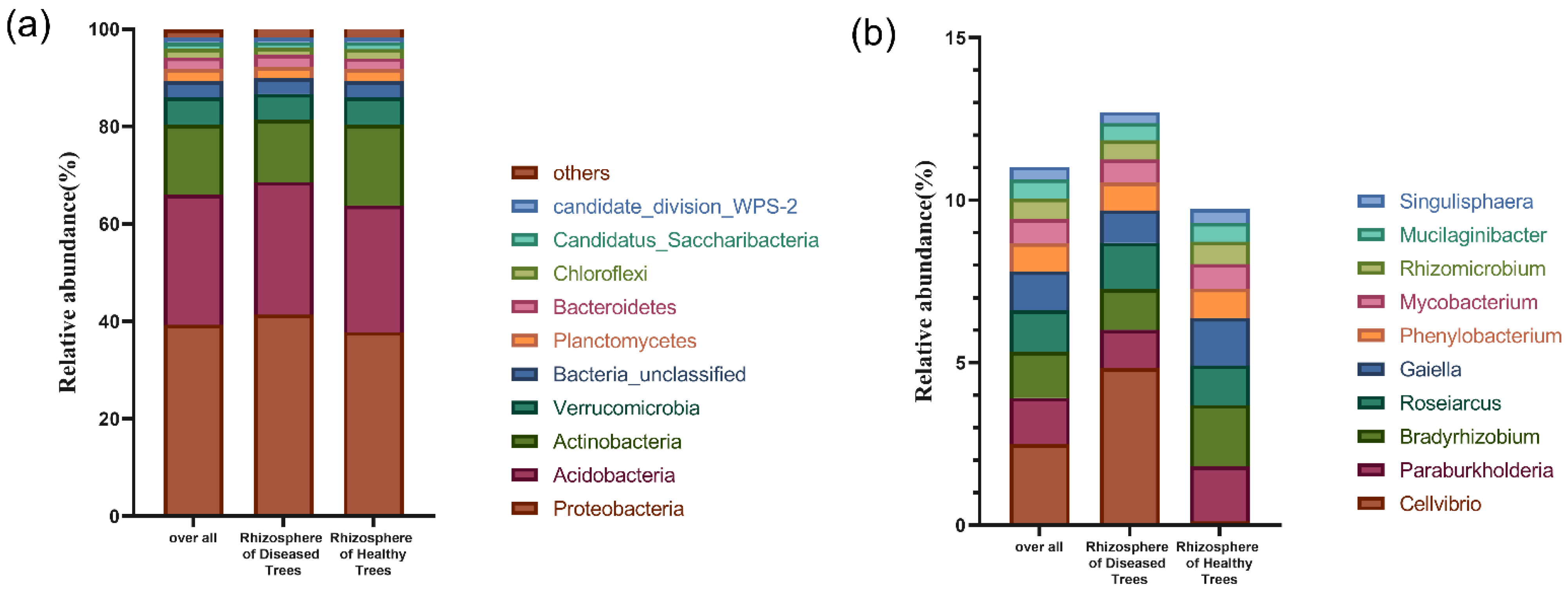
3.2.2. Microbial Community Structure in Rhizosphere Soil

The bacterial shared OTUs accounted for 93.6%, whereas unique OTUs represented 2.7% in the rhizosphere of healthy trees and 3.7% in the rhizosphere of diseased trees (Figure S1a). PCoA based on bacterial OTU data showed that the rhizosphere bacterial community structure was similar between healthy and diseased trees, as confirmed by PERMANOVA analysis, which found no significant differences (Figure S2a).
Rhizosphere fungal OTUs between diseased and healthy pine trees accounted for 75%. Each group had its unique set of OTUs, representing 14.6% in healthy trees and 10.4% in diseased trees (Figure S1b). Similar to the bacterial community structure, the fungal community structure of rhizosphere soil did not differ between healthy and diseased trees (Figure S2b).
A total of 31 bacterial phyla were identified from rhizosphere soil, with Proteobacteria being the most abundant (39.29%), followed by Acidobacteria (26.77%), Actinobacteria (14.33%), Verrucomicrobia (5.58%), Planctomycetes (2.52%), Bacteroidetes (2.32%), Chloroflexi (1.85%), and Candidatus_Saccharibacteria (1.27%) (Figure 1a). These dominant phyla, with a combined relative abundance of 90.80%, accounted for the majority of the bacterial community. Interestingly, the abundance of Actinobacteria was significantly higher in rhizosphere soil of healthy hosts compared to that of diseased hosts (Figure S5; p < 0.05), which was positively correlated with the enzyme activity of β-cellobiosidase (CEL) and β-glucosidase (GLS) (p < 0.05; Pearson = 0.773; and Pearson = 0.665) (Table S4).
At the genus level, 569 genera were identified, representing 17.58% of the sequences. The most prevalent genus was Cellvibrio (2.48%), followed by Paraburkholderia (1.44%), Bradyrhizobium (1.42%), Roseiarcus (1.27%), and Gaiella (1.20%) (Figure 1b). Notably, the abundance of Gaiella was significantly higher in rhizosphere soil of healthy hosts compared to that of diseased hosts (Figure S5; p < 0.05), which showed positive correlation with the enzyme activity of β-cellobiosidase (CEL) (p < 0.05; Pearson = 0.739) (Table S4).
A total of 10 fungal phyla were identified from the rhizosphere soil. The dominant fungal phylum was Mortierellomycota (45.86%), followed by Ascomycota (22.99%) and Basidiomycota (19.03%) (Figure 2a). Minor phyla, including Mucoromycota, Kickxellomycota, Chytridiomycota, Zoopagomycota, Glomeromycota, Rozellomycota, and Basidiobolomycota, constituted ≤0.3% of the sequences. Notably, the rhizosphere of diseased hosts showed a higher abundance of Mortierellomycota, and the rhizosphere of healthy hosts had a higher abundance of Mucoromycota (Figure S5; p < 0.05).
At the species level, 50.32% of the sequences were assigned to 191 fungal species. The most prevalent species included Mortierella humilis (40.25%), M. minutissima (4.61%), and Lactarius salmonicolor (1%) (Figure 2b). Interestingly, the abundance of M. humilis was significantly higher in the rhizosphere of diseased hosts compared to that of healthy hosts (Figure S5; p < 0.05).

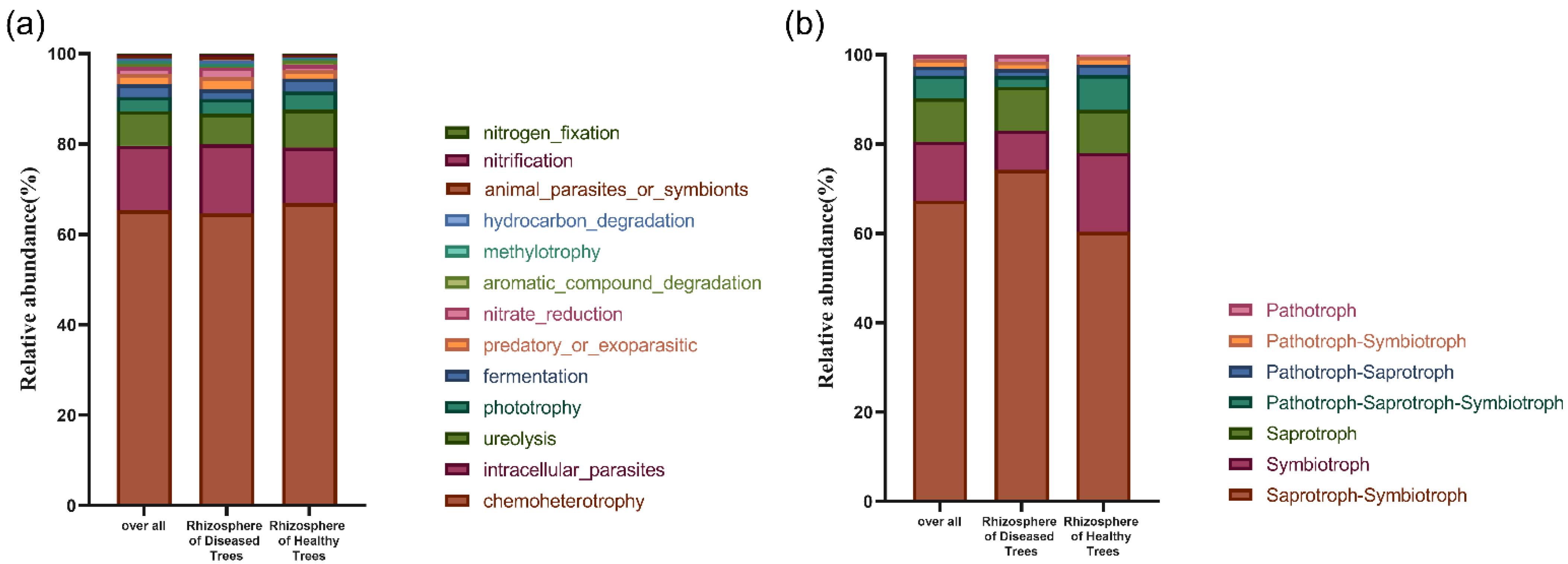
3.2.3. Microbial Community Potential Functional Structures in Rhizosphere Soil

FAPROTAX analysis, in which 503 OTUs (9.83%) were included, revealed that the most prevalent bacterial functional group was chemoheterotrophy involved in the carbon cycle (60.65%), followed by intracellular parasites (14.30%), ureolysis (7.67%), phototrophy (3.13%), fermentation (2.80%), predatory or exoparasitic (2.26%), and nitrate reduction (1.61%) (Figure 3a).
Significant differences were found in the abundance of specific bacterial functional groups in rhizosphere soil between healthy and diseased hosts. Rhizosphere soil of diseased hosts exhibited a significantly higher abundance of intracellular parasites, whereas that of healthy hosts had a significantly higher abundance of ureolysis (Figure S6; p < 0.05), which was positively correlated with the abundance Gaiella (p < 0.05; Pearson = 0.842). (Table S4).
A total of 497 of the total fungal OTUs (35.86%) were included in the FUNGuild analysis. The results showed that the most prevalent functional (nutritional) mode was saprotroph–symbiotroph (67.32%), followed by symbiotroph (13.24%), saprotroph (9.64%), pathotroph–saprotroph–symbiotroph (5.04%), pathotroph–saprotroph (2.07%), pathotroph–symbiotroph (1.68%), and pathotroph (0.99%) (Figure 3b).
The rhizosphere soil of diseased hosts had a higher abundance of saprotroph–symbiotroph than that of healthy hosts, whereas symbiotroph was lower in that of diseased hosts (Figure S6; p < 0.05). Principal Coordinate Analysis (PCoA) showed that the functional structures of rhizosphere bacteria and fungi were similar between healthy and diseased trees, as confirmed by PERMANOVA analysis, which revealed no significant differences in the structures (Figure S3).

3.3. Mycorrhizal Fungal Infection Rate and Fungal Community in Fine Root

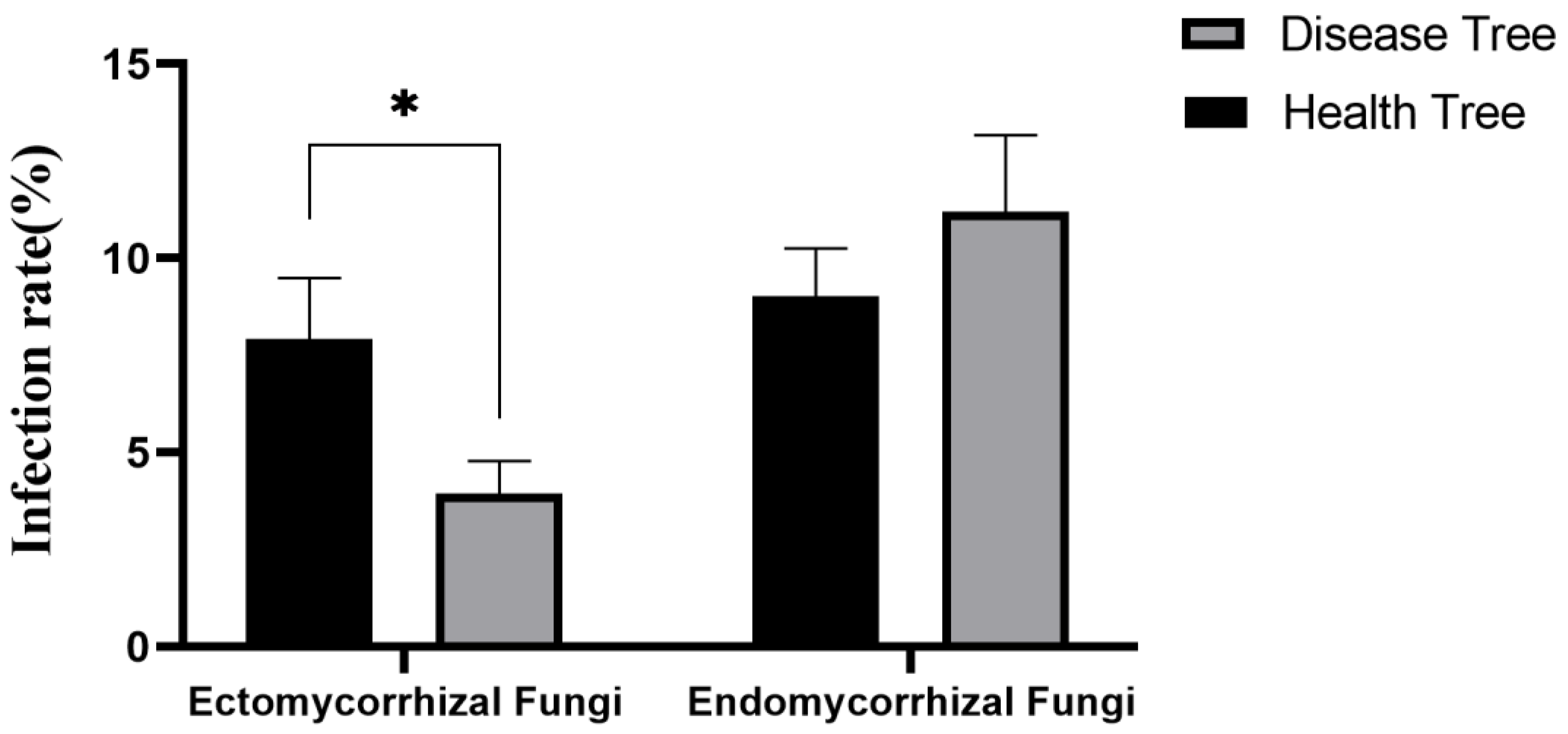
3.3.1. Mycorrhizal Fungal Infection Rate

Infection rates of ectomycorrhizal and endophytic mycorrhizal fungi were examined. In healthy hosts, the infection rates of ectomycorrhizal fungi and endophytic mycorrhizal fungi were 7.89% and 8.81%, respectively. Diseased hosts showed infection rates of 3.93% for ectomycorrhizal fungi and 11.19% for endophytic mycorrhizal fungi (Figure 4). A significant difference was found in the infection rate of ectomycorrhizal fungi between healthy and diseased hosts (p < 0.05), with higher rates observed in healthy hosts. However, no significant differences was observed in the infection rates of endophytic fungi between healthy and diseased hosts. The infection rate of ectomycorrhizal fungi was positively correlated with enzyme activity of β-cellobiosidase (CEL) and β-glucosidases (GLS) (p < 0.05; Pearson = 0.605; and Pearson = 0.572) (Tables S3 and S4).

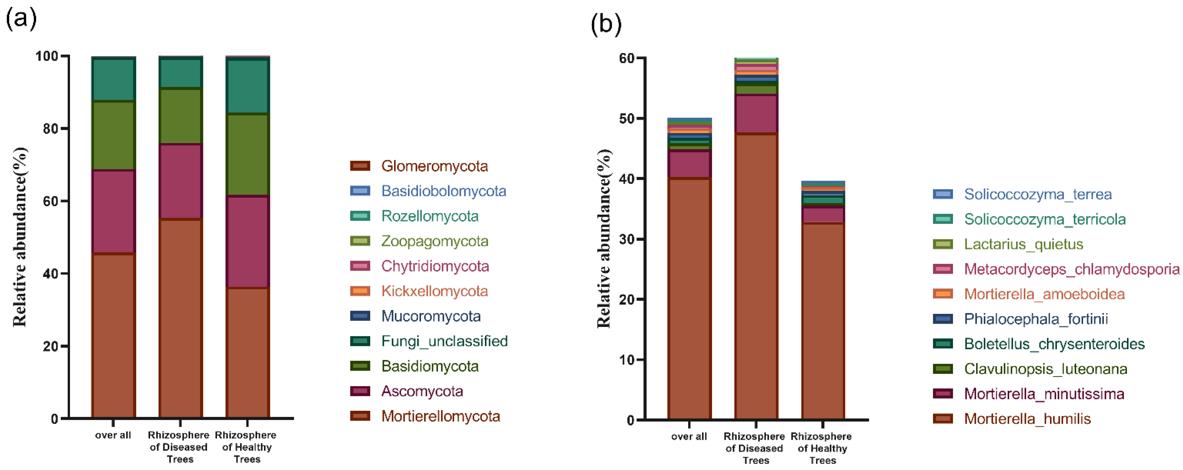
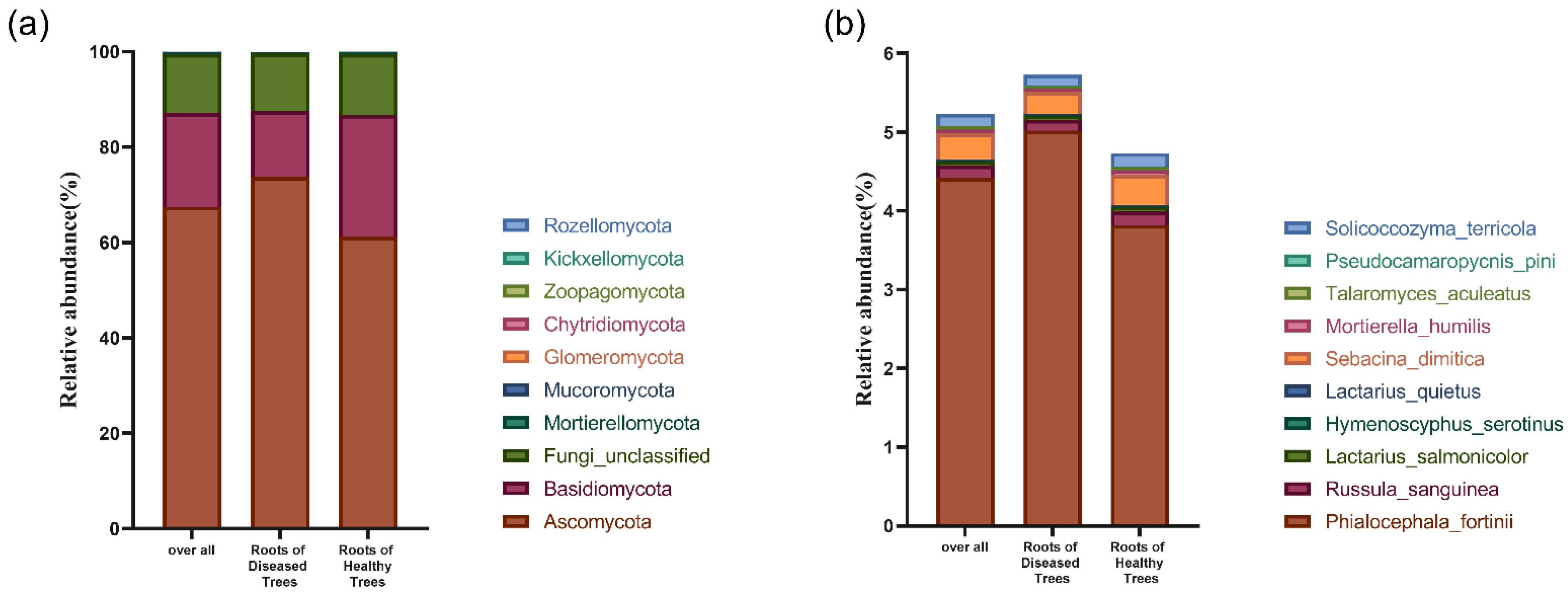
3.3.2. Fungal Community Structure in Fine Root

A total of 1192 fungal operational taxonomic units (OTUs) were identified from the root. However, no significant differences were found in any of the diversity indices between healthy and diseased fine root samples (Table S3). PCoA analysis based on OTU data showed that the community structure of fine root fungi was similar between healthy and diseased trees (Figure S2), as confirmed by PERMANOVA analysis, which revealed no significant differences in the structure.
A total of 10 fungal phyla were identified from the fine roots. The dominant phylum was Ascomycota (67.47%), followed by Basidiomycota (19.67%), and Mortierellomycota (0.35%). Minor phyla, including Kickxellomycota, Chytridiomycota, Zoopagomycota, Glomeromycota, and Rozellomycota, constituted ≤0.3% of the sequences (Figure 5a). Notably, Glomeromycota was significantly more abundant in the fine roots of healthy hosts (Figure S5; p < 0.05).
At the species level, 14.48% of the sequences were assigned to 170 species. The most prevalent species included Phialocephala fortini (4.42%), Mortierella humilis (0.33%), Russula sanguinea (0.15%), Solicoccozyma terricola (0.15%), and Penicillium adametzii (0.13%) (Figure 5b). The fine roots of diseased hosts had a higher abundance of Trichoderma hamatum, whereas Heterocephalacria arrabidensis and Penicillium daejeonium were lower in those of diseased hosts (Figure S5).

3.3.3. Fungal Potential Functional Structures in Fine Root of Diseased and Healthy Trees

A total of 436 (36.58%) OTUs were included in the FUNGuild analysis. The most prevalent nutritional mode was symbiotroph (32.72%), followed by pathotroph–saprotroph (24.11%), saprotroph (15.42%), pathotroph–symbiotroph (12.68%), pathotroph–saprotroph–symbiotroph (11.72%), saprotroph–symbiotroph (2.81%), and pathotroph (0.54%) (Figure 6).
The fine roots of diseased tree had a higher abundance of pathotroph–symbiotroph and a lower abundance of symbiotroph (Figure S6; p < 0.05). PCoA analysis revealed that there were no significant differences in the functional structures of rhizosphere fungi between healthy and diseased trees, as confirmed by PERMANOVA analysis (Figure S3).

4. Discussion

This study examined the fungal communities in the fine roots and microbial communities in the rhizosphere soil of healthy and diseased P. thunbergii trees affected by pine wilt disease (PWD) in natural field conditions. Surprisingly, no significant differences were found in rhizosphere bacterial community diversity between healthy and diseased trees. However, a notably higher richness of the rhizospheric fungal community in healthy trees was observed compared to diseased trees. These findings contrasted with previous studies by Zhang et al. (2021) and Deng et al. (2022). Zhang reported no significant differences in species richness of rhizosphere fungi and bacteria in P. massoniana infected with PWN in Zhejiang province, located in the south of China. Conversely, Deng observed significantly higher diversity indices (Pielou_e, Shannon and Simpson) of rhizosphere fungi and bacteria in P. koraiensis infected with PWN in Liaoning province, situated in the north of China. The dissimilarities observed in these studies may be attributed to variations in the degree of disease infection, tree species, and geographical location [43,44]. The reduction in richness index may lead to the alteration of the community and functional structure of the host, which could affect the ecological functions, such as nutrient cycling and biological invasion control, thereby impacting the health of the host [45].
Our study revealed a higher abundance of phylum Actinobacteria and genus Gaiella in the rhizosphere of healthy P. thunbergii trees compared to diseased ones. These taxa were positively correlated with β-cellobiosidase (CEL) enzyme activities. Actinobacteria is known to produce cellulase, which aids in cellulose degradation [46,47], and Gaiella play a crucial role in organic matter decomposition and carbon cycling [48]. The higher activities of β-cellobiosidase (CEL) and β-glucosidase (GLS) enzymes involved in carbon cycling in the rhizosphere of healthy trees indicate a more active decomposition of organic matter. Most Actinobacteria are heterotrophic, relying on organic compounds such as starch, sugars, cellulose, and organic acids for growth. Reduced secretion of soluble sugars and total sugars by pine roots during the later stages of infection may contribute to the decreased abundance of Gaiella and Actinobacteria [49,50]. Interestingly, the abundance of the ureolysis functional group in rhizosphere bacteria was positively correlated with Gaiella, known for its contribution to nutrient cycling, especially nitrogen decomposition [51]. The decrease in ureolysis in the rhizospheric diseased trees could have caused a reduction in soil CO2 amount, which may have led to a decrease in the abundance of Gaiella [52]. The rhizosphere of diseased hosts showed a higher abundance of fungal phylum Mortierellomycota and species M. humilis. Soil pH significantly influenced the composition of Mortierellomycota in the rhizosphere of moso bamboo [53], and a positive correlation between soil pH and the abundance of Mortierellomycota was observed in Panax notoginseng plantation [54]. In our previous study, the soil pH around diseased hosts was higher than around healthy hosts [22], potentially contributing to the increased abundance of Mortierellomycota in the rhizosphere of a diseased tree [55]. Numerous species of Mortierellomycota have been identified as a potential pathogen of arecanut palm root rot [56]. The decreased resistance of a tree in the rhizosphere of a diseased tree and pathogen enrichment could explain the increased abundance of Mortierellomycota. Some species of Mortierella genus have the capability to produce antibiotics and have shown potential as antagonists against plant pathogens [57]. M. humilis has been reported to be useful for the production of Se-NPs with a broad spectrum of antipathogenic activities [58]. Tea plants with gray blight have altered root exudates that recruit a beneficial rhizosphere microbiome to prime immunity against aboveground pathogen infection [59], which could be a possible explanation for the increased abundance of M. humilis in the rhizosphere of diseased hosts. Conversely, the abundance of Mucoromycota was significantly higher in healthy hosts compared to diseased ones. Mucoromycota includes both pathogens and symbiotic fungi, establishing relationships with non-vascular plants, gymnosperms, and angiosperms, producing ectomycorrhizae and endomycorrhizae [60]. The decrease in elemental C, H, N, and ash content in PWN-infected pines may lead to a reduction in nutrient secretion from the roots [61]. Changes in root secretion likely influence rhizosphere microorganisms and may be responsible for the reduction in Mucoromycota abundance in the rhizosphere of diseased trees The reduction in Mucoromycota abundance may lead to a lack of important nutrients in the host and decrease its resistance to foreign pathogens, thereby exacerbating pine wilt disease [62].
Plant roots host harbor a diverse range of fungi with various functional roles, such as plant pathogens, saprophytic fungi, and mycorrhizal fungi, impacting plant health and nutrition [63,64,65,66]. In our study, the abundance of Glomeromycota in healthy hosts was significantly higher than in diseased hosts. The vast majority of species in Glomeromycota are endophytic fungi that have a symbiotic relationship with 80% of higher plants. The needles and leaves die down during the course of the disease, and the decrease in photo-synthesis capacity leads to a decrease in the production of carbohydrates [67]. This may be the reason for the decrease in Glomeromycota abundance, which may also exacerbate the course of PWD. Our study revealed that Trichoderma hamatum, a saprophytic fungus, was significantly more abundant in the fine roots of diseased hosts. The reduced root secretions and increased saprophytic fungi in late stage of disease in susceptible pines could explain the higher abundance of T. hamatum in diseased hosts. Interestingly, T. hamatum has been reported to act as a biocontrol agent against soil pathogens and induces systemic resistance to foliar pathogens [68]. In our study, we observed that the infection rate of ectomycorrhizal fungi was significantly higher in healthy P. thunbergii trees compared to diseased ones. Similar results were found in P. tabulaeformis, where the infection rate of ectomycorrhizal fungi decreased with increasing levels of pine wilt disease [69]. Ectomycorrhizal fungi (ECMF), not only enhance plant host nutritional, disease resistance, and stress tolerance but also facilitate the recruitment and enrichment of other beneficial microorganisms in the rhizosphere. A reduction in ectomycorrhizal fungi can exacerbate the progression of PWD. Interestingly, we observed a positive correlation between the infection rate of ectomycorrhizal fungi and the enzyme activity of β-cellobiosidase (CEL) and β-glucosidases (GLS), indicating that the reduced ectomycorrhizal fungi may affect rhizosphere enzyme activity related to the carbon cycle. Plant roots release significant amounts of carbon produced through photosynthesis into the rhizosphere, primarily in the form of sugars, amino acids, and organic acids [70,71]. However, the development of pine wilt disease leads to decrease in the secretion of soluble sugars, total sugars, and proteins in the roots [49,50], which may lead to a reduction in ectomycorrhizal mycorrhizal fungi. The reduction in ectomycorrhizal fungi also contributes to decreased mycelial secretions and consequently affect rhizosphere enzyme activity [72]. Furthermore, rhizosphere enzyme activity can be influenced by stand development duration, soil nutrient stoichiometry, and the composition of rhizosphere fungal communities, as demonstrated in previous studies [26,73]. Additionally, fine roots and mycorrhizal fungi play a role in leaf litter decomposition by providing carbon to decomposers. However, they can also hinder decomposition through nutrient competition with saprophytic fungi [74]. This may be a contributing factor to the decreased rhizosphere soil enzyme activity related to the carbon cycle observed in our study.
In terms of bacterial function, the abundance of intracellular parasites was significantly higher in the rhizosphere of the diseased hosts. The resin in pine trees contains terpenoids with anthelmintic and antibacterial properties [75]. After PWN infection, tree reduction in tree resistance and resin production likely contributed to the increased abundance of in intracellular parasites. The abundance of ureolysis in healthy hosts was significantly higher than in diseased hosts and had significantly different effects on urease activities of different bacteria under different pH conditions [76]. Our previous study has shown that PWD causes changes in soil pH, which may be responsible for changes in ureolysis functional groups [22]. Additionally, PWN infection leads to changes in microbial communities, including certain taxa in plant needles, which, in turn, affects the microbial composition in litter and rhizosphere [22,23,77]. The relative abundance of symbiotroph in diseased hosts was significantly lower than in healthy hosts, which is influenced by fine root and rhizosphere fungi. During the development of PWD, nematode feeding on the epithelial cells of the resin tract of susceptible trees disrupts water conductance, leading to the death of the entire tree [78]. This disruption in nutrient supply can accelerate root decline, potentially resulting in reduced functions of both fine root and rhizosphere fungi. Notable, PWN infection induces physiological and biochemical changes in the host, affecting root secretions [66], which, in turn, have direct impacts on fine root and rhizosphere microorganisms [69]. Therefore, further research is necessary to explore the interactions between rhizosphere fungi and bacteria, given their shared ecological niche [23]. These interactions can have significant implications for understanding and managing plant wilt diseases. In addition, further research focusing on the different stages of disease is needed to fully understand how disease affects the host microbiota. It can be hypothesized that the changes in host microbiota increase as the disease period progresses. A comprehensive understanding of the interplay between functional groups of bacteria and fungi will be crucial in devising effective strategies for the prevention and management of plant wilt diseases.

5. Conclusions

In conclusion, PWN infection leads to notable changes in the rhizosphere. Specifically, the activities of key carbon enzymes, β-cellobiosidase and β-glucosidase, are reduced, indicating alterations in carbon cycling processes. Moreover, the richness of rhizospheric fungal community is diminished. Notably, PWN infection leads to an increase in the abundance of intracellular parasites group within bacterial functions and saprotroph-symbiotroph group within fungal functions. Additionally, the infection reduces ectomycorrhizal fungi infection rate in the fine roots, which may have implications for the reduction in nutrient and water uptake of the host tree. However, to gain a more comprehensive understanding of the disease’s effects on the host microbiota, further research is needed, especially focusing on different stages of PWD. Such studies would enhance our knowledge of the mechanisms underlying disease development and plant–microbe interactions in pine forests. Ultimately, this knowledge will be invaluable in devising effective strategies for the management and conservation of pine forests affected by pine wilt disease.

Supplementary Materials

The following supporting information can be downloaded at: https://www.mdpi.com/article/10.3390/f14091884/s1. Figure S1: Venn diagram showing the unique and shared Operational Taxonomic Units (OTUs) of bacteria (a) and fungi (b) in rhizosphere soil of healthy and diseased trees; Figure S2: Principal Coordinate Analysis (PCoA) showing the bacterial community structure (a) and fungal community structure (b) in rhizosphere soil of healthy and diseased trees. Principal Coordinate Analysis (PCoA) showing the fungal community structure (c) in fine roots of healthy and diseased trees; Figure S3: Principal coordinate analysis (PCoA) showing bacterial functional structure (a) and fungal functional structure (b) in rhizosphere soil of healthy and diseased trees. Principal coordinate analysis (PCoA) showing fungal functional structure (c) in fine roots of healthy and diseased trees; Figure S4: The images showing the status of pine root mycorrhizal infection using a Zeiss fluorescence electron microscope, with eyepiece magnification of 10× and objective 20× (a) and 40× (b) for ectomycorrhiza, eyepiece magnification of 10× and objective 20× (c) and 40× (d) for endomycorrhiza; Figure S5: Changes in abundance of Actinobacteria (a) and Gaiella (b) on rhizosphere bacteria, Mucoromycota (c), Mortierellomycota (d) and Motierella humills (e) on rhizosphere fungi and Glomeromycota (f), Trichoderma hamatum (g), Penicillium daejeonium (h), and Heterocephalacria arrabidensis (i) on fine root fungi in healthy and diseased trees; Figure S6: Changes in abundance of functional group intracellular parasites (a) and ureolysis (b) on rhizosphere bacteria, Symbiotroph (c) and Saprotroph-Symbiotroph (d) on rhizosphere fungi and symbiotroph (e) and pathotroph-symbiotroph (f) on fine root fungi in healthy and diseased trees; Table S1: Diversity indices of fungal community in roots of diseased and healthy trees; Table S2: One-Way ANOVA showing the differences in the rhizosphere soil enzyme activities of both healthy and diseased trees; Table S3: One-Way ANOVA showing the differences in the infection rate of ectomycorrhizal and endomycorrhizal fungi in healthy and diseased trees; Table S4: Pearson Correlation showing the differences in the rhizosphere healthy and diseased trees.

Author Contributions

H.S. conceived the ideas, designed methodology and received the funding; Z.J., Z.G., Y.L. (Yangchunzi Liao), L.D. and Y.L. (Yi Liu) collected the samples and performed lab work; Z.J. analyzed the data and writing of the manuscript. All authors have read and agreed to the published version of the manuscript.

Funding

This study was financially supported by the National Natural Science Foundation of China (grant No. 31870474), the Jiangsu Specially Appointed Professor Program (project 165010015), and the Priority Academic Program Development (PAPD) of Jiangsu Higher Education Institutions for economical support.

Data Availability Statement

The raw sequences were submitted to National Center for Biotechnology Information (NCBI), with access No. PRJNA974452.

Conflicts of Interest

The authors declare no conflict of interest. The funders had no involvement in the study design, data collection, analyses, interpretation, manuscript writing, or decision to publish the results.

References

  1. Fadiji, A.E.; Babalola, O.O. Metagenomics Methods for the Study of Plant-Associated Microbial Communities: A Review. J. Microbiol. Methods 2020, 170, 105860. [Google Scholar] [CrossRef] [PubMed]
  2. Brader, G.; Compant, S.; Vescio, K.; Mitter, B.; Trognitz, F.; Ma, L.-J.; Sessitsch, A. Ecology and Genomic Insights into Plant-Pathogenic and Plant-Nonpathogenic Endophytes. Annu. Rev. Phytopathol. 2017, 55, 61–83. [Google Scholar] [CrossRef] [PubMed]
  3. Verma, S.K.; White, J.F. Indigenous Endophytic Seed Bacteria Promote Seedling Development and Defend against Fungal Disease in Browntop Millet (Urochloa ramosa L.). J. Appl. Microbiol. 2018, 124, 764–778. [Google Scholar] [CrossRef]
  4. Agler, M.T.; Ruhe, J.; Kroll, S.; Morhenn, C.; Kim, S.-T.; Weigel, D.; Kemen, E.M. Microbial Hub Taxa Link Host and Abiotic Factors to Plant Microbiome Variation. PLoS Biol. 2016, 14, e1002352. [Google Scholar] [CrossRef]
  5. Omomowo, O.I.; Babalola, O.O. Bacterial and Fungal Endophytes: Tiny Giants with Immense Beneficial Potential for Plant Growth and Sustainable Agricultural Productivity. Microorganisms 2019, 7, 481. [Google Scholar] [CrossRef] [PubMed]
  6. Hartmann, A.; Rothballer, M.; Schmid, M. Lorenz Hiltner, a Pioneer in Rhizosphere Microbial Ecology and Soil Bacteriology Research. Plant Soil 2008, 312, 7–14. [Google Scholar] [CrossRef]
  7. Jacoby, R.P.; Chen, L.; Schwier, M.; Koprivova, A.; Kopriva, S. Recent Advances in the Role of Plant Metabolites in Shaping the Root Microbiome. F1000Research 2020, 9, F1000 Faculty Rev-151. [Google Scholar] [CrossRef]
  8. Dennis, P.; Miller, A.; Hirsch, P. Are Root Exudates More Important than Other Sources of Rhizodeposits in Structuring Rhizosphere Bacterial Communities? FEMS Microbiol. Ecol. 2010, 72, 313–327. [Google Scholar] [CrossRef]
  9. Jacoby, R.; Koprivova, A.; Kopriva, S. Pinpointing Secondary Metabolites That Shape the Composition and Function of the Plant Microbiome. J. Exp. Bot. 2021, 72, 57–69. [Google Scholar] [CrossRef]
  10. Bailly, A.; Weisskopf, L. The Modulating Effect of Bacterial Volatiles on Plant Growth: Current Knowledge and Future Challenges. Plant Signal. Behav. 2012, 7, 79–85. [Google Scholar] [CrossRef]
  11. Oldroyd, G. Speak, Friend, and Enter: Signalling Systems That Promote Beneficial Symbiotic Associations in Plants. Nat. Rev. Microbiol. 2013, 11, 252–263. [Google Scholar] [CrossRef] [PubMed]
  12. Kakkar, A.; Nizampatnam, N.; Kondreddy, A.; Pradhan, B.; Chatterjee, S. Xanthomonas Campestris Cell-Cell Signalling Molecule DSF (Diffusible Signal Factor) Elicits Innate Immunity in Plants and Is Suppressed by the Exopolysaccharide Xanthan. J. Exp. Bot. 2015, 66, 6697–6714. [Google Scholar] [CrossRef] [PubMed]
  13. Phillips, R.; Brzostek, E.; Midgley, M. The Mycorrhizal-Associated Nutrient Economy: A New Framework for Predicting Carbon-Nutrient Couplings in Temperate Forests. New Phytol. 2013, 199, 41–51. [Google Scholar] [CrossRef] [PubMed]
  14. Sulman, B.; Shevliakova, E.; Brzostek, E.; Kivlin, S.; Malyshev, S.; Menge, D.; Zhang, X. Diverse Mycorrhizal Associations Enhance Terrestrial C Storage in a Global Model. Glob. Biogeochem. Cycles 2019, 33, 501–523. [Google Scholar] [CrossRef]
  15. Kivlin, S.; Emery, S.; Rudgers, J. Fungal symbionts alter plant responses to global change. Am. J. Bot. 2013, 100, 1445–1457. [Google Scholar] [CrossRef] [PubMed]
  16. Ratcliffe, S.; Wirth, C.; Jucker, T.; van der Plas, F.; Scherer-Lorenzen, M.; Verheyen, K.; Allan, E.; Benavides, R.; Bruelheide, H.; Ohse, B.; et al. Biodiversity and Ecosystem Functioning Relations in European Forests Depend on Environmental Context. Ecol. Lett. 2017, 20, 1414–1426. [Google Scholar] [CrossRef] [PubMed]
  17. Smith, F.A.; Grace, E.J.; Smith, S.E. More than a Carbon Economy: Nutrient Trade and Ecological Sustainability in Facultative Arbuscular Mycorrhizal Symbioses. New Phytol. 2009, 182, 347–358. [Google Scholar] [CrossRef]
  18. Dwinell, L.D. The Pinewood Nematode: Regulation and Mitigation. Annu. Rev. Phytopathol. 1997, 35, 153–166. [Google Scholar] [CrossRef]
  19. Kim, B.-N.; Kim, J.H.; Ahn, J.-Y.; Kim, S.; Cho, B.-K.; Kim, Y.-H.; Min, J. A Short Review of the Pinewood Nematode, Bursaphelenchus Xylophilus. Toxicol. Environ. Health Sci. 2020, 12, 297–304. [Google Scholar] [CrossRef]
  20. Vicente, C.; Ikuyo, Y.; Mota, M.; Hasegawa, K. Pinewood Nematode-Associated Bacteria Contribute to Oxidative Stress Resistance of Bursaphelenchus Xylophilus. BMC Microbiol. 2013, 13, 299. [Google Scholar] [CrossRef]
  21. Wu, X.; Yuan, W.; Tian, X.; Fan, B.; Fang, X.; Ye, J.; Ding, X. Specific and Functional Diversity of Endophytic Bacteria from Pine Wood Nematode Bursaphelenchus Xylophilus with Different Virulence. Int. J. Biol. Sci. 2013, 9, 34–44. [Google Scholar] [CrossRef] [PubMed]
  22. Ma, Y.; Qu, Z.-L.; Liu, B.; Tan, J.-J.; Asiegbu, F.O.; Sun, H. Bacterial Community Structure of Pinus Thunbergii Naturally Infected by the Nematode Bursaphelenchus Xylophilus. Microorganisms 2020, 8, 307. [Google Scholar] [CrossRef] [PubMed]
  23. Liu, Y.; Qu, Z.-L.; Liu, B.; Ma, Y.; Xu, J.; Shen, W.-X.; Sun, H. The Impact of Pine Wood Nematode Infection on the Host Fungal Community. Microorganisms 2021, 9, 896. [Google Scholar] [CrossRef]
  24. Deng, S.; Katoh, M.; Guan, Q.; Yin, N.; Li, M. Interpretation of Forest Resources at the Individual Tree Level at Purple Mountain, Nanjing City, China, Using WorldView-2 Imagery by Combining GPS, RS and GIS Technologies. Remote Sens. 2014, 6, 87–110. [Google Scholar] [CrossRef]
  25. Deng, S.; Katoh, M.; Guan, Q.; Yin, N.; Li, M. Estimating Forest Aboveground Biomass by Combining ALOS PALSAR and WorldView-2 Data: A Case Study at Purple Mountain National Park, Nanjing, China. Remote Sens. 2014, 6, 7878–7910. [Google Scholar] [CrossRef]
  26. Qu, Z.-L.; Braima, A.; Liu, B.; Ma, Y.; Sun, H. Soil Fungal Community Structure and Function Shift during a Disease-Driven Forest Succession. Microbiol. Spectr. 2022, 10, e0079522. [Google Scholar] [CrossRef] [PubMed]
  27. Millberg, H.; Boberg, J.; Stenlid, J. Changes in Fungal Community of Scots Pine (Pinus Sylvestris) Needles along a Latitudinal Gradient in Sweden. Fungal Ecol. 2015, 17, 126–139. [Google Scholar] [CrossRef]
  28. Kikuchi, T.; Aikawa, T.; Oeda, Y.; Karim, N.; Kanzaki, N. A Rapid and Precise Diagnostic Method for Detecting the Pinewood Nematode Bursaphelenchus Xylophilus by Loop-Mediated Isothermal Amplification. Phytopathology 2009, 99, 1365–1369. [Google Scholar] [CrossRef]
  29. Saiya-Cork, K.R.; Sinsabaugh, R.L.; Zak, D.R. The Effects of Long Term Nitrogen Deposition on Extracellular Enzyme Activity in an Acer Saccharum Forest Soil. Soil Biol. Biochem. 2002, 34, 1309–1315. [Google Scholar] [CrossRef]
  30. Burns, R.G.; DeForest, J.L.; Marxsen, J.; Sinsabaugh, R.L.; Stromberger, M.E.; Wallenstein, M.D.; Weintraub, M.N.; Zoppini, A. Soil Enzymes in a Changing Environment: Current Knowledge and Future Directions. Soil Biol. Biochem. 2013, 58, 216–234. [Google Scholar] [CrossRef]
  31. Phillips, J.M.; Hayman, D.S. Improved Procedures for Clearing Roots and Staining Parasitic and Vesicular-Arbuscular Mycorrhizal Fungi for Rapid Assessment of Infection. Trans. Br. Mycol. Soc. 1970, 55, 158-IN18. [Google Scholar] [CrossRef]
  32. Li, Z.; Fu, J.; Zhou, R.; Wang, D. Effects of Phenolic Acids from Ginseng Rhizosphere on Soil Fungi Structure, Richness and Diversity in Consecutive Monoculturing of Ginseng. Saudi J. Biol. Sci. 2018, 25, 1788–1794. [Google Scholar] [CrossRef]
  33. Hui, N.; Jumpponen, A.; Francini, G.; Kotze, D.J.; Liu, X.; Romantschuk, M.; Strömmer, R.; Setälä, H. Soil Microbial Communities Are Shaped by Vegetation Type and Park Age in Cities under Cold Climate. Environ. Microbiol. 2017, 19, 1281–1295. [Google Scholar] [CrossRef] [PubMed]
  34. Schloss, P.D.; Westcott, S.L.; Ryabin, T.; Hall, J.R.; Hartmann, M.; Hollister, E.B.; Lesniewski, R.A.; Oakley, B.B.; Parks, D.H.; Robinson, C.J.; et al. Introducing Mothur: Open-Source, Platform-Independent, Community-Supported Software for Describing and Comparing Microbial Communities. Appl. Environ. Microbiol. 2009, 75, 7537–7541. [Google Scholar] [CrossRef] [PubMed]
  35. Needleman, S.B.; Wunsch, C.D. A General Method Applicable to the Search for Similarities in the Amino Acid Sequence of Two Proteins. J. Mol. Biol. 1970, 48, 443–453. [Google Scholar] [CrossRef] [PubMed]
  36. Olesen, S.W.; Duvallet, C.; Alm, E.J. dbOTU3: A New Implementation of Distribution-Based OTU Calling. PLoS ONE 2017, 12, e0176335. [Google Scholar] [CrossRef] [PubMed]
  37. Wang, Q.; Garrity, G.M.; Tiedje, J.M.; Cole, J.R. Naive Bayesian Classifier for Rapid Assignment of rRNA Sequences into the New Bacterial Taxonomy. Appl. Environ. Microbiol. 2007, 73, 5261–5267. [Google Scholar] [CrossRef] [PubMed]
  38. Tedersoo, L.; Sánchez-Ramírez, S.; Kõljalg, U.; Bahram, M.; Döring, M.; Schigel, D.; May, T.; Ryberg, M.; Abarenkov, K. High-Level Classification of the Fungi and a Tool for Evolutionary Ecological Analyses. Fungal Divers. 2018, 90, 135–159. [Google Scholar] [CrossRef]
  39. Louca, S.; Parfrey, L.W.; Doebeli, M. Decoupling Function and Taxonomy in the Global Ocean Microbiome. Science 2016, 353, 1272–1277. [Google Scholar] [CrossRef]
  40. Nguyen, N.H.; Song, Z.; Bates, S.T.; Branco, S.; Tedersoo, L.; Menke, J.; Schilling, J.S.; Kennedy, P.G. FUNGuild: An Open Annotation Tool for Parsing Fungal Community Datasets by Ecological Guild. Fungal Ecol. 2016, 20, 241–248. [Google Scholar] [CrossRef]
  41. Heberle, H.; Meirelles, G.V.; da Silva, F.R.; Telles, G.P.; Minghim, R. InteractiVenn: A Web-Based Tool for the Analysis of Sets through Venn Diagrams. BMC Bioinform. 2015, 16, 169. [Google Scholar] [CrossRef] [PubMed]
  42. Anderson, M.J.; Gorley, R.N.; Clarke, K.S.; Anderson, M.; Gorley, R.N.; Clarke, K.; Andersom, M. PERMANOVA+ for PRIMER: Guide to Software and Statistical Methods; Primer-E Limited.: Auckland, New Zealand, 2008. [Google Scholar]
  43. Zhang, W.; Wang, X.; Li, Y.; Liu, Z.; Li, D.; Wen, X.; Feng, Y.; Zhang, X. Pinewood Nematode Alters the Endophytic and Rhizospheric Microbial Communities of Pinus Massoniana. Microb. Ecol. 2021, 81, 807–817. [Google Scholar] [CrossRef] [PubMed]
  44. Deng, J.; Yu, D.; Zhou, W.; Zhou, L.; Zhu, W. Variations of Phyllosphere and Rhizosphere Microbial Communities of Pinus Koraiensis Infected by Bursaphelenchus Xylophilus. Microb. Ecol. 2022, 84, 285–301. [Google Scholar] [CrossRef] [PubMed]
  45. Sahib, M.R.; Pervaiz, Z.H.; Williams, M.A.; Saleem, M.; DeBolt, S. Rhizobacterial Species Richness Improves Sorghum Growth and Soil Nutrient Synergism in a Nutrient-Poor Greenhouse Soil. Sci. Rep. 2020, 10, 15454. [Google Scholar] [CrossRef]
  46. Taha, M.; Foda, M.; Shahsavari, E.; Aburto-Medina, A.; Adetutu, E.; Ball, A. Commercial Feasibility of Lignocellulose Biodegradation: Possibilities and Challenges. Curr. Opin. Biotechnol. 2016, 38, 190–197. [Google Scholar] [CrossRef] [PubMed]
  47. Zhao, Y.; Lu, Q.; Wei, Y.; Cui, H.; Zhang, X.; Wang, X.; Shan, S.; Wei, Z. Effect of Actinobacteria Agent Inoculation Methods on Cellulose Degradation during Composting Based on Redundancy Analysis. Bioresour. Technol. 2016, 219, 196–203. [Google Scholar] [CrossRef] [PubMed]
  48. Albuquerque, L.; da Costa, M.S. The Family Gaiellaceae. In The Prokaryotes: Actinobacteria; Rosenberg, E., DeLong, E.F., Lory, S., Stackebrandt, E., Thompson, F., Eds.; Springer: Berlin/Heidelberg, Germany, 2014; pp. 357–360. ISBN 978-3-642-30138-4. [Google Scholar]
  49. Shi, S.; Richardson, A.E.; O’Callaghan, M.; DeAngelis, K.M.; Jones, E.E.; Stewart, A.; Firestone, M.K.; Condron, L.M. Effects of Selected Root Exudate Components on Soil Bacterial Communities. FEMS Microbiol. Ecol. 2011, 77, 600–610. [Google Scholar] [CrossRef]
  50. Badri, D.V.; Chaparro, J.M.; Zhang, R.; Shen, Q.; Vivanco, J.M. Application of Natural Blends of Phytochemicals Derived from the Root Exudates of Arabidopsis to the Soil Reveal That Phenolic-Related Compounds Predominantly Modulate the Soil Microbiome. J. Biol. Chem. 2013, 288, 4502–4512. [Google Scholar] [CrossRef]
  51. Zhang, C.; Tayyab, M.; Abubakar, A.Y.; Yang, Z.; Pang, Z.; Islam, W.; Lin, Z.; Li, S.; Luo, J.; Fan, X.; et al. Bacteria with Different Assemblages in the Soil Profile Drive the Diverse Nutrient Cycles in the Sugarcane Straw Retention Ecosystem. Diversity 2019, 11, 194. [Google Scholar] [CrossRef]
  52. Caravaca, F.; Torres, P.; Díaz, G.; Roldán, A. Elevated CO2 Affects the Rhizosphere Microbial Community and the Growth of Two Invader Plant Species Differently in Semiarid Mediterranean Soils. Land Degrad. Dev. 2022, 33, 117–132. [Google Scholar] [CrossRef]
  53. Zhang, X.; Gao, G.; Wu, Z.; Wen, X.; Zhong, H.; Zhong, Z.; Bian, F.; Gai, X. Agroforestry Alters the Rhizosphere Soil Bacterial and Fungal Communities of Moso Bamboo Plantations in Subtropical China. Appl. Soil Ecol. 2019, 143, 192–200. [Google Scholar] [CrossRef]
  54. Shi, Y.; Qiu, L.; Guo, L.; Man, J.; Shang, B.; Pu, R.; Ou, X.; Dai, C.; Liu, P.; Yang, Y.; et al. K Fertilizers Reduce the Accumulation of Cd in Panax Notoginseng (Burk.) F.H. by Improving the Quality of the Microbial Community. Front. Plant Sci. 2020, 11, 888. [Google Scholar] [CrossRef] [PubMed]
  55. Li, H.; Ma, X.; Tang, Y.; Yan, C.; Hu, X.; Huang, X.; Lin, M.; Liu, Z. Integrated Analysis Reveals an Association between the Rhizosphere Microbiome and Root Rot of Arecanut Palm. Pedosphere 2021, 31, 725–735. [Google Scholar] [CrossRef]
  56. Solís-García, I.A.; Ceballos-Luna, O.; Cortazar-Murillo, E.M.; Desgarennes, D.; Garay-Serrano, E.; Patiño-Conde, V.; Guevara-Avendaño, E.; Méndez-Bravo, A.; Reverchon, F. Phytophthora Root Rot Modifies the Composition of the Avocado Rhizosphere Microbiome and Increases the Abundance of Opportunistic Fungal Pathogens. Front. Microbiol. 2021, 11, 3484. [Google Scholar] [CrossRef] [PubMed]
  57. Tagawa, M.; Tamaki, H.; Manome, A.; Koyama, O.; Kamagata, Y. Isolation and Characterization of Antagonistic Fungi against Potato Scab Pathogens from Potato Field Soils. FEMS Microbiol. Lett. 2010, 305, 136–142. [Google Scholar] [CrossRef] [PubMed]
  58. Hussein, H.G.; El-Sayed, E.-S.R.; Younis, N.A.; Hamdy, A.E.H.A.; Easa, S.M. Harnessing Endophytic Fungi for Biosynthesis of Selenium Nanoparticles and Exploring Their Bioactivities. AMB Express 2022, 12, 68. [Google Scholar] [CrossRef] [PubMed]
  59. Wang, Q.; Yang, R.; Peng, W.; Yang, Y.; Ma, X.; Zhang, W.; Ji, A.; Liu, L.; Liu, P.; Yan, L.; et al. Tea Plants With Gray Blight Have Altered Root Exudates That Recruit a Beneficial Rhizosphere Microbiome to Prime Immunity Against Aboveground Pathogen Infection. Front. Microbiol. 2021, 12, 774438. [Google Scholar] [CrossRef] [PubMed]
  60. Bonfante, P.; Venice, F. Mucoromycota: Going to the Roots of Plant-Interacting Fungi. Fungal Biol. Rev. 2020, 34, 100–113. [Google Scholar] [CrossRef]
  61. Reva, V.; Fonseca, L.; Lousada, J.L.; Abrantes, I.; Viegas, D.X. Impact of the Pinewood Nematode, Bursaphelenchus Xylophilus, on Gross Calorific Value and Chemical Composition of Pinus Pinaster Woody Biomass. Eur. J. For. Res. 2012, 131, 1025–1033. [Google Scholar] [CrossRef]
  62. Kowal, J.; Arrigoni, E.; Jarvis, S.; Zappala, S.; Forbes, E.; Bidartondo, M.I.; Suz, L.M. Atmospheric Pollution, Soil Nutrients and Climate Effects on Mucoromycota Arbuscular Mycorrhizal Fungi. Environ. Microbiol. 2022, 24, 3390–3404. [Google Scholar] [CrossRef]
  63. Yu, L.; Nicolaisen, M.; Larsen, J.; Ravnskov, S. Succession of Root-Associated Fungi in Pisum Sativum during a Plant Growth Cycle as Examined by 454 Pyrosequencing. Plant Soil 2012, 358, 225–233. [Google Scholar] [CrossRef]
  64. Dhyani, A.; Jain, R.; Pandey, A. Contribution of Root-Associated Microbial Communities on Soil Quality of Oak and Pine Forests in the Himalayan Ecosystem. Trop. Ecol. 2019, 60, 271–280. [Google Scholar] [CrossRef]
  65. Han, G.; Mannaa, M.; Kim, N.; Jeon, H.; Jung, H.; Lee, H.; Kim, J.; Park, J.; Park, A.; Kim, J.; et al. Response of Pine Rhizosphere Microbiota to Foliar Treatment with Resistance-Inducing Bacteria against Pine Wilt Disease. Microorganisms 2021, 9, 688. [Google Scholar] [CrossRef] [PubMed]
  66. Chu, H.; Wang, H.; Zhang, Y.; Li, Z.; Wang, C.; Dai, D.; Tang, M. Inoculation With Ectomycorrhizal Fungi and Dark Septate Endophytes Contributes to the Resistance of Pinus Spp. to Pine Wilt Disease. Front. Microbiol. 2021, 12, 687304. [Google Scholar] [CrossRef] [PubMed]
  67. Redecker, D.; Schüßler, A.; Stockinger, H.; Stürmer, S.L.; Morton, J.B.; Walker, C. An Evidence-Based Consensus for the Classification of Arbuscular Mycorrhizal Fungi (Glomeromycota). Mycorrhiza 2013, 23, 515–531. [Google Scholar] [CrossRef] [PubMed]
  68. Poveda, J.; Rodríguez, V.M.; Abilleira, R.; Velasco, P. Trichoderma Hamatum Can Act as an Inter-Plant Communicator of Foliar Pathogen Infections by Colonizing the Roots of Nearby Plants: A New Inter-Plant “Wired Communication”. Plant Sci. 2023, 330, 111664. [Google Scholar] [CrossRef] [PubMed]
  69. Chu, H.; Wang, C.; Wang, H.; Chen, H.; Tang, M. Pine Wilt Disease Alters Soil Properties and Root-Associated Fungal Communities in Pinus Tabulaeformis Forest. Plant Soil 2016, 404, 237–249. [Google Scholar] [CrossRef]
  70. el Zahar Haichar, F.; Heulin, T.; Guyonnet, J.P.; Achouak, W. Stable Isotope Probing of Carbon Flow in the Plant Holobiont. Curr. Opin. Biotechnol. 2016, 41, 9–13. [Google Scholar] [CrossRef]
  71. Sugiyama, A. Flavonoids and Saponins in Plant Rhizospheres: Roles, Dynamics, and the Potential for Agriculture. Biosci. Biotechnol. Biochem. 2021, 85, 1919–1931. [Google Scholar] [CrossRef]
  72. Zhang, L.; Zhou, J.; George, T.S.; Limpens, E.; Feng, G. Arbuscular Mycorrhizal Fungi Conducting the Hyphosphere Bacterial Orchestra. Trends Plant Sci. 2022, 27, 402–411. [Google Scholar] [CrossRef]
  73. Dong, H.; Ge, J.; Sun, K.; Wang, B.; Xue, J.; Wakelin, S.A.; Wu, J.; Sheng, W.; Liang, C.; Xu, Q.; et al. Change in Root-Associated Fungal Communities Affects Soil Enzymatic Activities during Pinus Massoniana Forest Development in Subtropical China. For. Ecol. Manag. 2021, 482, 118817. [Google Scholar] [CrossRef]
  74. Lang, A.K.; Jevon, F.V.; Vietorisz, C.R.; Ayres, M.P.; Hatala Matthes, J. Fine Roots and Mycorrhizal Fungi Accelerate Leaf Litter Decomposition in a Northern Hardwood Forest Regardless of Dominant Tree Mycorrhizal Associations. New Phytol. 2021, 230, 316–326. [Google Scholar] [CrossRef] [PubMed]
  75. Vilanova, C.; Marín, M.; Baixeras, J.; Latorre, A.; Porcar, M. Selecting Microbial Strains from Pine Tree Resin: Biotechnological Applications from a Terpene World. PLoS ONE 2014, 9, e100740. [Google Scholar] [CrossRef] [PubMed]
  76. Pommerening-Röser, A.; Koops, H.-P. Environmental pH as an Important Factor for the Distribution of Urease Positive Ammonia-Oxidizing Bacteria. Microbiol. Res. 2005, 160, 27–35. [Google Scholar] [CrossRef] [PubMed]
  77. Liu, K.; Meng, W.; Qu, Z.; Zhang, Y.; Liu, B.; Ma, Y.; Chang, L.; Sun, H. Changes in Bacterial Communities and Functions Associated with Litter Degradation during Forest Succession Caused by Forest Disease. Phytobiomes J. 2023. [Google Scholar] [CrossRef]
  78. Proença, D.N.; Francisco, R.; Kublik, S.; Schöler, A.; Vestergaard, G.; Schloter, M.; Morais, P.V. The Microbiome of Endophytic, Wood Colonizing Bacteria from Pine Trees as Affected by Pine Wilt Disease. Sci. Rep. 2017, 7, 4205. [Google Scholar] [CrossRef]
Figure 1. Relative abundance of bacterial phyla (a) and the top 10 most abundant genus (b) in rhizosphere soil of healthy and diseased trees.
Forests 14 01884 g001
Figure 2. Relative abundance of fungal phyla (a) and the top 10 most abundant fungal species (b) in rhizosphere soil of healthy and diseased trees.
Forests 14 01884 g002
Figure 3. Relative abundance functional groups of bacteria (a) and fungi (b) in rhizosphere soil of healthy and diseased trees.
Forests 14 01884 g003
Figure 4. The infection rate of ectomycorrhizal and endophytic mycorrhizal fungi in healthy and diseased trees. Bar shows mean with standard error (n = 90), * p < 0.05.
Forests 14 01884 g004
Figure 5. Relative abundance of fungal phyla (a) and the top 10 most abundant fungal species (b) in fine roots of healthy and diseased trees.
Forests 14 01884 g005
Figure 6. FUNGuild analysis showing predicted trophic mode in fine roots of healthy and diseased trees.
Forests 14 01884 g006
Table 1. Rhizosphere soil enzyme activities in the rhizosphere of both healthy and diseased trees.
RhizospHere, Soil C CycleN CycleP CycleS Cycle
XYLGLRCELGLSNAG PHO SUL
Healthy tree98.1 ± 17.7713.82 ± 5.3577.32 ± 17.07 *278.07 ± 20.43 *312.35 ± 37.762104.92 ± 343.067.52 ± 0.19
Diseased tree71.02 ± 4.835.53 ± 2.6133.73 ± 12.25139 ± 40.4313.28 ± 57.871822.14 ± 261.878.27 ± 0.62
The data are presented as the mean ± standard deviation. (n = 36) * p < 0.05 in the table indicates a significant difference among host status. Numbers indicate rhizosphere soil enzyme activities. XYL, β-xylosidase; GLR, β-D-glucuroniase; CEL, β-cellobiosidase; GLS, β-glucosidase; NAG, N-acetylglucosaminidase; PHO, phosphatase; and SUL, sulfatase.
Table 2. Diversity indices of rhizosphere microorganisms in diseased and healthy trees.
Diversity Indices
Samples Host StatusSobsShannonShannoneven
Rhizosphere bacteriaHealthy2433.67 ± 220.65.83 ± 0.220.75 ± 0.02
Diseased2367 ± 465.35.63 ± 0.760.72 ± 0.08
Rhizosphere fungiHealthy553 ± 23.42 *3.02 ± 0.460.46 ± 0.09
Diseased509 ± 40.752.54 ± 0.40.4 ± 0.06
The data are presented as the mean ± standard deviation. (n = 9) * p < 0.05 in the table indicate significant difference between host status.
Disclaimer/Publisher’s Note: The statements, opinions and data contained in all publications are solely those of the individual author(s) and contributor(s) and not of MDPI and/or the editor(s). MDPI and/or the editor(s) disclaim responsibility for any injury to people or property resulting from any ideas, methods, instructions or products referred to in the content.

