Article

Molecular Cloning and Functional Characterization of a Cytochrome P450 Enzyme (SaCYP736A167) Promoter from Santalum album

1
Guangxi Key Laboratory of Sugarcane Genetic Improvement, Sugarcane Research Institute, Guangxi Academy of Agricultural Sciences, Nanning 530007, China
2
Guangdong Provincial Key Laboratory of Applied Botany, South China Botanical Garden, Chinese Academy of Sciences, Guangzhou 510650, China
3
Cash Crops Research Institute, Guangxi Academy of Agricultural Sciences, Nanning 530007, China
*
Authors to whom correspondence should be addressed.
These authors contributed equally to this work.
Forests 2024, 15(10), 1705; https://doi.org/10.3390/f15101705
Submission received: 15 August 2024 / Revised: 18 September 2024 / Accepted: 25 September 2024 / Published: 26 September 2024
(This article belongs to the Section Genetics and Molecular Biology)

Abstract

:
The primary constituents of the essential oil derived from Santalum album L. are (Z)-α-santalol, (Z)-β-santalol, (Z)-α-exo-bergamotol, and (Z)-epi-β- santalol. SaCYP736A167 plays a pivotal role in the biosynthesis of these sesquiterpene alcohols within S. album, but the mechanisms governing the expression of the SaCYP736A167 gene is far from being deciphered. In this research, a promoter sequence of the SaCYP736A167 gene, spanning 2808 base pairs, was isolated from S. album. A bioinformatics analysis of the 2384-bp SaCYP736A167 promoter (PSaCYP736A167) showed that abundant stress-inducible cis-acting elements were distributed in different regions of PSaCYP736A167. The histochemical β-glucuronidase (GUS) staining of T1 transgenic Nicotiana tabacum plants harboring PSaCYP736A167 demonstrated that the predominant GUS activity was exhibited in the parenchyma cells of the stem cortex and phloem, suggesting that PSaCYP736A167 is a tissue-specific expression promoter. GUS fluorometric assays of transiently transgenic N. benthamiana leaves revealed that seven distinct segments of PSaCYP736A167 exhibited notably varied levels of GUS activity. A 936-base pair sequence upstream of the transcription initiation codon ATG constitutes the core promoter section of PSaCYP736A167. Our findings shed light on the regulatory mechanisms controlling the transcription of the SaCYP736A167 gene, potentially serving as a novel tissue-specific promoter for applications in transgenic plant biotechnology.

1. Introduction

Santalum album L., known as Indian sandalwood, is a tree species that exhibits slow growth and hemiparasitic characteristics that mainly grows in India, Indonesia, Australia, and the Pacific Islands [1,2]. Sandalwood is famous and precious for its high-priced essential oil that is synthesized in its mature heartwood of stems and roots. Sesquiterpene alcohols, such as (Z)-α-santalol, (Z)-β-santalol, (Z)-α-exo-bergamotol, and (Z)-epi-β-santalol, are the largest constituting ingredients (more than 90%) of natural sandalwood essential oil [3]. These sesquiterpenes, which provide the special fragrance and possess several pharmacological effects, are commercially used in perfumery, aromatherapy, and medicinal purposes [3]. Sandalwood sesquiterpenoids are known to be initially synthesized from the combination of isopentenyl diphosphate (IPP) and dimethylallyl diphosphate (DMAPP), two basic five-carbon units produced via the mevalonate (MVA) or methylerythritol phosphate (MEP) pathways, respectively. Subsequently, IPP and DMAPP are then catalyzed by farnesyl diphosphate synthase (FDS) to form farnesyl diphosphate (FPP) in the cytoplasm [4,5]. Then, FPP is cyclized by a santalene/bergamotene synthase (SaSSy) to yield a mixture of sesquiterpene hydrocarbons including α-santalene, β-santalene, α-exo-bergamotene, and epi-β-santalene [6,7], which are subsequently hydroxylated by cytochrome P450s to produce the corresponding sesquiterpene alcohols [8,9]. SaCYP736A167 is a stereo-selective P450 enzyme that produces (Z)-α-santalol, (Z)-β-santalol, (Z)-α-exo-bergamotol, and (Z)-epi-β-santalol, which matches authentic sandalwood oil [8]. Although the function of SaCYP736A167 in sandalwood has been identified, its regulatory mechanism remains to be elucidated further, until now.
A promoter is a DNA sequence positioned within the 5′ region of a gene. A promoter can be divided into three adjacent regions including a core promoter region, a proximal promoter region, and a distal region, based on their functions and nucleotide distances upstream from the transcription start site (TSS) [10,11]. The regions of the core promoter and proximal promoter not only contain the essential binding sites for RNA polymerase II, TFIID, and TFIIB to initiate the transcription of a gene, but also contain different regulatory elements for transcription factors. Since the promoter plays a fundamental role in gene regulation, the isolation and identification of a promoter is indispensable for comprehending the regulatory mechanisms governing gene expression [12,13].
Here, the promoter segment of the SaCYP736A167 gene, comprising 2808 base pairs, was successfully isolated and characterized. The tissue expression patterns were observed using histochemical GUS staining in the transgenic Nicotiana tabacum plants harboring the 2384-bp SaCYP736A167 promoter. In addition, the core promoter regions were analyzed using GUS fluorometric assays. These data offer essential insights for comprehending the transcriptional control of the SaCYP736A167 gene, potentially positioning it as an innovative promoter for applications in transgenic plant biotechnology.

2. Materials and Methods

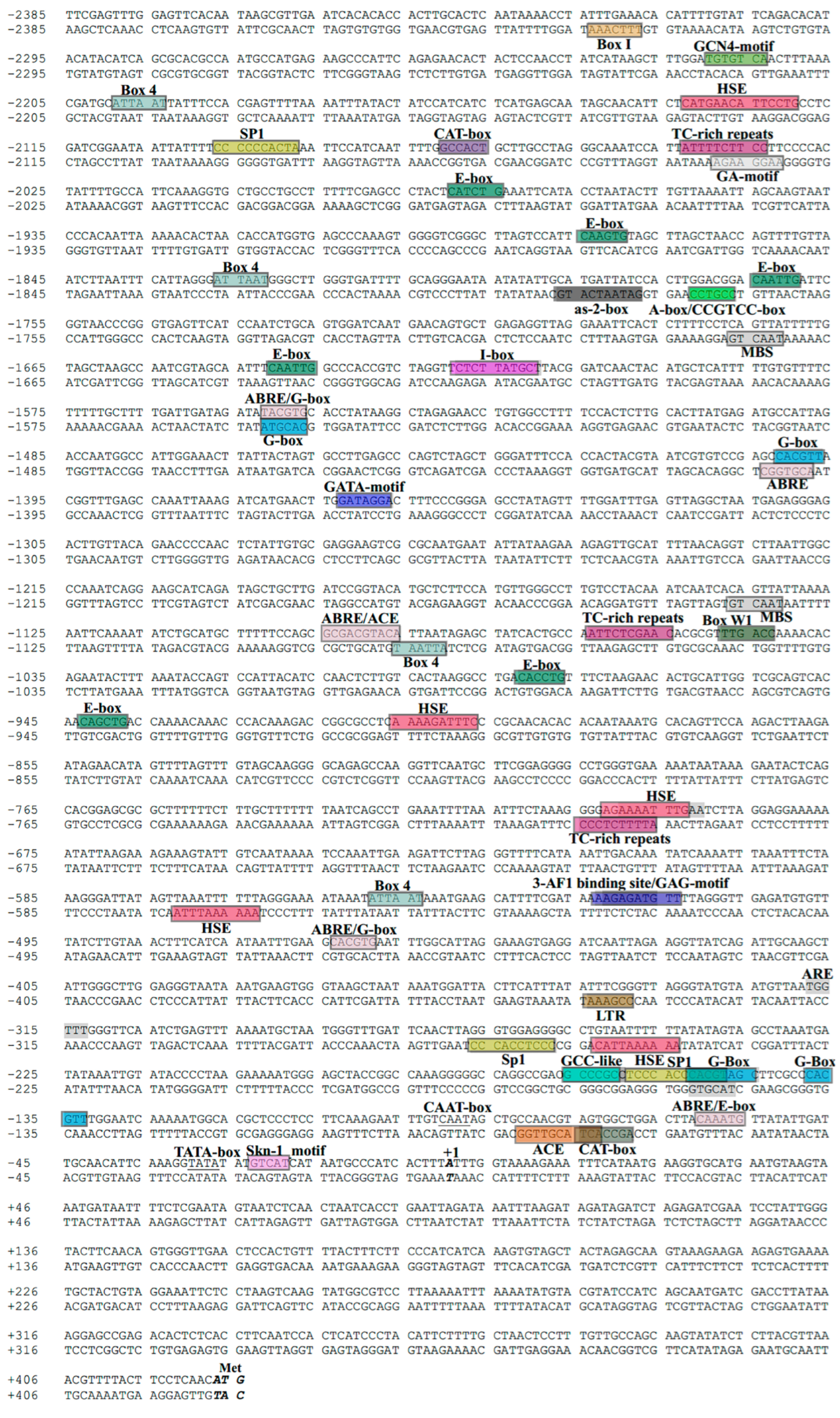
2.1. Extraction and Analysis of the SaCYP736A167 Gene Promoter

Genomic DNA was derived from the foliage of a mature Santalum album tree (aged seven years) using the DNAout kit by Tiandz (Beijing, China), in accordance with the protocol provided. Briefly, 0.5 g of leaf tissue was ground into a powder using liquid nitrogen, and was then added to DNAout Solution A and mixed well. Next, pre-warmed Solution B was added, followed by incubation in a 65 °C water bath for 5 min. Subsequently, 200 μL of chloroform was added, vortexed for 30 s, and then centrifuged at 12,000 rpm for 2 min. The supernatant was transferred to a new 1.5 mL centrifuge tube, and Solution C was added and mixed well. After centrifugation at 12,000 rpm, the plant DNA solution was obtained. The promoter of the SaCYP736A167 gene was identified through the thermal asymmetric interlaced polymerase chain reaction (tail-PCR) method [14,15] with the genomic DNA as template. Tail-PCR random degenerate primers (AD1 to AD5) were designed for the tail-PCR methods described, and the gene-specific primers SaCYP736A167 tail 1 to SaCYP736A167 tail 3 were designed near the 5′ flanking sequences of SaCYP736A167 downloaded from NCBI. All the primers used to isolate the promoter of SaCYP736A167 are listed in Table S1. The online tool TSSP (http://www.softberry.com/berry.phtml?topic=tssp&group=programs&subgroup=promoter, accessed on 24 March 2024) was used to analyze the predicted promoter transcription start sites. The PlantCARE online tool (http://bioinformatics.psb.ugent.be/webtools/plantcare/html/, accessed on 26 March 2024) was utilized to examine the core regulatory cis-acting elements within the SaCYP736A167 promoter sequences that were isolated.

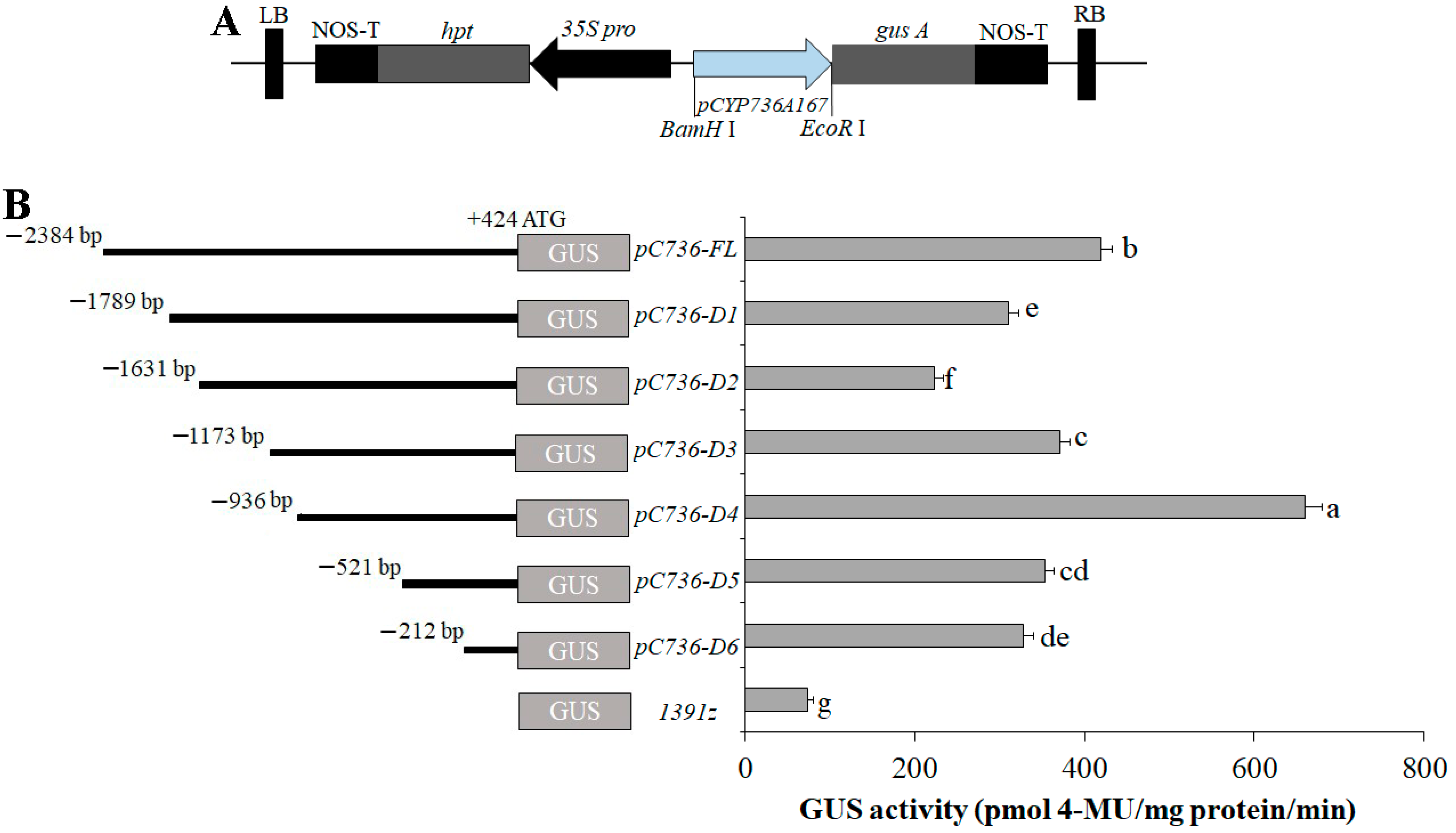
2.2. Fabrication of GUS-Containing Plasmids and Truncated Segments

GUS plasmid and deletion segments of the SaCYP736A167 promoter were constructed as described by Yan et al. [12]. Briefly, taking into account the concentration of cis-acting elements within the promoter region of the SaCYP736A167 gene, the 2384-base pair fragment (PSaCYP736A167), situated upstream from the ATG start codon within the SaCYP736A167 promoter region, was shortened to create seven distinct 5′-deletion fragments through the use of seven forward primers, each equipped with a BamHI restriction enzyme site, along with a single reverse primer that includes an EcoRI restriction enzyme site. (Figure 1A,B and Table S1). pC736D6 mainly contained the essential TATA-box and CAAT-box. Six other truncated fragments, in addition to containing the TATA-box and CAAT-box, also encompassed a variety of other cis-regulatory elements; for example, pC736D5 contained about 9 cis-regulatory elements (mainly G-box and SP1 box), pC736D4 contained about 15 cis-regulatory elements (mainly G-box, SP1 box, and HSE-elements), pC736D3 contained about 20 cis-regulatory elements (mainly G-box, SP1 box, HSE-elements, Box4, and ABRE-elements), pC736D2 contained about 28 cis-regulatory elements (mainly G-box, SP1 box, HSE-elements, Box4, ABRE-elements, and TC-rich repeats), pC736D1 contained about 30 cis-regulatory elements (mainly G-box, SP1 box, HSE-elements, Box4, ABRE-elements, TC-rich repeats, and E-box), and the full length 2384-bp sequence (pC736D6-FL) contained at least 45 cis-regulatory elements. The complete PSaCYP736A167 sequence and its six 5′-deletion variants were each cleaved by the BamHI/EcoRI enzymes. Subsequently, they were individually ligated into the pCAMBIA1391Z plasmid after enzymatic digestion with BamHI/EcoR1. The newly assembled plasmids containing the PSaCYP736A167–GUS fusion were introduced into Escherichia coli DH5a cells, and subsequently underwent sequencing to verify the integrity of the constructs.

2.3. Transformation and Identification of Successfully Modified Transgenic Lines of N. tabacum

The authenticated recombinant plasmids in conjunction with the pCAMBIA1391Z devoid vector (negative control—the GUS gene is not preceded by any promoter driving its expression) were subjected to transformation into Agrobacterium tumefaciens EHA105 and, following inoculation, into N. tabacum leaves using the leaf disk transformation–regeneration method [16,17]. PCR amplification was conducted with gene-specific primers (gDNApC736F/gDNAGUSR; Table S1) and genomic DNA as the substrate to ascertain the integration of the PSaCYP736A167 sequence within the transformed plants (Figure 1B). Three T1 generation transgenic lines, validated by PCR, were chosen for subsequent experimental procedures.
The confirmed recombinant plasmids including the full-length PSaCYP736A167 and its six 5′-deletion derivatives, as well as pCAMBIA1391Z empty plasmid (serving as negative control), were introduced into Agrobacterium tumefaciens EHA105 and were subsequently transiently transformed into N. benthamiana leaves, as described by Leonelli et al. [18], respectively. The transiently transformed plants were cultured for 12 h in the dark, and were then grown in an incubator at 28 °C under 16 h light/8 h darkness photoperiod (80 μmol m−2 s−1) condition for 48 h. Finally, the transiently transformed plant leaves were collected for GUS staining and GUS fluorometric assays.

2.4. Histochemical Analysis of β-Glucuronidase (GUS) Activity and Fluorometric Quantification of GUS Activity Assays

The transgenic organs of N. tabacum at the age of six weeks old were collected for histological GUS staining employing a β-glucuronidase (GUS) staining kit (Real times, Beijing, China) strictly following the maker’s prescribed methods. Briefly, GUS staining solution was added to the samples until they were fully submerged, then they were placed in a 37 °C incubator and incubated in the dark for at least 12 h. Subsequently, the samples were decolorized with 70% ethanol until the background was colorless. Images of GUS-stained samples were examined and documented using a stereo microscope (Leica S9, Leica, Solms, Germany).
The fluorometric GUS assays were executed following the procedure outlined by Jefferson et al. [19] and Yan et al. [12]. Approximately 100 milligrams of N. benthamiana leaves, following transient transformation, were ground into a fine powder using liquid nitrogen and were subsequently processed in a newly prepared GUS extraction buffer (50 mM potassium phosphate buffer (pH 7.0), 10 mM EDTA, 0.1% sodium laurylsarcosine, 0.1% Triton X-100, and 10 mM β-mercaptoethanol). After centrifugation at 12,000 rpm and a temperature of 4 °C for a duration of 10 min, the supernatants were used for fluorescence analysis to determine 4-methylumbelliferone (4-MU) levels, with 4-MUG (4-methyl-umbelliferyl-glucuronide) serving as the substrate. Fluorescence intensity was recorded with a versatile microplate reader (EnSpire, PerkinElmer, Waltham, MA, USA), utilizing 365 nm for excitation and 455 nm for emission wavelengths. Protein concentration was ascertained through the Bradford assay, standardizing against a bovine serum albumin (BSA) reference (Sigma-Aldrich, St. Louis, MO, USA) [20]. The GUS activity was standardized to nM of 4-MU generated per minute per milligram of protein. Each sample was subjected to three separate experimental trials to ensure reliability.

2.5. Statistical Analyses

Statistical analysis of the data was conducted employing SPSS software, version 19.0 (IBM Corp., Armonk, NY, USA). The significance of GUS activity differences among the 1391z control and the seven PSaCYP736A167 (pC736) promoter deletions was evaluated using one-way analysis of variance (ANOVA), supplemented by Duncan’s multiple range test. When the difference reaches a level of p < 0.05, it is considered that there is a significant difference between the groups. Error bars represent the mean ± standard error.

3. Results

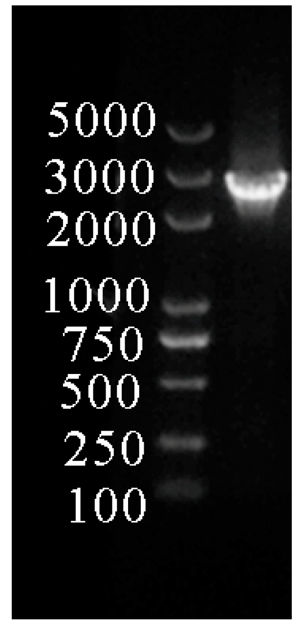
3.1. Characterization and Sequencing of the SaCYP736A167 Promoter Region

Employing sandalwood genomic DNA as a template, a 2808-base pair fragment located preceding the translation initiation codon ATG of the SaCYP736A167 gene was successfully isolated using the tail-PCR method (Figure 2) and was then sequenced. The sequence was considered as a putative SaCYP736A167 gene promoter (GenBank accession number: MH319768.1) and the 2384-bp fragment upstream from the start codon ATG was named as PSaCYP736A167 and was used for subsequent analysis. The transcription start site of PSaCYP736A167 was predicted using the online tool TSSP, and was indicated as +1; the TATA box and CAAT-box were located at −30-bp to −27 bp and −91 bp to −88 bp upstream of the transcription start site, respectively (Figure 3). The online tool PlantCARE’s analysis of PSaCYP736A167 showed that it contains abundant photoreactive elements, including a 3-AF1 binding site, ACE, i-box, Box I, G-box, box 4, GAG-motif, GA-motif, Sp1, GATA-motif, and as-2-box (Table 1 and Figure 3). Moreover, abscisic acid response related element (ABRE), heat stress response (HSE), TC-rich repeats involved in defense and stress, anaerobic induction related element (ARE), fungal inductor response Box W1, low-temperature induction-associated element (LTR), and wound-responsiveness related WUN-motif were all found in PSaCYP736A167. In addition, several important boxes that were potential binding sites of certain transcription factors also existed, such as G-box, W-box, MBS, and GCC-like box (Table 1 and Figure 3).

3.2. Tissue-Specific Expression Patterns of PSaCYP736A167

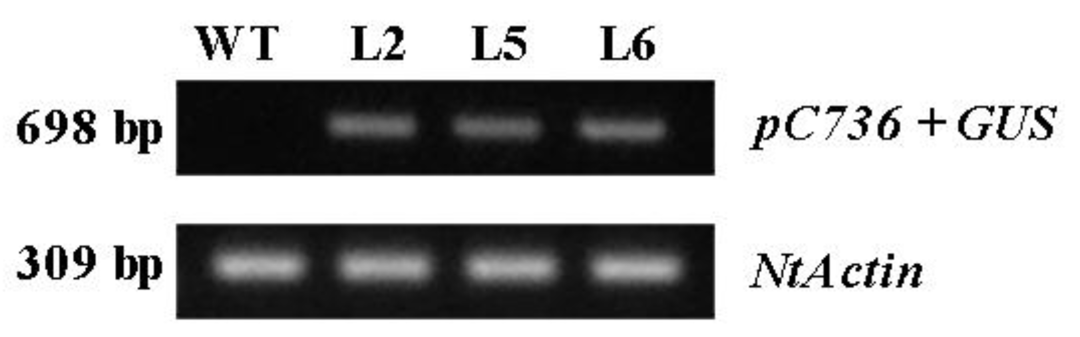
3.2.1. Screening of Positively Transgenic Tobacco Lines

PCR was executed with gene-specific primers (gDNApC736F/gDNAGUSR; Table S1) and genomic DNA as substrate to verify the presence of PSaCYP736A167 in the transformed N. tabacum plants. Three lines including L2, L5, and L6, which exhibited a positive PCR amplification result, were selected for tissue expression pattern analysis (Figure 4).

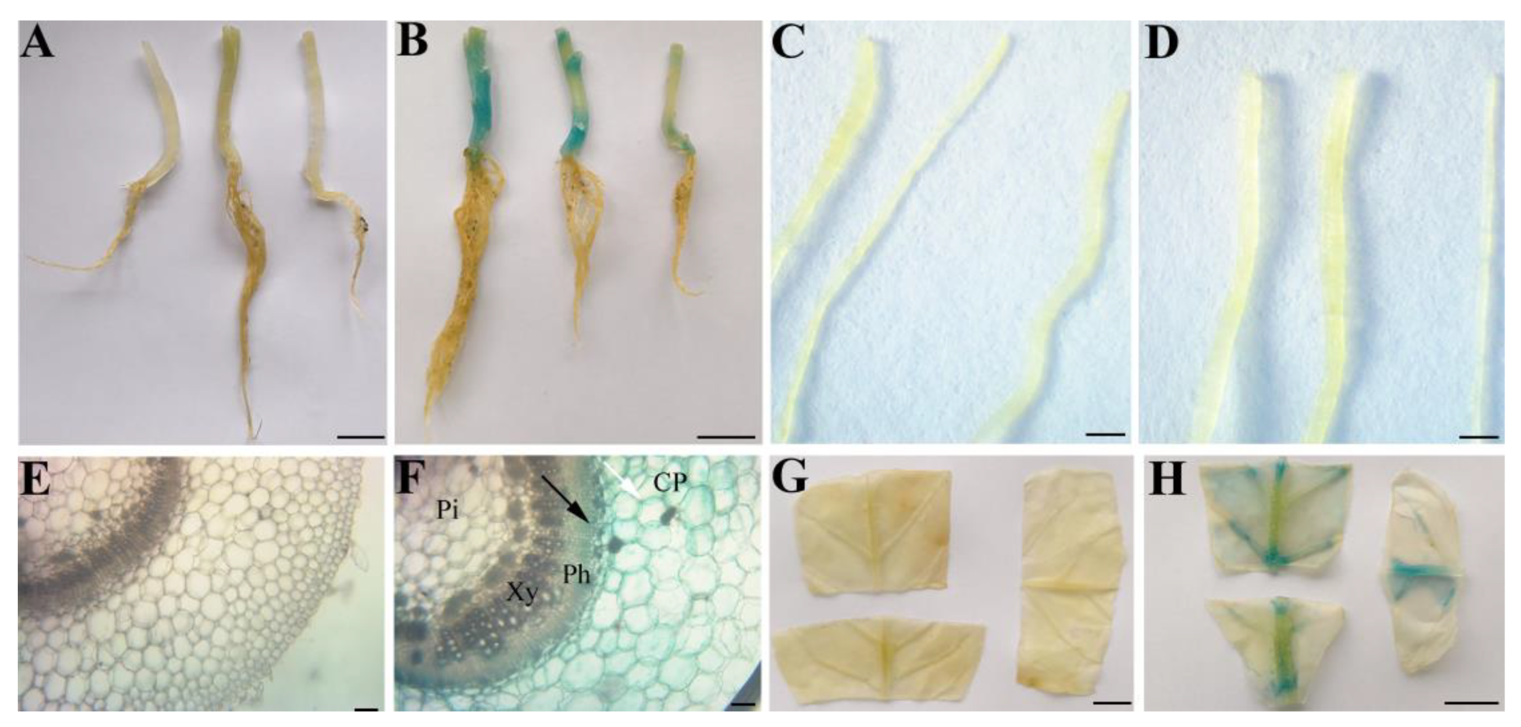
3.2.2. The Tissue-Specific Expression Profiles of PSaCYP736A167

To explore the tissue-specific expression profiles of the PSaCYP736A167 gene, β-glucuronidase (GUS) activity was measured in 6-week-old T1 transgenic N. tabacum plants harboring PSaCYP736A167 in the tissues of roots, stems, and leaves. Intense GUS activity, assessed through histochemical staining, was found in the tissues of stems and leaf veins, but not roots (Figure 5B,D,H). To further observe the tissue expression specificity, the stem was sliced and the GUS activity was observed under a stereo microscope; the findings indicated that GUS activity was predominantly observed in parenchyma cells of stem cortex and phloem, but not in the pulp and xylem of the stem (Figure 5F). In comparison, the T1 transgenic control plants (WT) harboring the 1391Z empty vector displayed no GUS staining in any of the tissues examined (Figure 5A,C,E,G).

3.3. Transient Expression Verified the Activity of PSaCYP736A167

The GUS activity was detected in the tobacco leaves that transiently expressed PSaCYP736A167::GUS. The results showed that high GUS expression, assessed through histochemical staining, was observed in the transiently transformed N. benthamiana leaves harboring PSaCYP736A167 (Figure 6B). In contrast, GUS staining was not exhibited in the CK that contained the empty 1391Z vector (Figure 6A). These results indicated that PSaCYP736A167 has a strong promoter activity.

4. Deletion Analysis of PSaCYP736A167 in Transiently Expressed N. tabacum Leaves

To pinpoint the core promoter segments of PSaCYP736A167, the GUS activity was measured in the leaves of N. benthamiana that had been transiently expressing the complete PSaCYP736A167 sequence along with a series of its truncated variants. GUS fluorometric assays demonstrated that PSaCYP736A167-D4 had the highest GUS activity, followed by full-length PSaCYP736A167, while PSaCYP736A167-D2 had the lowest GUS activity (significantly reduced by 66.11% compared to PSaCYP736A167-D4), and the GUS activity of the other four deletion segments (including PSaCYP736A167-D1, PSaCYP736A167-D3, PSaCYP736A167-D5, and PSaCYP736A167-D6) was all lower than PSaCYP736A167-D4 but higher than PSaCYP736A167-D2 (Figure 7). These results indicated that the core promoter regions of PSaCYP736A167 may be the segment between the start codon ATG and −936-bp, namely the segment of PSaCYP736A167-D4 (Figure 7).

5. Discussion

SaCYP736A167 is one of the critical encoding genes for the biosynthesis of sesquiterpenes, i.e., (Z)-α-santalol, (Z)-β-santalol, (Z)-α-exo-bergamotol, and (Z)-epi-β-santalol [8]. The transcriptional regulation of the SaCYP736A167 gene is mainly dependent on the cis-acting element in its promoter. The analysis of the 2384-bp segment of the cloned SaCYP736A167 promoter revealed that apart from abundant light-responsive elements, other inducible cis-acting elements also exist, such as the ABRE element associated with abscisic acid signaling, the HSE element related to heat stress tolerance, the LTR element responsive to low temperatures, the TC-rich repeats implicated in defense and stress responses, and the WUN-motif linked to wound-induced reactions (Table 1 and Figure 3). These findings suggested that apart from the light, the expression of SaCYP736A167 was mainly induced by abiotic stresses like heat, low temperature, defense, stress, wound, as well as the drought-related hormone ABA. The previous studies also showed that abiotic stresses are the important inducers in gene expression and secondary metabolite accumulation. For example, a like low temperature (8 °C) could significantly increase the content of phenolic acids, flavonoids, carotenoids, and glucosinolates in Kale (Brassica oleracea var. acephala) [21,22]; drought stress increased the amount of methylchavicol, methyleugenol, β-myrcene, and α-bergamotene through promoting the expression levels of chavicol O-methyl transferase and eugenol O-methyl transferase (EOMT) in Ocimum basilicum c.v. Keshkeni luvelou [23,24].
It is worth noting that a tissue-specific cis-acting element, as-2-box (involved in shoot-specific expression), is also found in PSaCYP736A167 (Table 1 and Figure 3). To identify the tissue-specific expression patterns of the SaCYP736A167 gene, the T1 transgenic N. tabacum plants harboring the full-length PSaCYP736A167 were observed under a stereo microscope; the intense GUS activity was found mainly in the parenchyma cells of the stem cortex and phloem, but not in other tissues such as the pulp and xylem of the stem (Figure 5F). This result suggested that the SaCYP736A167 gene may have a tissue-specific expression in the cells of the stem cortex and phloem; however, this needs to be further confirmed using hybridization in situ in a sandalwood tree.
The core promoter region performs the uppermost function across the gene transcriptional regulation [25,26,27]. To characterize the core promoter region of PSaCYP736A167, full-length PSaCYP736A167 was truncated into seven segments and was then transiently expressed into N. benthamiana leaves, respectively. The results of the quantitative detection of GUS activity showed that PSaCYP736A167-D4 (from ATG to −936 bp) had the highest GUS activity among all seven segments including full-length PSaCYP736A167 and the other five deletion segments (including PSaCYP736A167-D1, PSaCYP736A167-D2, PSaCYP736A167-D3, PSaCYP736A167-D5, and PSaCYP736A167-D6) (Figure 7). Apart from CAAT box and TATA box, which are essential for the transcriptional complex binding, at least two G-box (−152 bp to −147 bp and −138 bp to −133 bp) and one GCC-like box (−166 bp to −160 bp) were also found in the PSaCYP736A167-D4 segment (Figure 7). Numerous documents have verified that bHLH family factor members are more likely to bind to the G-box cis-element to regulate its target gene expression [28,29,30], while ERF subfamily proteins are more inclined to bind to the GCC-box (GCCGCC) element of its target genes [31,32,33]. So, we could predict that some members from the bHLH transcription factor family and the ERF subfamily could all regulate the expression of SaCYP736A167. On the other hand, the PSaCYP736A167-D4 segment, which was a core promoter region that may independently and effectively orchestrate the expression of the SaCYP736A167 gene, can be used as an alternative tissue-specific promoter for transgene expression. In particular, it can be employed to drive the synthesis genes or transcription factors involved in the production of secondary metabolites with significant economic value, specifically those synthesized in the cells of the stem cortex and phloem. For instance, it can be utilized to drive the expression of genes involved in the biosynthesis and regulation of paclitaxel [34,35], as well as to stimulate the expression of the genes responsible for the biosynthesis of bioactive components in Cinnamomum cassia [36,37].

6. Conclusions

In the present study, a putative promoter sequence of the SaCYP736A167 gene, spanning 2808 base pairs, was successfully isolated and characterized. The PlantCARE analysis of the 2384-base pair SaCYP736A167 promoter (PSaCYP736A167) revealed that it encompasses a variety of light-responsive elements, along with numerous stress-responsive elements and distinct transcription factor binding sites, including the G-box and GCC-like motifs. Histochemical GUS staining from transgenic N. tabacum showed that full-length PSaCYP736A167 was a parenchyma cell of the stem cortex and phloem-specific expression promoter. The 936-bp segment upstream of the ATG start site defines the core promoter domain for PSaCYP736A167. Our findings yield key insights into the transcriptional control mechanisms of the SaCYP736A167 gene, offering a beneficial promoter resource for transgenic plant breeding.

Supplementary Materials

The following supporting information can be downloaded at: https://www.mdpi.com/article/10.3390/f15101705/s1, Table S1: Primer sequences used in this study.

Author Contributions

Conceptualization, G.M. methodology, Y.Z.; software, Y.Z. and H.Y. validation, Y.Z.; formal analysis, H.Y.; investigation, Y.Z; resources, G.M.; data curation, Y.Z. and H.Y.; writing—original draft preparation, H.Y.; writing—review and editing, H.Y. and R.W.; visualization, L.Q., H.Z. and F.X.; supervision, G.M.; project administration, G.M. and H.Y.; funding acquisition, G.M. All authors have read and agreed to the published version of the manuscript.

Funding

This research was funded by the National Natural Science Foundation of China (grant number 32060358), Guangdong Key Areas Biosafety Project (2022B1111040003), and Guangdong Science and Technology Plan Project (2023B1212060046), and the Guangxi Natural Science Foundation Project (2019GXNSFAA185005).

Data Availability Statement

The original contributions presented in the study are included in the Supplementary Materials, further inquiries can be directed to the corresponding authors.

Conflicts of Interest

The authors declare no conflicts of interest.

References

  1. Jones, C.G.; Keeling, C.I.; Ghisalberti, E.L.; Barbour, E.L.; Plummer, J.A.; Bohlmann, J. Isolation of cDNAs and functional characterisation of two multi-product terpene synthase enzymes from sandalwood, Santalum album L. Arch. Biochem. Biophys. 2008, 477, 121–130. [Google Scholar] [CrossRef] [PubMed]
  2. Yan, H.; Li, M.; Xiong, Y.; Wu, J.; Teixeira da Silva, J.; Ma, G. Genome-Wide Characterization, Expression Profile Analysis of WRKY Family Genes in Santalum album and Functional Identification of Their Role in Abiotic Stress. Int. J. Mol. Sci. 2019, 20, 5676. [Google Scholar] [CrossRef] [PubMed]
  3. Baldovini, N.; Delasalle, C.; Joulain, D. Phytochemistry of the heartwood from fragrant Santalum species: A review. Flavour Fragr. J. 2010, 26, 7–26. [Google Scholar] [CrossRef]
  4. Srivastava, P.L.; Daramwar, P.P.; Krithika, R.; Pandreka, A.; Shankar, S.S.; Thulasiram, H.V. Functional Characterization of Novel Sesquiterpene Synthases from Indian Sandalwood, Santalum album. Sci. Rep. 2015, 5, 10095. [Google Scholar] [CrossRef]
  5. Nagegowda, D.A. Plant volatile terpenoid metabolism: Biosynthetic genes, transcriptional regulation and subcellular compartmentation. FEBS Lett. 2010, 584, 2965–2973. [Google Scholar] [CrossRef]
  6. Jones, C.G.; Moniodis, J.; Zulak, K.G.; Scaffidi, A.; Plummer, J.A.; Ghisalberti, E.L.; Barbour, E.L.; Bohlmann, J. Sandalwood fragrance biosynthesis involves sesquiterpene synthases of both the terpene synthase (TPS)-a and TPS-b subfamilies, including santalene synthases. J. Biol. Chem. 2011, 286, 17445–17454. [Google Scholar] [CrossRef]
  7. Rani, A.; Ravikumar, P.; Reddy, M.D.; Kush, A. Molecular regulation of santalol biosynthesis in Santalum album L. Gene 2013, 527, 642–648. [Google Scholar] [CrossRef]
  8. Celedon, J.M.; Chiang, A.; Yuen, M.M.S.; Diaz-Chavez, M.L.; Madilao, L.L.; Finnegan, P.M.; Barbour, E.L.; Bohlmann, J. Heartwood-specific transcriptome and metabolite signatures of tropical sandalwood (Santalum album) reveal the final step of (Z)-santalol fragrance biosynthesis. Plant J. 2016, 86, 289–299. [Google Scholar] [CrossRef]
  9. Cirino, P.C.; Diaz-Chavez, M.L.; Moniodis, J.; Madilao, L.L.; Jancsik, S.; Keeling, C.I.; Barbour, E.L.; Ghisalberti, E.L.; Plummer, J.A.; Jones, C.G.; et al. biosynthesis of sandalwood oil: Santalum album CYP76F cytochromes P450 produce santalols and bergamotol. PLoS ONE 2013, 8, e75053. [Google Scholar] [CrossRef]
  10. Hernandez-Garcia, C.M.; Finer, J.J. Identification and validation of promoters and cis-acting regulatory elements. Plant Sci. 2014, 217–218, 109–119. [Google Scholar] [CrossRef]
  11. Yasmeen, E.; Wang, J.; Riaz, M.; Zhang, L.; Zuo, K. Designing artificial synthetic promoters for accurate, smart, and versatile gene expression in plants. Plant Commun. 2023, 4, 100558. [Google Scholar] [CrossRef] [PubMed]
  12. Yan, H.; Xiong, Y.; Teixeira da Silva, J.A.; Pang, J.; Zhang, T.; Yu, X.; Zhang, X.; Niu, M.; Ma, G. Molecular cloning and functional characterization of bisabolene synthetase (SaBS) promoter from Santalum album. Forests 2020, 11, 85. [Google Scholar] [CrossRef]
  13. Villao-Uzho, L.; Chávez-Navarrete, T.; Pacheco-Coello, R.; Sánchez-Timm, E.; Santos-Ordóñez, E. Plant promoters: Their identification, characterization, and role in gene regulation. Genes 2023, 14, 1226. [Google Scholar] [CrossRef] [PubMed]
  14. Liu, Y.-G.; Mitsukawa, N.; Oosumi, T.; Whittier, R.F. Efficient isolation and mapping of Arabidopsis thaliana T-DNA insert junctions by thermal asymmetric interlaced PCR. Plant J. 1995, 8, 457–463. [Google Scholar] [CrossRef]
  15. Liu, Y.; Huang, N. Efficient amplification of insert end sequences frombacterial artificial chromosome clones by thermal asymmetric interlaced PCR. Plant Mol. Biol. Rep. 1998, 16, 0735–9640. [Google Scholar] [CrossRef]
  16. Makhzoum, A.; Petit-Paly, G.; Pierre, B.S.; Bernards, M.A. Functional analysis of the DAT gene promoter using transient Catharanthus roseus and stable Nicotiana tabacum transformation systems. Plant Cell Rep. 2011, 30, 1173–1182. [Google Scholar] [CrossRef]
  17. Horsch, R.B.; Fry, J.E.; Hoffmann, N.L.; Wallroth, M.; Eichholtz, D.; Rogers, S.G.; Fraley, R.T. A simple and general method for transferring genes into plants. Science 1985, 227, 1229–1231. [Google Scholar] [CrossRef] [PubMed]
  18. Leonelli, L.; Erickson, E.; Lyska, D.; Niyogi, K.K. Transient expression in Nicotiana benthamiana for rapid functional analysis of genes involved in non-photochemical quenching and carotenoid biosynthesis. Plant J. 2016, 88, 375–386. [Google Scholar] [CrossRef]
  19. Jefferson, R.K.T.B.M. GUS fusion: Beta-glucuronidase as a sensitive and versatile gene fusion marker in higher plants. EMBO J. 1987, 6, 3901–3907. [Google Scholar] [CrossRef]
  20. Bradford, M.M. A rapid sensitive method for the quantitation of microgram quantities of protein utilizing the principle of protein dye binding. Anal. Biochem. 1976, 72, 248–254. [Google Scholar] [CrossRef]
  21. Ljubej, V.; Karalija, E.; Salopek-Sondi, B.; Šamec, D. Effects of short-term exposure to low temperatures on proline, pigments, and phytochemicals level in Kale (Brassica oleracea var. acephala). Horticulturae 2021, 7, 341. [Google Scholar] [CrossRef]
  22. Muthusamy, M.; Lee, S.I. Abiotic stress-induced secondary metabolite production in Brassica: Opportunities and challenges. Front. Plant Sci. 2024, 14, 1323085. [Google Scholar] [CrossRef]
  23. Espinosa-Leal, C.A.; Mora-Vásquez, S.; Puente-Garza, C.A.; Alvarez-Sosa, D.S.; García-Lara, S. Recent advances on the use of abiotic stress (water, UV radiation, atmospheric gases, and temperature stress) for the enhanced production of secondary metabolites on in vitro plant tissue culture. Plant Growth Regul. 2022, 97, 1–20. [Google Scholar] [CrossRef]
  24. Abdollahi Mandoulakani, B.; Eyvazpour, E.; Ghadimzadeh, M. The effect of drought stress on the expression of key genes involved in the biosynthesis of phenylpropanoids and essential oil components in basil (Ocimum basilicum L.). Phytochemistry 2017, 139, 1–7. [Google Scholar] [CrossRef]
  25. Collins, J.M.; Wang, D. Cis-acting regulatory elements regulating CYP3A4 transcription in human liver. Pharm. Genom. 2020, 30, 107–116. [Google Scholar] [CrossRef]
  26. Cvetesic, N.; Lenhard, B. Core promoters across the genome. Nat. Biotechnol. 2017, 35, 123–124. [Google Scholar] [CrossRef]
  27. Haberle, V.; Stark, A. Eukaryotic core promoters and the functional basis of transcription initiation. Nat. Rev. Mol. Cell Biol. 2018, 19, 621–637. [Google Scholar] [CrossRef] [PubMed]
  28. Hong, G.-J.; Xue, X.-Y.; Mao, Y.-B.; Wang, L.-J.; Chen, X.-Y. Arabidopsis MYC2 Interacts with DELLA proteins in regulating sesquiterpene synthase gene expression. Plant Cell 2012, 24, 2635–2648. [Google Scholar] [CrossRef]
  29. Kazan, K.; Manners, J.M. MYC2: The Master in Action. Mol. Plant 2013, 6, 686–703. [Google Scholar] [CrossRef]
  30. Toledo-Ortiz, G.; Huq, E.; Quail, P.H. The Arabidopsis basic/helix-loop-helix transcription factor family. Plant Cell 2003, 15, 1749–1770. [Google Scholar] [CrossRef]
  31. Zhu, D.; Wu, Z.; Cao, G.; Li, J.; Wei, J.; Tsuge, T.; Gu, H.; Aoyama, T.; Qu, L.-J. TRANSLUCENT GREEN, an ERF family transcription factor, controls water balance in Arabidopsis by activating the expression of aquaporin genes. Mol. Plant 2014, 7, 601–615. [Google Scholar] [CrossRef]
  32. Xu, Z.S.; Chen, M.; Li, L.C.; Ma, Y.Z. Functions and application of the AP2/ERF Transcription Factor Family in Crop Improvement. J. Integr. Plant Biol. 2011, 53, 570–585. [Google Scholar] [CrossRef] [PubMed]
  33. Tang, Q.; Wei, S.; Zheng, X.; Tu, P.; Tao, F. APETALA2/ethylene-responsive factors in higher plant and their roles in regulation of plant stress response. Crit. Rev. Biotechnol. 2024, 1–19. [Google Scholar] [CrossRef] [PubMed]
  34. Yu, C.; Guo, H.; Zhang, Y.; Song, Y.; Pi, E.; Yu, C.; Zhang, L.; Dong, M.; Zheng, B.; Wang, H.; et al. Identification of potential genes that contributed to the variation in the taxoid contents between two Taxus species (Taxus media and Taxus mairei). Tree Physiol. 2017, 37, 1659–1671. [Google Scholar] [CrossRef] [PubMed]
  35. Howat, S.; Park, B.; Oh, I.S.; Jin, Y.-W.; Lee, E.-K.; Loake, G.J. Paclitaxel: Biosynthesis, production and future prospects. New Biotechnol. 2014, 31, 242–245. [Google Scholar] [CrossRef] [PubMed]
  36. Zhou, W.; Liang, Z.; Li, P.; Zhao, Z.; Chen, J. Tissue-specific chemical profiling and quantitative analysis of bioactive components of Cinnamomum cassia by combining laser-microdissection with UPLC-Q/TOF–MS. Chem. Cent. J. 2018, 12, 71. [Google Scholar] [CrossRef]
  37. Ye, P.; Su, J.; Lin, J.; Li, Y.; Wu, H. Identification of a cinnamoyl-CoA reductase from Cinnamomum cassia involved in trans-cinnamaldehyde biosynthesis. Planta 2024, 259, 138. [Google Scholar] [CrossRef]
Figure 1. Schematic presentation of the GUS vector and truncated segment construction. (A) The vector of pCAMBIA1391Z-PSaCYP736A167::GUS. (B) Schematic presentation of seven truncated promoters of PSaCYP736A167 and the 1391z control.
Forests 15 01705 g001
Figure 2. Agarose gel electrophoresis map of the cloned SaCYP736A167 promoter.
Forests 15 01705 g002
Figure 3. The nucleotide sequence along with the inferred cis-acting elements within PSaCYP736A167. The transcription start site and the ATG codon are presented in bold italics, and are indicated as +1 and +423 bp to +425 bp, respectively. The TATA and CAAT-box are indicated with an underline, and are labeled with −30 bp to −27 bp and −91 bp to −88 bp, respectively.
Forests 15 01705 g003
Figure 4. PCR amplification screening of transgenic Nicotiana tabacum plants. The actin gene from N. tabacum (NtActin) served as an internal control. The wild type (WT) represented the T1 transgenic control plants harboring the vacant 1391z plasmid, while L2, L5, and L6 represented the three T1 transgenic N. tabacum lines harboring PSaCYP736A167.
Forests 15 01705 g004
Figure 5. Photo of GUS staining in different tissues of transgenic Nicotiana tabacum. (A) Stems and roots in WT lines; (B) stems and roots in transgenic lines; roots in WT (C) and transgenic lines (D); stem section in WT (E) and transgenic lines (F); leaves in WT (G) and transgenic lines (H). ((A,B,G,H) Bar = 1 cm; (C,D) Bar = 0.1 mm; (E,F) Bar = 0.01 mm); Cp, cortical parenchyma; Pi, pith; Ph, phloem; Xy, xylem. White arrow indicates cortical parenchyma cell, and black arrow indicates phloem cells.
Forests 15 01705 g005
Figure 6. Histochemical GUS staining in N. benthamiana leaves of transient transformants. (A) pCAMBIA1391Z (CK). (B) pCAMBIA1391Z-PSaCYP736A167::GUS (Bar = 2 cm).
Forests 15 01705 g006
Figure 7. Deletion analysis of the SaCYP736A167 promoter. (A) Schematic representation of the pCAMBIA1391z-PSaCYP736A167::GUS plasmid used for transient transformation. (B) Schematic representation of different PSaCYP736A167 (pC736) deletions (left panel) and the GUS activity within the transiently transformed N. benthamiana leaves (right panel). The average of at least three independent experiments with triplicate samples is shown for each construct. Different lowercases indicated significance at p < 0.05. Error bars indicate SE.
Forests 15 01705 g007
Table 1. Main cis-acting elements of PSaCYP736A167.
Cis-Acting ElementSignal SequenceStart PositionFunction
3-AF1 binding siteAAGAGATATTT−647light-responsive element
A-boxCCGTCC−1918cis-acting regulatory element
ABRETACGTG−1598cis-acting element involved in
the abscisic acid responsiveness
ACGTGGC−1572
GCGACGTACA−1155
CACGTG−526
ACEGCGACGTACA−1155cis-acting element involved
in light responsiveness
ACTACGTTGG−197
ARETGGTTT−492cis-acting regulatory element essential for anaerobic induction
Box 4ATTAAT−2211, −1863,
−1164, −621
part of a conserved DNA module involved in light responsiveness
Box ITTTCAAA−2452light responsive element
Box W1TTGACC−1202fungal elicitor responsive element
CAT-boxGCCACT−2159cis-acting regulatory element
related to meristem expression
CCGTCC-
box
CCGTCC−1923cis-acting regulatory element related to meristem specific activation
G-boxCACGTA−1603, −298cis-acting regulatory element involved in light responsiveness
TACGTG−1598
CACGTT−1568, −312
CACGTG−526
GAG-motifAGAGATG−648part of a light responsive element
GA-motifAAGGAAGA−2198part of a light responsive element
GATA-motifGATAGGA−1427part of a light responsive element
GCN4_motifTGTGTCA−2370cis-regulatory element involved
in endosperm expression
GCC-likeGCCCGCC−284-
HSECNNGAANNTTCNNG−2277cis-acting element involved
in heat stress responsiveness
AAAAGATTTC−984
AGAAAATTTG−828
AAAAAATTTA−607
AAAAAATTAC−386
I-boxCTCTTATGCT−1711part of a light responsive element
LTRCCGAAA−471cis-acting element involved in
low-temperature responsiveness
MBSTAACTG−1838, −1298MYB binding site involved
in drought inducibility
Skn−1_motifGTCAT−67cis-acting regulatory element required for endosperm expression
Sp1CCCCCCACTA−2133light responsive element
CCCACCTCCC−363
TCCCACCCACGT−291
TC-rich repeatsATTTTCTTCC−2187cis-acting element involved in defense and stress responsiveness
ATTCTCGAAC−1186
ATTTTCTCCC−834
as-2-boxGATAATGATG−1912involved in shoot-specific
expression and light responsiveness
Disclaimer/Publisher’s Note: The statements, opinions and data contained in all publications are solely those of the individual author(s) and contributor(s) and not of MDPI and/or the editor(s). MDPI and/or the editor(s) disclaim responsibility for any injury to people or property resulting from any ideas, methods, instructions or products referred to in the content.

