Article

Taxonomic Studies on Five Species of Sect. Tuberculata (Camellia L.) Based on Morphology, Pollen Morphology, and Molecular Evidence

1
College of Forestry, Guizhou University, Guiyang 550025, China
2
College of Forestry, Jiangxi Agricultural University, Nanchang 330045, China
3
Jiangxi Provincial Key Laboratory of Conservation Biology, Jiangxi Agricultural University, Nanchang 330045, China
4
Key Laboratory of National Forestry and Grassland Administration on Biodiversity Conservation in Karst Mountainous Areas of South-Western China, Guizhou Academy of Forestry, Guiyang 550005, China
*
Author to whom correspondence should be addressed.
Forests 2024, 15(10), 1718; https://doi.org/10.3390/f15101718
Submission received: 30 August 2024 / Revised: 23 September 2024 / Accepted: 25 September 2024 / Published: 28 September 2024
(This article belongs to the Section Forest Ecophysiology and Biology)

Abstract

:
Sect. Tuberculata Chang in the genus Camellia (Theaceae Mirb.) is named after the “tubercle-like projections on the surface of the capsule and ovary”. Due to complex morphological variations in these taxon and insufficient field investigations, the interspecies relationships are unclear, the species’ definitions are vague, and the names are confusing. This is not conducive to the conservation and study of these species. Therefore, herein, we systematically explore the taxonomic status of five sect. Tuberculata species using morphological, pollen morphological, and molecular phylogenetic methods. The results showed that (1) the morphological characteristics of the flower, fruit, and leaves of C. anlungensis and C. leyeensis are similar. Furthermore, the pollen characteristics and pollen wall ornamentation show that there is no significant difference between the two species; (2) there are significant differences between C. acutiperulata and C. anlungensis in terms of leaf shape (elliptic vs. obovate), calyx characteristics (sepal apex pointed vs. sepal oblong), and fruit shape (subglobose folds with shallow verruculose vs. flat folds and verruculose protuberances with pronounced internal cleavage); (3) C. pyxidiacea and C. rubituberculata differ in flower color (white or light color vs. red) and fruit verrucae (obviously deeply cleft vs. shallowly uncracked); (4) a phylogenetic tree based on the chloroplast genome shows that C. anlungensis and C. leyeensis form a single clade (BS = 100%, PP = 1.0) and are on a different branch, with C. acutiperulata on clade II (BS = 100%, PP = 1.0), and C. pyxidiacea and C. rubituberculata clustered on different branches of clade I (BS = 99%, PP = 1.00). Considering the above results together, we propose that C. leyeensis should be treated as a homonym of C. anlungensis, and C. acutiperulata, C. pyxidiacea, and C. rubituberculata should be considered as separate species. Clarifying the taxonomic status of these five species not only advances our understanding of the significance and complexity of the systematic classification of the genus Camellia but also has important implications for diversity conservation and population genetics.

1. Introduction

Section Tuberculata belongs to the genus Camellia (Theaceae Mirb.) and is named after its special morphological characteristic of “fruits that show verrucose protuberances” [1]. The species was first discovered in Sichuan Province, China, in 1939 by the famous botanist professor Chien [2], who named it “Camellia tuberculata Chien”. Subsequently, some unknown species with verrucose protuberances were gradually discovered [3,4,5,6,7]. These species were divided into section Heterogenea Sealy in 1958 [8]. Chang Hongda, a famous plant taxonomist, established sect. Tuberculata based on the tuberculation of the fruit in 1981 through morphology and field studies [1]. Since then, the classification sect. Tuberculata has been considered a monophyletic group in molecular phylogenetic, morphological, and micro-morphological studies [4,7,9]. However, at the species level, most of the taxonomic treatments were carried out based on only a few plant specimens and insufficient field surveys, which resulted in confusing interspecies relationships, interspecies definitions, and nomenclature. Therefore, to determine the taxonomic relationships of these species in sect. Tuberculata, it is necessary to conduct taxonomic research on the taxonomic groups with taxonomic conflicts.
In the recent decade, leaf micromorphology and molecular studies have been carried out on the plants of the sect. Tuberculata species (C. anlungensis Chang, C. leyeensis Chang and Y. C. Zhong, C. acutiperulata Chang and Ye, C. pyxidiacea Xu, F. P. Chen and C. Y. Deng and C. rubituberculata Chang and Yu). For example, Jiang et al. considered the combined application of C. leyeensis with C. anlungensis and treated C. pyxidiacea, C. rubituberculata, and C. acutiperulata as independent species based on leaf micromorphological studies [8]. Jiang et al. constructed a Bayesian phylogenetic tree of Camellia genus based on the joint matrix of several chloroplast genes (matK, rbcL, ycf1, trn L-F), showing that C. anlungensis was not in the same branch as C. acutiperulata [9]. Transcriptome phylogenetic studies of 116 species of the genus Camellia showed that C. leyeensis, C. acutiperulata, C. rubituberculata, and C. anlungensis should be treated as different species [10]. Recently, Xiao conducted the first study on the pollen morphology of 13 species of sect. Tuberculata plants and the results showed that there are interspecific differences among the pollen fractions of sect. Tuberculata plants, which may provide an auxiliary basis for the evolution of the sect. Tuberculata [11]. Ran et al. reported the chloroplast genomes of 13 sect. Tuberculata plants, which showed that C. pyxidiacea, C. anlungensis, and C. leyeensis are clustered on a clade (BS = 100%, PP = 1.00) [12]. However, there are still some challenges regarding these species, such as unclear interspecific relationships, the absence of key taxonomic traits, and so on, which is due to the absence of insufficient field investigations and incomplete research on the morphology and systematics at the population level. Therefore, it is necessary to adopt comprehensive methods and multi-disciplinary evidence for species with significant taxonomic conflicts and many taxonomic problems.
Five species of sect. Tuberculata are narrowly distributed throughout the Panjiang River Basin, China. C. anlungensis was discovered in 1981 by Chang in Lekang township, Wangmo County, Guizhou Province. Its main morphological characteristics are obovate, leathery leaves, a cuneate base, and subglobose capsules [13]. C. rubituberculata was discovered in Qinglong County, Guizhou Province, China, in 1984 and was named by Chang based on its tuberculate pericarp and red flowers [5]. In the same year, another species was found by Chang in Longlin County, Guangxi Province, and called C. acutiperulata based on its long elliptical leaf blade and pointed sepal apex [5]. In 1987, a new species was found in Luoping County, Yunnan Province, China, and named C. pyxidiacea based on its pericarp surfaces with verrucous protuberances and folds, as well as deep grooves in the transverse cleavage [14]. In 1991, a species was discovered in Leye County, Guangxi Province, and named C. leyeensis based on its leathery, elliptic leaves with broadly cuneate base and sometimes slightly rounded, globose capsule with pleated pericarp and pilose seed epidermis [15]. Due to difficulties related to the collection of the five plants in the taxa belonging to sect. Tuberculata, previous studies on these species have mainly relied on incomplete specimens in the collections for taxonomic studies. Many specimens lack key “flower and fruit” data, which has resulted in unclear interspecific delimitation. Still, the taxonomic problems remain unresolved. Therefore, in this study, we conducted a comparative analysis of five species using traditional morphology, micro-morphology, and specimen and phylogenetic studies of chloroplast genomes to elucidate the taxonomic status of five species of sect. Tuberculata.

2. Materials and Methods

2.1. Collection of Materials

This study was conducted by field sampling five species of plants in sect. Tuberculata from the Panjiang River Basin, China. We collected fresh pollen and preserved it in an electron microscope fixative. Healthy leaves were preserved in bags filled with silica gel, and all samples were sent to the laboratory for study. The specimens were deposited in the Tree Herbarium of the College of Forestry, Guizhou University (GZAC). The collected and sample information are shown in Table 1.

2.2. Experimental Research Method

2.2.1. Data Statistics

During the field survey, data were collected using traditional methods for determining the morphological characteristics of these species, including observations of the species’ habitats, branches, leaves, flowers, and fruits in their natural states. The texture and color of leaves, type of margin, leaf base and veins, size, number, and color of the petals, and shape, size, and color of the fruit were recorded. At least 10 specimens were selected from each population to measure the above morphological data, which were statistically analyzed in Excel 2010.

2.2.2. Micromorphological of Pollen

Selected formaldehyde acetic acid ethanol fixative (FAA) fixed anthers were dehydrated with a gradient of 30%–100% ethanol, and full anthers were selected under a deconstructing microscope and glued to the conductive double-sided adhesive tape on the sample stage. The anther wall was punctured with a deconstructing needle to release the pollen. Representative pollen grains were observed under a JSM-6490LV scanning electron microscope (SEM) by coating and spraying them with gold. Photographs were taken to preserve the characteristics of the pollen grains, such as the overall view, polar view, equatorial view, and pollen wall ornamentation. Ten pollen grains were selected for each species. For pollen description terminology, refer to the Handbook of Palynology, Introduction to Sporology, Illustrated Pollen Terminology and other related works [16,17,18].

2.2.3. Chloroplast DNA Acquisition and Genome Assembly

Fresh plant leaves were harvested, and total deoxyribonucleic acid (DNA) was extracted using a modified cetyltrimethyllammonide bromide (CTAB) method [19]. The DNA quality and concentration were determined using agar gel electrophoresis. Fragments that passed the quality tests were sequenced using the Illumina high-throughput sequencing platform. The DNA was then randomly interrupted, end-repaired, and joined to construct sequencing libraries [20,21]. The low-quality data were tailored using Trimmomatic v.0.39 software. After filtering, the qualified complete genome sequences were compared with those in the experimental group. Finally, cp DNA was obtained by online annotation, comparison, and manual correction using C. rubituberculata Chang and Yu (MZ424202) as the reference sequence [22]. Using Prodigal v26.3, we annotated the coding sequence (CDS) region of the chloroplast genome; HMMER v3.4 software predicts rRNA, and ARAGORN v1.2.38 predicts tRNA. Using Blast v2.6, we aligned and annotated the assembled sequences and manually inspected the two differential genes with the annotation results to obtain the best gene structure annotation. OGDRAW was used (https://chlorobox.mpimp-golm.mpg.de/OGDraw.html, accessed on 29 August 2024) [23] to map the chloroplast genome. Finally, the obtained sequences were uploaded to the National Center of Biotechnology Information GenBank (NCBI) to obtain the GenBank accession number.

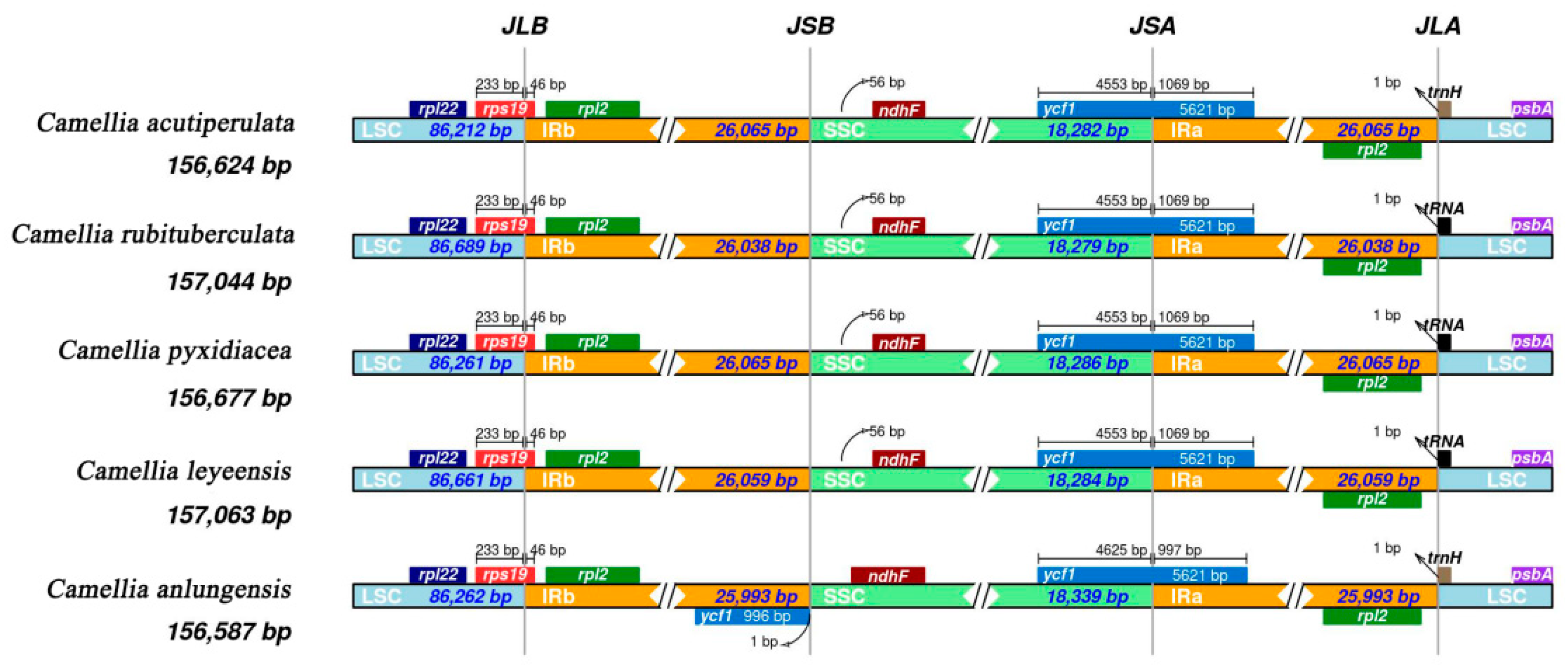
2.2.4. Expansion and Contraction of the IR Boundary

The chloroplast genome sequences of the five species were selected and analyzed for the expansion and contraction of the IRscope region (IR) boundaries using IRscope (https://irscope.shinyapps.io/irapp/, accessed on 29 August 2024) [24] online software, and the data were plotted for comparison.

2.2.5. Phylogenetic Tree

The chloroplast genome sequences of 19 species (containing five sequences that were newly sequenced) were downloaded from NCBI. A total of 18 cp genomes from Camellia species and a sister species, Stewartia sichuanensis (S. Z. Yan), J. Li, and T. L. Ming (ON853833), were used as an outgroup. Sequence comparisons were performed using the MAFFT 7 software [25]. In IQ-TREE v1.6.12, the phylogenetic tree was reconstructed using the maximum likelihood (ML) method [26] and manually corrected with MEGA X [27]. Using the best tree-building model. The optimal model (GTR+I+G) was determined using MrModeltest v2.3. The Bayesian (BI) analyses were performed using the MrBayes v3.2.7 software [28] and landscaped in the iTOL v6 (https://itol.embl.de/, accessed on 29 August 2024) online tool [29]. Based on the protein-coding genes, the same phylogenetic tree was constructed for the whole chloroplast genome.

3. Results

3.1. Morphological Studies

Statistical analyses were carried out on some of the morphological data (Figure 1 and Figure 2 and Table 2) of the five species. The annual branches were not ribbed; the leaf blades among these species were similar; the flowers were white, except for those of C. rubituberculata, which were red; there were obvious differences in the petals, bracts, and calyces; the fruit diameter and pericarp thickness of C. rubituberculata were greater than those of the other four species; and the morphological characteristics of C. anlungensis and C. leyeensis were very similar. The differences among the morphological features of the five species are shown in Table 2. Therefore, based on the morphology, we concluded that C. anlungensis and C. leyeensis are the same species, and the other three species are independent.

3.2. Micromorphological Characteristics of Pollen

The results of the pollen morphological characteristics of the electron microscope scans of five species of C. Tuberculata showed the following (Figure 3 and Table 3): Comparative analyses showed that the pollen of C. anlungensis and C. leyeensis had the same pollen shape, the same outer wall ornamentation, and were similar in the polar and equatorial axes and the germination furrow and the differences between the remaining three species were also not obvious.
C. anlungensis: the pollen grain morphology was oblate spherical, trilobate suborbicular in the polar view, oblong ellipsoid in the equatorial view, pollen grains were 30.21–32.27 μm, and the pollen germination pore was of a three-pore furrow type; the outer wall ornamentation was wrinkled and wave-like to granular with obvious inter-ridges, and the reticulation was deep and narrow. There were three germination grooves in the pollen micromorphology. The germination groove, about 26.83–27.75 μm, was shallow and broad, wider and narrower from both ends to the middle, and prominent in the middle.
C. leyeensis: the pollen grain morphology was oblate spherical, with very few subglobose pollen grains; the pollen shape was trilobate and suborbicular; pollen grains were 29.41–31.67 μm; the pollen germination pore was of a three-pore furrow type; and the outer wall ornamentation was wrinkled and wave-like to granular with thick and obvious ridges, warty grains, and obvious reticulation. There were three germination grooves in the pollen micromorphology. The germination groove, about 26.94–27.73 μm, was shallow and broad, wider and narrower from both ends to the middle, and prominent in the middle.
C. acutiperulata: the pollen grain morphology was oblate globose, trilobate ortho-triangular in the polar view, ellipsoid in the equatorial view, pollen grains were 27.54–28.51 μm, and the pollen germination pore was of the three-pore furrow type; the outer wall ornamentation was crinkled wave-like to granular with short, thick and obvious reticulation ridges, inter-reticulation stripes, and an inconspicuous reticulation mesh. There were three germination grooves in the pollen micromorphology. The germination groove, about 24.54–25.12 μm, was shallow and broad, wider and narrower from both ends to the middle, and prominent in the middle.
C. pyxidiacea: the pollen grain morphology was oblate spherical, trilobate suborbicular in the polar view, ellipsoid in the equatorial view, pollen grains were 30.91–31.82 μm, and the pollen germination pore was of the three-pore furrow type; the outer wall ornamentation was wrinkled and wave-like, with thick and short reticulation ridges, tight inter-ridges, narrow and shallow interridge stripes, and narrow and deep reticulation eyes. There were three germination grooves in the pollen micromorphology. The germination groove, about 22.11–23.17 μm, was shallow and broad, wider and narrower from both ends to the middle, and prominent in the middle.
C. rubituberculata: the pollen grain morphology was oblate spherical, trilobate deltoid in the polar view, sori-ellipsoid in the equatorial view, pollen grains were 28.27–29.84 μm, and the pollen germination pore was of the three-pore furrowed type; the outer wall ornamentation was crinkled and furrowed, with obvious and clear mesh ridges that were tall and the interridge stripes were wide and deep, and the mesh was deep. There were three germination grooves in the pollen micromorphology. The germination groove, about 26.85–27.35 μm, was shallow and broad, wider and narrower from both ends to the middle, and prominent in the middle.

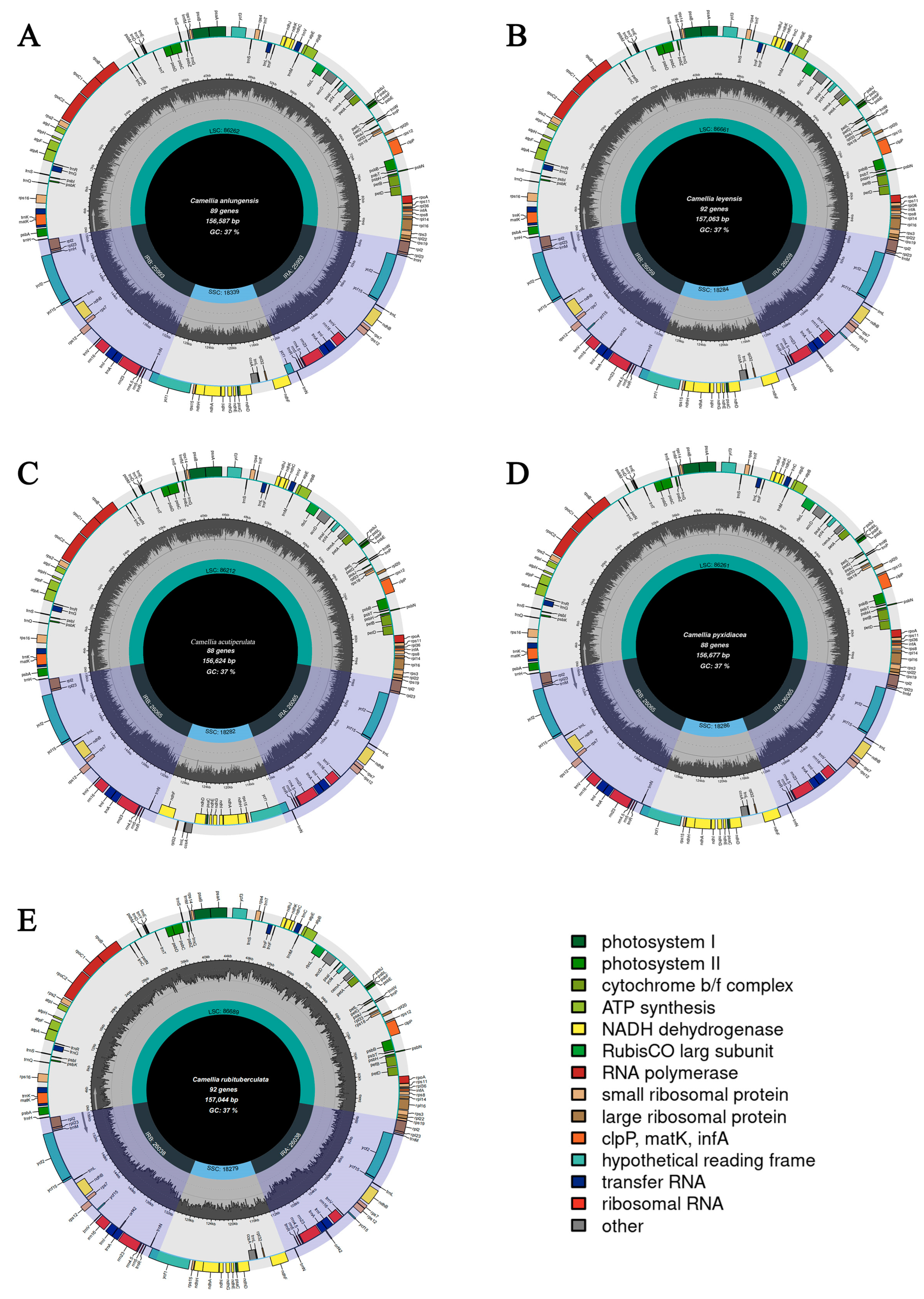
3.3. Features of Chloroplast Genomes

The whole chloroplast genomes of the five species were submitted to the NCBI GenBank, and their accession numbers were obtained (Table 4). A comparison of these chloroplast genomes showed (Figure 4 and Table 4) that their lengths ranged from 156,624 bp to 157,063 bp. They consisted of LSC regions (86,212–86,689 bp), SSC regions (18,276–18,286 bp), and IR regions (51,986–52,130 bp). The total number of genes was 130–136, which included 87–91 CDSs, 35–37 tRNAs, and 8 rRNAs. The chloroplast GC content ranged from 37.30% to 37.33%, and the GC content varied only slightly among the regions, with the IR region having a higher GC content than the other regions.
Although the size of IR regions is more highly conserved than other regions, the contraction and expansion of IR boundaries are thought to play an important role in genome size. Analysis of the contraction and expansion of these IR boundaries revealed (Figure 5) that the rpl2/rps19 genes in these five species had a high degree of conservation with identical gene lengths and locations. However, the ycf1 gene of C. anlungensis was found within the IR region, and the distance between the ndhF gene and the IRb region was less than 56 bp. The trnH gene in C. anlungensis and C. acutiperulata was located in the LSC region, whereas C. leyeensis, C. pyxidiacea, and C. rubituberculata contained tRNA genes within the LSC region.

3.4. Phylogenetic Analysis

A phylogenetic tree was constructed using the maximum likelihood and Bayesian methods on the nineteen cp genomes, which revealed that the five species were clustered on the main branch, consisting of clade I and clade II, with high support values (BS = 100%, PP = 1.0) (Figure 6). C. anlungensis, C. leyeensis, C. pyxidiacea, and C. rubituberculata were on clade I (BS = 58%, PP = 0.62), while C. atuberculata, C. neriifolia, C. acutiperulata, and C. rhytidocarpa were located on clade II. C. anlungensis and C. leyeensis formed an independent clade with a significantly high bootstrap value (BS = 100%, PP = 1.0) (Figure 6). At the same time, the phylogenetic tree constructed based on protein-coding gene sequences showed that five species clustered in one clade of sect. Tuberculata. C. anlungensis and C. leyeensis clustered in one branch. And the remaining three plants are clustered in different branches with high support (Figure 7).

3.5. Taxonomic Treatment

Through field studies, we classified five species of sect. Tuberculata with distinct taxonomic positions, employing comprehensive approaches encompassing morphology, specimen, pollen morphology, and molecular systematic research. The results of this research show that C. leyeensis should be merged with C. anlungensis and categorized as C. anlungensis, while C. acutiperulata, C. pyxidiacea, and C. rubituberculata are regarded as separate species.
  • Camellia anlungensis Chang
Camellia anlungensis H. T. Chang, Tax. Gen. Camellia 48, 1981, in Act. Scl. Nat. Univ. Sunyats; 30 (4): 88, 1991; H. T. Chang and B. Bartholomew. Camellias 69, 1984; Flora of Guizhou 5: 16, pl. 3: 3–4, 1988.
Type: China, Guizhou, Anlong, Guizhou Team, Botanical Institute, Zhang Zhisong, Zhang Yongtian 5952; Ceheng, Luxiong Logging Farm, Cao Ziyu 1227, 1259; Wangmu, Guizhou Team 1757 (Type in Bot. Inst. Acad. Sin Peking) (PE, GZBI) (Figure 8A).
Additional specimens examined: China, Guizhou, Wangmo, Lekang, 400–500 m, 4 November 2021, LZ20211204 (GZAC).
Camellia leyeensis H. T. Chang & Y. C.; Zhong in Act. Scl. Nat. Univ. Sunyats; 30 (4): 89, 1991.
Type: China, Guangxi, Leye, Y. C. Zhong 890806 (SYS, GXFS, KUN) (Figure 8B).
Additional specimens examined: China, Guangxi, Leye, Yachang, 600–750 m, 13 April 2021, LZ20210413, LZ20210414, LZ20210415 (GZAC).
Morphological description: perennial shrub to small tree; the plant height is 2–4 m; the diameter at breast height is 4–10 cm; the trunk is yellowish-brown; the height below the branches is 1.5–3 m; the leaves alternate; the shoots do not have ribs and are glabrous; the leaves are thin, leathery, and obovate, 9.14–12.54 cm long, and 2.78–4.45 cm wide; the apex is sharply acute; the base is broadly cuneate, with light green above, dark obscured, and light yellowish green below, with no black glandular dots; there are 6–9 pairs of lateral veins, curved to the tip of the leaf near the margin and obvious on both the upper and lower surfaces; the margins are sparsely serrulate; the serration is almost symmetrical on both sides; there are 20–31 pairs of serration; the teeth are 2–4 mm apart; the petiole length is 0.4–0.8 cm; the flowers are white; the calyx segments are ovate and hairy, with a single one at the top of the branch, and nearly sessile; there are 9–13 petals; the petals are obovate–oblong, 2.08–2.95 cm long, and 0.97–1.54 cm wide; the outer filament is whorl-free; the anthers are deep yellow; the filaments are creamy white and coherent at base; the ovary is glabrous; there are 3–4 locules and 3–4 styles, 2.4–2.8 cm long; the capsule is subsessile; the carpophore is 0.26–0.32 mm long; the fruit is 1.4–2.5 cm in diameter; the pericarp has many folds and verrucose projections and is 1.2–2.3 mm at its thickest; there is one seed per locule; the seeds are dark brown and lanceolate, semi-ellipsoid, and 13–16 mm; the flowering period is from October to November.
Distribution and habitat: The species is distributed in Lekang Village, Wangmo, Guizhou, China (Figure 9), where most plants grow on slopes and river valleys at 400–500 m above sea level, within mixed-wood forests in the mountains, sometimes forming groups. It is also distributed in Yachang Village, Leye, Guangxi, China, where most plants grow on slopes at 600–750 m above sea level, within mixed-wood forests in the mountains, sometimes forming groups.
  • Camellia autoregulates Chang and C. X. Ye
Camellia acutiperulata H. T. Chang & C. X. Ye in Act. Scl. Nat. Univ. Sunyats, 1984 (2): 79, 1984; 30 (4): 88, 1991, in clavi; Flora of Guangxi 1: 771, 1991.
Type: China, Guangxi, Longlin, Zeng Pei, Xie Qingjian 17023 (SYS) (Figure 8C).
Additional specimens examined: China, Gangxi, Longlin, Jinzhongshan, 800–900 m, 4 November 2022, LZ20221103, LZ20221104, and LZ20221105 (GZAC).
Morphological description: perennial shrub to small tree; the shoots are glabrous; the leaves are thin, leathery, and elliptic; the apex is acute or slightly obtuse, 0.28–13.69 cm long, and 3.32–4.73 cm wide, the base is cuneate to rounded and glabrous, with dry dark green above and slightly shiny green below; there are 6–7 pairs of lateral veins, all slightly elevated on the abaxial surface; the margins are sparsely serrulate or subentire in the lower half; the petiole length is 5–10 mm; the flowers are white; there are 1–2 axillary sessile leaves and 5–6 bracts, 3–5 mm long; the apex is pointed, glabrous, or midrib pilose; there are 5–7 sepals that are broadly ovate and 1.4–1.7 cm long; the margin is membranous, the apex is pointed, glabrous, or long velutinous at the tip; there are 9–12 petals, which are elliptic, 3.4–4.0 cm long, and 1.3–1.9 cm wide; the filaments are glabrous; the outer whorls are nearly free or slightly connate; the ovary is glabrous; there are 3–4 glabrous locules, and 3–4, free styles, 2.0–2.3 cm long; the capsule is subglobose and 1.3–2.1 cm in diameter; the pericarp is approximately 3.1–4.3 mm thick, verruculose, and raised; there are 1–2 seeds per locule, which are orbicular, bald, and glabrous; the flowering period is from November to December.
Distribution and habitat: This species is distributed in Longlin, Guangxi, China (Figure 9), where most plants grow on steep slopes at 800–900 m above sea level, within mixed-wood forests in the mountains, sometimes forming small groups.
  • Camellia pyxidiacea Xu, F. P. Chen and C. Y. Deng
Camellia pyxidiacea Xu, F. P. Chen & C. Y. Deng in Guihaia 7(1): 19–21, 1987; H. T. Chang in Act. Sci. Nat. Univ. Sunyats; 30 (4): 88; 1991, in clavi.
Type: China, Yunnan, Luoping, Xu Zhaoran SJ5225 (SYS) (Figure 8D).
Additional specimens examined: China, Yunnan, Luoping, Yehou Valley, 800–900 m, 4 December 2021; LZ20211204, LZ20211205, and LZ20211206 (GZAC).
Morphological description: shrub to small tree; the shoots are glabrous and brown; the leaves are leathery, elliptic, 8.14–13.08 cm long, and 3.94–4.88 cm wide; the apex is neo-acute; the margins are serrulate, with dry olive-green above, slightly glossy, and glabrous, and yellowish-green and glabrous underneath; the primary veins are slightly convex above and distinctly elevated below; there are 8–10 pairs lateral veins, which are conspicuous above and slightly convex below; reticulate veins are conspicuous on the leaf surface, with a petiole length of 6–12 mm, and glabrous; there is a single white flower axillary near the tip; the petal apex is pink; the 7–9 calyces are larger, hairy at the apex, semi-orbicular, or ovate, 3–6 mm long, and 4–9 mm wide; there are 6–8 petals, which are obovate, apically lobed, basally coherent, 2.43–2.88 cm long and 0.95–1.58 cm wide; the petal apex is hairy; the filaments are white, yellow, and glabrous; there are 3–4 styles, which are slender and 2.5–3.4 cm long; the capsule is subglobose, and 4.84–5.18 cm in diameter; the ovary has 3–4 locules; the pericarp is longitudinally lobed, the surface is densely tuberculate and wrinkled, with transverse deep grooves; the pericarp is often dehiscent along these transverse grooves; the seeds are hairy and triangular–orbicular or sub-semi-orbicular; the flowering period is in October; fruit ripening occurs in the following year in October–November.
Distribution and habitat: This species is distributed in Yehou Valley, Luoping, Yunnan, China (Figure 9), where most plants grow on steep slopes at 800–900 m above sea level, within mixed-wood forests in the mountains, sometimes forming groups.
  • Camellia rubituberculata Chang
Camellia rubituberculata Chang, Lin & Liu, in Act. Scl. Nat. Univ. Sunyatseni, 23(2): 82, 1984; H. T. Chang; 30 (4): 88. 1991, in clavi.; Flora of Guizhou 5: 10; 1988.
Type: China, Guizhou, Qinglong, Lin Mengjia, and Lu Qiming 155710 (SYS, GZTI) (Figure 8E).
Additional specimens examined: China, Guizhou, Qinglong, Longtou Village, 1200–130 m, 11 April 2021; LZ20210411, LZ20210412, and LZ20210413 (GZAC).
Morphological description: perennial shrub to subtree 5–10 m high; the annual shoots are glabrous; the leaves are thin, leathery, long ovate, 10.1–11.4 cm long, and 3.5–4.8 cm wide; the blade apex is slightly pointed or acute mucronate; the base is broadly cuneate; the overall appearance of the leaf blade is olive green, glabrous, and dark green adaxially, with 7–8 pairs of lateral veins clearly visible on both sides; the flowers are red, terminal or axillary, and 4–7 cm in diameter; the bracts are persistent with imbricate; there are 6–8 bracts, which are semi-orbicular or suborbicular, 2.79–3.25 cm long, 2.15–3.24 cm wide, and basally connate; the petal apices are shallowly columnar; the outer filament is whorl-free; the anthers are dark yellow; the filaments are creamy white, basally connate, and united with petals; the capsule is verrucose globose; the seed coat is 3-4 cleft, with a thickness of 1.22 cm; the fruit diameter is 4.84–5.18 cm; the ovary has 3–4 locules, containing 3–11 seeds, and is mostly triangular, ovate, obovate, or trapezoidal–obovate; the seeds are brown, triangular–orbicular, or sub-semi-orbicular; the flowering period is from October to November.
Distribution and habitat: This species is distributed in Longtou Village, Qinglong, Guizhou, China (Figure 9), and most plants grow on slopes at 1200–1300 m above sea level, within mixed-wood forests in the mountains, sometimes forming groups.

4. Discussion

Many taxonomic conflicts are caused by differences in definition or incomplete taxonomic studies. We conducted extensive field surveys and specimen studies for the sect. Tuberculata. A series of morphological data were measured at the population level, including the leaves, fruit, and other morphological characteristics. The results show that C. pyxidiacea and C. rubituberculata have differences in flower color, the numbers of petals and stigmas, capsule morphology, etc., which supports Min and Chang et al. [3,4,5] views that there are significant morphological differences between the two species. At the same time, the above morphological characteristics are very similar between C. anlungensis and C. leyeensis. It is noteworthy that we also found some other morphological features that have not previously been given attention, such as stigmas, trunks, and pericarp thickness, which also have certain taxonomic values among species. Thus, we found that plant species with leaf shape, petiole length, and fruit size were rather stable between populations, suggesting that these traits are key features of the plant group.
Although the study of pollen morphology is becoming more common, most researchers have only studied the external morphology of pollen and have seldom studied its internal morphology. The morphology of the pollen and analysis showed that the differences in pollen morphology at the germination furrow, polar, and equatorial axes were not significant among the five species, while some differences were detected. For example, C. anlungensis had the largest values at the polar axis, equatorial axis, and emergence groove; the outer wall ornamentation of C. rubituberculata and C. pyxidiacea was crinkled and wavy, whereas that of C. anlungensis, C. acutiperulata, and C. leyeensis was crinkled and wavy to granular [30,31]. The study of pollen grain morphology can not only aid in the characterization of species morphology but also provide strong evidence for species classification.
The whole chloroplast genome not only enriches the molecular phylogenetic database of wild plant resources but also plays an important role in evaluating the genetics and evolution of species and their origins [11]. The results of the chloroplast genome phylogenetic trees show that C. anlungensis and C. leyeensis were merged (BS = 100%, PP = 1.0), but C. pyxidiacea, C. rubituberculata, and C. acutiperulata were located on different branches, further supporting that they should be treated as independent species. In addition, in the field investigation, we found that some species with close geographical distribution may have resulted in natural hybridization. Therefore, we will increase the study of nuclear gene fragments in the next study to analyze their network relationships. Based on our fieldwork, the Panjiang Basin of China was the most concentrated and dominant distribution area for the five species. These sites highly overlap, and it is hypothesized that a high degree of natural hybridization may exist. The two species may have evolved over different periods.
Based on the above research, we consider that C. leyeensis and C. anlungensis should be merged, supporting the view of Min et al. [3]. Furthermore, C. acutiperulata, C. pyxidiacea, and C. rubituberculata should be restored to the status of separate species, supporting the view of Chang et al. [5].

5. Conclusions

In this study, morphology, pollen morphology, and chloroplast genomes were analyzed to investigate five species of sect. Tuberculata in the Panjiang Basin of China. The results showed that C. leyeensis should be treated under the single name of C. anlungensis, while C. acutiperulata, C. pyxidiacea, and C. rubituberculata should be regarded as independent species. This study not only increases our understanding of Camellia species within the distribution area of the Panjiang Basin but also clarifies the classification of these plants in sect. Tuberculata, providing a foundation for resource development and protection.

Author Contributions

Conceptualization, X.X. and Z.L.; methodology, X.X. and Z.R.; software, L.H. and X.X.; validation, Z.L. and M.T.; investigation, Z.R. and X.X.; resources, C.Y.; data processing, Z.R.; writing—original draft preparation, X.X. and Z.R.; writing—review and editing, Z.L., Z.R. and X.X.; visualization, Z.L.; supervision, M.T. and Z.L.; project administration, Z.L.; funding acquisition, Z.L. All authors have read and agreed to the published version of the manuscript.

Funding

This work was supported by the National Natural Science Foundation of China (32400179) and the Guizhou Provincial Basic Research Program (Natural Science) 2022 (072).

Data Availability Statement

GenBank accession numbers: OQ556869, OP0586859, NC_050354, MZ766253, and OK046127.

Conflicts of Interest

The authors declare no conflicts of interest.

References

  1. Chang, H.T. A Taxonomy of the Genus Camellia; Editorial Department of Journal of Sun Yatsen University: Guangzhou, China, 1981; pp. 47–52. [Google Scholar]
  2. Chien, S.S. Four new ligneous plants of Szechuan. Contrib. Biol. Lab. Sci. Soc. China Bot. 1939, 12, 89–100. [Google Scholar]
  3. Min, T.L.; Zhong, Y.C. A revision of genus Camellia sect. Tuberculata. Acta Bot. Yunnanica 1993, 15, 123–130. [Google Scholar]
  4. Min, T.L. A systematic synopsis of the genus Camellia. Acta Bot. Yunnanica 1999, 21, 149–159. [Google Scholar]
  5. Chang, H.T.; Ren, S.X. Diagnosis on the systematic development of Camellia VI. revised on sect. Tuberculata of Camellia. Suppl. J. Sun Yatsen Univ. 1996, 2, 55–60. [Google Scholar]
  6. Chang, H.T. New record of Camellia from South China. Acta Sci. Nat. Univ. Sun Yatseni 1984, 23, 77–82. [Google Scholar]
  7. Sealy, J.R. A Revision of the Genus Camellia; The Royal Horticulture Society: London, UK, 1958; pp. 1–239. [Google Scholar]
  8. Jiang, B.; Peng, Q.F.; Shen, Z.G.; Moller, M.; Pi, E.X.; Lu, H.F. Taxonomic treatments of Camellia (Theaceae) species with secretory structures based on integrated leaf characters. Plant Syst. Evol. 2010, 290, 1–20. [Google Scholar] [CrossRef]
  9. Jiang, Z.D. Preliminary Study of Molecular Phylogenetics and Biogeography of the Genus Camellia L. Based on Chloroplast DNA. Ph.D. Thesis, Zhejiang Sci-Tech University, Hangzhou, China, 2017. [Google Scholar]
  10. Wu, Q.; Tong, W.; Zhao, H.J.; Ge, R.H.; Li, R.P.; Huang, J.; Li, F.D. Comparative transcriptomic analysis unveils the deep phylogeny and secondary metabolite evolution of 116 Camellia plants. Plant J. 2022, 111, 406–421. [Google Scholar] [CrossRef]
  11. Xiao, X. Revision of the Classification and Comprehensive Evaluation of the Ornamental Value of Plants in the Camellia L. Sect. Tuberculate. Master’s Thesis, Guizhou University, Guiyang, China, 2023. [Google Scholar]
  12. Ran, Z.H.; Li, Z.; Xiao, X.; An, M.T.; Yan, C. Complete chloroplast genomes of 13 species of sect. Tuberculata Chang (Camellia L.): Genomic features, comparative analysis, and phylogenetic relationships. BMC Genom. 2024, 25, 108. [Google Scholar] [CrossRef]
  13. Lin, M.J.; Lu, Q.M. New record of Guizhou Camellia. Act. Sci. Nat. Univ. Sunyats. 1984, 2, 81–83. [Google Scholar]
  14. Xu, Z.R.; Chen, F.; Deng, C.Y. A new species of sect. Tuberculata. Guihaia 1987, 19–21. [Google Scholar]
  15. Chang, H.T.; Ren, S.X. A classification on the section Tuberculata of Camellia. Act. Sci. Nat. Univ. Sunyats. 1991, 30, 86–91. [Google Scholar]
  16. Erdtman, G. Handbook of Palynology; Science Press: Beijing, China, 1978. [Google Scholar]
  17. Wang, K.F.; Wang, X.Z. Introduction to Sporology; Peking University Press: Beijing, China, 1983. [Google Scholar]
  18. Halbritter, D.D.; Ulrich, D.S.; Grímsson, D.F.; Weber, P.D.; Zetter, P.D.; Hesse, P.D.; Buchner, D.R.; Svojtka, M.M.; Frosch-Radivo, A. Illustrated Pollen Terminology; Cambridge International Law Journal: Cambridge, UK, 2018. [Google Scholar]
  19. Li, J.L.; Wang, S.; Yu, J.; Wang, L.; Zhou, S.L. A modified CTAB protocol for plant DNA extraction. Chin. Bull. Bot. 2013, 48, 72–78. [Google Scholar]
  20. Bolger, A.M.; Lohse, M.; Usadel, B. Trimmomatic: A flexible trimmer for Illumina sequence data. Bioinformatics 2014, 30, 2114–2120. [Google Scholar] [CrossRef]
  21. Jin, J.J.; Yu, W.B.; Yang, J.B.; Song, Y.; Li, D.Z. GetOrganelle: A fast and versatile toolkit for accurate de novo assembly of organelle genomes. Genome Biol. 2020, 21, 241. [Google Scholar] [CrossRef]
  22. Lohse, M.; Drechsel, O.; Bock, R. Organellar Genome DRAW (OGDRAW): A tool for the easy generation of highquality custom graphical maps of plastid and mitochondrial genome. Curr. Genet. 2007, 52, 267–274. [Google Scholar] [CrossRef]
  23. Amyiryousefi, A.; Hyvonen, J.; Poczai, P. IRscope: An online program to visualize the junction sites of chloroplast genomes. Bioinformatics 2018, 34, 3030–3031. [Google Scholar] [CrossRef]
  24. Katoh, K.; Standley, D.M. MAFFT: Iterative refinement and additional methods. Methods Mol. Biol. 2014, 1079, 131–146. [Google Scholar]
  25. Nguyen, L.T.; Schmidt, H.A.; Haeseler, A.; Minh, B.Q. IQTREE: A fast and effective stochastic algorithm for estimating maximum-likelihood phylogenies. Mol. Biol. Evol. 2015, 32, 268–274. [Google Scholar] [CrossRef]
  26. Kumar, S.; Stecher, G.; Li, M.; Knyaz, C.; Tamura, K. MEGA X: Molecular evolutionary genetics analysis across computing platforms. Mol. Biol. Evol. 2018, 35, 1547–1549. [Google Scholar] [CrossRef]
  27. Huelsenbeck, J.P.; Ronquist, F. MRBAYES: Bayesian inference of phylogenetic trees. Bioinformatics 2001, 17, 754–755. [Google Scholar] [CrossRef]
  28. Letunic, I.; Bork, P. Interactive Tree of Life (iTOL) v5, an online tool for phylogenetic tree display and annotation. Nucleic Acids Res. 2021, 49, 293–296. [Google Scholar] [CrossRef]
  29. Efroni, I.; Eshed, Y.; Lifschitz, E. Morphogenesis of simple and compound leaves: A critical review. Plant Cell 2010, 22, 1019–1032. [Google Scholar] [CrossRef]
  30. Wei, Z.X.; Zavada, M.S.; Min, T.L. Pollen morphology of Camellia (Theaceae) and ITS taxonomic significance. Acta Bot. Yunnanica. 1992, 14, 275–282. [Google Scholar]
  31. Hu, Z.M.; Zhao, C.H.; Zhao, Y.Y.; Liu, J.X. Pollen morphology of Liliaceae and its systematic significance. Palynology 2021, 45, 531–568. [Google Scholar] [CrossRef]
Figure 1. Main morphological characteristics of five plants of sect. Tuberculata ((A) C. anlungensis, (B) C. leyeensis, (C) C. acutiperulata, (D) C. pyxidiacea and (E) C. rubituberculata; 1. habitat, 2. flower, 3. fruit, 4. petals, 5. calyxes, 6. pistils).
Forests 15 01718 g001
Figure 2. Main morphological characteristics of fruit and leaves five plants of sect. Tuberculata ((A) C. anlungensis, (B) C. leyeensis, (C) C. acutiperulata, (D) C. pyxidiacea, and (E) C. rubituberculata; 1. capsules, 2. seeds, 3. leaves).
Forests 15 01718 g002
Figure 3. Scanning electron microscopic observation of the five species pollen morphology of sect. Tuberculata ((A) C. anlungensis, (B) C. leyeensis, (C) C. acutiperulata, (D) C. pyxidiacea, (E) C. rubituberculata; 1. equatorial view, 2. polar view, 3. pollen aperture, 4. pollen wall ornamentation).
Forests 15 01718 g003
Figure 4. Gene map of the five species chloroplast gemome of sect. Tuberculata. ((A) C. anlungensis, (B) C. leyeensis, (C) C. acutiperulata, (D) C. pyxidiacea, (E) C. rubituberculata).
Forests 15 01718 g004
Figure 5. Analysis of the cp genome IR region boundaries of five species.
Forests 15 01718 g005
Figure 6. Phylogenetic evolutionary tree of cp genomes (Maximum Likelihood (ML) and Bayesian (BI) trees, BS ≥ 50% and PP ≥ 0.95 are indicated above branches as BS/PP; Species marked in red are target species).
Forests 15 01718 g006
Figure 7. Construction of a phylogenetic tree based on the PCGs (Maximum Likelihood (ML) and Bayesian (BI) trees, BS ≥ 50% and PP ≥ 0.95 are indicated above branches as BS/PP; Species marked in red are target species).
Forests 15 01718 g007
Figure 8. Information on the type specimen of five species.
Forests 15 01718 g008
Figure 9. Distribution map of five plant species in the Panjiang River Basin in China.
Forests 15 01718 g009
Table 1. Plant collection information for the five species of sect. Tuberculata.
Species NameLocationSpecimen NumberElevation (m)Longitude and Latitude
C. anlungensisWangmo County, Guizhou, ChinaGZAC, LZ20211204449106°16′40.69″ E, 25°2′51.65″ N
C. leyeensisLeye County, Guangxi, ChinaGZAC, LZ20210413684106°17′31.48″ E, 24°51′29.98″ N
C. acutiperulataLonglin County, Guangxi, ChinaGZAC, LZ20221103851104°52′39.66″ E, 24°39′31.59″ N
C. pyxidiaceaXingyi City, Guizhou, ChinaGZAC, LZ20211204837104°32′18.18″ E, 24°47′53.12″ N
C. rubituberculataXingren County, Guizhou, ChinaGZAC, LZ202104111297105°21′08.10″ E, 25°43′50.08″ N
Table 2. Comparison of morphological characteristics of five species of sect. Tuberculata.
TraitC. anlungensisC. leyeensisC. acutiperulataC. pyxidiaceaC. rubituberculata
Leaf typeLeaves thin, leathery, obovateLeaves thin, leathery, obovateLeaves thin, coriaceous, ellipticLeaves thin, coriaceous, ellipticLeaves thin coriaceous, elliptic
Length (cm)9.14–12.549.95–12.3110.28–13.698.14–13.0810.08–11.43
Width (cm)2.78–4.452.79–3.853.32–4.733.94–4.883.51–4.82
Flower typeObovateObovateEllipticEllipticElliptic
ColorWhiteWhiteWhitePink petal apexesRed
Length (cm)2.08–2.952.27–3.103.44–4.012.43–2.882.79–3.25
Width (cm)0.97–1.540.93–1.631.35–1.940.95–1.582.15–3.24
Number of petals11–1310–129–126–96–8
SepalOvateOvateOvate, apex pointed, margin membranousOvateSemicircular
Number of calyces5–75–85–76–87–9
Fruit shapeCapsule verrucose, subglobose, epidermis with verrucose projectionsCapsule verrucose, subglobose, epidermis with verrucose projectionsCapsule verrucose, subglobose, testa transversely lobedFruit-compressed globose, skin with verrucose protuberance and obvious partingCapsule verrucose, globose, seed coat shallowly undulated
Diameter (cm)1.4–2.041.35–1.91.3–2.12.65–3.814.84–5.18
Shell thickness (mm)6.3–7.75.8–7.33.1–4.33.2–0.6712.2–14.1
Table 3. Comparison of pollen micromorphological characteristics of five species of sect. Tuberculata.
SpeciesPollen ShapePolar ViewEquatorial ViewPolar Axis (P)/μmEquatorial Axis E/μmGermination PoresPollen AperturePollen Wall OrnamentationRidge
Long/μmWide/μm
C. anlungensisoblate sphericalTriangularOval30.21–32.2736.09–37.8826.83–27.757.14–7.75ColpusUnevenWavy
C. leyeensisoblate sphericalTriangularOval29.41–31.6734.74–36.5126.94–27.736.73–7.42ColpusUnevenWavy
C. acutiperulataoblate sphericalTriangularOval27.54–28.5133.66–35.4724.54–25.126.71–7.13ColpusUnevenWavy
C. pyxidiaceaoblate sphericalTriangularOval30.91–31.8231.87–33.2122.11–23.176.59–6.97ColpusUnevenWavy
C. rubituberculataoblate sphericalOvalOval28.27–29.8432.69–34.5726.85–27.358.28–8.74ColpusUnevenWavy
Table 4. Summary of complete chloroplast genomes of five species of sect. Tuberculata.
C. acutiperulataC. pyxidiaceaC. anlungensisC. rubituberculataC. leyeensis
Genome size (bp)156,624156,677156,587157,044157,063
GC (%)37.3337.3337.3337.3137.30
LSC size (bp)86,21286,26186,26286,68986,661
SSC size (bp)18,28618,28318,28118,27918,276
IR size (bp)52,13052,13051,98652,07652,118
GC in LSC (%)35.3635.3535.3135.3135.31
GC in SSC (%)30.5930.6230.6030.6230.60
GC in IR (%)42.9542.9542.9642.9842.96
GC in CDS (%)37.6137.5337.5437.6537.55
1st position GC (%)45.3745.1945.2445.4245.26
2nd position GC (%)38.0437.9437.9738.0037.97
3rd position GC (%)29.4329.4329.4029.5329.40
Length of CDS79,50079,76779,67180,15580,175
Number of genes130132134136136
Number of CDS8787899191
Number of tRNAs3537373737
Number of rRNAs88888
GenBank IDOQ556869OP058659NC_050354MZ766253OK046127
Disclaimer/Publisher’s Note: The statements, opinions and data contained in all publications are solely those of the individual author(s) and contributor(s) and not of MDPI and/or the editor(s). MDPI and/or the editor(s) disclaim responsibility for any injury to people or property resulting from any ideas, methods, instructions or products referred to in the content.

