Article

Comparative Study on Vascular Bundle Morphological Characteristics of Parts of Branches, Culms, and Rhizomes of Oligostachyum sulcatum

1
School of Materials and Chemistry, Anhui Agricultural University, Hefei 230036, China
2
Institute of New Bamboo and Rattan Based Biomaterials, International Center for Bamboo and Rattan, Beijing 100102, China
3
Key Laboratory of National Forestry and Grassland Administration, Beijing for Bamboo and Rattan Science and Technology, Beijing 100102, China
4
School of Physical Science and Technology, ShanghaiTech University, 393 Middle Huaxia Road, Shanghai 201210, China
*
Authors to whom correspondence should be addressed.
Forests 2024, 15(10), 1752; https://doi.org/10.3390/f15101752 (registering DOI)
Submission received: 30 August 2024 / Revised: 30 September 2024 / Accepted: 3 October 2024 / Published: 5 October 2024
(This article belongs to the Section Forest Ecophysiology and Biology)

Abstract

:
This study presents a comprehensive analysis of the vascular bundle morphology, tangential and radial diameters, and distribution frequency of different parts of Oligostachyum sulcatum, elucidating their structural and functional significance. Electron microscopy images revealed distinct vascular bundle characteristics in the different parts, including the vascular bundles in both parts of the rhizomes, the middle parts of the internodes, and the middle and inner parts of the branches, which were semi-open. The vascular bundles in the outer parts of both internodes and branches were semi-differentiated and undifferentiated. The vascular bundles in the inner parts of internodes were open. Statistical analysis showed significant variations in tangential and radial diameters among these parts, reflecting their diverse mechanical and physiological functions. The internodes exhibited the largest tangential and radial diameters, suggesting a critical role in mechanical support. In contrast, the branches had the smallest diameter, indicating that vascular bundle differentiation is influenced by growth conditions. The vascular bundle frequency was the highest in branches and the lowest in inside-sticks. This study provides theoretical references for the adaptive strategies and growth regulation mechanisms of O. sulcatum.

1. Introduction

Oligostachyum sulcatum Z. P. Wang et G. H. Ye, a bamboo species belonging to the subfamily Bambusoideae of the family Poaceae, possesses a monopodial rhizome system. It is an endemic species in China [1,2,3]. Compared to wood, O. sulcatum possesses high strength, good toughness, and great hardness, making it an ideal candidate for engineering materials [4]. Additionally, O. sulcatum can also be used in the production of paper [5], activated carbon [6], decorative bamboo flooring, and other bamboo composite panels [7,8], which enhances its economic value. It has a short growth cycle and high yield, which can effectively alleviate the imbalance between the supply and demand of wood. It can also be used as a substitute for plastic products to reduce the use of these environmental pollutants [9].
Compared to other bamboo species, O. sulcatum exhibits some unique characteristics. Current research on O. sulcatum primarily focuses on its biology, ecology, high-yield cultivation techniques, and its use in the form of bamboo shoots [10,11]. However, there is relatively little research on the anatomical characteristics of O. sulcatum, which influence to the production and application of bamboo materials. The anatomical structure of bamboo mainly includes the vascular bundle system and parenchyma tissues [12]. In-depth studies of these anatomical structures can provide a comprehensive understanding of the characteristics of O. sulcatum, thereby providing a theoretical basis for its scientific and rational development and application. In the study of bamboo microstructures, the tangential and radial diameters of bamboo vascular bundles are directly related to the load-bearing capacity of bamboo. They significantly influence the mechanical properties of bamboo [13,14,15,16], which are particularly important in structural applications. Moreover, the distribution frequency of vascular bundles is another crucial indicator. This has a significant impact on the mechanical properties, structural stability, processing adaptability, and macroscopic physical properties of bamboo. A higher vascular bundle distribution frequency correlates with better mechanical properties [16,17], which is particularly important in applications with high structural requirements, such as construction and furniture manufacturing. In summary, the tangential diameter, radial diameter, and distribution frequency of vascular bundles are indispensable key factors in the microstructure of bamboo. They play a decisive role in the comprehensive performance and application potential of bamboo [18]. Furthermore, the basic morphological characteristics of bamboo vascular bundles are of great significance for bamboo classification. Grosser et al. [19] classified bamboo vascular bundles into four types: double broken-waist (type IV), broken-waist (type III), tight-waist (type II), and open types (type I). Recently, domestic scholars [20,21,22] have further classified vascular bundles into seven types: double broken-waist, broken-waist, tight-waist, open, semi-open types, semi-differentiated, and undifferentiated. However, research on the anatomical structure of vascular bundles has mostly been limited to classification, and there has been a lack of comparative studies on the morphological characteristics of vascular bundles in different parts of the same bamboo species.
Therefore, in this study, the tangential diameter, radial diameter, and distribution frequency of vascular bundles in the branches, culms, and rhizomes of O. sulcatum were observed and statistically analyzed. The vascular bundles in different parts were classified, and a Z-test was used to analyze the differences in diameters and distribution frequency of vascular bundles in different parts. This study provides theoretical references for the adaptive strategies and growth regulation mechanisms of O. sulcatum.

2. Materials and Methods

2.1. Materials and Sampling Methods

The O. sulcatum samples used in this study were collected from the International Bamboo and Rattan Center at the Taiping Experimental Center, Huangshan, Anhui, China (longitude 117′13′′ to 118′53′′ E, latitude 29′24′′ to 30′31′′ N). Three-year-old bamboo plants were sampled and labeled as No. 1, No. 2, and No. 3. The sampling record sheet of O. sulcatum is shown in Table 1.
Square blocks were cut from the different parts of the collected O. sulcatum. The dimensions of the bamboo block were 15 mm in length (longitudinal direction), 20 mm in width (tangential direction), and t mm in thickness (radial direction, corresponding to the bamboo culm wall thickness). These parts included the coming parts of the rhizome, the going parts of the rhizome, the internode at 1.5 m, the inside-stick at 1.5 m, and the branch (middle living branch). The rhizome extending from the mother bamboo is referred to as the coming parts of the rhizome, while the rhizome extending from the offspring bamboo is referred to as the going parts of the rhizome. The internode at 1.5 m refers to the section of the bamboo culm, or stem, between two nodes at a height of 1.5 m from the ground. The inside-stick at 1.5 m is the solid part within the culm, positioned just at the node at the same height or slightly higher. Additionally, a segment was taken from the third internode of the middle living branch. The specific locations of these sampling points are illustrated in Figure 1.

2.2. Measurement of Vascular Bundle Size and Frequency

The prepared samples were submerged in a beaker filled with water, which was then placed in a microwave (M1-L213 B, Midea, Hefei, China, 700 W output power). The samples were heated at 90 °C for 10 min, removed, and then cooled with fresh water for 2 min. This process was repeated more than 30 times until the samples sank to the bottom of the beaker, indicating successful softening [23]. Using a rotary microtome (KD-2508, Kedee, Jinhua, China), 30 μm thick sections were cut from the coming parts of the rhizome, the going parts of the rhizome, the internode at 1.5 m, the inside-stick, and the branch. The sections were then stained with 1% safranin solution, decolorized with distilled water, dehydrated with alcohol, clarified with xylene, and mounted with Canada balsam. Finally, the slides were labeled for examination.
The prepared sections were observed under an optical microscope (Nikon Eclipse E200, Nikon, Tokyo, Japan) using an MD90 microscope camera attached to the eyepiece lens. The eyepiece magnification was 10× and the objective magnification was 4× (Nikon E plan achromat 4×, WD 30), resulting in a total magnification of 40×. The radial and tangential diameters of the vascular bundles were measured and recorded using the Mshot image analysis system (version 9. 3. 3. 1). The system is an integrated hardware and software solution designed for professional microscope digital imaging applications. Its design focuses on high performance and user-friendliness. The system controls the camera through software, enabling functions such as real-time photography, video recording, measurement and statistics, real-time fluorescence multi-channel synthesis, and image processing. The number of vascular bundles within a 1 mm2 was counted to determine the distribution frequency [24] (a vascular bundle entirely within the selected area counted as 1, intersecting one edge counted as 0.5, and intersecting a corner counted as 0.25).

2.3. Scanning Electron Microscopy (SEM) Observation of Micromorphology

Samples sized 5 mm × 5 mm × 5 mm were placed in a beaker filled with water and microwaved for 15 min (G80W23CSP-Z, Galanz, Foshan, China) under 90 °C. The samples were then polished on the cross-section using a sliding microtome (Leica SM2010R, Leica, Wetzlar, Germany; A-35, Feather microtome blades, Feather Safety Razor Co., Ltd., Osaka, Japan). Before gold coating, the samples were dehydrated to the critical point and then sputter-coated with gold for 90 s in a vacuum chamber using a sputter coater at an 8 mA current (E-1010, Quorum Technologies, Emsworth, UK). The polished cross-sections were observed under a scanning electron microscope (GeminiSEM 360, Carl Zeiss, Oberkochen, Germany) at an accelerating voltage of 3 kV.

2.4. Feature Analysis

A normality test was conducted on the collected data using IBM SPSS Statistics (version 27. 0. 1. 0) (90 sets of data collected from 3 samples, with 30 observations and measurements for each sample). The results indicated that the data conformed to the normal distribution. Since the sample size was greater than 30 and the data conformed to a normal distribution, the Z-test for two-sample variance analysis [25] in Microsoft Excel (version 16. 0. 17928. 20114) was introduced to assess the significance of differences in the morphological characteristics of vascular bundles between different parts of the same O. sulcatum specimen. This aimed to compare the variances in tangential diameter, radial diameter, and distribution frequency among different parts. For each pairwise comparison, the null hypothesis stated no difference in the variable, while the alternative hypothesis stated a difference in the variable between the two parts. The Z-value between the two parts was calculated using Equation (1). The p-value associated with the Z-value was determined using the standard normal distribution table. A p-value less than 0.01 indicated a highly significant difference, less than 0.05 indicated a significant difference, and greater than 0.05 indicated no significant difference.
Z = x ¯ 1 x ¯ 2 σ 1 2 n 1 + σ 2 2 n 2
Notes: x1 and x2 represent the average value of the two parts, σ1 and σ2 represent the variance of the two parts, and n1 and n2 represent the number of samples of the two parts.

3. Results

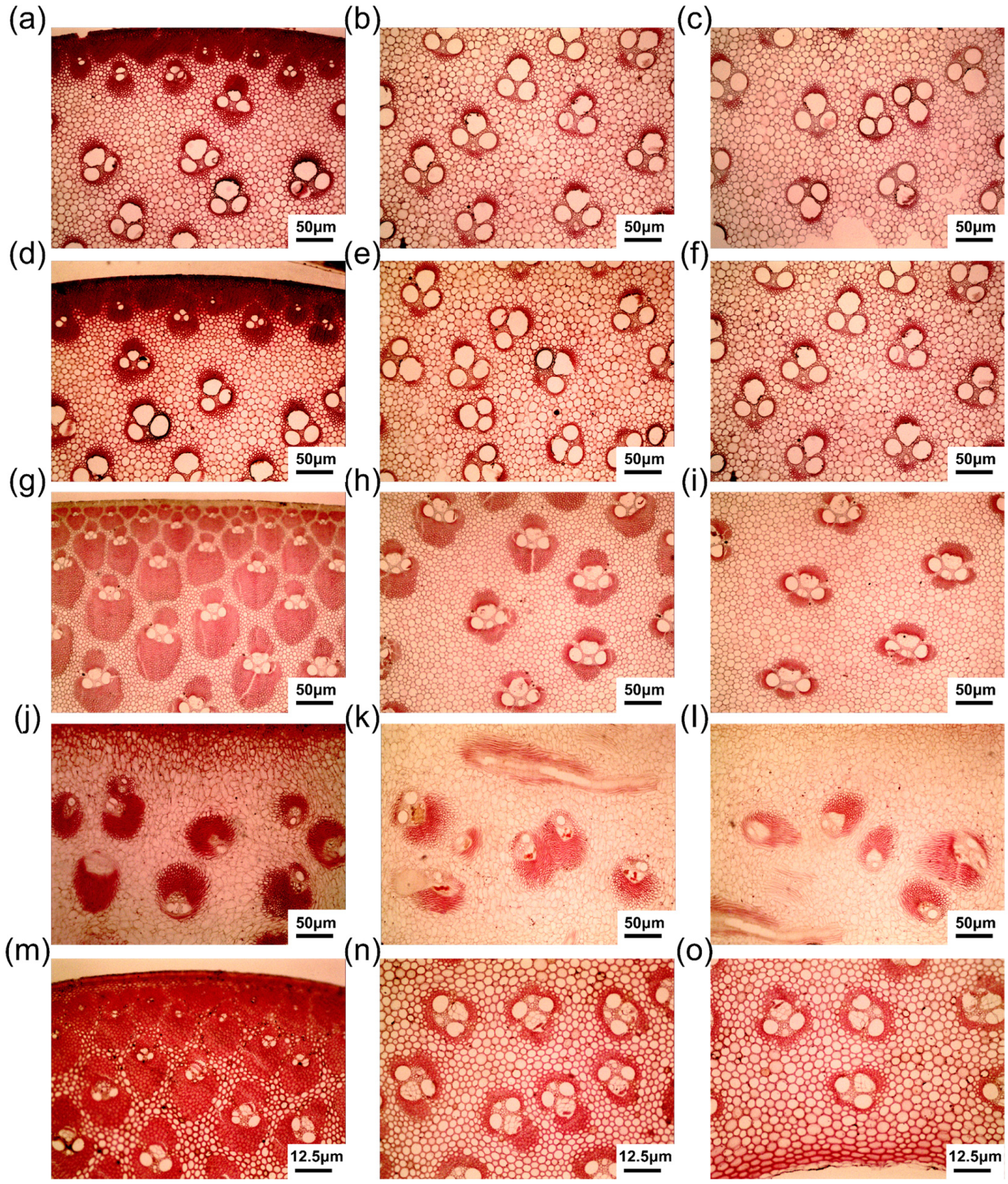
3.1. Radial Variation in Vascular Bundle Morphological Characteristics

The vascular bundle morphology in different parts of O. sulcatum exhibits radial variability (except for the inside-stick). The regions near the outer side of the bamboo culm are referred to as bamboo green, whereas the regions near the inner side of the culm are called bamboo yellow.
As shown in Figure 2a, the tangential diameter of the vascular bundles gradually increases from the outer to the inner parts along the radial direction of the bamboo wall. The tangential diameter of the vascular bundles in the internodes first increases sharply and then rises slowly from the outer to the inner parts of the bamboo (Figure 2a). The increasing trend of tangential diameter in the vascular bundles is approximately the same for both rhizomes (Figure 2a).
Unlike the tangential diameter, the radial diameter of the vascular bundles in different parts shows significant variations along the radial direction (Figure 2b). In the internodes, the radial diameter of vascular bundles gradually decreases from the outer to the inner parts along the radial direction of the bamboo wall (Figure 2b). This is mainly attributed to the large inner fiber groups in the vascular bundles of the bamboo green (Figure 3g), which significantly increases the radial diameter of the vascular bundles, while the bamboo yellow only has four smaller fiber sheaths (a protective layer composed of fibers) (Figure 3i). Along the radial direction of the bamboo wall, the radial diameter of the vascular bundles in the coming parts of the rhizomes gradually increases from the outer to the inner parts, while in branches and the going parts of the rhizomes, they first increase and then decrease (Figure 2b). Notably, both parts of the rhizomes show a gradual increase in the radial diameters of vascular bundles from the outer to the middle parts, which may be related to their sieve tubes. The underdeveloped vascular bundles in the rhizomes result in smaller fiber sheaths, so the size of sieve tubes (tubular structures in the primary phloem) significantly affects the radial diameter of the vascular bundles (Figure 3a–f).
The distribution frequency of the vascular bundles gradually decreases from the outer to the inner parts along the radial direction of the bamboo wall in different parts (Figure 2c). The vascular bundle distribution frequency in the internodes first decreases sharply and then declines slowly from the outer to the inner parts (Figure 2c). The decreasing trend of vascular bundle distribution frequency in other parts is more gradual (Figure 2c), indicating a relatively uniform distribution of the vascular bundles in the bamboo wall of these parts.
In conclusion, as evidenced by Figure 2 and Figure 3, the radial variations in vascular bundles exhibit significant differences among different parts of O. sulcatum.

3.2. Comparative Analysis of Vascular Bundle Types

The vascular bundle morphology of the coming parts of the rhizome and the going parts of the rhizome can be clearly observed in Figure 3a–f and Figure 4a,b. The vascular bundle morphology within the bamboo walls of both parts of the rhizomes is essentially identical, exhibiting semi-open vascular bundles. As subterranean stem tissues, both parts of the rhizome lack fiber groups in their vascular bundles and possess relatively large vessels and sieve tubes. This is mainly due to the subterranean organs that are mechanically sustained by the soil and do not require many fibers [26]. However, the large-diameter vessels and sieve tubes can enhance transport efficiency.
The types of vascular bundles in the internodes differ from the outer to inner parts of bamboo. Therefore, we collected electron microscopy images (Figure 4c,d) and optical microscope images (Figure 3g–i) of the vascular bundles from the bamboo green to bamboo yellow of the internodes. The vascular bundle morphology of the bamboo green internodes is shown in Figure 3g and Figure 4c, categorizing them as undifferentiated and semi-differentiated vascular bundles. They typically possess a large inner fiber group and are densely arranged. This structure effectively protects the conductive tissues while providing higher strength to resist external damage to the bamboo culm. The vascular bundle morphology in the middle part of the internodes is shown in Figure 3h, categorizing it as a semi-open vascular bundle. The vascular bundles in the bamboo yellow internodes is shown in Figure 3i and Figure 4d, classifying it as an open vascular bundle. Compared to bamboo green, the bamboo yellow region contains more parenchyma tissues. This loose parenchyma tissue acts as a good cushion, increasing the toughness of the bamboo and achieving a perfect balance between strength and flexibility.
Since the inside-sticks (diaphragms) are internal structures of bamboo, there is no distinction among the inner, middle, and outer parts. The vascular bundle morphology in the inside-stick region is irregular, as shown in Figure 3j–l and Figure 4e. Most of the bundles inside the diaphragm originate from the inner part of the culm but some bundles from the periphery also bend radially and pass into the diaphragm. This consists of a ground tissue of shorter and longer parenchyma cells and is lined with rows of heavily sclerified cells. The small and mostly round vascular bundles in the diaphragm consist mainly of conducting cells surrounded by supporting tissue [19]. All the bundles together form an irregular interwoven texture. This type of vascular bundle morphology has not yet been classified. Thus, the vascular bundle morphology in the inside-stick of O. sulcatum is unique and does not belong to any vascular bundle types previously described by scholars [27].
This vascular bundle morphology is closely related to its mechanical properties and functions. As a monocotyledonous plant, bamboo lacks the vascular ray system present in the stems of dicotyledonous plants, making it difficult for substance transfer to occur between vascular bundles in the internodes. At the nodes, vascular bundles repeatedly branch out to form a complex network system, enabling lateral material transport [28]. To adapt to this transport function, the fiber content is significantly reduced at the nodes. Consequently, the fiber sheaths on both sides of the vascular bundles entering the inside-stick are usually undeveloped or poorly developed [28]. Compared to the fiber sheath area of vascular bundles in the internodes, the fiber sheath area in the inside-stick is smaller, as the primary function of the inside-stick is conductive transport. The formation of this microscopic structure is closely related to its mechanical tasks.
Figure 3m–o and Figure 4f show the morphology of vascular bundles in the branches. As observed in Figure 3m, undifferentiated and semi-differentiated vascular bundles are present in the outer parts of branches. Vascular bundles in the middle part and inner part of branches are semi-open vascular bundles (Figure 3n,o and Figure 4f). Compared to both parts of rhizomes, the diameter of vessels and sieve tubes in the branches is smaller, indicating a greater reliance on subterranean stem tissues for upward nutrient transport. Additionally, the number of vascular bundles and the area of fiber sheaths in the branches are more than those in both parts of rhizomes, enhancing their ability to resist external damage.

3.3. Comparative Analysis of Diameters of Vascular Bundles

In this study, the tangential diameter and radial diameters of the vascular bundles in different parts of the bamboo were measured (Table 2), reflecting the structural and functional diversity within the plant’s anatomy. Figure 5 presents the data distribution of the tangential and radial diameters of the vascular bundles in different parts of O. sulcatum.
According to Table 2, the part with the largest average vascular bundle diameters is the internodes, while the smallest is found in the branches. This difference is caused by the growth conditions. The branches are located higher so they are more susceptible to stress damage caused by bending moments under wind loads compared to the internodes. Therefore, bamboo branches require more vascular bundles to enhance their strength. However, the wall thickness of the branches is smaller, resulting in smaller diameters of the internal vascular bundles compared to the internodes. The vascular bundles in the inside-sticks exhibit relatively large standard deviations (79.4 μm and 101.8 μm, respectively) and ranges (383.2 μm and 413.7 μm, respectively) in both the tangential and radial diameters due to their unique morphology. The coming and going parts of rhizomes, being parts of the rhizome tissues and having similar properties, also show similar data distribution characteristics in the tangential and radial diameters of the vascular bundles.
Two-sample Z-tests and a p-value analysis were conducted on the tangential diameters of the vascular bundles in different parts of O. sulcatum to assess the significance of differences between the samples (Figure 5a,d). The results indicate that for the tangential diameters of vascular bundles, the p-value between the coming parts of the rhizomes and the inside-sticks is 0.8132, suggesting no significant difference (p > 0.05); similarly, the p-value between the going parts of the rhizomes and the inside-sticks is 0.2846, also indicating no significant difference (p > 0.05). In contrast, the p-values among the coming and going parts of the rhizomes, the internodes and going parts, the internodes and inside-sticks, and the internodes and branches are all less than 0.01, showing highly significant differences between these parts. For the radial diameters of vascular bundles, significant differences are noted between the coming and going parts of the rhizomes, internodes, and branches, but not between inside-sticks; significant differences are also observed between the going parts of the rhizome, inside-sticks, and branches, but not with internodes. Additionally, significant differences exist between internodes, inside-sticks, and branches, as well as between inside-sticks and branches (p < 0.001). Overall, there are highly significant differences in the vascular bundle diameters of different parts of O. sulcatum.
The box plots (Figure 5b,e) reveal the distribution of the vascular bundle tangential and radial diameters across different parts of the bamboo. The tangential diameters of the coming parts and the going parts of the rhizomes are quite similar, with a narrow distribution range and an average of around 400, suggesting a uniform vascular bundle structure in these parts. However, the radial diameter of the vascular bundles in the coming parts of rhizomes is slightly smaller than that in the going parts, with a slightly smaller standard deviation as well. The internodes exhibit the largest tangential and radial diameters and the widest distribution range, indicating their crucial role in supporting function within the bamboo structure, with a complex and variable structure. The distribution ranges of tangential and radial diameters of the vascular bundles in the inside-stick are second only to the internode. This is due to their special shapes. Branches have the smallest diameter, with an average of 194.8 μm and 212.3 μm and the smallest standard deviation, indicating a more uniform vascular bundle structure.
The kernel frequency estimate plot (Figure 5c,f) further demonstrates the probability frequency distribution of vascular bundle diameters among the different parts. The frequency distributions of the coming and going parts, and the branches are more concentrated, indicating less variability in their diameters and a more uniform structure. In contrast, the internodes and inside-sticks show a more dispersed frequency distribution, particularly the internodes, which exhibit the greatest variability in diameter, reflecting their structural complexity and diversity. The distribution in the inside-sticks is more gradual. Notably, the distribution of radial diameters in the going parts of the rhizome is more dispersed compared to the coming parts, indicating greater flexibility in adapting to environmental changes. The concentrated distribution in the branches emphasizes their efficiency in function.
In conclusion, the vascular bundle diameters of different parts of O. sulcatum exhibit significant structural differences, mainly due to the diverse functional demands and growing conditions of the different parts. Understanding the diameters of the vascular bundles in different parts of O. sulcatum reveals variations in physiological function and ecological adaptability. These findings not only provide new insights into the growth mechanisms of O. sulcatum but also offer important data support for structural research and functional analysis of bamboo plants. Further study of these distribution characteristics will enhance our understanding of the adaptive strategies and growth regulation mechanisms of bamboo plants under various environmental conditions.

3.4. Comparative Analysis of Vascular Bundle Distribution Frequency

Table 3 and Figure 6 illustrate significant variations in the vascular bundle distribution frequency across different parts of O. sulcatum. The two-sample Z-tests and p-values analysis (Figure 6a) indicate that the mean differences between all paired samples are statistically significant, with p-values well below 0.05, demonstrating distinct characteristics among these samples.
Table 3 and Figure 6b presents the measurements and box plots of vascular bundle distribution frequencies in various parts of O. sulcatum, including the coming parts of the rhizome, the going parts of the rhizome, the internodes, the inside-sticks, and the branches. The branches exhibit the highest vascular bundle frequency, with an average of 11.8 per mm2, peaking at 16.0 per mm2, and the greatest standard deviation of 1.8 per mm2, indicating substantial variability. The internodes follow, with an average frequency of 2.8 per mm2, a range of 6.5 per mm2, and a standard deviation of 1.5 per mm2. The coming and going parts of the rhizome have relatively lower and more uniform frequencies, with standard deviations of 0.5 per mm2 and 0.5 per mm2, respectively. The inside-stick sections show the lowest frequency and variability, with an average of 1.4 per mm2 and a standard deviation of 0.6 per mm2, highlighting their structural stability.
The kernel frequency distribution curves from Figure 6c reveal symmetric unimodal distributions for both the coming and going parts of the rhizome, with remarkably similar distribution characteristics. The internode section displays a more flattened distribution curve with a wider range, indicating greater variability. The inside-stick sections exhibit low variability and uniformity, with a relatively low peak. The branches’ kernel frequency distribution shows high peaks and a broad range for vascular bundle frequency, reflecting their high frequency and variability.
In summary, the differences in vascular bundle distribution frequencies across various parts of O. sulcatum reflect adaptational variations in function and structure. Such differences affect the mechanical properties of bamboo such as strength, hardness, and modulus of elasticity [13,14]. Understanding these characteristics can help in selecting the most suitable part of bamboo for a particular application. Meanwhile, these findings can ensure the effective use of O. sulcatum. For example, stronger parts can be used for structural materials, while other parts can be used for applications such as pulp or fiberboard.

3.5. Comparative Analysis of Vascular Bundle Morphological Characteristics

This study compares and analyzes the morphological characteristics of vascular bundles in various bamboo species, as listed in Table 4. O. sulcatum exhibits an average tangential diameter of 438.82 μm and an average radial diameter of 374.24 μm. These dimensions indicate that the radial and tangential diameters of O. sulcatum’s vascular bundles are smaller than those of most other bamboo species analyzed [29,30,31,32,33,34]. This suggests a more compact vascular bundle structure, potentially conferring stronger mechanical properties.
Furthermore, the vascular bundle frequency in O. sulcatum is relatively high, second only to Dendrocalamus sinicus, indicating a denser vascular bundle arrangement. Many studies [13,14,35] have reported a significant positive correlation between the distribution frequency of vascular bundles in bamboo and its mechanical properties. Therefore, this could imply that O. sulcatum possesses greater mechanical strength and distinct physiological characteristics compared to bamboo types with lower vascular bundle frequencies.
The anatomical characteristics of O. sulcatum, particularly its fiber dimensions and distribution frequency, highlight its potential as a robust material for various structural applications. The most commonly used bamboo species for structural purposes are Bambusa pervariabilis and Phyllostachys pubescens. They are often utilized in scaffolding, bridges, and housing in many parts of Asia [36]. The mechanical properties of bamboo, such as its high tensile strength (Phyllostachys bamboo is 144.81 MPa) [37], make it an attractive alternative to traditional construction materials.
Its balanced tangential diameter, small radial diameter, and high fiber distribution frequency make O. sulcatum a qualified candidate for high-strength, lightweight construction materials and other applications where durability and minimal deformation are required. Further research into the specific mechanical properties of O. sulcatum, such as its compressive and flexural strength, would be beneficial to fully understand its potential in structural applications. This detailed analysis underscores the significance of O. sulcatum in comparison to other bamboo species, providing valuable insights for its utilization in scientific and industrial contexts. Future studies could focus on comparing the mechanical properties of O. sulcatum with those of other bamboo species commonly used in construction, such as Bambusa pervariabilis or Phyllostachys pubescens, to further elucidate its potential advantages as a structural material.

4. Conclusions

The radial variations in vascular bundle morphological characteristics exhibit significant differences among different parts of O. sulcatum. Morphological classification of vascular bundles reveals that the vascular bundles in both parts of the rhizomes, the middle parts of internodes, and the middle and inner parts of branches are semi-open. The vascular bundles in the outer parts of both internodes and branches are semi-differentiated and undifferentiated. The vascular bundles in the inner parts of internodes are open vascular bundles.
The detailed comparative analysis of the vascular bundle morphology in different parts of O. sulcatum has revealed significant structural and functional variations. The vascular bundles of the coming and going parts of rhizomes possess smaller fiber sheaths, indicating that they do not have significant mechanical support functions. The internodes, with the largest tangential and radial diameters, play a pivotal role in supporting the bamboo structure, as evidenced by their complex and variable vascular bundle morphology. The distinct shape of vascular bundles in the inside-stick regions serves as an important pathway for lateral substance transfer within the internodes. Branches exhibit the smallest vascular bundle diameters, and provide less mechanical support than the internode. However, their dense distribution of vascular bundles enhances their resistance to external damage. The tangential diameter analysis indicated highly significant differences among the internodes, branches, and other parts, reflecting the diverse functional demands and growth conditions of the bamboo. The radial diameter analysis further highlighted the mechanical and physiological adaptations of the internodes to withstand greater mechanical stress. The distribution frequency analysis confirmed the structural and functional variations, with branches showing the highest frequency and variability, supporting their critical role in nutrient transport and structural complexity.
Overall, this study provides a comprehensive understanding of the morphological characteristics and adaptive strategies of O. sulcatum, offering valuable data for the research and application of bamboo materials in various engineering contexts. The unique structural features of this bamboo species highlight its potential as a sustainable alternative to traditional materials, emphasizing the importance of continued research into its mechanical properties and ecological adaptability.

Author Contributions

Data curation, K.Z.; Funding acquisition, Methodology, Y.W.; Supervision, L.Z., J.W., G.T., Z.J. and Y.W.; Writing—original draft, P.Z.; Writing—review and editing, W.G. All authors have read and agreed to the published version of the manuscript.

Funding

This work was carried out with the financial support of the Research and Demonstration of Key Technologies for “Bamboo as Substitute for Plastic” in Pilot Member States of the International Bamboo and Rattan Organization (INBAR) “New Value-added Technology of Full Utilizing Small-diameter Bamboo as Substitute for Plastic” (23-1396-2023091101).

Data Availability Statement

The datasets generated during and/or analyzed during the current study are available from the corresponding author upon reasonable request.

Conflicts of Interest

The authors declare that they have no known competing financial interests or personal relationships that could have appeared to influence the work reported in this paper.

References

  1. Liu, W.; Hui, C.; Wang, F.; Wang, M.; Liu, G. Review of the resources and utilization of bamboo in China. In Bamboo—Current and Future Prospects; IntechOpen: London, UK, 2018. [Google Scholar]
  2. Wu, J.; Guo, Q. Bamboo resources and distribution in China. Text. Sci. Res. 2017, 3, 76–78. [Google Scholar]
  3. Dai, X.; Xu, C.; Dai, Q. Bamboo resources and research progress. J. Shandong For. Sci. Technol. 2009, 39, 107–111. [Google Scholar]
  4. Sharma, B.; Gatóo, A.; Bock, M.; Ramage, M. Engineered bamboo for structural applications. Constr. Build. Mater. 2015, 81, 66–73. [Google Scholar] [CrossRef]
  5. Shamsuri, A.; Main, N.M. Review on the paper making process from bamboo as a paper product. Prog. Eng. Appl. Technol. 2021, 2, 965–971. [Google Scholar]
  6. Tsubota, T.; Morita, M.; Kamimura, S.; Ohno, T. New approach for synthesis of activated carbon from bamboo. J. Porous Mater. 2016, 23, 349–355. [Google Scholar] [CrossRef]
  7. Zhou, D. Rapidly rising and transcending of the straw-based panel industry in China. China For. Prod. Ind. 2016, 43, 3–8. [Google Scholar]
  8. Zhu, G.; Shen, W. Global timber resources and China’s timber market prospects. China Wood-Based Panels. 2022, 29, 36–40. [Google Scholar]
  9. Zhao, X.; Ye, H.; Chen, F.; Cheng, H.; Gao, Z.; Qi, C.; Wang, G. Analysis of the bamboo button industry and processing technology based on bamboo as a substitution for plastic. J. Bamboo Res. 2024, 43, 19–27. [Google Scholar]
  10. Fan, Y. Studies on Biological Characteristics of Oligostachyum oedogonatum. Master’s Thesis, Nanjing Forestry University, Nanjing, China, 2013. [Google Scholar]
  11. Luo, H. Rapid propagation cultivation experiment of Oligostachyum sulcatum. Pract. For. Technol. 2004, 7, 24–25. [Google Scholar]
  12. Liese, W. Anatomy and properties of bamboo. In Proceedings of the International Bamboo Workshop, Hangzhou, China, 6–14 October 1985; pp. 196–208. [Google Scholar]
  13. Zhang, X.; Li, J.; Yu, Z.; Yu, Y.; Wang, H. Compressive failure mechanism and buckling analysis of the graded hierarchical bamboo structure. J. Mater. Sci. 2017, 52, 6999–7007. [Google Scholar] [CrossRef]
  14. Huang, D.; Zhou, A.; Li, H.; Su, Y.; Chen, G. Experimental study on the tensile properties of bamboo related to its distribution of vascular bundles. Key Eng. Mater. 2012, 517, 112–117. [Google Scholar] [CrossRef]
  15. Wei, X.; Zhou, H.; Chen, F.; Wang, G. Bending flexibility of moso bamboo (Phyllostachys edulis) with functionally graded structure. Materials 2019, 12, 2007. [Google Scholar] [CrossRef] [PubMed]
  16. Lo, T.Y.; Cui, H.; Tang, P.W.C.; Leung, H.C. Strength analysis of bamboo by microscopic investigation of bamboo fibre. Constr. Build. Mater. 2008, 22, 1532–1535. [Google Scholar] [CrossRef]
  17. Li, H.; Shen, S. The mechanical properties of bamboo and vascular bundles. J. Mater. Res. 2011, 26, 2749–2756. [Google Scholar] [CrossRef]
  18. Ji, S.; Mou, Q.; Li, T.; Li, X.; Cai, Z.; Li, X. The Novel Applications of Bionic Design Based on the Natural Structural Characteristics of Bamboo. Forests 2024, 5, 1205. [Google Scholar] [CrossRef]
  19. Grosser, D.; Liese, W. On the anatomy of Asian bamboos, with special reference to their vascular bundles. Wood Sci. Technol. 1971, 5, 290–312. [Google Scholar] [CrossRef]
  20. Jiang, X.; Li, Q. Preliminary observation on vascular bundles of domestic bamboos. J. Sichuan Agric. Coll. 1983, 1, 7–67. [Google Scholar]
  21. Wen, T.; Zhou, W. A preliminary study on the anatomical morphology of bamboo vascular bundles in China (I). J. Bamboo Res. 1984, 1, 1–21. [Google Scholar]
  22. Fang, W.; Huang, J.; Lu, M.; Qian, L.; Fu, W. Comparative anatomy on seventeen species of tufted bamboos. J. Zhejiang For. Coll. 1998, 15, 225–231. [Google Scholar]
  23. Hao, R.; Liu, W.; Chen, X. Investigation on bamboo chip bending process by microwave-heating softening. J. Cent. South Univ. For. Technol. 2014, 34, 111–113. [Google Scholar]
  24. Li, J.; Lu, W.; Liu, Y.; Ge, M. The stereological microscopy is applicable for wood anatomy. J. Northeast For. Univ. 1986, 3, 92–98. [Google Scholar]
  25. How to Understand Z-Test and T-Test? Available online: https://zhuanlan.zhihu.com/p/49468324 (accessed on 15 September 2024).
  26. Gong, Z.; Zhao, H. Study on the anatomical structure of bamboo rhizomes. J. Nanjing Norm. Univ. 1988, 4, 81–88. [Google Scholar]
  27. Li, S.; Yang, S.; Shang, L.; Liu, X.; Ma, J.; Ma, Q.; Tian, G. 3D visualization of bamboo node’s vascular bundle. Forests 2021, 12, 1799. [Google Scholar] [CrossRef]
  28. Ding, Y.; Liese, W. On the nodal structure of bamboo. J. Bamboo Res. 1995, 14, 24–32. [Google Scholar]
  29. Zhan, H.; Niu, C.; Li, C.; Wang, C.; Wang, S. Chemical and Anatomical Properties of Dendrocalamus giganteus Sheaths as Pulp Fiber. For. Prod. J. 2017, 67, 474–480. [Google Scholar] [CrossRef]
  30. Pu, X.; Du, F. Anatomical studies on the culm and variation of Dendrocalamus sinicus. J. Southwest For. Coll. 2003, 23, 1–5. [Google Scholar]
  31. Xiang, E.; Liu, X.; Tian, G.; Ma, J.; Yang, S.; Wang, Y. Anatomical characteristics of four sympodial bamboo in Guangxi autonomous region. Trans. China Pulp Pap. 2019, 34, 1–6. [Google Scholar]
  32. Huang, X.; Xie, J.; Hao, J.; Qin, J.; Chen, B. Variation in anatomical characteristics of bamboo, Bambusa rigida. Sains Malays. 2015, 44, 17–23. [Google Scholar]
  33. Su, W.; Fang, S.; Peng, Y.; Yu, Y.; Zhang, D. Fiber forms and tissue measurements of Bambusa sinospinosa, Bambusa blumeana and Dendrocalamus yunnanicus stem. J. Zhejiang For. Coll. 2011, 28, 5. [Google Scholar]
  34. Liu, X.; Hu, F.; Wang, Y.; Fei, B.; Zhou, X.; Zhang, L.; Gao, L.; Lu, J. Main anatomical characteristics of Bamdusa pervariadilis × Dendrocalamopsis daii. J. Anhui Agric. Univ. 2013, 40, 1–4. [Google Scholar]
  35. Zhang, K.; Yu, L.; Dai, F.; Chen, Y.; Jiang, Z.; Wang, Y.; Tian, G. Bamboo Structure and Its Impact on Mechanical Properties: A Case Study of Bambusa arundinaceae. Forests 2024, 15, 762. [Google Scholar] [CrossRef]
  36. Chung, K.; Yu, W. Mechanical properties of structural bamboo for bamboo scaffoldings. Eng. Struct. 2002, 24, 429–442. [Google Scholar] [CrossRef]
  37. Liu, P.; Zhou, Q.; Jiang, N.; Zhang, H.; Tiam, J. Fundamental Research on Tensile Properties of Phyllostachys Bamboo. Results Mater. 2020, 7, 100076. [Google Scholar] [CrossRef]
Figure 1. Sampling diagram for O. sulcatum.
Forests 15 01752 g001
Figure 2. Radial variation in vascular bundles morphological characteristics in different parts. (a) Radial variation in tangential diameter of vascular bundles in different parts. (b) Radial variation in radial diameter of vascular bundles in different parts. (c) Radial variation in distribution frequency of vascular bundles in different parts.
Forests 15 01752 g002
Figure 3. Optical microscope images of vascular bundles in different parts of the cross-section. (a) Outer, (b) middle, and (c) inner parts of the coming parts of the rhizome. (d) Outer, (e) middle, and (f) inner parts of the going parts of the rhizome. (g) Outer, (h) middle, and (i) inner parts of the internode. (jl) Inside-stick. (m) outer, (n) middle, and (o) inner parts of the branch.
Forests 15 01752 g003
Figure 4. Scanning electron microscope images of vascular bundles in different parts of the cross-section. (a) Coming parts of the rhizome. (b) Going parts of the rhizome. (c) Outer part of the internode. (d) Inner part of the internode. (e) Inside-stick. (f) Branch. FB: fiber; MV: metaxylem vessel; PC: parenchyma; Ph: phloem; PV: protoxylem vessel.
Forests 15 01752 g004
Figure 5. Comparison of vascular bundle diameters data from different parts of bamboo. Significance analysis of differences in vascular bundle (a) tangential and (d) radial diameters between different parts; boxplot comparison of vascular bundle (b) tangential and (e) radial diameters between different parts from different parts (the points represent outliers). Kernel density plot comparison of vascular bundle (c) tangential and (f) radial diameters from different parts.
Forests 15 01752 g005
Figure 6. Comparison of vascular bundle distribution frequency data from different parts of bamboo. (a) Significance analysis of differences in vascular bundle distribution frequency between different parts; (b) boxplot comparison of vascular bundle distribution frequency from different parts. (c) Kernel density plot comparison of vascular bundle distribution frequency from different parts.
Forests 15 01752 g006
Table 1. Sampling record sheet of O. sulcatum.
InformationPlants Number
No. 1No. 2No. 3
age (years)333
height (m)12.4012.5313.00
height to the first branch (m)3.305.705.66
Branches (middle living branches)height of the sampled branch (m)6.106.306.25
branch length (cm)133.00107.00115.00
internode length (cm)8.306.445.30
internode diameter (mm)7.387.906.77
Culm (ground stem) at 1.5 Minternode length (cm)35.5038.8043.00
outer diameter (mm)south and north79.4970.8877.74
east and west78.0574.0877.01
inner diameter (mm)south and north63.4059.0863.21
east and west63.1262.2465.11
Rhizome (underground stem)third internode of coming parts of rhizomelength (mm)63.7948.7952.75
diameter (mm)30.4619.5213.20
third internode of going parts of rhizomelength (mm)57.2658.7236.74
diameter (mm)20.1418.7013.97
Table 2. Measurement data of vascular bundle diameters in different parts.
PartsAVG (μm)Max (μm)Min (μm)RG(μm)SD (μm)QTY
Coming parts
of rhizome
tangential386.6494.4243.2251.250.990
radial477.5634.3371.4262.952.890
Going parts
of rhizome
tangential406.7510.0278.3231.648.990
radial517.6738.1353.0385.072.690
Internodetangential492.5629.2222.8406.375.490
radial520770.2177.6592.6102.190
Inside-sticktangential390.3578.1194.9383.279.430
radial466.4665.5251.7413.7101.830
Branchtangential194.8263.2112.9150.330.790
radial212.3293.5135.9157.532.790
Notes: AVG represents average; Max represents maximum; Min represents minimum; RG represents range; SD represents standard deviation QTY represents quantity.
Table 3. Measurement data of vascular bundle distribution frequency in different parts.
PartsAVG (Per mm2)Max (Per mm2)Min (Per mm2)RG (Per mm2)SD (Per mm2)QTY
Coming parts of rhizome2.33.71.02.70.590
Going parts of rhizome1.93.51.02.50.590
Internode2.87.20.76.51.590
Inside-stick1.42.50.02.50.630
Branch11.816.08.27.71.890
Table 4. Comparison of vascular bundle characteristics among different bamboo species.
Bamboo SpeciesTangential Diameter (μm)Radial Diameter (μm)Distribution Frequency
(Per mm2)
O. sulcatum438.8374.24.0
Dendrocalamus giganteus [29]862.8702.71.1
Dendrocalamus sinicus [30]670.0680.05.9
Bambusa chungii [31]513.0550.03.4
Dendrocalamus minor [31]423.0578.03.2
Bambusa textilis [31]473.0523.04.0
Bambusa rigida [32]762.9640.21.6
Bambusa sinospinosa [33]710.1742.41.0
B. pervariadilis × D. daii [34]778.4700.11.5
Disclaimer/Publisher’s Note: The statements, opinions and data contained in all publications are solely those of the individual author(s) and contributor(s) and not of MDPI and/or the editor(s). MDPI and/or the editor(s) disclaim responsibility for any injury to people or property resulting from any ideas, methods, instructions or products referred to in the content.

