Article

Study of Selective Modification Effect of Constructed Structural Color Layers on EUROPEAN Beech Wood Surfaces

1
College of Furnishings and Industrial Design, Nanjing Forestry University, Nanjing 210037, China
2
Jiangsu Co-Innovation Center of Efficient Processing and Utilization of Forest Resources, Nanjing Forestry University, Nanjing 210037, China
3
Nantong Tongzuo Furniture Museum, Nantong 226002, China
*
Author to whom correspondence should be addressed.
Forests 2024, 15(2), 261; https://doi.org/10.3390/f15020261
Submission received: 28 December 2023 / Revised: 22 January 2024 / Accepted: 26 January 2024 / Published: 29 January 2024
(This article belongs to the Section Wood Science and Forest Products)

Abstract

:
In this study, the brush method was used to construct structural color layers on the surface of European beech wood, which has wide rays. The purpose was to expand the research on the structural color modification of wood surfaces and to promote its industrial application. By comparing the structural color layers constructed through brushing on beech wood and glass surfaces, the construction speed on the wood surface was significantly faster than that on the glass surface, which was mainly attributed to the porous structure and hydrophilicity of the wood, which made the solvents to be absorbed quickly, greatly improving construction efficiency. At the same time, the wide-ray regions of the European beech wood showed distinct and excellent structural color modification effects. This specific effect was not only reflected in faster construction speeds than other regions, but also in a complete and full-color block. Moreover, by changing the particle size, raw material, and structure of the microspheres, and by brushing several times, the special construction phenomenon and decorative effect still existed. By characterizing the surface morphology and roughness of beech wood, it was found that the surface of the wide rays was flatter than other anatomical structural regions, which was more conducive to the self-assembly of microspheres and the formation of a structural color layer. The results of this study will help to advance the development of technologies such as structural color-selective modification of wood surfaces.

1. Introduction

In the wood processing industry, dyeing and induced discoloration are often used to improve the color of wood, which can enhance its visual quality and decorative effect [1,2,3]. However, in the presence of ultraviolet and visible lights, the wood itself and chemical dyes or pigments can undergo photocatalytic degradation, resulting in fading and discoloration [4,5,6,7]. In order to improve the color durability of wood and its products, they are often protected by adding light stabilizers and by other means [8,9,10]. In current research, according to the principle of color generation, in addition to chemical colors generated by color-emitting groups and color-assisting groups, etc., there is structural color generation due to the microscopically ordered structure and its modulation of light of substances [11,12]. Such structural color-production phenomenon exists widely in nature, such as in the elytra of beetle, peacock feathers, and opal [13,14]. Structural color is widely used in the field of color modification on the surface of paper, fiber, and fabric because their color is derived from their microstructure and is not easily faded [15,16,17].
Depending on the principle of the structural color finishing of wood, there are two categories: top–down and bottom–up [18,19]. The top–down stencil printing method involves the delignification of the wood surface, followed by embossing and drying of wood fibers in a softened state using a structured stencil. When the stencil is removed, the structured pattern which can produce structural color is reproduced on the surface of the wood. The advantage of this method is that the substrate for producing structural color comes from the wood and does not have the issue of a low bond strength [19]. Compared to the top–down method, the bottom–up self-assembly method of the structural color layer has the advantages of a fast construction, a simple process, and the suitable for a large-area construction [20]. Submicron spheres in an emulsion dispersion are often used as a raw material, and the microspheres are arranged in an orderly manner during the dispersant removal process, eventually drying and forming a structural color layer on the wood surface [18,20]. The raw materials for microspheres usually include two types: organic and inorganic. Organic raw materials include polystyrene (PSt), polydopamine (PDA), etc., while the inorganic raw material is usually silica [21,22,23]. The ordered structure composed by microspheres is a three-dimensional photonic crystal. The crystal structure has photonic bandgap characteristics, which can reflect the visible light of specific wavelengths to form structural colors [24,25,26]. By adjusting the size of the microspheres, the optical properties of a photonic crystal structure can be changed to produce diffraction effects on different wavelengths of visible light, revealing a variety of colors [20].
The most frequently used method for the structural color modification of wood surfaces is thermally assisted gravity deposition. In a study on the behavior of microspheres on wood surfaces for constructing structural color layers through thermally assisted gravity deposition, it was found that the constructs underwent a transformation from the colloidal liquid phase to the coexistence of liquid and crystalline phases, and finally to the ordered photonic crystal phase [18]. When emulsion containing PSt microspheres with a uniform size is applied dropwise to wood, the microspheres remain in Brownian motion in the initial state. With the evaporation of the dispersant, a large number of microspheres gather toward the edge of the liquid surface, and the microspheres self-assemble and form a photonic crystal structure under the joint action of capillary force on the liquid surface, electrostatic repulsion between microspheres, buoyancy force, and gravity. And, with the continuous evaporation of emulsion, the ordered structure gradually thickens and dries, finally forming a structural color layer on the wood surface. The microscopic characteristics of the structural color layer show that not all microspheres in the emulsion are self-assembled at the liquid surface, but some are disordered and deposited by the adsorption of water on wood, filling the cell cavities and forming the disordered structure. Finally, the ordered and disordered structures together form the structural color layer on the wood surface. During the construction process, the microspheres and the substrate properties have a significant influence on the formation of the structural color layer, as well as its optical and color-generating properties [18,20].
Unlike substrates such as silicon chips and metals, wood has a wide variety of cells and tissues, which are mechanically processed to form a complex surface with rough microscopic scales, and different species of wood have different macro- and micro-anatomical structural characteristics, resulting in beautiful natural textures [27,28]. When structural color layers are constructed on wood using drip coating, the amount of coating per unit area is usually controlled at a high level (40.8 μL/cm2) in order to obtain a continuous and uniformly produced structural color layer [20]. Although such a structural color layer has good uniformity, the number of microspheres participating in self-assembly is too large, the resulting coating is too thick and masks the grain of the wood, and the emulsion-drying and microsphere’s self-assembly times are long. In the traditional wood surface finishing field, manual finishing methods such as brushing, scraping, and rubbing are often used, as well as mechanical finishing methods such as spraying, curtain, and roller coating [29,30]. Among them, manual finishing methods have the advantages of simple tools and flexible methods, which are suitable for application in the scientific research on structural color finishing on wood surfaces.
In our preliminary research, the selected wood species included aspen, yellow poplar, hard maple, and kang duan, all of which are light-colored porous wood [31]. Herein, in order to expand the research on the structural color modification of wood surfaces and to promote its industrial application, the traditional brushing method was used to construct a structural color-modified layer on the surface of European beech wood. The difference between beech and these woods is that it is a semi-porous material with rich, wide rays, which can form beautiful patterns on the longitudinal section. The original intention of the study was to attempt to modify wood with wide rays using structural colors. It was found that the brushing method could not only construct the structural color layer on the wood surface of beech, but also construct it quickly. At the same time, the structural color layer had a selective coating effect on the longitudinal ray region of beech wood, which produced structural color before other anatomical regions, and the structural color layer had more colors after drying.

2. Materials and Methods

2.1. Materials and Substrates

The styrene (St) monomer for the polymerization of PSt microspheres and polystyrene-methyl methacrylate-acrylic acid (P(St-MMA-AA)) microspheres was purchased from Shanghai Macklin Biochemical Technology Co., Shanghai, China. The copolymers methyl methacrylate (MMA), acrylic acid (AA), initiator ammonium persulfate (APS), and emulsifier sodium dodecylbenzene sulfonate (SDBS) were purchased from Shanghai Lingfeng Chemical Reagent Co., Shanghai, China, and the stabilizer ammonium bicarbonate was purchased from Sinopharm Chemical Reagent Co., Shanghai, China. These reagents were analytical reagents (AR) and were used in the state they were received in, without purification. Ultrapure water was used in all experiments. The wood species used to construct structural color layer on its surface was beech (Fagus sylvatica L.). The timber of beech was purchased at the timber market in Nanjing, China. The air-dried density of the wood was approximately 0.59 g/cm3, and the moisture content of the wood after being air-dried was about 6%, which was calculated by baking three samples to absolute dryness.

2.2. Synthesis of Monodisperse PSt and P(St-MMA-AA) Microspheres

The homogeneous structure of PSt microspheres and the shell-core structure of P(St-MMA-AA) microspheres were prepared in the laboratory through emulsion polymerization. The prepared emulsions were applied directly to the construction of structural color modification layers on the wood surface without any treatments. The emulsion polymerization reactions of both microspheres were all performed in a three-port flask with three ports connected to a thermometer, an overhead stirrer, and a spherical condenser, respectively. For the synthesis of PSt microspheres, a certain amount of APS and SDBS was first dissolved in 100 mL of water and added to the flask, followed by 40 mL of anhydrous ethanol, and these ingredients were stirred well at 400 rpm and heated to 75 °C or 70 °C in an oil bath. After that, some St was added, and the reaction was continued for 6 h before the stopping of heating and stirring [20]. The solid content of the emulsion was determined to be about 14.7% using the quantitative emulsion drying method.
P(St-MMA-AA) microspheres were prepared with reference to the soapless emulsion polymerization method used in study of Wang et al. [32] by adding a certain amount of St, MMA, and AA to a flask, followed by the addition of ammonium bicarbonate as a stabilizer, SDBS as an emulsifier, and 100 mL of water. These mixtures were thoroughly stirred at 400 rpm and heated in an oil bath to 70 °C. The polymerization reaction was then initiated by the addition of APS, and the reaction was continued for 8 h. The solid content of the emulsion obtained by this method was about 12.3%.

2.3. Construction of Structural Color Layer on Wood Surface Using Brushing Method

The wood species used in this study for the construction of the structural color layer was European beech. Before constructing the structural color layer, the sawn timber was first slit and surface-treated. According to the angle between the wood growth ring line and cut surface, the boards were cut into quarter-sawn boards (45°~90°) and plain-sawn boards (0°~45°), and the specimen size was about 40 mm × 50 mm × 10 mm. The reason why we need to consider the radial and tangential sections is that the beech wood has broad wood ray characteristics. As rays are arranged radially from the pith of the wood to the bark, different textures appear on the radial and tangential sections of beech wood. The construction of structural color layers on these two longitudinal sections yields different results. In order to obtain a radial or tangential surface, before sawing the wood longitudinally, we first observed its cross-section. After identifying the growth rings, we cut the wood longitudinally according to the angle between the saw blade and rings. The surface of the cut wood was sanded with 320 grit sandpaper to remove the cut marks and burrs formed by the circular sawing machine and to ensure a certain flatness of the surface. After sanding, the wood was cleaned with compressed air for dust cleaning. This study referred to the process of manually brushing paint on the surface of wood. When the brush-coating method was used to provide emulsion for structural color construction on the wood surface, a woolen brush was dipped into the emulsion containing microspheres and used to apply the paint back and forth across the wide surface of the sanded wood to ensure that the surface was completely painted. Before finishing, we brushed once along the longitudinal grain of the wood to ensure uniform application of the emulsion. With this method, the applied amount of emulsion per square centimeter was about 5 μL. After painting, the wood was placed at room temperature to allow for the emulsion to dry and form a structural color layer. To ensure complete drying of the coating, the drying time was set to 20 min. In order to clarify the process of constructing the structural color layer by brushing, a digital camera was used to take pictures of the construction process.

2.4. Characterization

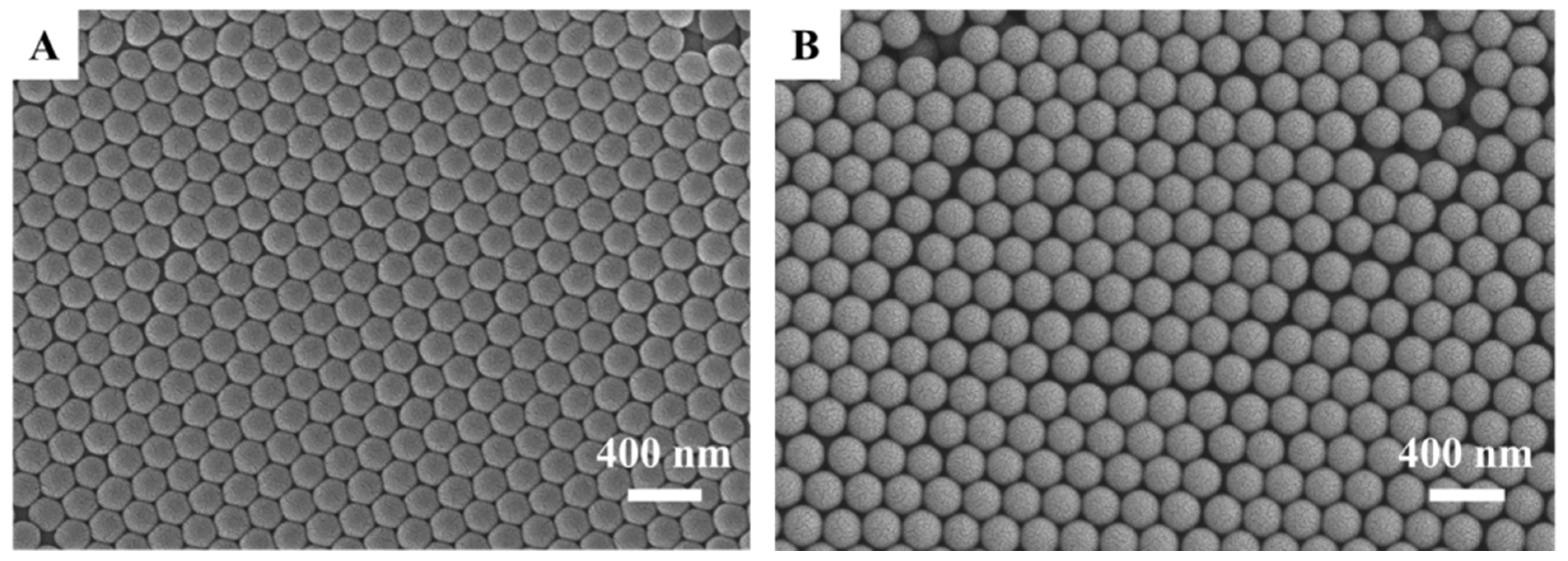
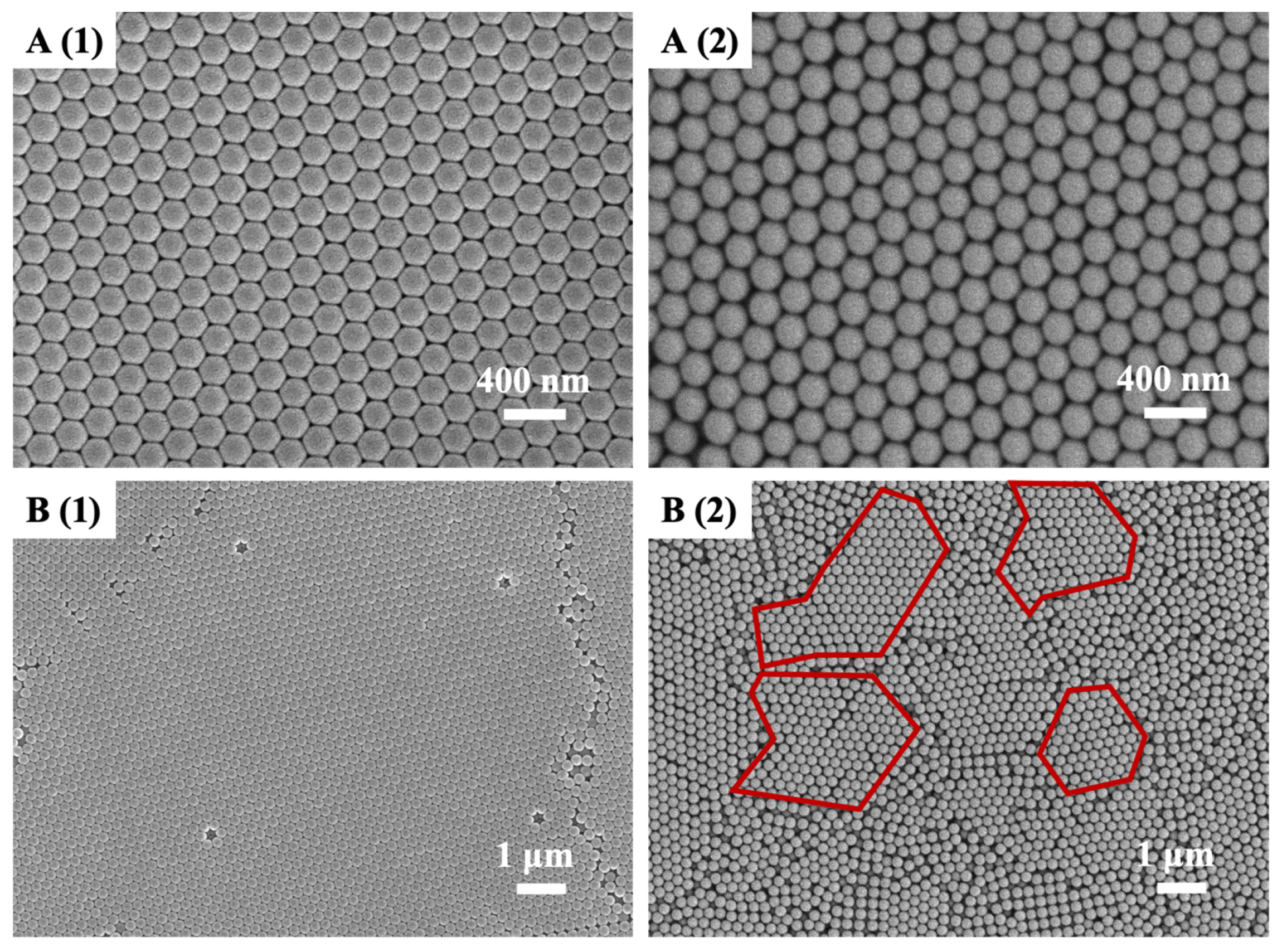
2.4.1. Characterization of the Surface Morphology of Microspheres, Structural Color Layers, and Wood

A portable microcamera (TipscopeCAM, Convergence Technology Co., Ltd., Wuhan, China) and a scanning electron microscope (TM-1000, Hitachi, Ltd., Tokyo, Japan) were used to observe the European beech wood sections and to clarify the macroscopic and microscopic anatomical structural features of the radial and tangential sections of the wood. The microscopic morphology and structure of the microspheres and their structural color layer were observed through cold field emission scanning electron microscopy (Regulus 8100, Hitachi, Ltd., Tokyo, Japan). For the microspheres synthesized using different emulsion polymerization reactions, no less than 300 microspheres were randomly selected from their electron micrographs, and their diameters were measured using the scribing method using ImageJ software (version 1.52a), and the average diameters and coefficients of variation of the microspheres in the dry state were obtained through calculations.

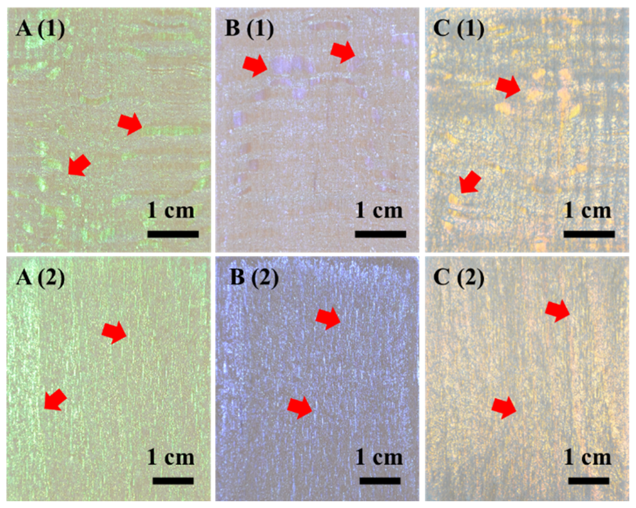
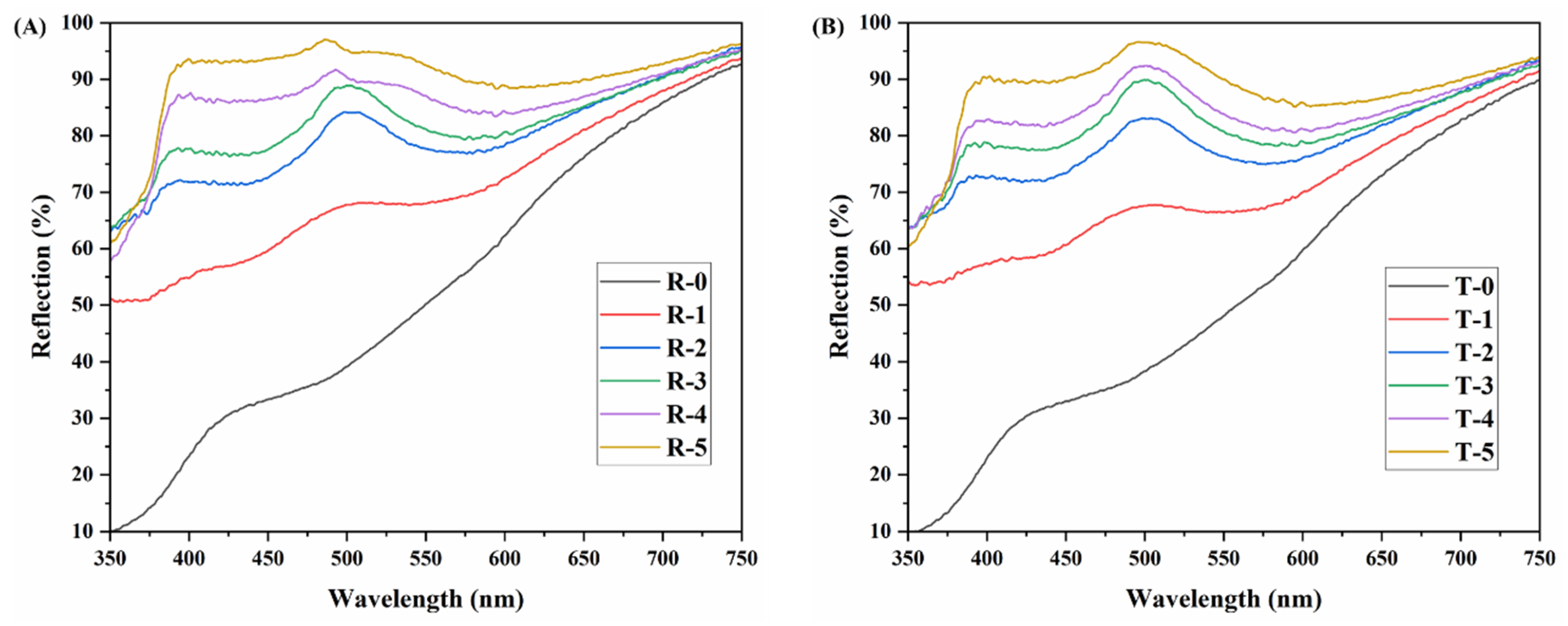
2.4.2. Photographs and Visible Light Reflection Spectra of Structural Color Layers

The structural color layer construction process and color changes on the wood surface were recorded with a digital single-lens reflex camera (D7000, Nikon Imaging (China) Sales Co., Ltd., Shanghai, China), and the reflectance spectra of the wood and the structural color layer were obtained using a UV-vis spectrophotometer (U3900, Hitachi, Ltd., Tokyo, Japan).

2.4.3. Characterization of Wood Surface Roughness

In order to analyze the selective modification effect of structural color on the surface of beech wood, a stylus surface roughness tester (JB-4C, Shanghai Taiming Optical Instrument Co., Ltd., Shanghai, China) was used to measure the surface roughness of radial and tangential sections of the sanded wood. The stylus radius was 2 μm. As the rays on the radial section of beech wood were lines or patches, an obvious ray tissue was selected on the surface of the wood. And the surface roughness was measured from axial (A) and radial (R) directions through the ray tissue when the radial section of the wood was tested, which was represented by RA1~RA10 and RR1~RR5. When testing along the axial direction, the sampling length was set to 0.8 mm, and the starting point of each test was moved about 1 mm along the radial transverse compared with the last test, making a total of 10 tests. For testing along the radial direction, the sampling length was adjusted to 2.5 mm, making a total of 5 tests, with each test’s starting point extending more than the last along the axial transverse shift by about 0.2 mm. When the surface roughness of the tangential section was tested, because the beech ray tissue showed widely distributed axial spindle spots, the testing was carried out along the tangential (T) direction with a sampling length of 0.8 mm, and it was represented by TT1~TT3. Each test’s starting point extended more than the last along the axial transverse shift by about 0.2 mm three times in total. The schematic diagram of the surface roughness test is shown in Figure 1.

3. Results and Discussion

3.1. Construction of Structural Color Layer on the Surface of European Beech Wood Using PSt Microspheres

The microscopic morphology of the two kinds of microspheres synthesized in this study is shown in Figure 2. The average particle size of the microspheres was calculated by measuring the diameters of 300 microspheres in the SEM images using the scribe measurement function in ImageJ software. When the reaction temperature of the emulsion polymerization was 70 °C, the size of microspheres was about 183.3 nm, with a coefficient of variation of 0.021; when the reaction temperature was increased to 75 °C, the size of the microspheres was about 207.4 nm with a coefficient of variation of 0.028. As the reaction temperature increased, the polymerization reaction started at a faster rate and became more vigorous, and the particle size of the microspheres increased. The microspheres prepared under both conditions had good monodispersity, and the coefficient of variation was less than 0.08, which meet the requirements of self-assembling in constructing microstructurally ordered photonic-crystal structural color layers.
Structural color layers were constructed on the wood surface using the brush coating method. Like the drop coating, which has been widely used to construct structural color on wood surfaces, the brush-coating method is also a bottom–up approach, and the microspheres migrate to the air–liquid and solid–liquid interfaces and self-assemble through the evaporation of the emulsion and the water absorption of cellulose, hemicellulose, and other biomass in the wood. It was also due to the water absorption of the wood that the structural color layer was constructed faster when the same brushing method was used but on a different substrate, as shown in Figure 3. This was similar to the rapid construction of the structural color layer using the spray method [33]. When the structural color layer was constructed on the surface of glass and wood via the brushing of emulsion containing the microspheres with a size of 207.4 nm, the structural color layer on the wood surface was constructed within 5 min at the same ambient temperature, but the structural color layer on the glass surface took about 20 min to be constructed. Although the wood enabled a rapid construction of the structural color layers, the color saturation of the structural color layers was low due to the microstructure of the wood surface. The cut cellular cavities on the wood surface had an uneven structure, as well as complex and rough substrates for the self-assembly of microspheres [17,34].
The SEM images of the structural color layers constructed on glass and wood surfaces using the brush-coating method are shown in Figure 4. When the two structural color layers were magnified to a high magnification, i.e., focused only on a very small (4 μm × 3 μm, Figure 4A) range in the coating, it was found that there was not much difference between the structures of the two pictures, indicating that in such a scale range, the microspheres were ordered and self-assembled on the emulsion surface by various forces such as electrostatic adsorption and capillary forces [35,36], forming a face-centered cubic-like structure [32,33]. However, when the two structural color layers were observed at a lower magnification (13 μm × 9 μm, Figure 4B), it was found that the size of the ordered structure of microspheres in the structural color layer on the glass surface was larger than that on the wood surface, and the large area of the structural color layer on the glass surface was also formed by the close fitting of this larger scale photonic crystal structure (Figure 4(B(1))). In contrast, the structural color layer on wood surface had smaller size of ordered structure and some areas were in disorder as shown in Figure 4(B(2)). The red wireframe circles parts of the ordered structure. It was assumed that the coexistence of ordered and disordered structural color layers was due to the rough cellular structure of the wood surface on the one hand, and the rapid construction of the structural color layer on the wood surface on the other hand, which did not allow for sufficient time for the microspheres to closely adhere to each other, and this was due to the water absorption of the wood surface.

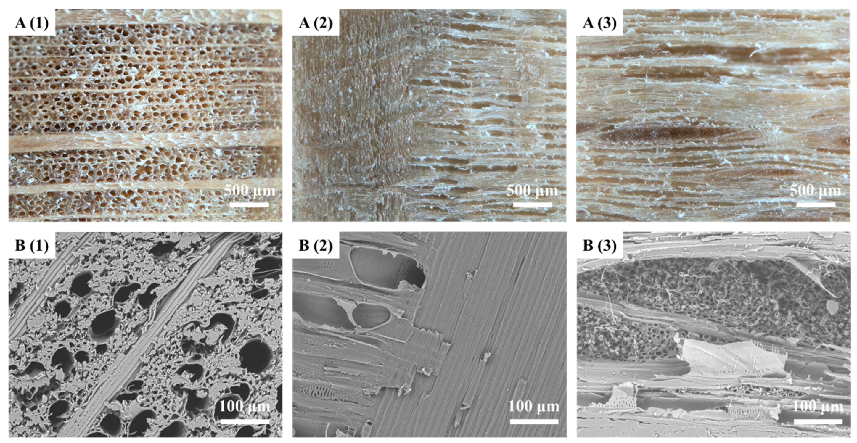
3.2. Special Finishing Properties of the Structural Color Layers on Ray Areas of Beech Wood Surfaces

As shown in the red arrows in Figure 3 and Figure 5, the structural color layer on the surface of beech showed a peculiar decorative effect when the brushing method was applied. We noticed that the structural color layer was brighter and more saturated on the wide rays of the longitudinal section of the wood (the parts are marked with red arrows in the pictures) than on other surface areas. In order to clarify whether this phenomenon was related to the microspheres used to construct the structural color layers, the particle size and type of raw material of microspheres were adjusted as shown in Figure 5. When PSt microspheres with an average size of about 183.3 nm were used to self-assemble the structural color layer on wood surface, the structural color layer was violet, while the special modification effect on the broad ray surface remained (Figure 5B). Then, the microspheres were replaced with P(St-MMA-AA) microspheres synthesized using the method used by Wang et al. [32]. These microspheres had shell-core structure, with the shell layer composed of P(MMA-AA) wrapped around the hard PSt core. The shell layer had more carboxyl groups and helped to form more hydrogen bonds between the microspheres and the wood surface. The average particle size of the P(St-MMA-AA) microspheres employed in this study was about 217 nm, and the same method of brushing was used to construct a structural color layer on the wood surface, which produced tangerine color and still showed brighter color on the surface of the broad-ray regions (Figure 5C). The above two results are evidence to infer that the particle size, microstructure, and active groups of PSt-based microspheres are not major factors influencing the special modification effect of the structural color layer on the wide-ray region of the beech wood surface.
Furthermore, the pure PSt microsphere emulsion was used as a raw material to brush the radial section and tangential section of the European beech wood five times, respectively, and the obtained structural color layers were photographed and analyzed using a UV-vis spectrophotometer, as shown in Figure 6. In the study of multiple brushings of the radial section of the wood, after the initial brushing of the sanded wood surface with the emulsion, a heterogeneous layer of the structural color was built up on the wood surface, and it was observed that the structural color layer on the wide-ray areas showed a bright green color (Figure 6(A(2))). The structural color layer in other areas did not show any significant color changes than the wide rays, but it was off-white in general and had much less color saturation than that on the wide rays, with some spots of green. The color of the wood below the finishing layer was still faintly visible. When the second and third brushings were performed directly on the surface of the painted wood, the structural color layer on the wood surface showed significant changes, having a large area of green (Figure 6(A(3),A(4))). Meanwhile, the vivid color effect of the finishing layer on the surface of the wide rays was still very prominent, and the visual effect appeared as bright patches. When the structural color layer was constructed by painting the specimen for the fourth and fifth times (Figure 6(A(5),A(6))), the photos show that the color of the layers gradually became less pronounced, with only a very light green color. And the color of the wood was no longer visible, changing to grayish white.
In the study of multiple brushings of the tangential section of European beech wood (Figure 6B), it was found that the selective modification effect was not obvious due to the fact that the wide rays were finely distributed in fusiform spots in this section (Figure 6(B(1))), with a length of about 2–5 mm and width of less than 1 mm. Therefore, the special finishing effect of the structural color layer on the wide-ray area of the tangential section was still visible during the first and second brushings (Figure 6(B(2),B(3))) but not after the subsequent three brushings (Figure 6(B(4)–B(6))). Moreover, the original color of wood the had been covered up, the finishing layer was no longer flat, the structural coloring effect was getting worse, and, finally, only uneven off-white and mottled light green finishing effects were visible.
When the visible-light reflection spectrum was used to characterize the structural color modification layers as shown in Figure 7, it was found that the reflection spectral curve of the two sections of unmodified wood was rising monotonically, which indicated that the wood had a reflection effect on visible light in the wavelength range of 425~750 nm, resulting in the visual effect of yellow-brown wood (R-0 and T-0 in Figure 7). The structural color layer constructed on the surface of the wood diffracted the visible light, creating a corresponding reflection peak at a specific wavelength. After the first brushing, the reflectance of two sections in the visible wavelength range increased to more than 50% (R-1 and T-1 in Figure 7). This was due to the white color of the PSt microspheres themselves, so the structural color layer constructed by them reflected visible light more strongly, and both reflectance spectral curves showed a lower peak at 506.5 nm, which corresponds to the green structural color. After the second and third brushings, the light-reflection ability of the structural color layers was obviously stronger. The height of the reflection peak increased significantly, and the central position of the peak moved to 501 nm (R-2, R-3, T-2, and T-3 in Figure 7). The changes in the reflectance spectra were consistent with the photographs, that is, the structural color layers were more complete, and the color was brighter. And there was a slight blue shift in the color, which was presumed to be related to the thickening of the ordered structural layer formed by the second brushing. However, within the scope of this study, the optical effect of the structural color layer reached the best state during the third brushing, that is, the relative height of the reflection peak of the two curves reached the maximum (about 13%) (R-3 and T-3 in Figure 7).
When the fourth and fifth brushings were performed, the reflection curve continued overall toward a higher reflectance for the structural color layer of the tangential section of the wood, with the peak reflectance at 501 nm of 92.4% and 96.5% (T-4 and T-5 in Figure 7), respectively, which had gentler slopes of the peak shape relative to the structural color layer constructed in the second and third brushings. This was consistent with the greenish gray color in the optical photos. In contrast, the reflectance spectral curves for the structural color layer obtained by the fourth and fifth brushings on the radial section were not exactly the same. The reflectance peaks of the curves also moved towards a higher reflectance of 91.7% and 97.1%, respectively. But the position of the reflectance peaks moved towards a lower wavelength range to 493 nm and 486 nm (R-4 and R-5 in Figure 7). In addition, the peak shapes were deformed into compound peak shapes, with a gentle peak at 535 nm after the appearance of the reflection peak, which then slowly decreased. Moreover, the relative peak height under the highest reflection peak of these two reflection curves was low, and the half-peak width was small. Combined with the photos of the structural color layer in the radius section, it can be speculated that the peak shapes were mainly due to the fact that the structural color layers in the wide-ray regions still had a good structural coloration effect, while the structural color layer in other regions had a poor effect.

3.3. Influence of Anatomical Characteristics of European Beech Wood on the Selective Modification of Structural Color Layers

Knowledge of the macroscopic and microscopic anatomy of the beech wood can facilitate a systematic analysis of the selective modification effect of structural color layers. European beech wood has a fine and homogeneous structure with a clean-cut surface and slightly pronounced growth rings [37]. As shown in Figure 8, the pores of the vessels are small and numerous in the cross-section, densely distributed and large in size within the growth ring, and very few at the growth ring boundaries. The parenchymatous tissue was not visible to the naked eye and appeared to exhibit a scattered-agglomerated state under the microscope. The wood rays were obvious under the naked eye as a non-stacked state and densely arranged, and they can be divided into two categories: single-rowed rays 1 to 18 cells high, multi-rowed rays 6 to hundreds of cells high or more, and 2 to 19 cells wide. In the radial section, the wood rays are in broken band patches of varying widths, and in the tangential section, they are in an aesthetically pleasing fusiform pattern.
It can be seen from Figure 8B that, compared with other diffuse porous woods such as aspen and hard maple [31], the wide rays of beech wood were its typical anatomical structure. From the three-section micrographs of beech, it could be seen that the rays, especially the wide rays, were made up of closely fitting ray cells of similar shape. In addition to the ray tissue, it was widely distributed with vessels which had a much larger inner diameter than the ray cells. In previous studies, it was found that the roughness of the wood surface would affect the formation of ordered structures by microspheres [31]. Therefore, the ray region provided excellent substrate flatness for the structural color layer construction, while the microstructure of other areas mainly composed of vessels and wood fibers was more uneven, which was not conducive to the self-assembly of microspheres into well-ordered structures. By comparing the SEM images of radial and tangential sections of beech wood (Figure 8(B(2),B(3))), it can be observed that the wood ray tissue on the radial section was a plane composed of flat arranged cells, while on the tangential section, it was formed into a structure rich in small holes composed of cross-section with hundreds of ray cells. Therefore, the main reason why the visible-light reflection spectral curve of the structural color layers on the radial section produced sharp peaks after multiple brushing tests (Figure 7A) was that the wide rays in the radial section were in the form of blocks or strips of horizontally arranged cells, while in the tangential section, they were in the form of spindle-shaped spots where the ray cells were dissected along the end face. It was found that the cell and tissue morphology and properties of the wood’s wide rays were the main reasons for the specific modifications achieved by the structural color layers on them. It was the difference between the cells and tissues of the European beech wood’s wide rays and other axially distributed cells and tissues such as vessels and wood fibers that led to the selective modification phenomenon.
Although it could be seen from the SEM images that the ray region of European beech had a smoother surface than other anatomical characteristics, it was more suitable for brush coating to construct the structural color layer. However, it is still important to note that the wood sample used to observe the anatomy in the SEM was cut with a sharp blade to obtain a good cutting surface. However, before brushing the finishing layer, the radial or tangential section of the wood needed to be sawed and sanded, and these treatments produced cutting and ploughing on the wood surface [38], which also made it impossible to clearly distinguish the microscopic anatomical features, such as ray tissue, through SEM. Therefore, a stylus surface roughness tester was used to characterize the sanded wood surface, as shown in Figure 1 and Figure 9. The reason why the numbers of measurements in each section and direction are not equal is that the aim of measuring the roughness was to show that the ray area had better flatness than others, rather than to characterize the overall roughness of the wood surface, and the size of the ray tissue was not the same in different sections and directions.
Figure 9A,B show the track curves obtained through roughness measurements in the area around a long-strip ray tissue in the radial section, and the horizontal axis represents the data point recorded using the roughness measurement instrument. As can be seen from the track curve obtained from ten tests along the axis direction (Figure 9A), the surface roughness of RA1~9 from 740 to 2036 was better than that of other ranges, and for RA10, the surface roughness ranges were from 1100 to 2264. These flatter areas derived from the profile of ray tissue, and troughs of varying depths and widths in other test areas outside these ranges, could be presumed to represent the vessel profiles of the wood formed by the sanding process. A similar situation existed in the trajectory curve obtained when the surface roughness near the ray tissue was tested along the radial direction (Figure 9B), where the surface roughness of RR2~5 in the range of 220 to 2570 was better than the other regions of the four curves. Meanwhile, for RR1, only a small range was smoother. This was because, in this test, the range passed by the stylus of the tester was at the edge of the ray tissue, and the track of the RR1 curve shows the surface of the long side of the wide ray tissue in the radial section and the joint parts with vessels and wood fibers. Figure 9C shows three roughness test results on the tangential section of beech wood which crosses a wood ray area. Although the ray tissues were small in size and formed by the truncated surface of multiple ray cells after sanding, they were still better than other tissues in terms of flatness. The ranges of the trajectory curves obtained in the three tests from 1858 to 2313 (TT1), 1371 to 2202 (TT2), and 1988 to 2455 (TT3), respectively, formed a continuous curve with smaller troughs. The reason why the flat range appeared and ended in different positions and lengths was the spindle shape of the wide ray tissue. TT1 and TT3 were tested through the ends of the spindle, while TT2 was tested through the middle of the spindle.
The reason why the troughs of the track curves in Figure 9A–C were different in sharpness was mainly because of the sampling length set during the roughness measurement. The surface roughness tester used in this study could select a variety of sample lengths, such as 0.8 mm and 2.5 mm. Therefore, although the horizontal coordinate length of the three trajectory curves (RA, RR, TT) was the same, it only indicated the testing points taken by the instrument but did not indicate the actual measurement length. In one test, the accompanying software system sampled five times. These were when the 0.8 mm sampling length was selected, and the route length of a single test was 4 mm; for the 2.5 mm sampling length, the test route length was 12.5 mm. Through multiple tests of the surface roughness of the two sections of beech wood, it was seen that the ray tissues on the wood surface after sawing and sanding with a 320# sandpaper still had better flatness than other anatomical structural areas, as shown by SEM photos. This was precisely the decisive factor for the faster construction and better coloration of the modified layer on the ray tissue area when the brush-coating method was used. It can be seen that wood species, material characteristics, and anatomical structures affect the construction of structural color modification layers on wood surfaces, and they can produce specific modification effects.

4. Conclusions

In this study, emulsion containing PSt-based microspheres was applied to beech wood surface using the brush coating method to construct structural color layers. Moreover, the reasons for the specificity of construction speed and the color generation effect of the structural color layer are discussed in this paper. The following conclusion can be drawn:
(1)
A small amount of emulsion can be applied to the beech wood surface by brushing, which can rapidly construct the structural color layer.
(2)
Since the wood surface was water-absorbent, the time required to construct the structural color layer on the wood surface under the same external environment was much shorter than that required for the surface of non-absorbent materials such as glass.
(3)
A selective construction effect was shown on the wide rays of beech. The construction speed of the structural color layer was faster, and the optical color effect was better.
(4)
The special modification still existed even when the particle size, synthetic material, structure, and chemical functional groups of the microspheres were changed.
(5)
It was found that the flatness of the profile of the wide rays was the main driver of the specific modification of the structural color layer.

Author Contributions

Conceptualization, J.H., Y.L., J.W. and W.X.; methodology, J.H. and Y.L.; data curation, J.H.; writing—original draft preparation, J.H.; writing—review and editing, J.H.; visualization, J.H. All authors have read and agreed to the published version of the manuscript.

Funding

This work was supported by Natural Science Foundation of Jiangsu Province (grant number, BK20230403, Jiangsu Provincial Department of Science and Technology); Postgraduate Research & Practice Innovation Program of Jiangsu Province (grant number KYCX23_1196, Jiangsu Education Department); and Qing Lan Project (Jiangsu Education Department).

Data Availability Statement

The raw and processed data required to reproduce these findings cannot be shared at this time as the data also form part of an ongoing study.

Acknowledgments

The authors acknowledge the valuable support from Nanjing Forestry University.

Conflicts of Interest

The authors declare no conflicts of interest.

References

  1. Li, Y.; Li, J. Fabrication of reversible thermoresponsive thin films on wood surfaces with hydrophobic performance. Prog. Org. Coat. 2018, 119, 15–22. [Google Scholar] [CrossRef]
  2. Esteves, B.; Marques, A.V.; Domingos, I.; Pereira, H. Heat-induced colour changes of pine (Pinus pinaster) and eucalypt (Eucalyptus globulus) wood. Wood Sci. Technol. 2008, 42, 369–384. [Google Scholar] [CrossRef]
  3. Hu, J.; Li, Y.; Liu, Y.; Guo, H.; Li, L. Evaluation of the dyeing properties of basswood veneer treated by dichlorotriazine reactive dye based on gray correlation analysis. Bioresources 2016, 11, 466–481. [Google Scholar] [CrossRef]
  4. Liu, Y.; Shao, L.; Gao, J.; Guo, H.; Chen, Y.; Chen, Q.; Via, B.K. Surface photo-discoloration and degradation of dyed wood veneer exposed to different wavelengths of artificial light. Appl. Surf. Sci. 2015, 331, 353–361. [Google Scholar] [CrossRef]
  5. Srinivas, K.; Pandey, K.K. Photodegradation of thermally modified wood. J. Photochem. Photobiol. B Biol. 2012, 117, 140–145. [Google Scholar] [CrossRef]
  6. Nguyen, T.T.; NguyenTH, V.; Ji, X.; Yuan, B.; Trinh, H.M.; Do KT, L.; Guo, M. Prediction of the color change of heat-treated wood during artificial weathering by artificial neural network. Eur. J. Wood Wood Prod. 2019, 77, 1107–1116. [Google Scholar] [CrossRef]
  7. Zhu, T.; Ren, K.; Sheng, J.; Zhang, Q.; Li, J.; Lin, J. Natural dye extracted from Dalbergia cochinchinensis residue with water fastness, mildew resistance and permeability properties for wood staining. Wood Sci. Technol. 2022, 56, 969–988. [Google Scholar] [CrossRef]
  8. Salla, J.; Pandey, K.K.; Srinivas, K. Improvement of UV resistance of wood surfaces by using ZnO nanoparticles. Polym. Degrad. Stab. 2010, 97, 592–596. [Google Scholar] [CrossRef]
  9. Miklečić, J.; Turkulin, H.; Jirouš-Rajković, V. Weathering performance of surface of thermally modified wood finished with nanoparticles-modified waterborne polyacrylate coatings. Appl. Surf. Sci. 2017, 408, 103–109. [Google Scholar] [CrossRef]
  10. Saha, S.; Kocaefe, D.; Boluk, Y.; Pichette, A. Enhancing exterior durability of jack pine by photo-stabilization of acrylic polyurethane coating using bark extract. Part 1: Effect of UV on color change and ATR–FT-IR analysis. Prog. Org. Coat. 2011, 70, 376–382. [Google Scholar] [CrossRef]
  11. Wang, C.; Lin, X.; Schäfer, C.G.; Hirsemann, S.; Ge, J. Spray synthesis of photonic crystal based automotive coatings with bright and angular-dependent structural colors. Adv. Funct. Mater. 2020, 31, 2008601. [Google Scholar] [CrossRef]
  12. Zhao, Y.; Xie, Z.; Gu, H.; Zhua, C.; Gu, Z. Bio-inspired variable structural color materials. Chem. Soc. Rev. 2012, 41, 3297–3317. [Google Scholar] [CrossRef]
  13. Kinoshita, S.; Yoshioka, S. Structural colors in nature: The role of regularity and irregularity in the structure. ChemPhysChem 2005, 6, 1442–1459. [Google Scholar] [CrossRef] [PubMed]
  14. Srinivasarao, M. Nano-optics in the biological world: Beetles, butterflies, birds, and moths. Chem. Rev. 1999, 99, 1935–1961. [Google Scholar] [CrossRef] [PubMed]
  15. Yuan, W.; Zhang, K.Q. Structural evolution of electrospun composite fibers from the blend of polyvinyl alcohol and polymer nanoparticles. Langmuir 2012, 28, 15418–15424. [Google Scholar] [CrossRef] [PubMed]
  16. Chai, L.; Zhou, L.; Liu, G.; Li, Y.; Fan, Q.; Shao, J. Interface–gravity joint self-assembly behaviors of P(St-MAA) colloidal microspheres on polyester fabric substrates. J. Mater. Sci. 2017, 52, 5060–5071. [Google Scholar] [CrossRef]
  17. Tang, H.; Yang, Z.; Tian, Y.; Song, L.; Shen, H.; Zhang, X. Fabrication of crack-free non-iridescent structural color coatings with good stability on calcium silicate board. Opt. Mater. 2021, 120, 111478. [Google Scholar] [CrossRef]
  18. Liu, Y. Self-assembly of poly(styrene-methyl methacrylate-acrylic acid) (P(St-MMA-AA)) colloidal microspheres on wood surface by thermal-assisted gravity deposition. Wood Sci. Technol. 2021, 55, 403–417. [Google Scholar] [CrossRef]
  19. Huang, D.; Wu, J.; Chen, C.; Fu, X.; Brozena, A.H.; Zhang, Y.; Gu, P.; Li, C.; Yuan, C.; Ge, H.; et al. Precision imprinted nanostructural wood. Adv. Mater. 2019, 31, 1903270. [Google Scholar] [CrossRef]
  20. Hu, J.; Liu, Y.; Xu, W.; Wu, Z.; Pang, X. Construction of rainbow-like structural color coatings on wood surfaces based on polystyrene microspheres. Forests 2022, 14, 76. [Google Scholar] [CrossRef]
  21. Wang, J.; Pang, F.; Fu, Q.; Ge, J. Fabrication of anti-counterfeiting patterns with angle-dependent colors by silkscreen printing and UV-curable photonic crystal inks. Sci. China Mater. 2023, 66, 1623–1631. [Google Scholar] [CrossRef]
  22. Shen, H.; Lin, Q.; Tang, H.; Tian, Y.; Zhang, X. Fabrication of temperature and alcohol-responsive photonic crystal hydrogel and its application for sustained drug release. Langmuir 2022, 38, 3785–3794. [Google Scholar] [CrossRef]
  23. Wang, F.; Guo, F.; Xue, Y.; Luo, H.; Zhu, J. Rapid fabrication of large-scale structurally colored PS@SiO2 films with enhanced self-cleaning capability. J. Coat. Technol. Res. 2021, 18, 489–499. [Google Scholar] [CrossRef]
  24. Li, S.; Jia, L.; Wang, L.; Li, P.; Fan, X.L. Preparation of robust fully encapsulated structural color fabrics and their application to display. Dye. Pigment. 2023, 212, 111150. [Google Scholar] [CrossRef]
  25. Lu, X.; Huang, H.; Wang, X.; Li, G.; Chai, L.; Zhou, L.; Shao, J. Quasi-ordered nanosphere-based photonic crystals with high-fastness structural colors via screen printing: Implications for textile printing and dyeing. ACS Appl. Nano Mater. 2022, 5, 16108–16115. [Google Scholar] [CrossRef]
  26. Li, M.; Lyu, Q.; Peng, B.; Chen, X.; Zhang, L.; Zhu, J. Bioinspired colloidal photonic composites: Fabrications and emerging ap plications. Adv. Mater. 2022, 34, 2110488. [Google Scholar] [CrossRef] [PubMed]
  27. Strobel, K.; Nyrud, A.Q.; Bysheim, K. Interior wood use: Linking user perceptions to physical properties. Scand. J. For. Res. 2017, 32, 798–806. [Google Scholar] [CrossRef]
  28. Conceição, W.S.; Matos, R.S.; Bufalino, L.; Ramos, G.Q.; Zayas, F.G.; Filho, H.D.F. Micromorphology and fractal evaluation of Dinizia excelsa Ducke wood under three different cut conditions by atomic force microscopy. Measurement 2021, 179, 109490. [Google Scholar] [CrossRef]
  29. Salca, E.A.; Krystofiak, T.; Lis, B.; Hiziroglu, S. Glossiness evaluation of coated wood surfaces as function of varnish type and exposure to different conditions. Coatings 2021, 11, 558. [Google Scholar] [CrossRef]
  30. Feng, X.; Wu, Z.; Sang, R.; Wang, F.; Zhu, Y.; Wu, M. Surface design of wood-based board to imitate wood texture using 3D printing technology. BioResources 2019, 14, 8196–8211. [Google Scholar] [CrossRef]
  31. Liu, Y.; Hu, J.; Xu, W. Surface roughness of wood substrates after grinding and its influence on the modification effect of structural color layers. Forests 2023, 14, 2213. [Google Scholar] [CrossRef]
  32. Wang, J.; Wen, Y.; Ge, H.; Sun, Z.; Zheng, Y.; Song, Y.; Jiang, L. Simple fabrication of full color colloidal crystal films with tough mechanical strength. Macromol. Chem. Phys. 2006, 207, 596–604. [Google Scholar] [CrossRef]
  33. Cui, L.; Zhang, Y.; Wang, J.; Ren, Y.; Song, Y.; Jiang, L. Ultra-fast fabrication of colloidal photonic crystals by spray coating. Macromol. Rapid Commun. 2009, 30, 598–603. [Google Scholar] [CrossRef]
  34. Aguirre, C.I.; Reguera, E.; Stein, A. Colloidal photonic crystal pigments with low angle dependence. ACS Appl. Mater. Interfaces 2010, 11, 3257–3262. [Google Scholar] [CrossRef]
  35. Vogel, N.; Retsch, M.; Fustin, C.A.; del Campo, A.; Jonas, U. Advances in colloidal assembly: The design of structure and hierarchy in two and three dimensions. Chem. Rev. 2015, 115, 6265–6311. [Google Scholar] [CrossRef] [PubMed]
  36. McGorty, R.; Fung, J.; Kaz, D.; Manoharan, V.N. Colloidal self-assembly at an interface. Materialstoday 2010, 13, 34–42. [Google Scholar] [CrossRef]
  37. Burgert, I.; Eckstein, D. The tensile strength of isolated wood rays of beech (Fagus sylvatica L.) and its significance for the biomechanics of living trees. Trees 2001, 15, 168–170. [Google Scholar] [CrossRef]
  38. Zhang, J.; Sun, P.; Luo, B.; Li, L.; Liu, H. Surface creation and cutting characteristics during sugar maple wood scratching with spherical cone grit. Eur. J. Wood Wood Prod. 2021, 79, 679–689. [Google Scholar] [CrossRef]
Figure 1. Testing method of European beech wood surface roughness: (A) the schematic diagram of the surface roughness testing; (B) testing along radial (R) and axial (A) directions on the radial section; (C) testing along tangential (T) direction on the tangential section of the wood. The arrows indicate the direction of stylus movement.
Forests 15 00261 g001
Figure 2. PSt microspheres synthesized at different temperatures using emulsion polymerization method described in this study; (A) synthesized at 70 °C; (B) synthesized at 75 °C.
Forests 15 00261 g002
Figure 3. The process by which the emulsion containing PSt microspheres (the average particle size was 207.4 nm) was applied to glass and wood surfaces via brush coating and the formation of structural color modification layers; (A) structural color layer on the surface of glass; (B,C) structural color layer on the radial section of wood, where the angle between the tangent line of the growth ring and the surface was about 80° and 50°, respectively; (D) structural color layer on the tangential section of wood, where the angle between the tangent line of the growth ring and the surface was about 10°. The arrows represent some ray areas on the surface of wood.
Forests 15 00261 g003
Figure 4. The surface micromorphologies of wood and glass surface structural color layers were observed via FSEM; (A(1),B(1)) structural color layers on glass surface; (A(2),B(2)) structural color layers on wood surface. The red wireframes indicate some ordered arrangement of microspheres.
Forests 15 00261 g004
Figure 5. The structural color layers formed by the self-assembly of PSt microspheres and P(St-MMA-AA) microspheres on the wood surfaces showed obvious selective modification effect in the wood ray regions, including the radial section (1) and tangential section (2); (A) the particle size of the PSt microspheres composed of the green structural color layer was about 207.4 nm; (B) the particle size of the PSt microspheres composed of the violet structural color layer was about 183.3 nm; (C) the particle size of the P(St-MMA-AA) microspheres composed of the tangerine structural color layer was about 217 nm. The arrows represent some ray areas on the surface of wood.
Forests 15 00261 g005
Figure 6. Five-times construction of structural color layers on radial section (A) and tangential section (B) of European beech wood via brush coating; (1) uncoated wood; wood after the (2) first brushing, (3) second brushing, (4) third brushing, (5) fourth brushing, and (6) fifth brushing.
Forests 15 00261 g006
Figure 7. Visible-light reflection spectrum of the five-times construction of structural color layers in radial section (A) and tangential section (B) of European beech wood by brush coating.
Forests 15 00261 g007
Figure 8. Structural characteristics of three sections of European beech wood observed using microcamera (A) and SEM (B); (A(1),B(1)) the transverse sections; (A(2),B(2)) the radial section; (A(3),B(3)) the tangential section.
Forests 15 00261 g008
Figure 9. The trace curves obtained by measuring the surface roughness around the wide ray tissues on the radial and tangential planes: (A) ten tests of the strip ray tissue surface region along the axial direction in the radial section; (B) five tests of the strip ray tissue surface region along the radial direction in the radial section; (C) three tests of the fusiform ray tissue along the tangential direction in the tangential section. The dotted boxes represent the roughness curve of the wood ray area.
Forests 15 00261 g009
Disclaimer/Publisher’s Note: The statements, opinions and data contained in all publications are solely those of the individual author(s) and contributor(s) and not of MDPI and/or the editor(s). MDPI and/or the editor(s) disclaim responsibility for any injury to people or property resulting from any ideas, methods, instructions or products referred to in the content.

