Article

Construction of a Genetic Transformation System for Populus wulianensis

1
Key Laboratory of National Forestry and Grassland Administration on Conservation and Utilization of Warm Temperate Zone Forest and Grass Germplasm Resources, Shandong Provincial Center of Forest and Grass Germplasm Resources, Jinan 250102, China
2
State Key Laboratory of Tree Genetics and Breeding, College of Biological Sciences and Technology, Beijing Forestry University, Beijing 100083, China
3
College of Agronomy and Agricultural Engineering, Liaocheng University, Liaocheng 252000, China
*
Authors to whom correspondence should be addressed.
Forests 2024, 15(8), 1474; https://doi.org/10.3390/f15081474
Submission received: 7 May 2024 / Revised: 16 August 2024 / Accepted: 19 August 2024 / Published: 22 August 2024
(This article belongs to the Section Genetics and Molecular Biology)

Abstract

:
Transgenic technology is a potent tool for verifying gene functions, and poplar serves as a model system for genetically transforming perennial woody plants. However, the current poplar genetic transformation system is limited to a few genotypes. In this study, we developed an efficient transformation system based on the Agrobacterium-mediated transformation of Populus wulianensis, a rare and endangered tree species endemic to Shandong Province. Aseptic seedlings of P. wulianensis were used as experimental materials, and the optimal medium for inducing adventitious buds was explored as 1/2(NH4NO3) MS + 0.05 mg/L naphthalene acetic acid (NAA) + 0.5 mg/L 6-benzylaminopurine (6-BA), resulting in up to 35 adventitious buds. The selection resistance critical pressure of 300 mg/L for timentin can effectively inhibit the growth of Agrobacterium while promoting the induction of adventitious buds in leaves. The critical screening pressure for kanamycin for producing resistant adventitious buds and inducing resistant rooting seedlings was 100 mg/L. We optimized several independent factors, which significantly enhanced the efficiency of genetic transformation. The leaves were infected with Agrobacterium suspension diluted twice by adding 100 μmol/L acetylsyringone (β-AS) (OD600 = 0.6) for 15 min, followed by co-culture in the dark for 3 d. Using this improved transformation system, we obtained transgenic P. wulianensis clones overexpressing the enhanced green fluorescent protein (EGFP) gene through direct organogenesis. Among the 112 resistant buds obtained, 17 developed resistant rooting in seedlings. Eight positive plants were identified through DNA, RNA, and protein level analyses, with a positivity rate of 47.06%. This study provides a foundation for developing and utilizing P. wulianensis germplasm resources and lays the groundwork for resource improvement.

1. Introduction

The reproduction of woody plants is characterized by long growth cycles and seasonal dormancy [1]. Tissue culture technology addresses the challenge of asexual reproduction and provides a basis for studying gene function and physiological characteristics.
Plant genetic transformation serves to explore gene function and to develop plant varieties with specific traits, particularly in long-generation trees. The establishment of this plant genetic improvement method has assisted the application of traditional breeding methods [2] and accelerates forest tree breeding [3]. Agrobacterium tumefaciens-mediated transformation is an effective technique for introducing exogenous DNA into plant genomes, facilitating gene function and breeding studies [4,5]. Since 1986, when Parsons et al. [6] first demonstrated the successful expression of exogenous genes in poplar using transgenic technology, the field of poplar genetic engineering has rapidly advanced. The successful genome sequencing of Populus trichocarpa in 2006 marked the transition of genomic research in forest trees into the post-genomic era [7]. In recent years, various poplar species have developed genetic transformation systems, achieving significant breakthroughs in gene function research and other areas [8,9].
Agrobacterium-mediated genetic transformation is affected by numerous factors such as plant genotype, explant type, strain type, vector-receptor affinity, Agrobacterium infection mode, pre-culture time, bacterial fluid concentration, infection time, co-culture time, and antibiotic selection [10]. Therefore, to establish an efficient genetic transformation system, it is essential to coordinate these factors with the induction of resistant adventitious buds before conducting the experiments. Studies have indicated that various types of explants, including leaves, petioles, hypocotyls, stem internodes, stem segments, roots, cell suspension cultures, and buds [8,9,11], can enhance the efficiency of Agrobacterium-mediated transformation in Populus [7,12]. For instance, Yu et al. [13] achieved a transformation yield of 16.28% in male diploid P. tomentosa by screening transformed leaves with 30 mg/L kanamycin (Kan) after 12 h of pre-culture using an Agrobacterium concentration (OD600) of 0.4, 20 min of infection time and 24 h of co-culture. Wen et al. [14] achieved a transformation frequency exceeding 50% in callus transformation by adding 100 µM acetylsyringone (β-AS) to an Agrobacterium tumefaciens suspension with OD600 of 0.6, infecting for 15 min and co-culturing for 2 d, with explants pre-cultured for 6 d. Ma et al. [15] obtained an average transformation efficiency of 23.60% for P. alba var. pyramidalis leaf explants by inducing leaf callus with 90 mg/L hygromycin, screening callus-induced adventitious buds twice for resistant transformants, and optimizing factors such as an Agrobacterium OD600 of 0.3–0.5, 10 min of infection, addition of 100 µM AS, and the use of different vectors.
P. wulianensis S. B. Liang & X. W. Li, a member of Salicaceae, is a dioecious, deciduous hardwood [16] and, similar to other poplar species, is valuable for timber. However, its small wild population, limited habitat, high risk of extinction, and sensitivity to human activities warrant its classification as a PSESP. The declining effective population size and increasing isolation of this species have enhanced the genetic drift and reduced gene flow, resulting in a loss of genetic diversity [17,18]. This loss has severely reduced the ability of species to withstand harsh environments, ultimately accelerating their extinction. The transgenic technology enables rapid and targeted genetic improvement in P. wulianensis, facilitating the development of new varieties that can adapt to climate change and meet evolving needs. However, establishing a culture medium for leaf redifferentiation to induce adventitious buds in P. wulianensis and breaking through its efficient genetic transformation system are currently unresolved. In this study, we selected the aseptic seedlings of P. wulianensis, an endemic and endangered tree species in Shandong, screened the orthogonal combinations of leaf-induced adventitious buds and the main experimental conditions of Agrobacterium-mediated genetic transformation, and established and optimized the regeneration system and the genetic transformation system of P. wulianensis, in order to provide reference for the study of genetic transformation of other woody plants and lay a foundation for molecular assisted breeding in the later stage.

2. Materials and Methods

2.1. Source of Materials

2.1.1. Plant Materials of P. wulianensis

The leaf redifferentiation test of P. wulianensis used 3–4 mature, healthy leaves below the apical bud of the aseptic seedlings stored in our laboratory as experimental materials, and 15–20 leaves were inoculated in each medium, with three replicates. After inoculation, the leaves were grouped for aseptic culture, maintaining a controlled temperature of (25 ± 5) °C during the day and (18 ± 5) °C at night, with a light cycle of 16 h per day and a light intensity of 3000–5000 LX. The medium was changed every 10 d, and after 35 d of culture, the number of regenerated buds was recorded, and the health state of leaf growth was evaluated.
The materials for antibiotic screening experiments were as above, and the leaves were divided into three parts (leaf base with petiole, leaf middle, and leaf tip) (Figure 1); 3–4 samples were placed in each petri dish with three replications for each gradient concentration. The optimal differentiation medium selected from the above tests was used, and the leaves were cultivated in the dark for 8 days; the culture conditions were unchanged.

2.1.2. Strain and Plasmid

The strain used in the experiment was Agrobacterium tumefaciens GV3101, and the plant expression vector was PEZR-(K)-LC, which is a binary expression vector carrying the Kan resistance gene and fluorescent protein gene (EGFP) (Figure 2).

2.2. Methods

2.2.1. Leaf Adventitious Bud Induction

Four base media, Naphthalene acetic acid (NAA) (Sigma, Darmstadt, Germany), 6-Benzylaminopurine (6-BA) (Sigma, Darmstadt, Germany), and dark culture time, were selected as the influencing factors, with three levels for each factor. The design used an orthogonal table, L9(34) (Table 1). This study analyzed the effects of these different factors at various levels on leaf redifferentiation of P. wulianensis.

2.2.2. Antibiotic and Bacteriostatic Agent Concentration Screening Experiment

The effect of timentin (Coolaber, Beijing, China) on Agrobacterium GV3101 growth was tested at concentrations ranging from 0 mg/L (control) to 250 mg/L in 50 mg/L increments. The effect of timentin on the induction of adventitious buds in P. wulianensis leaves was tested on three parts of the treated leaves (Figure 1). The leaf middle was tested at concentrations ranging from 0 mg/L to 850 mg/L in 50 mg/L increments, while the other two parts were tested at concentrations ranging from 0 mg/L to 900 mg/L in 100 mg/L increments. The effect of Kan (Sigma, Darmstadt, Germany) on the induction of adventitious buds in leaves was tested on three parts of the treated leaves at concentrations ranging from 0 mg/L to 200 mg/L in 25 mg/L increments. Additionally, the above adventitious buds were selected and separately inoculated with different concentrations of Kan (0, 25, 50, 75, 100, 125, 150, 175, and 200 mg/L) and timentin (0, 100, 200, 300, 400, 500, 600, 700, and 800 mg/L) on a rooting medium of 1/2MS + 0.05 mg/L NAA + 15 g/L sucrose + 7.0 g/L agar powder [19], for adventitious buds induction and rooting critical pressure. The medium was changed every 7–10 d in the above experiments, and the differentiation of induced adventitious buds on leaves and their subsequent rooting were counted after 30 d.
To test the antibacterial effect of the bacteriostatic agent timentin, Agrobacterium tumefaciens GV3101 was activated and inoculated into 1 mL of YEB liquid medium (containing 50 mg/L Kan and 25 mg/L rifampicin) and shaken with reference to the method of Jia et al. [20]. The 20 μL liquid was spread on a solid medium (1/2(NH4NO3) MS + 0.05 mg/L NAA + 0.5 mg/L 6-BA) containing different concentrations of timentin for culture. After 4 d of inverted dark culture incubation at 28 °C, Agrobacterium growth was observed.

2.2.3. Genetic Transformation of P. wulianensis Leaf Discs

The plant expression vector PEZR-(K)-LC was transferred into Agrobacterium GV3101 using the electric shock transformation method, and the bacteria were preserved after Polymerase chain reaction (PCR) identification. The activation culture of Agrobacterium is shown in Section 2.2.2, and after activation, Agrobacterium was inoculated into 100 mL of medium for mass amplification. The OD600 = 0.6 bacterial solution was centrifuged and resuspended in 2 times the liquid medium of 1/2(NH4NO3) MS without hormones, antibiotics, or sucrose. We used the leaf disc transformation method for genetic transformation. The middle of 3 to 4 leaves under the apical leaf was taken, cut transversely, placed in the above Agrobacterium suspension with 100 μmol/L β-AS for 15 min, and shaken approximately 10 times every 2 min. After imbibition, the leaves were blotted dry with sterile filter paper and placed abaxially facing up into a solid co-culture medium without antibiotics (1/2(NH4NO3) MS + 0.05 mg/L NAA + 0.5 mg/L 6-BA + 100 μmol/L β-AS). They were incubated in the dark at 25 °C for 3 d, and then, the leaves were transferred to a solid medium containing antibiotic screening agents (1/2(NH4NO3) MS + 0.05 mg/L NAA + 0.5 mg/L 6-BA + 100 mg/L Kan + 300 mg/L timentin) for the resistant adventitious buds screening test. The medium was changed every 3 d, and after two changes, it was changed once every 7 d. The data on resistant adventitious buds were recorded after 50 d. The resistant buds were transferred to a solid medium (1/2 MS + 0.05 mg/L NAA + 100 mg/L Kan + 300 mg/L timentin) for further cultivation. After 35 d, the data on resistant rooted seedlings were recorded, and robust-rooted Kan-resistant plants were selected as the primary transformed plants.

2.2.4. Evaluation of Transformation Factors on Genetic Efficiency

A single-factor gradient experimental design was employed in the optimization test of the genetic transformation system. All factors, except the experimental ones, followed the genetic transformation methods outlined earlier. The single factor of bacterial concentration (OD600 = 0.6 bacterial solution was diluted 0, 2, 4 times with 1/2(NH4NO3) MS without any hormone and sucrose liquid medium), β-AS addition concentration (0, 100, and 200 μmol/L), infection time (5, 15 and 20 min), and dark culture (co-culture) time (1, 3, and 6 d) were used to investigate the genetic transformation efficiency. In this case, the average number of Kan-resistant adventitious buds cultured for 35 d was used as an indicator of transformation efficiency.

2.2.5. PCR and RT-PCR Analyses

The DNA from the resistant rooted seedling and wild-type (WT) plant strains was extracted using the CTAB method [21]. PCR detection was performed using the PEZR-(K)-LC vector primer sequence 35SF/EGFPR (5′-GACGCACAATCCCACTATC-3′ and 5′-GGTGCTCAGGTAGTGGTTGT-3′). The total PCR amplification system volume was 30 μL, including 15 μL of TakaraTM Premix Taq (Takara, Dalian, China), 0.6 μL each of the 10 μmol/L upstream and downstream primers, and 0.6 μL of DNA, reached 30 μL with ddH2O. The gene amplification procedure included pre-denaturation at 95 °C for 3 min, followed by denaturation at 95 °C for 10 s, annealing at 56 °C for 30 s, and extension at 72 °C for 1 min, set up for 30 cycles, and finally, full extension at 72 °C for 10 min. The amplification products were stored at 4 °C and analyzed using 1.2% agarose gel electrophoresis. The total RNA was extracted from the leaves of resistant lines using a Plant RNA Extraction Kit (Vazyme, Nanjing, China) 1 month after transplantation. The RNA reverse transcription was performed using the PrimeScript™ IV 1st strand cDNA Synthesis Mix Kit (Takara, Dalian, China). Actin (5′-AAACTGTAATGGTCCTCCCTCCG-3′ and 5′-GCATCATCACAATCACTCTCCGA-3′) was used as the internal reference gene, following the TaKaRaTM Premix Taq reaction system. Primer pairs for the EGFP gene (5′-GAAGAACGGCATCAAGGT-3′ and 5′-GCTCAGGTAGTGGTTGTC-3′) were designed to undergo semi-quantitative PCR to detect the relative expression levels of the integrated exogenous gene.

2.2.6. Observation of EGFP Expression

We removed the lower epidermis of the leaves of transgenic EGFP and WT lines for fluorescence expression. Leaf epidermis were observed with an Axio Imager 2 (Zeiss, Oberkochen, Germany) fluorescence microscope. EGFP fluorescence was observed with 473 nm laser excitation and emission between 487 nm and 521 nm, and 4′,6-Diamidino-2-phenylindole (DAPI) fluorescence was observed with 360 nm laser excitation and emission 460 nm.

2.2.7. Statistical Analysis

The efficiency of genetic transformation was calculated using the formula positive rate = identification of positive plants/total number of resistant rooted seedlings × 100%. Data processing and graphing were performed using Excel 2010 (Microsoft, Redmond, WA, America) and -Prism 9 (GraphPad, San Diego, CA, America). SPSS Statistics 26 software (IBM, Armonk, NY, America) was used for the analysis of variance (ANOVA) and multiple comparisons of the experimental data. Duncan’s test was employed for comparison, with the least significant difference level (p) set at 0.01.

3. Results

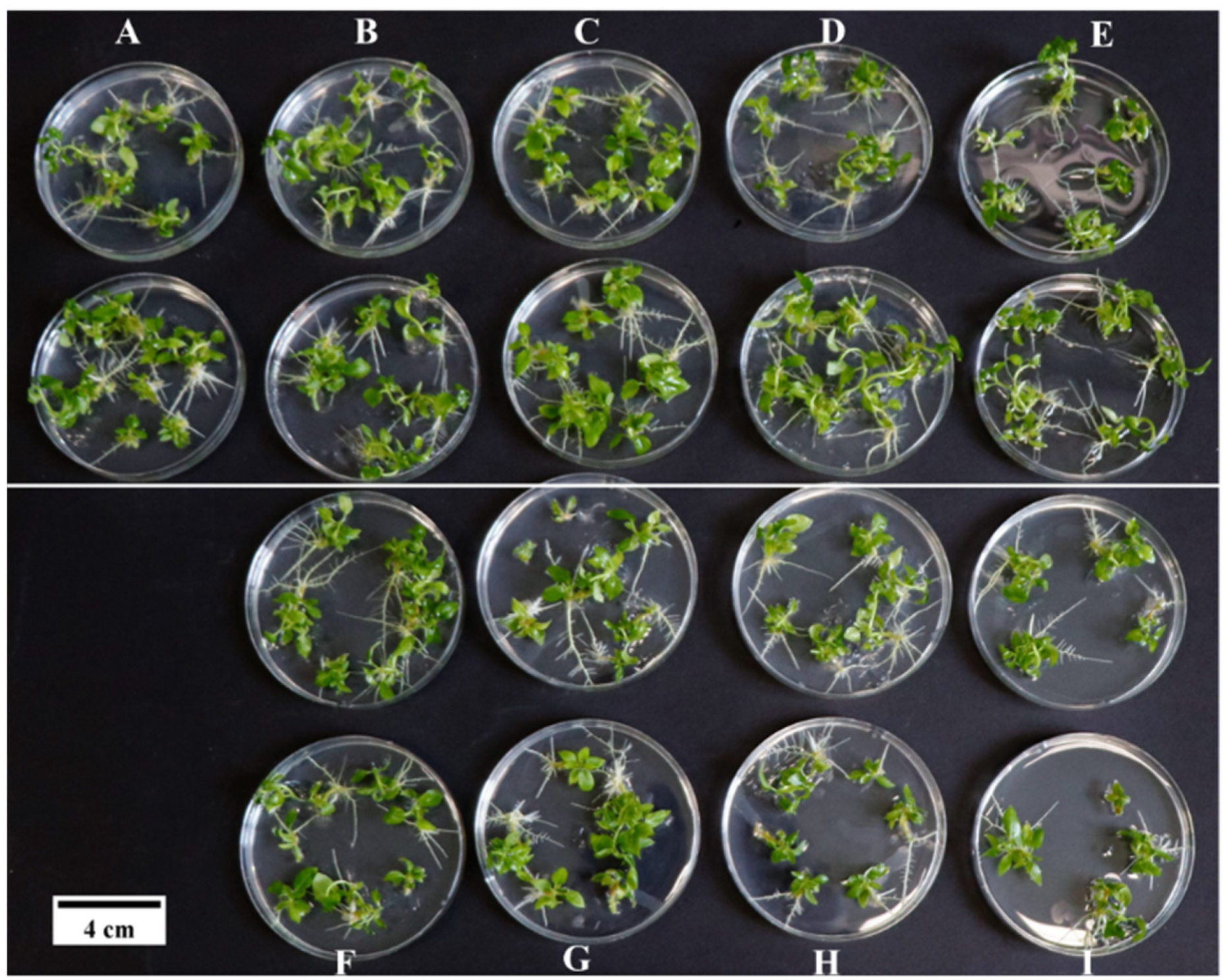
3.1. Effects of Medium Components on the Induction of Adventitious Buds of P. wulianensis Leaves

The multifactorial ANOVA results for the number of adventitious buds induced by the leaves of P. wulianensis with different treatments. The F values for factors A, C, and D were 77.43, 15.39, and 49.29, respectively, reaching a highly significant level (p < 0.01). Conversely, the NAA concentration had no significant effect on the number of regenerated buds (Table 2 and Table S1). This suggested that the base medium had the most significant influence on P. wulianensis leaf redifferentiation, followed by dark culture time and 6-BA hormone concentration, and the NAA hormone concentration had the least effect.
Further multiple comparative analyses of the three factors (base medium, dark culture time, and 6-BA hormone concentration) revealed (Table S2) that leaf inoculation into medium 1 induced the highest number of adventitious buds at 38, demonstrating a highly significant difference compared with media 2 and 3. Therefore, 1/2(NH4NO3) MS was selected as the optimal base medium in this study.
The analysis of the effects of dark culture treatments on leaf adventitious bud regeneration (Table S2) revealed that 1 (0 d) of dark culture significantly differed in the number of adventitious buds compared to 2 (4 d) and 3 (8 d). However, the difference between days 2 (4 d) and 3 (8 d) was not significant. The number of induced adventitious buds was highest after 8 d of dark culturing (23 buds). Subsequently, the dark treatment time was set to 8 d.
The analysis of 6-BA addition concentration on leaf adventitious bud regeneration (Table S2) revealed a highly significant difference in the number of induced adventitious buds in treatment 3 (1.0) compared to treatments 1 (0.1) and 2 (0.5). However, the difference between treatments 1 (0.1) and 2 (0.5) was not statistically significant. Treatment 3 (1.0) had the lowest number of induced adventitious buds. Therefore, the subsequent addition of 6-BA concentration was 0.5 mg/L.
The optimal medium combination for inducing adventitious buds was A1B2C2D3, which corresponded to 1/2(NH4NO3) MS + 0.05 mg/L NAA + 0.5 mg/L 6-BA. Under these conditions, 35 adventitious buds per leaf were induced after 8 d of dark culture (Table 2 and Figure 3).

3.2. Screening of Critical Antibiotic Concentrations

In plant genetic transformation systems, two types of antibiotics are commonly used: bacteriostatic agents (which inhibit Agrobacterium growth) and screeners (which identify transformed agents). The selection of antibiotics and their critical concentrations are crucial for establishing a genetic transformation system. In this experiment, the vector used was PEZR-(K)-LC, with Kan as the selective antibiotic marker. Timentin was selected as the bacteriostatic agent for genetic transformation mediated by Agrobacterium tumefaciens.

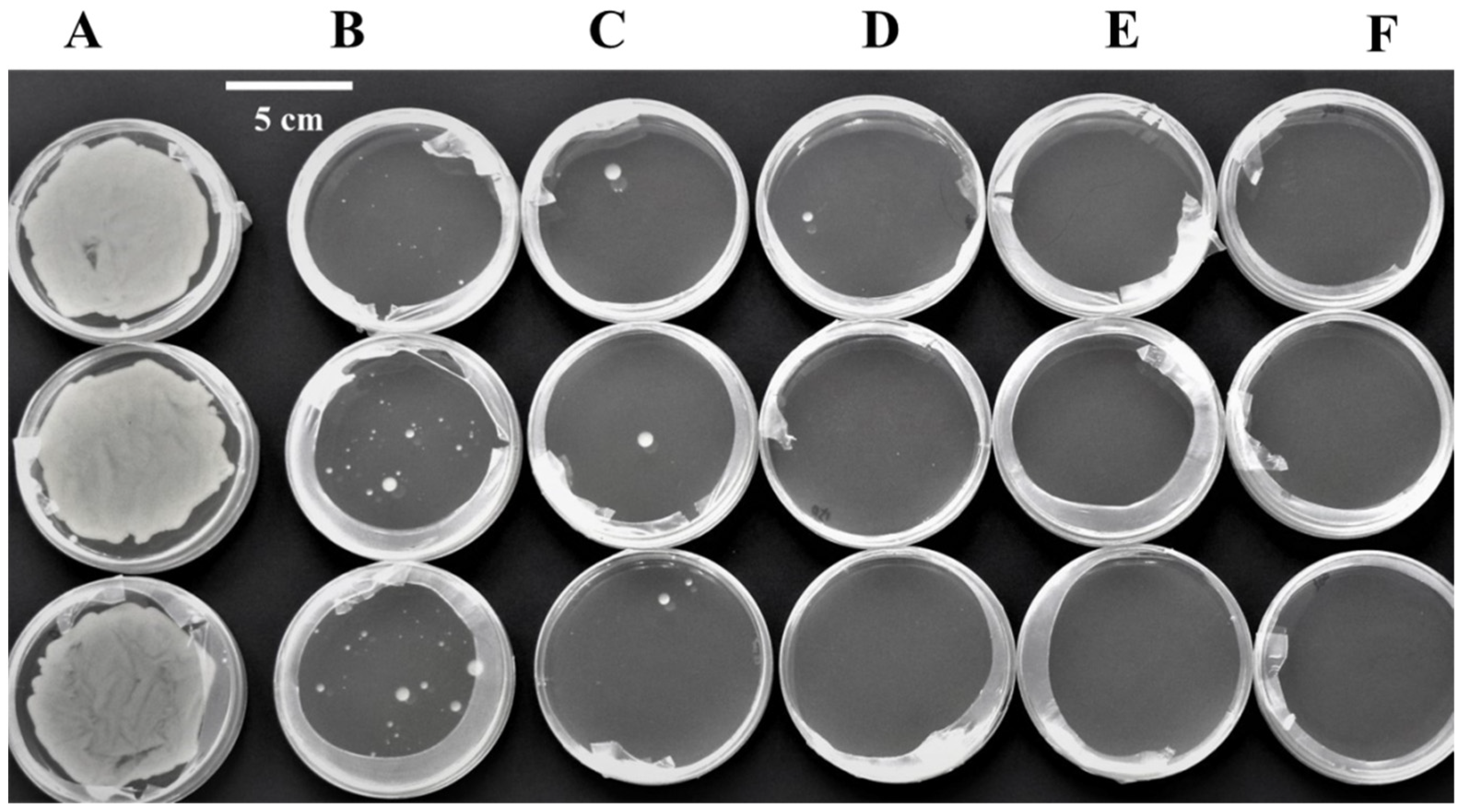
3.2.1. Determination of Critical Screening Pressure for Timentin

The inhibitory effect of timentin on Agrobacterium tumefaciens GV3101 increased significantly with higher timentin concentrations (Figure 4). No bacterial spots were observed in the culture medium at concentrations ≥ 200 mg/L. Consequently, the timentin concentration for the subsequent genetic transformation test was set at ≥200 mg/L.
To study the effect of timentin on the induction of adventitious buds in leaves, various antibiotic concentrations were added to the optimal medium. The number of differentiated buds in different parts of the leaves (base, middle, and tip) initially increased and then decreased with increasing antibiotic concentrations until no more adventitious buds were induced (Figure 5).
When using the middle of the leaves as explants, the number of clustered buds was 32.3 at 0 mg/L bacteriostatic agent concentration, and it reached a maximum of 50.3 at 300 mg/L. However, at bacteriostatic agent concentrations of ≥450 mg/L, the number of differentiated adventitious buds significantly decreased (Figure 5i and Table 3). Therefore, the optimal timentin concentration for this purpose was 300 mg/L.
When using the leaf base as explants, the number of induced adventitious buds was 14.7 at 0 mg/L timentin concentration and peaked at 44 at 400 mg/L, the highest observed level. However, the number of differentiated adventitious buds significantly decreased at bacteriostatic agent concentrations ≥ 700 mg/L (Figure 5ii and Table 3). Therefore, the optimal timentin concentration for leaf-base explants was determined to be 400 mg/L.
When using the leaf tip as the explant, the number of induced adventitious buds was 16.0 at 0 mg/L and peaked at 24.3 at 300 mg/L, the highest timentin concentration. However, the number of differentiated adventitious buds significantly decreased when the bacteriostatic agent concentration was ≥500mg/L (Figure 5ii and Table 3). Therefore, the optimal timentin concentration for leaf tip explants was determined to be 300 mg/L.
The analysis of timentin’s effect on adventitious bud rooting revealed that although the rooting rate remained at 100% with increasing bacteriostatic agent concentrations, the growth of rooted seedlings was affected. Growth slowed notably at timentin concentrations ≥ 400 mg/L (Figure 6). Consequently, the timentin concentration for the subsequent induction of root formation in leaf adventitious buds was set at 300 mg/L.

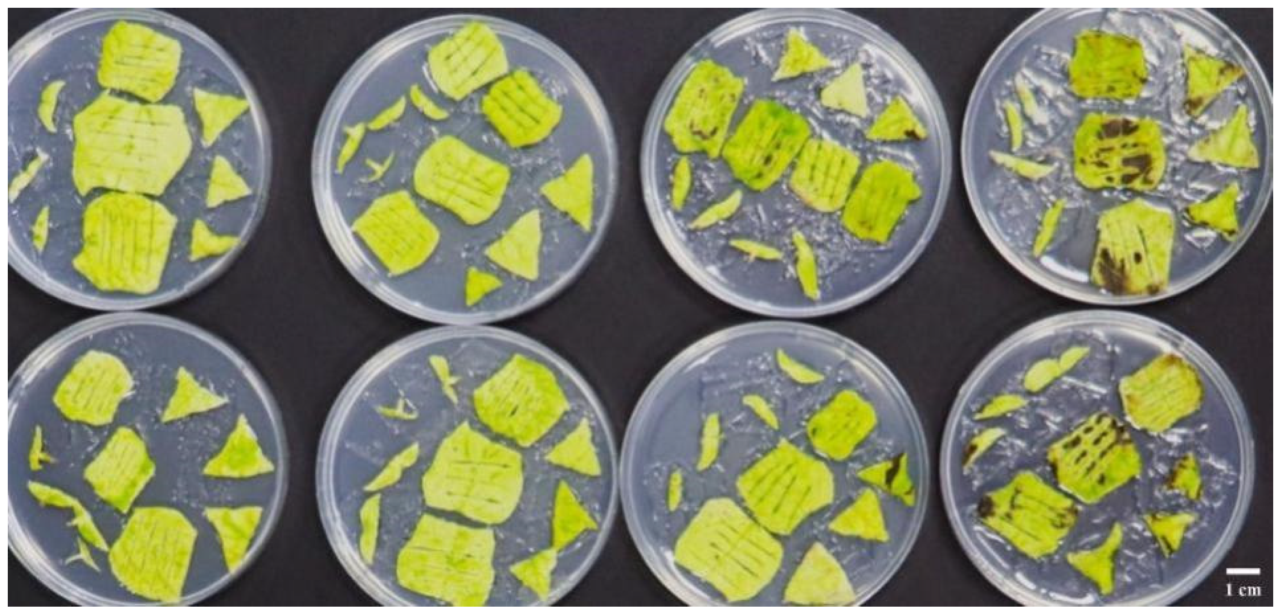
3.2.2. Determination of Kan Critical Screening Pressure

To determine the critical pressure of P. wulianensis on Kan, the tests were conducted on Kan concentrations to induce adventitious buds and rooting in leaves (base, middle, and tip). The results revealed that Kan significantly inhibited adventitious bud induction in P. wulianensis leaves (Figure 7 and Table 4), with an inhibitory effect increasing at higher concentrations.
When the leaf base was used as the explant, few adventitious buds formed at a Kan concentration of 125 mg/L, and the base only enlarged at 150 mg/L without differentiated buds. When the middle of the leaf was used as the explant, a small number of adventitious buds appeared at a Kan concentration of 75 mg/L, whereas at 100 mg/L, the leaf blades yellowed without differentiated buds. Using the leaf tip as the explant, 25 mg/L Kan produced a few adventitious buds, and 50 mg/L led to yellowing of leaves without buds. These results indicated that the critical Kan concentrations were 150 mg/L for the leaf base, 100 mg/L for the leaf middle, and 50 mg/L for the leaf tip as genetic transformation materials.
Kan also inhibited the adventitious rooting of P. wulianensis, with the inhibitory effect increasing with concentration (Figure 8). In the control group without Kan, the rooting rate was 100%, and the plants were healthy and green. At a Kan concentration of 25 mg/L, the rooting rate decreased to half that of the control group, and seedling growth was slightly slower than that of the control. At 75 mg/L, the rooting rate was 23.08%, with curled and vitrified leaves. At a concentration of 100 mg/L, the antibiotic completely inhibited adventitious bud rooting, resulting in dwarfed plants. At concentrations exceeding 100 mg/L, the adventitious buds ceased growth and extension, whereas the leaves exhibited yellowing, drying, and death. The optimal Kan concentration for rooting P. wulianensis adventitious buds was 100 mg/L (Table 4).

3.3. Optimization of a Genetic Transformation System for P. wulianensis

The effect of Agrobacterium on the transformation efficiency of P. wulianensis was also studied. Using a high concentration (OD600 = 0.6) infected an average of one adventitious bud per leaf disc, with the discs fading green and becoming necrotic (Figure 9). The inhibitory efficiency of timentin against Agrobacterium was low, along with the substantial Agrobacterium propagation. When the concentration of Agrobacterium was OD600 = 0.3, an average of five adventitious buds were induced in each leaf disc. The regenerated buds exhibited good growth and produced a lower and controllable amount of Agrobacterium. However, at an Agrobacterium concentration of OD600 = 0.15, it was challenging to induce resistant buds in the leaf discs, which eventually dried and died (Table 5). Consequently, the optimal Agrobacterium concentration for infection was determined to be OD600 = 0.3.
The duration of Agrobacterium infection significantly affected P. wulianensis transformation (Table 6). A 15-min infection resulted in the highest differentiation efficiency, with an average of six resistant adventitious buds per leaf disc. Shorter infection times led to fewer regeneration buds, whereas longer times dried up leaf discs, reducing regeneration bud numbers. Hence, a 15-min infection was deemed optimal in this study.
The addition of β-AS to both the infection solution and the co-culture solid medium significantly influenced the transformation rate of P. wulianensis leaves (Table 7). The number of resistant buds increased with increasing β-AS concentrations. However, at 200 μmol/L, regenerated resistant buds exhibited brown spots and poor growth. At 100 μmol/L, leaf discs remained green, and regenerated buds grew healthily and abundantly, making this the final selected concentration.
In the study on the effect of co-culture (dark culture) on P. wulianensis transformation efficiency, we observed that after 1 d of dark culture, leaf discs did not produce adventitious buds, gradually withered, and died later (Figure 10). The highest transformation rate occurred after 3 d of co-culture, with an average of 10 resistant adventitious buds per leaf disc. Prolonged co-cultivation beyond three days led to flourishing Agrobacterium populations, causing leaf disc deterioration and a significant decrease in resistant adventitious bud production (Table 8). The optimal co-culture time was determined to be 3 d.

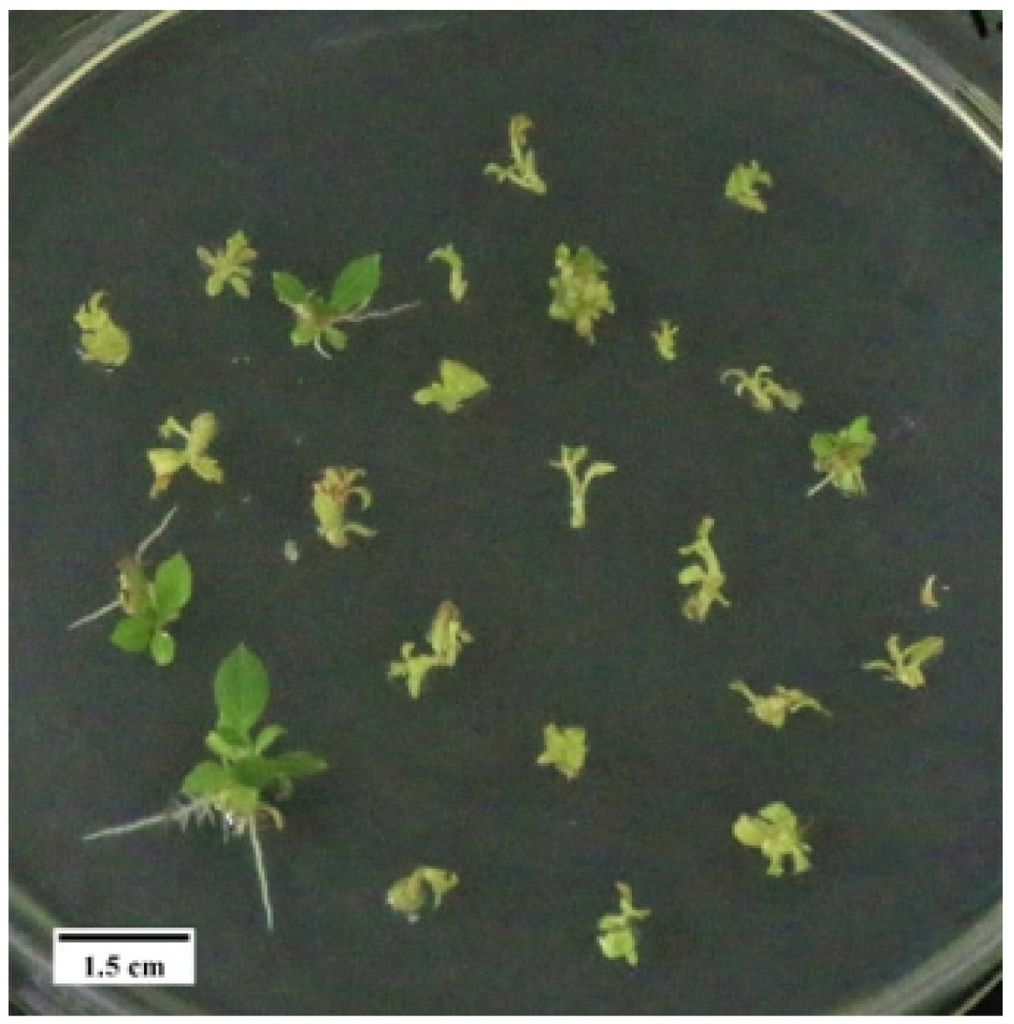
3.4. Verification of a Genetic Transformation System for P. wulianensis

A genetic transformation system for P. wulianensis was established as follows: leaves were infected with MS liquid medium resuspended to an OD600 of 0.3 for 15 min and co-cultured on differentiation medium for 3 d, followed by transfer to adventitious bud screening medium containing Kan 100 mg/L and timentin 300 mg/L. The medium was changed every 3 d for the first week and every 7 d from the second week. After 35 d of culture, the resistant adventitious buds induced from the leaves were transferred to the resistant rooting medium containing Kan 100 mg/L and timentin 300 mg/L. After 30 d, resistant rooted seedlings were selected for refinement and transplantation, and DNA and RNA levels were examined after an additional 20 d of growth.
To validate the transformation system of P. wulianensis, we used a PEZR-(K)-LC plasmid carrying the EGFP gene. Following the established genetic transformation system, we obtained 112 Kan-resistant adventitious buds that grew on the rooting medium, resulting in 17 resistant rooted seedlings (Figure 11). After transplantation (Figure 12A), the genomic DNA was extracted from the plants for PCR detection. All eight rooted seedlings showed amplified bands of the expected sizes (729 bp), indicating successful integration of the EGFP gene into the P. wulianensis genome. The proportion of positive plants was 47.06% of the total number of root seedlings (Figure 12B). Semi-quantitative PCR confirmed the presence of target bands in all eight transgenic-positive plants, with no target bands found in WT plants (Figure 13). In order to detect whether EGFP protein was expressed, we tore off the epidermis of the leaves of overexpressed EGFP gene and WT strains for fluorescence microscopy. The results in Figure 14 show that fluorescent signals were found at the location of the nuclei of the DAPI-stained cells in the leaves of plants overexpressing the EGFP gene, which were not found in the WT strain. All of the above results suggested the stable expression of the exogenous EGFP gene in the P. wulianensis genome.

4. Discussion

4.1. Comparison of Adventitious Bud Regeneration System of Poplar

The organogenesis pathway has an impact on genetic transformation. The existing poplar transformation systems generally use two main methods for bud induction. One method is indirect induction: induce cell division and callus growth on the cut surfaces of the explants, followed by bud induction from the callus [15]. Yevtushenko et al. [12] inoculated infested explants into a healing-induction medium supplemented with 6-BA and NAA for cultivation and then transferred them to the bud differentiation medium for screening of resistant adventitious buds after 2 to 3 weeks. This indirect induction way can effectively reduce the false positive rate of P. nigra NM6 transgene but also the transformation efficiency of poplar. For example, the use of Agrobacterium tumefaciens to infect the 40–50 d seedling-aged stem segments of P. trichocarpa and then screening the resistant indeterminate buds after the induction of callus, with a maximum transformation efficiency of only 5.7% [22]. In addition, callus induction is time-consuming and complex, and methods involving the callus stage may result in plants that differ from the maternal plant owing to somatic clonal variation [23]. Another approach is direct induction: induce buds directly from Agrobacterium-infected poplar-sliced explants [2,24]. For example, Li et al. [25] improved the formulation of shoot regeneration medium (SRM) and inoculated Agrobacterium-infected tissue culture seedlings explants into SRM medium, which could directly differentiate into buds without going through the redifferentiation stage of callus. This not only effectively reduced the transgenic cycle of P. trichocarpa to 2 months but also increased the genetic transformation efficiency to 26.7%. In addition, the integration of exogenous genes based on the direct/indirect organogenesis pathway resulted in a high level of gene chimerism. In order to reduce the problem of this transformation, Zhan et al. [26] also proposed a “ root first and then bud ” method, in which Agrobacterium tumefaciens was used to infect the leaf discs of P. kitakamiensis, and the rooting hormone (1.5 mg/L IBA) was used to induce the differentiation of the leaf discs to produce adventitious roots, and then cells were induced to differentiation adventitious buds to form complete resistant plants. The proportion of rooted adventitious buds obtained in this way was as high as 55%, and the positive rate was as high as 76.5%. In this study, direct induction through orthogonal tests revealed that the average number of adventitious buds induced by leaves exceeded 35 when using medium (1/2(NH4NO3) MS + 0.05 mg/L NAA + 0.5 mg/L 6-BA) and dark culture for 8 d (Table 2), and the tissue culture seedlings could be successfully sub-cultured. The average number of induced adventitious buds in the leaves of the poplar and P. tremuloides hybrids ranged from 3 to 20 (Table 9). For example, the average numbers of induced adventitious buds in leaf differentiation of P. tomentosa [27], P. tremuloides [28], P. leucopyramidalis [29], and P. tremula × P. tremuloides [30] were 3.4, 10, 8.2, and 2.73, respectively. Therefore, this study system is one of the best systems among the poplar tree species currently available.

4.2. Comparison of Poplar Genetic Transformation Systems

The efficiency of poplar genetic transformation is closely related to plant genotypes. Numerous studies have shown significant differences in the transformation rates between genotypes, even within the same plant species. Table 10 demonstrates the varying positive yields in genetic transformation using leaf discs from both P. tremuloides and P. tremuloides hybrids. Long et al. [36] achieved an optimized genetic transformation efficiency of 9.38% using P. tomentosa as the test material. In contrast, Hu et al. [37] found that P. tomentosa transformed more easily, with a transformation efficiency as high as 36.32%. Yu et al. [13] obtained 86 resistant plants through genetic transformation using male diploid poplar as the test material, 14 of which were identified as positive single plants through molecular analyses. In addition, when using P. dabidiana × P. bolleana leaves as exosomes for genetic transformation, different studies reported significant differences in transformation efficiencies: 9.3% [2]; 30.0% [4]; and 93.3% [38]. Ma et al. [15] sequenced the genome of P. alba var. pyramidalis during genomic research, achieving an average positive transformation rate of 23.6% using leaf disc indirectly induced genetic transformation. For the same poplar, P. tremuloides [39], the positive transformation rate through leaf-induced indirect pathways was as high as 94.17%, possibly because of varying degrees of sensitivity to Agrobacterium among different genotypes.
This study successfully integrated the EGFP gene into the genomic DNA of P. wulianensis using leaf discs, achieving stable expression with a positive transformation rate of 47.06%. Establishing a genetic transformation system for P. wulianensis not only enriched the germplasm resources of P. tremuloides but also provided transgenic “tools” for molecular plant breeding of poplar and other woody plants. This system can also serve as a technical and theoretical basis for genetic engineering.

4.3. Analysis of Factors Affecting Conversion Efficiency

The genetic transformation plays a crucial role in plant molecular biology and functional genomic research. Since the establishment of the first successful poplar transformation system [6], poplar has been transformed for genetic improvement and gene function studies more frequently than any other woody species [42,43]. Several factors influence transformation efficiency and regeneration during poplar genetic transformation, such as explants material, bacterial type, growth stage, the concentration of added β-AS, concentration of the bacteriostatic agent, co-culture time, and duration of Agrobacterium infection. The concentration of the added inducer is an important influencing factor. Rhizobium infects only the damaged parts of the plant, and the cells release some inducers similar to phenols or acidic polysaccharides when the plant undergoes damage and repair. Therefore, the cellular damage signals from the environment are transmitted to Agrobacterium cells through outer membrane protein when necessary, which ultimately leads to the expression of the vir genes and T-DNA transfer [41]. The most effective inducer is AS, which can activate the vir gene on the Agrobacterium plasmid and improve the transformation rate [15]. Li et al. [25] used the leaf blade of hybrid poplar (P. nigra var. betulifolia × P. trichocarpa) as the material, they found that AS and pH were the main factors affecting the efficiency of the genetic transformation with the help of GUS reporter genes, and the concentration of AS should be 25–30 μmol/L, while too high concentration would reduce the genetic transformation efficiency. In addition, the addition of 100 μmol/L AS to the medium of infiltration-transformed P. alba var. pyramidalis promoted the effective knockdown and efficient overexpression of the target gene [15]. Additionally, the bacteriostatic agent is also an important influencing factor. Bacteriostatic agents effectively kill or inhibit Agrobacterium in transformed explants; they can also affect the growth, isolation, and differentiation of plant cells. Higher concentrations of bacteriostatic agents generally result in better bacteriostatic effects but also increase the side effects of the explants [44]. In a study on tea tissue culture seedlings, bacteriostatic agents induced clumping of seedlings. Yang et al. [45] observed that Cefquinome and carbenicillin significantly reduced the survival rate of tea explants, the clumped bud induction rate of single explants, and the proliferation rate of clumped buds, showing a concentration-dependent inhibitory effect. Similar effects were also noted in Nicotiana tabacum [46] and Fraxinus profunda [47].
In this study, timentin was used as a bacteriostatic agent in the genetic transformation of P. wulianensis leaf discs. The results showed that timentin at concentrations ≥ 200 mg/L not only effectively inhibited the Agrobacterium growth but also increased the number of induced adventitious buds in all parts of the leaf blade (leaf base, middle, and tip). This positive effect is similar to timentin’s promotion of tufted bud growth rate and seedling formation rate in tea tissue-cultured seedling explants [45], its effectiveness in inducing secondary embryogenesis in Chrysanthemum [48], and promotion of adventitious bud differentiation in N. tabacum leaf discs (127% increase after one month of culture) [46]. This effect may be attributed to the chemical structure of timentin, which resembles growth hormones, such as 2,4-D and NAA, thereby exhibiting hormonal effects in tissue culture [49].

4.4. False Positive Exclusion Analysis

In plant genetic transformation, chimeric buds comprising transgenic and non-transgenic parts form with a certain probability [50,51]. PCR is commonly used to identify transgenic plants because it is a powerful and direct tool for detecting target genes inserted into the genome [2]. In this study, we successfully detected transgenic poplar plants using PCR with an optimized vector and gene cross-specific primers, confirming the successful insertion of the EGFP gene into the poplar genome in eight transgenic poplar lines (Figure 12A). Direct identification of resistant tissue culture leaves can yield “false-positive” results due to Agrobacterium residues. Using Agrobacterium-specific primers, such as the tzs gene [24,52], or sequences other than plasmid T-DNA, such as the aadA gene [14], for contamination identification may inadvertently exclude positive plants. In this study, resistant seedlings underwent transplantation for refinement and were grown under natural light for approximately 1 month. This approach ensured that the gradual dilution of Agrobacterium did not interfere with the identification results. Additionally, the DNA and RNA levels were verified to ensure the accuracy of the experimental results. In addition, based on the efficient genetic transformation system of P. wulianensis obtained in this experiment, it has been successfully applied to the genetic transformation of PwuWRKY48 gene [53].

5. Conclusions

In this study, we established a genetic transformation system using P. wulianensis as a genotype for the first time. First, we established the optimal medium (1/2(NH4NO3) MS + 0.05 mg/L NAA + 0.5 mg/L 6-BA) for efficient leaf redifferentiation of adventitious buds and also explored the effects of Kan and timentin on the induction of adventitious buds and adventitious bud-induced rooting of P. wulianensis leaves. Meanwhile, we determined and optimized a number of factors affecting the efficiency of genetic transformation, including the concentration of a bacterial solution (OD600 = 0.3), infection time (15 min), co-culture time (3 d), and β-AS (100 μmol/L), etc. Finally, we established a genetic transformation system of P. wulianensis with high efficiency and stable expression of exogenous genes and verified its transformation of exogenous EGFP gene, resulting in a high positive transformation yield of 47.06%. These findings can facilitate genetic improvements in breeding programs and provide a foundation for future gene research.

Supplementary Materials

The following supporting information can be downloaded at https://www.mdpi.com/article/10.3390/f15081474/s1, Table S1: Variance analysis of effects of different factors on the regeneration number of P. wulianensis adventitious buds; Table S2: Duncan multiple comparisons of the effects of the three factors on the induction of adventitious buds in P. wulianensis.

Author Contributions

Conceptualization, J.X. and Y.L.; methodology, Y.H., R.W., L.G., Y.M., T.S., X.X. and Y.Z.; investigation, Y.W.; writing—original draft preparation, Y.W. and C.S.; writing—review and editing, J.X.; supervision, J.X. and Y.L.; funding acquisition, Y.W. and Y.L. All authors have read and agreed to the published version of the manuscript.

Funding

We gratefully acknowledge financial support for this research from the Youth Project of Shandong Provincial Natural Science Foundation (ZR2020QC165), the Subject of Key R & D Plan of Shandong Province (2021LZGC02304), and the Innovation Team for Conservation and Utilization of Precious Tree Species Germplasm project of the Department of Natural Resources of Shandong Province (LZYZZ202398).

Data Availability Statement

The data presented in this study are available on request from the corresponding author.

Conflicts of Interest

The authors declare no conflicts of interest.

Abbreviations

MSMurashige and Skoog medium
NAANaphthalene acetic acid
6-BA6-Benzylaminopurine
EGFPEnhanced green fluorescent protein
DAPI4′,6-Diamidino-2-phenylindole
PCRPolymerase chain reaction
β-ASAcetylsyringone
WTWild type

References

  1. Franklin, O.; Palmroth, S.; Näsholm, T. How eco-evolutionary principles can guide tree breeding and tree biotechnology for enhanced productivity. Tree Physiol. 2014, 34, 1149–1166. [Google Scholar] [CrossRef] [PubMed]
  2. Wang, H.; Wang, C.; Liu, H.; Tang, R.; Zhang, H. An efficient Agrobacterium-mediated transformation and regeneration system for leaf explants of two elite aspen hybrid clones Populus alba × P. berolinensis and Populus davidiana × P. bolleana. Plant Cell Rep. 2011, 30, 2037–2044. [Google Scholar] [CrossRef]
  3. Flachowsky, H.; Hanke, M.V.; Peil, A.; Strauss, S.H.; Fladung, M. A review on transgenic approaches to accelerate breeding of woody plants. Plant Breed. 2009, 128, 217–226. [Google Scholar] [CrossRef]
  4. Han, X.; Ma, S.; Kong, X.; Takano, T.; Liu, S. Efficient Agrobacterium-Mediated Transformation of Hybrid Poplar Populus davidiana Dode × Populus bollena Lauche. Int. J. Mol. Sci. 2013, 14, 2515–2528. [Google Scholar] [CrossRef] [PubMed]
  5. John, E.; Maqbool, A.; Malik, K. Optimization of Agrobacterium tumefaciens mediated transformation in Populus deltoids. Pak. J. Bot. 2014, 46, 1079–1086. [Google Scholar]
  6. Parsons, T.J.; Sinkar, V.P.; Stettler, R.F.; Nester, E.W.; Gordon, M.P. Transformation of Poplar by Agrobacterium tumefaciens. Nat. Biotechnol. 1986, 4, 533–536. [Google Scholar] [CrossRef]
  7. Yin, D.M. A review of the genomic and gene cloning studies in trees. Hereditas 2010, 32, 677–684. [Google Scholar] [CrossRef]
  8. Chen, Y.N.; Hu, C.J.; Zu, G.Q.; Hu, J.J.; Yin, D.M. Thirty Years of Agrobacterium-Mediated Genetic Transformation of Populus. Sci. Silvae Sin. 2022, 58, 114–129. [Google Scholar]
  9. Ding, L.P.; Wang, H.Z.; Wei, J.H. Progress and Prospect of Research in Transgenic Poplar. For. Res. 2016, 29, 124–132. [Google Scholar]
  10. Niedbała, G.; Niazian, M.; Sabbatini, P. Modeling Agrobacterium-Mediated Gene Transformation of Tobacco (Nicotiana tabacum)—A Model Plant for Gene Transformation Studies. Front. Plant Sci. 2021, 12, 695110. [Google Scholar] [CrossRef]
  11. Movahedi, A.; Zhang, J.; Amirian, R.; Zhuge, Q. An Efficient Agrobacterium-Mediated Transformation System for Poplar. Int. J. Mol. Sci. 2014, 15, 10780–10793. [Google Scholar] [CrossRef]
  12. Yevtushenko, D.P.; Misra, S. Efficient Agrobacterium-mediated transformation of commercial hybrid poplar Populus nigra L. × P. maximowiczii A. Henry. Plant Cell Rep. 2010, 29, 211–221. [Google Scholar] [CrossRef] [PubMed]
  13. Yu, Z.C.; Ling, C.; Chen, Y.N.; Li, S.X.; Yin, D.M.; Li, X.P. Establishment of regeneration system and study on genetic transformation for male diploid Populus tomentosa. J. Nanjing For. Univ. (Nat. Sci. Ed.) 2022, 46, 187–196. [Google Scholar]
  14. Wen, S.S.; Ge, X.L.; Wang, R.; Yang, H.F.; Bai, Y.E.; Guo, Y.H.; Zhang, J.; Lu, M.Z.; Zhao, S.T.; Wang, L.Q. An efficient Agrobacterium-mediated transformation method for hybrid Poplar 84K (Populus alba × P. glandulosa) using Calli as explants. Int. J. Mol. Sci. 2022, 23, 2216. [Google Scholar] [CrossRef]
  15. Ma, J.; Wan, D.; Duan, B.; Bai, X.; Bai, Q.; Chen, N.; Ma, T. Genome sequence and genetic transformation of a widely distributed and cultivated poplar. Plant Biotechnol. J. 2019, 17, 451–460. [Google Scholar] [CrossRef]
  16. Liang, S.B.; Li, X.W. A new species of populus from Shandong. Bull. Bot. Res. 1986, 6, 135–137. [Google Scholar]
  17. Wu, Q.; Zang, F.; Xie, X.; Ma, Y.; Zheng, Y.; Zang, D. Full-length transcriptome sequencing analysis and development of EST-SSR markers for the endangered species Populus wulianensis. Sci. Rep. 2020, 10, 16249. [Google Scholar] [CrossRef]
  18. Wu, Q.; Zang, F.; Ma, Y.; Zheng, Y.; Zang, D. Analysis of genetic diversity and population structure in endangered Populus wulianensis based on 18 newly developed EST-SSR markers. Glob. Ecol. Conserv. 2020, 24, e01329. [Google Scholar] [CrossRef]
  19. Wang, Y.; Wu, D.; Liu, L.J.; Han, Y.; Sun, T.; Wang, N.; Xie, X.M.; Dong, X.; Lu, Y.Z. Establishment of generation system for Populus wulianensis and control of vitrification of its test-tube seedlings. Mol. Plant Breed. 2019, 17, 6434–6446. [Google Scholar] [CrossRef]
  20. Jia, T.; Tang, T.; Cheng, B.C.; Zhou, L.; Peng, Y. Development of two protocols for Agrobacterium-mediated transformation of white clover (Trifolium repens) via the callus system. 3 Biotech 2023, 13, 150. [Google Scholar] [CrossRef]
  21. Porebski, S.; Bailey, L.G.; Baum, B.R. Modification of a CTAB DNA extraction protocol for plants containing high polysaccharide and polyphenol components. Plant Mol. Biol. Rep. 1997, 15, 8–15. [Google Scholar] [CrossRef]
  22. Ma, C.; Strauss, S.H.; Meilan, R. Agrobacterium-mediated transformation of the genome-sequenced poplar clone, Nisqually-1 (Populus trichocarpa). Plant Mol. Biol. Rep. 2004, 22, 311a. [Google Scholar] [CrossRef]
  23. Giri, C.C.; Shyamkumar, B.; Anjaneyulu, C. Progress in tissue culture, genetic transformation and applications of biotechnology to trees: An overview. Trees 2004, 18, 115–135. [Google Scholar] [CrossRef]
  24. Song, C.; Lu, L.; Guo, Y.; Xu, H.; Li, R. Efficient Agrobacterium-Mediated Transformation of the Commercial Hybrid Poplar Populus Alba × Populus glandulosa Uyeki. Int. J. Mol. Sci. 2019, 20, 2594. [Google Scholar] [CrossRef]
  25. Li, S.J.; Zhen, C.; Xu, W.J.; Wang, C.; Cheng, Y.X. Simple, rapid and efficient transformation of genotype Nisqually-1: A basic tool for the first sequenced model tree. Sci. Rep. 2017, 7, 2638. [Google Scholar] [CrossRef]
  26. Zhan, X.C.; Kawai, S.; Katayama, Y.; Morohoshi, N. A new approach based on the leaf disc method for Agrobacterium mediated transformation and regeneration of aspen. Plant Sci. 1997, 123, 105–112. [Google Scholar] [CrossRef]
  27. Li, C.L.; Wang, X.J.; Ding, Q.Q.; Wu, H. Optimization of leaf regeneration and genetic transformation system of Populus tomentosa. Bull. Bot. Res. 2016, 36, 177–183. [Google Scholar]
  28. Huang, D.; Dai, W. Direct regeneration from in vitro leaf and petiole tissues of Populus tremula ‘Erecta’. Plant Cell Tissue Organ Cult. (PCTOC) 2011, 107, 169–174. [Google Scholar] [CrossRef]
  29. Li, J.H.; Xing, S.Y.; Jiao, Z.; Liu, X.G.; Zhang, Y.P. Tissue culture and regeneration system from leaves of Populus leucopyramidalis in vitro. Chin. Agric. Sci. Bull. 2008, 24, 60–64. [Google Scholar]
  30. Wang, B.; Ling, B.L.; Zhang, J.F.; Hong, X.L.; Mu, C.L. Establishment of in vitro regeneration system of Poplar Hybrid 717 and 353. Acta Bot. Boreali-Occident. Sin. 2009, 29, 704–710. [Google Scholar]
  31. Huang, S.; Gao, K.; Miao, D.Y.; An, X.M. Establishment of regeneration and genetic transformation system of elite clone GM107 of Populus tomentosa. Mol. Plant Breed. 2022, 21, 1–22. [Google Scholar]
  32. Li, K.Y.; Fan, J.F.; Zhao, Z.; Zhou, Y.X.; Gao, J.S. Prioritization of the system of regeneration and genetic transformation of Populus 84K. J. Northwest A F Univ. (Nat. Sci. Ed.) 2007, 35, 90–96. [Google Scholar]
  33. Zhang, Y.; Fan, J.F.; Gao, J.S.; Zhou, Y.X. Establishment of an efficient regeneration system for the Populus adenopoda × the poplar 84K. North. Hortic. 2010, 9, 152–156. [Google Scholar]
  34. Wang, H.L. Establishment of efficient tissue culture regeneration system of Populus. North. Hortic. 2010, 13, 174–175. [Google Scholar]
  35. Wu, R.H.; Dai, J.L.; Bai, X.D.; Bai, Y.E. Establishment of the regeneration system of Populus hopeiensis leaf induced adventitious buds. J. Inn. Mong. Agric. Univ. (Nat. Sci. Ed.) 2021, 42, 34–38. [Google Scholar]
  36. Long, C.; Pang, X.M.; Cao, G.L.; Liu, Y.; Zhang, Z.Y. A study on the efficient protocol for transforming MdSPDS1 gene into Populus tomentosa Carr. J. Beijing For. Univ. 2010, 32, 21–26. [Google Scholar]
  37. Hu, L.; Lu, H.; Liu, Q.; Chen, X.; Jiang, X. Overexpression of mtlD gene in transgenic Populus tomentosa improves salt tolerance through accumulation of mannitol. Tree Physiol. 2005, 25, 1273–1281. [Google Scholar] [CrossRef]
  38. Zhang, B.Y.; Su, X.H.; Zheng, S.X. Establishment of a highly efficient plant regeneration system of Populus davidiana × P. bolleana and study of its genetic stability. J. Beijing For. Univ. 2008, 30, 68–73. [Google Scholar]
  39. Tsai, C.J.; Podila, G.K.; Chiang, V.L. Agrobacterium-mediated transformation of quaking aspen (Populus tremuloides) and regeneration of transgenic plants. Plant Cell Rep. 1994, 14, 94–97. [Google Scholar] [CrossRef]
  40. Zhang, B.Y.; Su, X.H.; Huang, Q.J.; Zhang, X.H.; Hu, Z.M. Regeneration of transgenic Poplar (Populus alba × P. glandulosa) expressing levansucrase from Bacillus subtilis. Sci. Silvae Sin. 2005, 41, 48–53. [Google Scholar]
  41. Dai, W.; Cheng, Z.-M.; Sargent, W. Plant regeneration and Agrobacterium-mediated transformation of two elite aspen hybrid clones from in vitro leaf tissues. Vitr. Cell. Dev. Biol.-Plant 2003, 39, 6–11. [Google Scholar] [CrossRef]
  42. Busov, V.B.; Brunner, A.M.; Meilan, R.; Filichkin, S.; Ganio, L.; Gandhi, S.; Strauss, S.H. Genetic transformation: A powerful tool for dissection of adaptive traits in trees. New Phytol. 2005, 167, 9–18. [Google Scholar] [CrossRef] [PubMed]
  43. Hu, J.; Wang, L.; Yan, D.; Lu, M.Z. Research and Application of Transgenic Poplar in China. In Challenges and Opportunities for the World’s Forests in the 21st Century; Fenning, T., Ed.; Springer: Dordrecht, The Netherlands, 2014; pp. 567–584. [Google Scholar]
  44. Rajesh, M.; Jeyaraj, M.; Sivanandhan, G.; Subramanyam, K.; Mariashibu, T.S.; Mayavan, S.; Kapil Dev, G.; Anbazhagan, V.R.; Manickavasagam, M.; Ganapathi, A. Agrobacterium-mediated transformation of the medicinal plant Podophyllum hexandrum Royle (syn. P. emodi Wall. ex Hook.f. & Thomas). Plant Cell Tissue Organ Cult. (PCTOC) 2013, 114, 71–82. [Google Scholar] [CrossRef]
  45. Yang, Y.P.; Li, Y.L.; Lu, J.L.; Zheng, X.Q. Antibiotics inhibition to Agrobaceterium rhizogenes and effect to tea multiple shoots. J. Tea Sci. 2015, 35, 437–442. [Google Scholar] [CrossRef]
  46. Nauerby, B.; Billing, K.; Wyndaele, R. Influence of the antibiotic timentin on plant regeneration compared to carbenicillin and cefotaxime in concentrations suitable for elimination of Agrobacterium tumefaciens. Plant Sci. 1997, 123, 169–177. [Google Scholar] [CrossRef]
  47. Stevens, M.E.; Pijut, P.M. Agrobacterium-mediated genetic transformation and plant regeneration of the hardwood tree species Fraxinus profunda. Plant Cell Rep. 2014, 33, 861–870. [Google Scholar] [CrossRef]
  48. Naing, A.H.; Kim, C.K.; Yun, B.J.; Jin, J.Y.; Lim, K.B. Primary and secondary somatic embryogenesis in Chrysanthemum cv. Euro. Plant Cell Tissue Organ Cult. (PCTOC) 2013, 112, 361–368. [Google Scholar] [CrossRef]
  49. Lin, J.J.; Assad-Garcia, N.; Kuo, J. Plant hormone effect of antibiotics on the transformation efficiency of plant tissues by Agrobacterium tumefaciens cells. Plant Sci. 1995, 109, 171–177. [Google Scholar] [CrossRef]
  50. Li, B.; Xie, C.; Qiu, H. Production of selectable marker-free transgenic tobacco plants using a non-selection approach: Chimerism or escape, transgene inheritance, and efficiency. Plant Cell Rep. 2009, 28, 373–386. [Google Scholar] [CrossRef]
  51. Cervera, M.; Pina, J.A.; Juárez, J.; Navarro, L.; Peña, L. A broad exploration of a transgenic population of citrus: Stability of gene expression and phenotype. Theor. Appl. Genet. 2000, 100, 670–677. [Google Scholar] [CrossRef]
  52. Akiyoshi, D.E.; Regier, D.A.; Jen, G.; Gordon, M.P. Cloning and nucleotide sequence of the tzs gene from Agrobacterium tumefaciens strain T37. Nucleic Acids Res. 1985, 13, 2773–2788. [Google Scholar] [CrossRef] [PubMed]
  53. Wang, Y.; Li, M.T.; Mu, Y.J.; Guan, L.S.; Wu, F.S.; Liu, K.; Li, M.; Wang, N.; Zhuang, Z.J.; Zhao, Y.C.; et al. PwuWRKY48 Confers Drought Tolerance in Populus wulianensis. Forests 2024, 15, 302. [Google Scholar] [CrossRef]
Figure 1. Diagram of the blade cutting area. A, B, and C represent the tip, middle, and base parts of the leaf, respectively.
Forests 15 01474 g001
Figure 2. Expression vector PEZR—(K)—LC map.
Forests 15 01474 g002
Figure 3. Adventitious bud induction. (A,B) were the best induction medium for leaf differentiation of adventitious buds and the optimal induction medium for 35 d elongation of adventitious buds, respectively.
Forests 15 01474 g003
Figure 4. Effects of timentin concentrations on Agrobacterium Growth. (AF) were 0, 50, 100, 150, 200, and 250 mg/L, respectively.
Forests 15 01474 g004
Figure 5. Effects of timentin concentrations on the induction of adventitious buds. (i,ii) indicate adventitious bud results in the tip part of the leaf and the middle/base part of the leaf differentiation after the addition of timentin, respectively. (A–S) timentin with concentrations of 0, 50, 100, 150, 200, 250, 300, 350, 400, 450, 500, 550, 600, 650, 700, 750, 800, and 850 mg/L was added to the medium.
Forests 15 01474 g005
Figure 6. Effects of timentin concentrations on the rooting of adventitious buds. (A–I) timentin with concentrations of 0, 100, 200, 300, 400, 500, 600, 700, and 800 mg/L was added to the medium. Two replicates were used for each gradient in the figure.
Forests 15 01474 g006
Figure 7. Effects of Kan concentrations on adventitious budding induction in the leaves. (A–I) Kan with concentrations of 0, 25, 50, 75, 100, 125, 150, 175, and 200 mg/L was added to the medium. Two replicates were used for each gradient in the figure.
Forests 15 01474 g007
Figure 8. Effects of Kan concentrations on adventitious budding induction from rooting. (A–I) Kan with concentrations of 0, 25, 50, 75, 100, 125, 150, 175, and 200 mg/L was added to the medium. Each gradient was repeated twice.
Forests 15 01474 g008
Figure 9. Leaf discs after infection with a high concentration of Agrobacterium (OD600 = 0.6) and culturing for 10 d.
Forests 15 01474 g009
Figure 10. Leaf discs were cultured for 1 d, and adventitious buds were differentiated.
Forests 15 01474 g010
Figure 11. Rooted seedlings for Kan resistance screening.
Forests 15 01474 g011
Figure 12. Transgenic P. wulianensis. (A,B) represent the electropherograms of seedlings transplanted, resistant rooting seedlings, and DNA molecular identification, respectively. M, Marker. PC, Positive plants. WT, Wild type. H2O, negative control. Numbers 1–17, transgenic lines L1–L17.
Forests 15 01474 g012
Figure 13. Semi-quantitative PCR detection of EGFP in transgenic P. wulianensis. L1–5, L13, L15, and L17 were identified as positive clonal clones at the DNA level. WT, wild type.
Forests 15 01474 g013
Figure 14. Monitoring of EGFP expression. The brightfield, EGFP fluorescence, DAPI fluorescence, and the combined field (merged EGFP fluorescence, DAPI fluorescence, and brightfield) were present for the fusion protein (EGFP) (up) and control (WT) (down), respectively. Arrows in the same direction indicate the expression of EGFP in the nucleus.
Forests 15 01474 g014
Table 1. Orthogonal factors for induction of regeneration of adventitious buds from sterile.
LevelBasal Medium
(A)
NAA (mg/L)
(B)
6-BA (mg/L) (C)Dark Culture Time (d)
(D)
1(1/2NH4NO3) MS0.010.10
21/2MS0.050.54
3MS0.101.08
Table 2. Orthogonal test results of adventitious bud regeneration in detached leaves.
CodeABCDAverage Number of Regenerated Buds
13(MS)2 (0.05)3 (1.0)1 (0 d)1.67 ± 0.72 e
23(MS)3 (0.10)1 (0.1)2 (4 d)17.33 ± 1.70 c
32(1/2MS)1 (0.01)3 (1.0)2 (4 d)10.00 ± 0.82 d
42(1/2MS)3 (0.10)2 (0.5)1 (0 d)3.33 ± 2.05 e
52(1/2MS)2 (0.05)1 (0.1)3 (8 d)15.00 ± 3.27 c
61(1/2(NH4NO3) MS)3 (0.10)3 (1.0)3 (8 d)22.33 ± 2.05 b
71(1/2(NH4NO3) MS)1 (0.01)1 (0.1)1 (0 d)19.00 ± 2.94 c
83(MS)1 (0.01)2 (0.5)3 (8 d)17.67 ± 1.70 c
91(1/2(NH4NO3) MS)2 (0.05)2 (0.5)2 (4 d)34.67 ± 3.30 a
Influencing factors A, B, C, and D were basic medium, NAA, 6-BA, and dark culture time, respectively. The numbers in front of the brackets represent the numbers of different levels under the factor. Different lowercase letters indicate significant differences at the 0.05 level based on LSD.
Table 3. Statistics of adventitious buds under timentin treatment.
CodeConcentration
mg/L
Middle of
the Leaf
Top of
the Leaf
Bottom of
the Leaf
A032.30 ± 1.15 cd16.00 ± 0.66 bc14.70 ± 0.18 e
B5037.70 ± 0.35 bc//
C10039.30 ± 0.98 bc17.70 ± 0.98 bc15.30 ± 1.12 e
D15041.30 ± 1.52 ab//
E20042.30 ± 1.34 ab20.001.25 ab24.00 ± 1.75 cd
F25049.30 ± 0.33 a//
G30050.30 ± 1.71 a24.30 ± 1.26 a35.70 ± 1.27 b
H35040.00 ± 1.11 bc//
I40034.00 ± 0.68 bc20.70 ± 1.31 ab44.00 ± 2.05 a
J45027.30 ± 1.28 d//
K50016.00 ± 0.53 e13.00 ± 1.09 cd26.00 ±1.02 c
L55014.70 ± 1.14 e//
M60011.70 ± 0.39 ef7.70 ± 0.88 d17.00 ± 0.72 de
N6508.00 ± 0.27 ef//
O7007.70 ± 1.02 ef5.70 ± 0.42 e12.70 ± 0.93 e
P7507.00 ± 0.83 ef//
Q8002.70 ± 0.57 f0.00 ± 0.003.70 ± 0.14 f
R8501.70 ± 0.24 f//
S900/0.00 ± 0.001.70 ± 0.70 f
The data represent the mean ± standard deviation of three replicates. Different lowercase letters indicate significant differences at the 0.05 level based on LSD.
Table 4. Adventitious bud differentiation and adventitious bud rooting of leaves under different concentrations of Kan.
Kan Concentration(mg/L)Indeterminate Number of BudsLeaf-Induced Growth StatusRooting Rate
(%)
Adventitious Buds Induce Rooting
033.30 ± 1.28 aAll three parts of the leaf blade were dark green and had the highest number of differentiated buds.100.00All rooted, green plants, healthy growth.
2515.70 ± 1.09 bAll three parts of the leaf blade were green, and 50% of the B part of the leaf produced adventitious buds.50.00Half rooted, and the other leaves were light green.
505.00 ± 0.27 cLeaf A was slightly yellow; the rest was green, and 30% of leaf B produced adventitious buds.42.67Some were rooted, whereas others were yellowed.
753.70 ± 0.33 cOnly the C part of the leaf produced adventitious buds, whereas the other parts of the leaf did not produce adventitious buds.23.08A small part of the leaves took root, and a small part of the leaves appeared vitrified.
1002.00 ± 0.18 cOnly a small part of the leaf C part produced adventitious buds, and the leaf B part appeared yellow.0.00The base was swollen; none of them took root, and the adventitious buds grew slowly.
1251.70 ± 0.15 cVery few leaves had adventitious bud differentiation in the leaf C part, and no adventitious buds were produced in the others.0.00A few had swollen bases; none of them took root, and adventitious buds had stagnated growth.
1500.00 ± 0.00Dehydration began to appear in all three parts of the leaves, and there was no redifferentiation or adventitious buds.0.00The base was not enlarged and did not take root, and the adventitious bud leaves were partially yellowed.
1750.00 ± 0.00The three parts of the leaf showed different degrees of whitening, with no redifferentiation or adventitious buds.0.00The base was not swollen and did not take root, and the adventitious bud leaves were curled and yellowed.
2000.00 ± 0.00The three parts of the leaves were dehydrated and whitish, and there were no adventitious buds.0.00The base was not swollen and did not take root, and the leaves of adventitious buds were partially dry.
The data represent the mean ± standard deviation of three replicates. Different lowercase letters indicate significant differences at the 0.05 level based on LSD.
Table 5. Effect of bacterial concentration on screening resistant buds of P. wulianensis.
OD600Number of Resistant BudsGrowth State
0.601.44 ± 0.51 bThe leaves gradually turned pale green and eventually withered, often displaying white plaques.
0.305.11 ± 0.92 aA few leaf edges showed signs of fading with faintly visible colonies.
0.153.89 ± 0.87 abSeveral leaves exhibited fading with faintly visible colonies.
The results are expressed as mean ± standard error of three independent experiments. Different letters within each variable indicate statistically significant differences at p < 0.05. Same below.
Table 6. Effect of infection time on transformation of P. wulianensis resistant buds.
Infection Time/minNumber of Resistant BudsGrowth State
53.36 ± 0.45 bLeaf disks remained green, with no colonies visible.
156.38 ± 1.28 aLeaf blade margins displayed reddish brown, whereas wounds exhibited multiple buds and pale mycorrhizae.
202.92 ± 0.31 bLeaf disks appeared dry with visible marginal mycorrhiza.
The results are presented as the means and standard errors from three independent experiments. Within each variable, values with different letters indicate statistically significant differences at the p < 0.05 level.
Table 7. Effect of β-AS on screening of resistant buds.
β-AS Concentration
/μmol·L−1
Number Of Resistant BudsGrowth State
02.94 ± 0.73 bDry spots appeared with yellowing of the leaf margins.
1005.95 ± 1.12 aLeaf disks remained green, with many buds forming around the wounds.
2006.45 ± 1.34 aBrown spots developed, with more buds forming around the wound.
The results are presented as the means and standard errors from three independent experiments. Within each variable, values with different letters indicate statistically significant differences at the p < 0.05 level.
Table 8. Effect of dark culture (co-culture) time on transformation.
Co-Culture Time/dNumber of Resistant BudsGrowth State
10.00 ± 0.00 cLeaf disks appeared greenish but dry without any visible adventitious buds.
36.11 ± 1.43 aYellowing was observed along the margins of the leaf disks, with green adventitious buds but no visible mycorrhizae.
62.95 ± 0.22 bLate leaf discs appeared dry and exhibited white fungal spots.
The results are presented as the means and standard errors from three independent experiments. Within each variable, values with different letters indicate statistically significant differences at the p < 0.05 level.
Table 9. Comparison of the number of adventitious buds induced by Sect. Populus leaves.
Populus Species/HybridExplantsAdventitious BudsReference
P. tomentosa (clone GM107)Leaf20.00Huang et al., 2023 [31]
P. alba × P. glandulosaLeaf20.00Li et al., 2007 [32]
P. adenopoda × poplar84KLeaf14.80Zhang et al., 2010 [33]
P. dabidiana × P. bolleanaLeaf12.10Wang et al., 2010 [34]
P. hopeiensisLeaf11.40Wu et al., 2021 [35]
P. tremuloidesLeaf10.00Huang et al., 2011 [28]
P. LeucopyramidalisLeaf8.20Li et al., 2008 [32]
P. tomentosa (male diploid)Leaf6.63Yu et al., 2022 [13]
P. tremula × P. albaLeaf4.27Wang et al., 2009 [30]
P. tomentosaLeaf3.40Li et al., 2016 [27]
P. tremula × P. tremuloidesLeaf2.73Wang et al., 2009 [30]
Table 10. Statistics of the genetic transformation of Populus tree species.
Populus Species/HybridPositive RateSelective Agent and Its Concentration(mg/L)Bacterial Inhibitor and Its Concentration (mg/L)Reference
P. dabidiana × P. bolleana9.30%25–50, Kan500, CefWang et al., 2011 [2]
P. tomentosa9.38%20–50, Kan150–250, CefLong et al., 2010 [36]
Male diploid P. tomentosa16.28%30, Kan200 Cef + 200 timentinYu et al., 2022 [13]
P. alba var. pyramidalis23.60%90, Hygromycin400, CefMa et al., 2019 [15]
P. dabidiana × P. bolleana30.00%50, KanUnmarkedHan et al., 2013 [4]
P. tomentosa36.32%30–50, Kan300, CefHu et al., 2005 [37]
P. alba × P. glandulosa cv. 84K51.54%50–60, Kan300, CefZhang et al., 2005 [40]
P. dabidiana × P. bolleana93.33%30–60, KanUnmarkedZang et al., 2008 [38]
P. canescens × P. grandidentata93.33%60–80, Kan250 Cef + 500 CarDai et al., 2003 [41]
P. tremuloides (clone 271)94.17%40–100, Kan300, CefTsai et al., 1994 [39]
Disclaimer/Publisher’s Note: The statements, opinions and data contained in all publications are solely those of the individual author(s) and contributor(s) and not of MDPI and/or the editor(s). MDPI and/or the editor(s) disclaim responsibility for any injury to people or property resulting from any ideas, methods, instructions or products referred to in the content.

