Article

Vitality and Inhibition Parameters in the Analysis of Dual Fungal Cultures as an Effective Tool in the Bio-Protection of Forest Ecosystems

by
Jan Pukalski
1,
Monika Olchawa-Pajor
2,
Paweł Jedynak
1,
Katarzyna Nawrot-Chorabik
3 and
Dariusz Latowski
1,*
1
Department of Plant Physiology and Biochemistry, Faculty of Biochemistry, Biophysics and Biotechnology, Jagiellonian University, Gronostajowa 7, 30-387 Krakow, Poland
2
Department of Environmental Protection, Faculty of Mathematics and Natural Sciences, University of Applied Sciences in Tarnow, Mickiewicza 8, 33-100 Tarnow, Poland
3
Department of Forest Ecosystems Protection, University of Agriculture in Krakow, 29 Listopada Ave. 46, 31-425 Krakow, Poland
*
Author to whom correspondence should be addressed.
Forests 2024, 15(9), 1510; https://doi.org/10.3390/f15091510
Submission received: 22 July 2024 / Revised: 17 August 2024 / Accepted: 27 August 2024 / Published: 28 August 2024
(This article belongs to the Section Forest Health)

Abstract

:
Using a pathogenic fungus and selected endophytic fungi of the ash tree, we propose a modified model of fungal dual cultures that allows us to obtain two new parameters particularly useful in the search for fungal pathogen control agents. The first of these, called the vitality parameter, is applicable to the study of both biotic and abiotic factors affecting fungal growth. It is measured as the ratio of fungal growth radius in the direction of a studied factor to the growth radius in the opposite direction. The second parameter, called the inhibition parameter, relates to biofactors and is the ratio of the vitality parameters of the two tested fungi in dual cultures. This parameter combines the information on the growth of both dual culture components, for the first time, as a one value. In order to correctly determine the values of both parameters, a required inoculation configuration in dual cultures and a method for calibrating the duration of such cultures have been developed. All this together creates a new tool for a more sophisticated look at the use of dual cultures in the search for means to control fungal pathogens, including those that threaten the stability of forest ecosystems.

1. Introduction

In recent decades, there has been an alarming increase in serious fungal diseases of trees. Several tree species such as ash (Fraxinus excelsior), elm (Ulmus spp.), American chestnut (Castanea dentata), cork oak (Quercus suber), and western white pine (Pinus monticola) are seriously threatened by pathogenic fungi [1,2,3,4,5]. For economic and ecological reasons, there is an urgent need to stop the development of pathogenic fungi leading to the death of trees. The choice of a general strategy in this area is also very important. The use of fungicides, due to their low selectivity, usually also negatively affects the populations of fungal species crucial to the proper functioning of forest ecosystems [6,7]. The removal of infected trees is also not an effective method of combating fungal diseases due to the presence of the infectious agent in other elements of forest ecosystems. The pathogenic fungi can survive and even develop in leaf litter, in fallen leaves and twigs, in the trunks and roots of cut trees, and often on other tree species, without always causing disease symptoms [8,9]. One of the newest and ecologically safest trends in combating pathogens is the use of other, harmless, microorganisms that are preferably beneficial to the host. The most attractive species are those that naturally inhabit the host and produce metabolites harmful to pathogenic species. In the quick and cheap screening of these species, dual cultures of the tested fungi with the pathogenic fungus are invaluable [10,11,12]. They allow us to determine the type and strength of interactions between the tested fungi and thus select those with the highest anti-pathogenic potential.
In this paper, we present a new improved scheme for studying the interactions between fungal dual cultures. It eliminates the influence of physical barriers on the tested culture, such as the edge of a dish or the physical presence of another, i.e., verified, organism, and focuses only on the strength and type of chemical interaction between the tested fungi. The newly developed model accurately determines the initial conditions of the dual culture. It uses the monoculture of the fungus under study only to calibrate the time limit, which allows a reliable interaction study. Moreover, the use of this scheme allows the introduction of two new parameters that characterize in detail the interaction between the test organisms. One is the vitality parameter, and the other is the inhibition parameter.
The scheme was applied to parameterize the biocidal potential of four selected fungal endophytes of the common ash (F. excelsior) against Hymenoscyphus fraxineus, which is a highly invasive fungus and the main factor attributed to ash dieback in Europe. We decided to study these fungal interactions due to the great threat which is posed by H. fraxineus. Ash dieback started in the early 1990s and was one of the most mysterious ecological problems until 2006, when the etiological agent was recognized [8,9]. Currently, the disease is present in almost all European countries and causes great ecological damage to forest ecosystems across the continent, as well as economical loss, since ash is a source of high-quality wood used in, e.g., the construction and furniture industries [1,8,9]. H. fraxineus is an invasive species from East Asia, which causes extensive necroses of xylem and bark necroses which may lead to mass tree mortality [1,8,9]. Combating the disease is difficult since its reservoir is deposited in the leaf litter [1,9], which is practically impossible to remove. The reliable approach is to analyze which endophytes colonize unaffected ash trees and check their effectivity against the pathogen. Related studies were previously performed by, e.g., Pukalski et al. [13], Kowalski and Bilański [14], Bilański and Kowalski [15], and Nawrot-Chorabik et al. [16], and based on the information from the mentioned works, we decided to check some promising endophytes with our scheme and parameters.

2. Materials and Methods
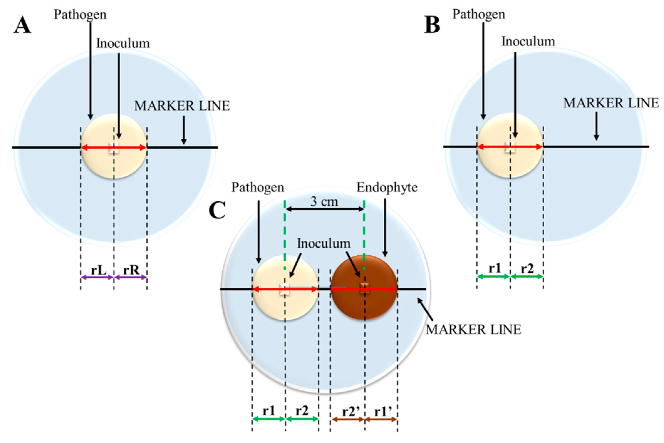
2.1. Inoculation

The fungal inoculum was a cubic agar block measuring 0.5 cm × 0.5 cm × 0.5 cm, cut from areas of the youngest mycelium from the mother culture. The inoculum was placed in an 8.7 cm diameter Petri dish on fresh malt extract agar (MEA Biomaxima, Lublin, Poland) with a layer thickness of 0.5 cm. In monocultures, inocula were placed in two variants. In variant 1, dedicated to calibration of growth effectivity, inocula were placed in the centre of the agar plates (c.a. 4.4 cm from the edges) (Figure 1A).
In variant 2, dedicated to calibration of time of undisturbed fungus growth, inocula were placed c.a. 3 cm from the closer edge of the plate (Figure 1B). For each variant, 10 repetitions were performed. The third experimental variant included dual cultures, where inocula of ash pathogen, H. fraxineus, strain 21508, and one of four Fraxinus excelsior endophyte fungi—Plenodomus biglobosus 13F ([13], GenBank acc. no. MT651609 and described as Pleospora sp. FeF80 by Kowalski and Bilański [14] with GenBank acc. No. MZ492945), Thielavia basicola FeC43 (GenBank acc. no. MW447029), Fusarium lateritium FeC44 (GenBank acc. no. MW447017), and Paracucurbitaria corni 608F (GenBank acc. no. MT547825, named FeC33 in work of Bilański and Kowalski [15])—were placed at two opposite sides of Petri dishes, c.a. 3 cm from the closer edge and at a distance of 3 cm between centres of each inoculum (Figure 1C). Due to the slow growth of the pathogen, the pathogenic fungus was inoculated 7 days in advance. The third experimental variant was carried out in eight to nine repetitions. All colonies of each variant were cultivated at 22 °C in the dark. The studied fungal strains were received from Professor Tadeusz Kowalski’s collection, Department of Forest Ecosystems Protection, University of Agriculture in Krakow.

2.2. Calibration of the Pathogen Growth in Monocultures

Variants 1 and 2 were used to calibrate the growth of the pathogen and differed with the initial position of the inoculum—central (variant 1) or asymmetrical (variant 2), mimicking the position in dual culture. Thus, in variant 1, radii measured along the plate’s diameter axis were described as left (“rL”) and right (“rR”) (Figure 1A), while asymmetrically placed, i.e., in variant 2, were described with “r1” (growth to the proximal edge of the dish) and “r2” radii (growth to the centre/distal edge of the dish) (Figure 1B). The radii of H. fraxineus were measured past one week after inoculation and every second day for 21 days. To analyze the differences between the mean values of the radii on each day, Student’s t-test with α = 0.05 was applied. Calibration had two purposes: firstly, to check if the pathogen’s inocula were capable of growing (especially important for strongly inhibited growth of the pathogen in dual culture); secondly, to verify the effect of the close proximity of the plate edge on the dynamics of fungus growth and thus to indicate the time in which r1 growth was disturbed in comparison to r2. Such calculation is essential to indicate which variant of calibration should be used, as well as for the reliability of the calculations of both the “vitality parameter” and the “inhibition parameter”.

2.3. The Analyses of H. fraxineus Growth Inhibition in Dual Cultures

For the analyses of H. fraxineus growth inhibition in dual cultures (i.e., variant 3), one of four F. excelsior endophytic fungal strains were tested. Three of them, P. biglobosus 13F, T. basicola FeC43, and P. corni 608F, were melanized, and one strain, F. lateritium FeC44, which was nonpigmented and previously revealed as the most strongly inhibiting fungus towards H. fraxineus [15], served as a good reference for validation of our model. Petri dishes with dual cultures in all variants were divided by a thin, straight marker line into two equal halves (Figure 1A–C). Importantly, the marker line crossed both centres of the fungal inocula. The radii of pathogen mycelia, from the centre of the inoculum toward the closer edge of the plate (r1) and toward the endophytic fungus (r2), were measured. An analogical scheme was applied to endophyte analysis with r1’ and r2’ (Figure 1C). Measurements were started with the time of endophyte inoculation (7 days after pathogen inoculation). Radii were measured along the marker line to an accuracy of 1 mm every second day for 21 days. To characterize the growth of the pathogen (or endophyte), we calculated the vitality parameter as follows:
vitality parameter = r2/r1 (or r2’/r1’)
To determine the growth ability of the endophyte (verified organism) while affecting the pathogen’s (tested organism) growth, we introduce the inhibition parameter:
inhibition   parameter   = verified   organism s   vitality   parameter tested   organism s   vitality   parameter

2.4. Software

To indicate statistically significant differences between the mean calculated parameters, one-way ANOVA was applied.
To prepare text and figures and to conduct statistical calculations, MS Office 365 ver. 2406 (compilation 16.0.17726.20206), Origin 2018, and Statistica ver. 13.3 software was used.

3. Results

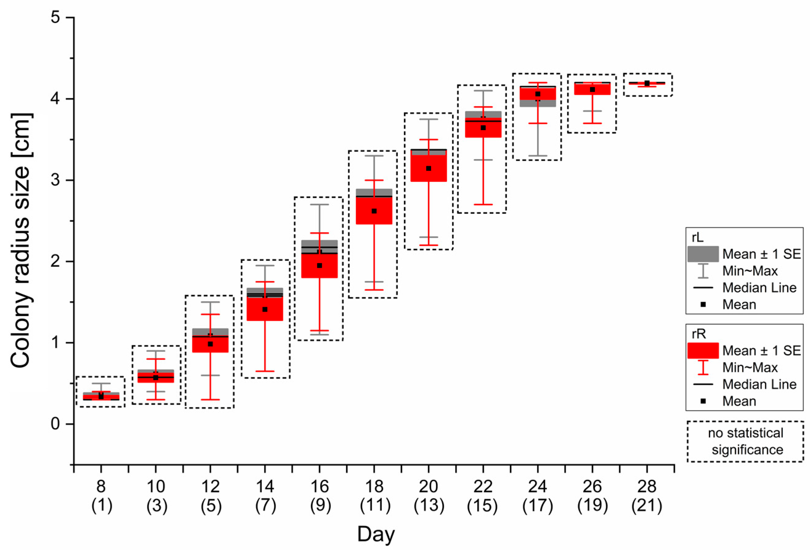
3.1. Pathogen’s Growth Measurement Calibration

The “rR” and “rL” or “r1” and “r2” radii of the pathogen were measured from the 8th day as a result of a 7-day shift between pathogen and endophyte inoculation. In variant 1 (monoculture with centrally placed inoculum, Figure 1A), there were no statistically significant differences between mean “rR” and “rL” values. Both “rR” and “rL” radii maximum lengths were 4.190 ± 0.021 cm and 4.195 ± 0.016 cm, respectively, on the 28th day of the experiment (which corresponds to the 21st day in dual-culture experiments) (Figure 2).
In variant 2 of the calibration experiment, in monocultures with asymmetrically placed inocula (Figure 1B), a statistically significant difference between mean “r1” and “r2” values (p = 0.048) was observed no earlier than the 18th day of the experiment, i.e., on the 11th day of radii measurement (Figure 3). On the 18th day, the “r1” mean value was 2.333 ± 0.173 cm, when the “r2” mean value was 2.611 ± 0.349 cm. From the 18th day (corresponding to the 11th day of the potential dual-culture experiment) until the 28th day (21st day of potential dual-culture experiment), the mean value of “r2” increased to 4.939 ± 0.480 cm, when “r1” reached a maximum value of 2.700 cm on the 24th day (the mycelium reached the edge of the dish) (Figure 3).

3.2. Vitality Parameter

In order to quantify the growth efficiency of the analyzed fungus under given dual-culture conditions (in our study, the effect of fungal ash endophytes on H. fraxineus), we propose to introduce a parameter that has not been used before, which we call the vitality parameter.
The vitality parameter we have proposed is a ratio of “r2” to “r1” values, thus determined by the growth asymmetry of the fungal colony. If nothing disturbs the growth of the colony, it expands symmetrically; thus, the r2/r1 ratio is about 1. Directional factors inhibiting the growth of the exposed side (r2) of the colony (e.g., the presence of a co-cultured organism, Figure 4A) decrease the vitality parameter, while stimulating factors (Figure 4B) increase the r2 value in comparison to r1, and thus increase the vitality parameter. The latter may be caused by stimulation of the tested organism by attractants, hormones, or nutrients secreted by the co-cultured organism.
For the co-cultures of H. fraxineus with four different fungal endophytes of F. excelsior conducted for 21 days, using measured r1 and r2, the vitality parameters for the pathogen were calculated (Figure 5).
Statistically significant differences in the vitality parameter values were detected from the 5th day from the start of the experiments (start of the co-culture cultivation) until the end of the observation. On the 5th day, the mean vitality parameter values were 0.50000 (0.49994) ± 0.065, 0.741 ± 0.048, 0.872 ± 0.085, and 0.943 ± 0.052 for H. fraxineus in co-cultures with FeC44, 13F, 608F, and FeC43 endophytes, respectively. The strongest inhibitory effect was observed for FeC44 (vitality parameter of H. fraxineus reduced to 0.339 ± 0.041in the 9th day) (Figure 5).

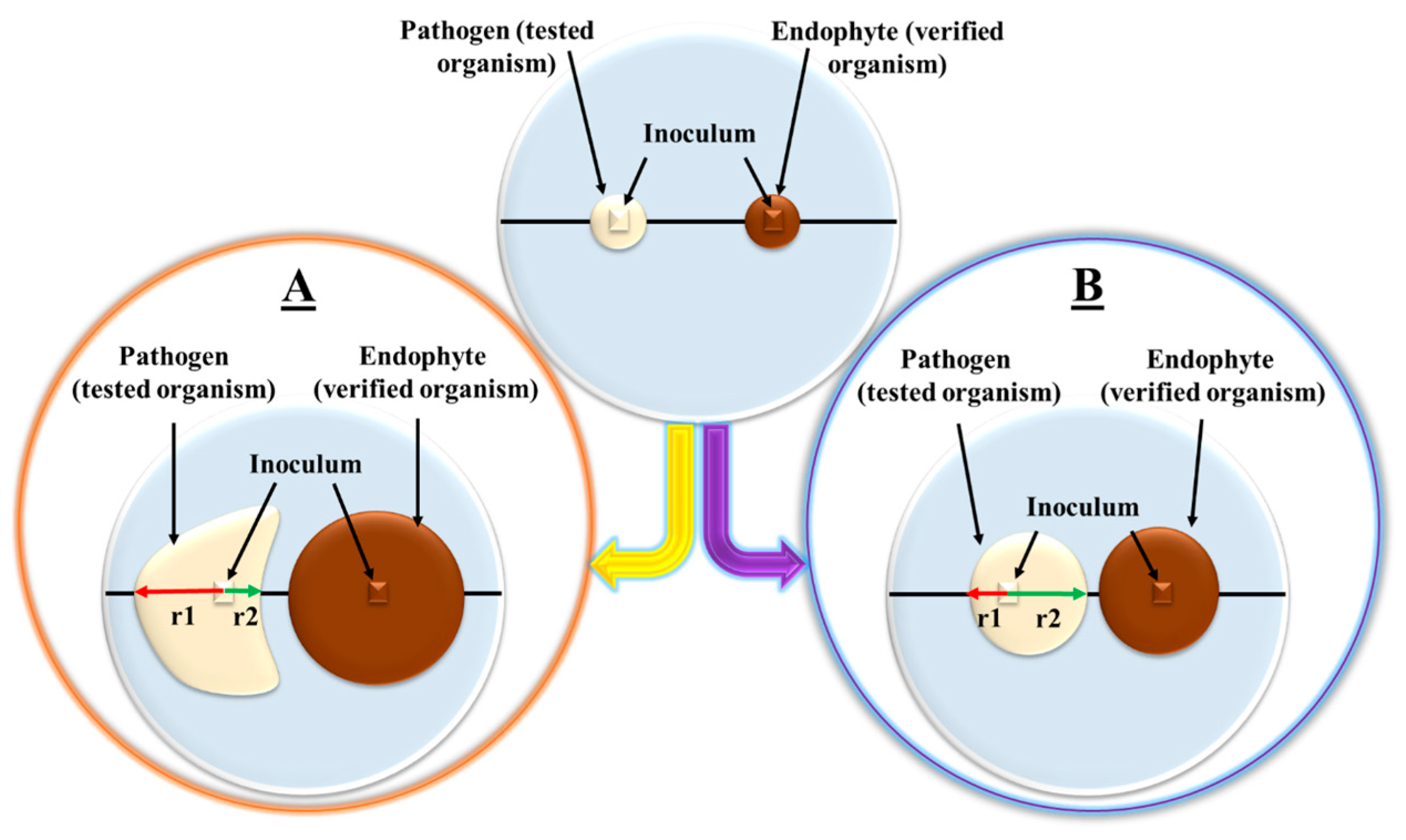
3.3. Inhibition Parameter as a Summary Factor in the Interaction between Organisms in Dual Cultures

For the study of interaction in dual cultures, we propose a new parameter referred to as the inhibition parameter. It combines information about the condition of both organisms in co-culture and is calculated as the ratio of the vitality parameters of the verified organism (in our studies the endophyte) to the vitality parameter of the tested organism (in our studies the pathogen). Equal vitality parameters for both organisms result in an inhibition parameter value of 1 and indicate no effect or a simultaneous inhibitory effect on both species. A higher vitality of the verified organism increases the inhibition parameter above one, thus indicating better growth of the verified organism in comparison to the growth of the tested organism. Alternatively, a lowered inhibition parameter indicates a possible inhibition of the verified organism by the tested organism.
In the example experiment, it was investigated among European ash endophytes which one would be decreasing the value of vitality parameter of H. fraxineus at the highest rate while maintaining relatively the highest vitality parameter of its own, thus increasing the inhibition parameter. As we conclude from the monoculture calibration, results after the 9th day of dual-culture experiments should not be taken into consideration (Figure 3). On the 9th day of the measurements, the mean value of the inhibition parameter was significantly the highest for dual cultures with FeC44 fungus (2.008 ± 0.352), the lowest for 608F (0.632 ± 0.186), and there was no significant difference in inhibition parameter between the 13F and FeC43 strains’ dual cultures with H. fraxineus (1.153 ± 0.390 and 1.112 ± 0.384, respectively) (Figure 6).

4. Discussion

Dual-culture studies are popular methods of analyzing the interactions between organisms, especially fungi [14,16,17]. They enable researchers to study the growth rates, inhibition zones, and accumulation and secretion of metabolites on the plate with solidified media [14,15,16,18,19]. Already in 1924, Porter described four types of interactions between growing fungal colonies as follows: (1) mutually intermingling (overgrowing); (2) slight inhibition; (3) growth around; and (4) inhibition at a distance [20]. In 1973, Fokkema applied a new scheme of dual cultures, where the growth reduction of Drechslera sorokiniana (fungal pathogen of rye) by saprophytic yeasts and yeast-like fungi was studied [21]. The effectivity of these fungi in combating D. sorokiniana was determined by calculating two parameters—the percentage of inhibition of radial growth of the pathogen and the zone of inhibition, the distance between both growing colonies (Figure 7A) [21].
In Fokkema’s measurements, the percentage of inhibition of radial growth inferred from the asymmetry of the colony in the presence of an antagonist and was calculated as the ratio of the difference between the reference radius (r1) (on the opposite, unexposed side of the mycelium) and the antagonist-exposed radius of the mycelium (r2), divided by r1 (Figure 7A) [21]. Both radii, r1 and r2, were measured along the diameter of the dish passing through the centres of the inocula (Figure 7A). Five years later, Royse and Ries used the same equation to calculate the percentage of inhibition of radial growth to determine the inhibition of Cytospora cincta, a pathogen of peach trees; however, they modified r1’s definition and measurement methodology [22]. r2 was measured the same as in Fokkema [21,22], but reference r1 was measured as the distance from the centre of the C. cincta inoculum to the farthest possible point of the colony measured toward the potential antagonist, and thus not along the diameter of the dish passing through the centres of the both inocula, but at an angle to that diameter (Figure 7B) [22]. A model with the same scheme was applied by Manandhar et al. in 1987 [23]; however, measurement method of r1, which should be an unaffected, reference radius of growth, in the methodologies of both Royse and Ries [22] and Manandhar et al. [23] seems dubious, as r1 is measured with the front of the mycelium of pathogen and thus affected by the presence of potential antagonists, e.g., via allelopathic interactions (Figure 7B).
In later studies, further improvements have been made and the reference radius, marked as Rc, was measured in monocultures of tested organisms, whereas Rm (analogue of r2) was determined in co-cultures (Figure 7C). The scheme was used by other researchers [14,15,16,24,25,26]. In 2010, Lahlali and Hijri applied the same scheme; however, fungi in the control monocultures were inoculated in the centre of the Petri dish [27]. Generally, the scheme of dual cultures with measuring reference radii on separated monocultures always requires additional parallel cultures, preferably in multiple repetitions, in order to eliminate individual differences between the organism in the monoculture and in the dual culture.
Here, we proposed new scheme of studies with dual-culture application. We show the necessity of using a monoculture of the test organism to calibrate the upper limit of the time at which a dual culture with that organism is a suitable test model. Moreover, the results obtained from measurements of the “rR” and “rL” (Figure 2) radii in variant 1 (Figure 1A) and the “r1” and “r2” (Figure 3) radii in variant 2 (Figure 1B) clearly indicate that in calibration monocultures, as well as in controls in other experiments [14,15,16,24,25,26], control inocula should be placed at the same distance from the edge of the plate as the inocula in the tested dual cultures (as shown in Figure 1B,C), rather than in the centre of the plate (as shown in Figure 1A). The “rL” radius (Figure 1A) does not correspond to the “r1” radius in dual cultures (Figure 1C). The increase in radius “r1” is significantly inhibited by the edge of the dish, which cannot be taken into account in a control or calibration culture with the inoculum placed in the centre of the plate (Figure 1A).
In this paper, for the first time, the effect of distance of the mycelium from the edge of the dish was experimentally demonstrated both in the control and in the dual culture. This shows that only the experimental scheme with the control inoculum placed asymmetrically, at the same distance from the edge of the dish as in the dual culture (Figure 1B), is a valid reference (Figure 3) for fungal growth in dual culture. The results of our analyses clearly indicate that, in order to ensure correct parameterization of interactions in dual cultures, the following guidelines should be taken into account when planning an experiment using the proposed scheme:
  • Inocula in dual cultures should be placed at the same distance from each other as from the plate edge to ensure that fungus will have the same distance to both physical barriers. The distances are strongly dependent on the Petri dish diameter; therefore, the calibration control must be carried out on a plate with exactly the same parameters as the dual cultures.
  • Asymmetrical monoculture (calibration variant 2 shown in Figure 1B) is essential to properly analyze data from dual-culture experiments.
  • In calibration monocultures, fungal inocula must be inoculated at the same distance from the plate edge as fungi in the dual-culture experiments to determine the end time of measurements in co-cultures.
  • In dual-culture experiments, radii measurements should be terminated at the last day with unaffected mycelial growth in both directions in calibration monocultures—in H. fraxineus growth studies, using the proposed scheme, it was the 16th day from the start of pathogen cultivation, i.e., the 9th day of the potential dual-culture experiment.
Using the proposed dual-culture scheme with a calibration culture allows for precise quantitative parameterization of both the response of the test organism and the interaction between the two organisms in the dual cultures. This can be achieved with the entirely new parameters described and used in this paper, i.e., the vitality and inhibition parameters. We tested the use of the new parameters in dual cultures of H. fraxineus with the same selected endophytes as Kowalski and Bilański [14,15].
The vitality parameter is sufficient to determine how strong the growth of a tested organism is affected by another organism, which we term the verified organism, and if it is a stimulation or inhibition of the growth. Additionally, that parameter may be applied in studies of monocultures exposed to various factors, not only to other organisms. It can be widely used for screening for positive as well as negative interactions between organisms. Moreover, it can be adjusted to determine the influence of, e.g., pure chemicals or physical sources of radiation on the colony. The results of our vitality parameter analysis (Figure 5) are consistent with the findings of Kowalski and Bilański [14,15], where F. lateritium FeC44 was considered capable of strongly reducing H. fraxineus growth (75% of the pathogen’s growth reduction), while P. biglobosus 13F (classified as Pleospora sp. FeF80 by Kowalski and Bilański [14]), T. basicola FeC43, and P. corni 608F exhibited mild (26%–50%) inhibition of H. fraxineus growth.
Importantly, statistically significant difference between values for H. fraxineus in dual cultures with the 608F and FeC43 strains was revealed on the 11th day of the co-culture experiments; however, the monoculture calibration showed that the result should not be taken into account (Figure 3), as the vitality parameter is reliable up to the 9th day, and in older cultures H. fraxineus reaches the edge of the Petri dish. Neglecting this fact will cause fallacy, as several factors will simultaneously affect fungal growth and the calculation of the vitality parameter.
Until this time, typically, interactions in dual-culture experiments have been presented by parameters describing the inhibition or stimulation of one [24,25,26] or two fungi (but as separate values) [14,15]. Inhibition zone (ZI) width, considered as the distance between mycelia of both fungi growing towards each other, is another parameter describing fungal interaction [21]. However, ZI may be unreliable, as it is dependent on the concentration of the toxic metabolites and its diffusion capacity in medium as well as in the case of fungi exhibiting high tolerance to co-cultured competitors or even mycoparasites [28,29,30,31,32,33,34]. Thus, it should be concluded that inhibition zone studies are essential for studying interaction types, but not for determining the effectivity of combating one organism with another. Additionally, it is often presented as a separated value and is not combined with parameters describing, e.g., the stimulation or inhibition of growth of one or both organisms. Now we propose the inhibition parameter, which combines information about the vitality parameters of both co-cultivated organisms, thus elucidating how strongly both organisms affects each other’s growth.
Considering both the vitality parameter (Figure 5) and inhibition parameter (Figure 6) values calculated from dual cultures of H. fraxineus with endophytes, it is clearly seen that the FeC44 strain is not only the most effective in decreasing the vitality parameter of the pathogen (Figure 5), but also its vitality parameter is over two times higher than the vitality parameter calculated for H. fraxineus (Figure 6). The pathogen’s vitality parameters in co-cultures with 608F and FeC43 show no significant differences (Figure 5). However, the calculation of inhibition parameters revealed that the growth of FeC43 in dual culture was significantly less affected than the growth of the 608F fungus (Figure 6); thus, the inhibition parameter provides additional information about the co-culture, and it is a sensitive tool for estimating interactions between organisms. Until now, we did not have a parameter providing such information. The vitality and inhibition parameters may be applied to analyze all types of ecological interactions, e.g., parasitism, concurrence, or cooperation, until there is a minimal distance between studied colonies, which gives a new quality to dual cultures, especially in microbiological research. The proposed parameters may be also correlated with, e.g., chemical, biochemical, or genetic analyses of growing fungi to further expand research and fully characterize interactions in co-cultures. Additionally, the great advantage of both the vitality and inhibition parameters is the fact that apart from calibration cultures, i.e., determining the correct measurement times for the organisms in question, no other additional control monocultures are required to calculate these parameters. Essential are organisms from dual cultures only and the vitality parameter (a component of the inhibition parameter) is calculated based on the radii of the same biological object on the same Petri dish. Thus, intra-individual differences are eliminated in the presented scheme and application of dual cultures in contrast to using the radii of two biological objects growing on two separate Petri plates. Another advantage of the proposed scheme and parameters is the fact that they can be applied on Petri plates of different sizes and with different media (in case of H. fraxineus it may be, e.g., MEA [14,15,16] as well as PDA medium enriched with ash shoot extract, which was also used in studies of the effectivity of ash endophytes in combating the pathogen [35]). Additionally, the results of our example experiment indicate F. lateritium FeC44, among the tested ash endophytic fungi, would be the most suitable biocontrol agent in combating H. fraxineus.

5. Conclusions

In this article a new scheme of studies with dual cultures application as well as two new parameters were proposed. In the presented models, monocultures of the tested organism are needed to perform calibration experiment only, which provide information, when dual-culture experiments should be terminated. Proposed parameters are vitality and inhibition parameters. Vitality parameter is sufficient to determine how strong the growth of tested organism is affected by another organism. Additionally, the parameter may be applied in studies of monocultures exposed to various substances, not only to other organisms. Until this time, the physical parameters of the growth in dual-cultures were presented as separated values for both organisms. Now we propose another parameter, named inhibition parameter, which combines information about vitality parameters of both co-cultivated organisms, thus, elucidating how strong both organisms affects each other’s growths. The vitality and inhibition parameters may be applied to analyze all types of ecological interactions e.g., parasitism, concurrence or cooperation, which gives a new quality to dual-cultures in, especially, microbiological research. Vitality and inhibition parameters can be used to analyze all kinds of ecological interactions, including the effective search for biocontrol agents in the protection of forest ecosystems against tree pathogens.In this work we have shown comprehensively the possibility of application of our novel scheme of dual-cultures in selection of European ash fungal endophyte’s strain, which would be the most promising biocontrol agent against H. fraxineus, invasive species causing ash dieback epidemy in Europe. As the result of the particular study, F. lateritium FeC44 strain was determined as the most effective in combating the pathogen.

Author Contributions

Conceptualization: D.L. and J.P.; methodology: D.L., J.P. and K.N.-C.; software: J.P. and M.O.-P.; validation: J.P. and M.O.-P.; formal analysis: D.L., J.P., P.J. and M.O.-P.; investigation: J.P., P.J. and D.L.; resources and data curation: J.P. and M.O.-P.; writing—original draft preparation: J.P.; writing—review and editing: D.L. and P.J.; visualization: J.P.; supervision: D.L.; project administration: D.L.; funding acquisition D.L. All authors have read and agreed to the published version of the manuscript.

Funding

This research was funded by a subsidy for statutory activities from the Polish Ministry of Education and Science No. 19000882.

Data Availability Statement

The raw data supporting the conclusions of this article will be made available by the authors on request.

Acknowledgments

We dedicate this paper to our late colleague Natalia Marcol-Rumak, student, who conducted a lot of the research described in this article during the last months of her life. Thanks to Tadeusz Kowalski and Piotr Bilański for providing the fungal strains. All authors have read and agreed to the published version of the manuscript.

Conflicts of Interest

The authors declare no conflicts of interest.

References

  1. Timmermann, V.; Børja, I.; Hietala, A.M.; Kirisits, T.; Solheim, H. Ash Dieback: Pathogen Spread and Diurnal Patterns of Ascospore Dispersal, with Special Emphasis on Norway. EPPO Bull. 2011, 41, 14–20. [Google Scholar] [CrossRef]
  2. Brasier, C.M. Ophiostoma Novo-Ulmi Sp. Nov., Causative Agent of Current Dutch Elm Disease Pandemics. Mycopathologia 1991, 115, 151–161. [Google Scholar] [CrossRef]
  3. Muñoz-Adalia, E.J.; Meijer, A.; Abel, J.; Colinas, C.; Aletà, N.; Guàrdia, M. Evaluation of Chestnut Susceptibility to Cryphonectria parasitica: Screening under Controlled Conditions. Agriculture 2021, 11, 1158. [Google Scholar] [CrossRef]
  4. Moricca, S.; Linaldeddu, B.T.; Ginetti, B.; Scanu, B.; Franceschini, A.; Ragazzi, A. Endemic and Emerging Pathogens Threatening Cork Oak Trees: Management Options for Conserving a Unique Forest Ecosystem. Plant Dis. 2016, 100, 2184–2193. [Google Scholar] [CrossRef] [PubMed]
  5. Warwell, M.V.; McDonald, G.I.; Hanna, J.W.; Kim, M.-S.; Lalande, B.M.; Stewart, J.E.; Hudak, A.T.; Klopfenstein, N.B. Armillaria altimontana Is Associated with Healthy Western White Pine (Pinus monticola): Potential in Situ Biological Control of the Armillaria Root Disease Pathogen, A. solidipes. Forests 2019, 10, 294. [Google Scholar] [CrossRef]
  6. Queloz, V.; Grünig, C.R.; Berndt, R.; Kowalski, T.; Sieber, T.N.; Holdenrieder, O. Cryptic Speciation in Hymenoscyphus albidus. For. Pathol. 2011, 41, 133–142. [Google Scholar] [CrossRef]
  7. Sieber, T.N. The Phyllosphere Mycobiome of Woody Plants. In Forest Microbiology; Elsevier: Amsterdam, The Netherlands, 2021; pp. 111–132. ISBN 978-0-12-822542-4. [Google Scholar]
  8. Coghlan, A. Are Europe’s Ash Trees Finished? Available online: http://www.newscientist.com/article/dn22449-are-europes-ash-trees-finished.html (accessed on 31 October 2012).
  9. Kowalski, T.; Bilański, P.; Holdenrieder, O. Virulence of Hymenoscyphus albidus and H. fraxineus on Fraxinus excelsior and F. pennsylvanica. PLoS ONE 2015, 10, e0141592. [Google Scholar] [CrossRef] [PubMed]
  10. Pratt, J.E.; Niemi, M.; Sierota, Z.H. Comparison of Three Products Based on Phlebiopsis gigantea for the Control of Heterobasidion annosum in Europe. Biocontrol Sci. Technol. 2000, 10, 467–477. [Google Scholar] [CrossRef]
  11. Grabka, R.; d’Entremont, T.W.; Adams, S.J.; Walker, A.K.; Tanney, J.B.; Abbasi, P.A.; Ali, S. Fungal Endophytes and Their Role in Agricultural Plant Protection against Pests and Pathogens. Plants 2022, 11, 384. [Google Scholar] [CrossRef]
  12. Akram, S.; Ahmed, A.; He, P.; He, P.; Liu, Y.; Wu, Y.; Munir, S.; He, Y. Uniting the Role of Endophytic Fungi against Plant Pathogens and Their Interaction. J. Fungi. 2023, 9, 72. [Google Scholar] [CrossRef] [PubMed]
  13. Pukalski, J.; Marcol, N.; Wolan, N.; Płonka, P.M.; Ryszka, P.; Kowalski, T.; Latowski, D. Detection of a Pheomelanin-like Pigment by EPR Spectroscopy in the Mycelium of Plenodomus biglobosus. Acta Biochim. Pol. 2020, 67, 295–301. [Google Scholar] [CrossRef] [PubMed]
  14. Kowalski, T.; Bilański, P. Fungi Detected in the Previous Year’s Leaf Petioles of Fraxinus excelsior and Their Antagonistic Potential against Hymenoscyphus fraxineus. Forests 2021, 12, 1412. [Google Scholar] [CrossRef]
  15. Bilański, P.; Kowalski, T. Fungal Endophytes in Fraxinus excelsior Petioles and Their in Vitro Antagonistic Potential against the Ash Dieback Pathogen Hymenoscyphus fraxineus. Microbiol. Res. 2022, 257, 126961. [Google Scholar] [CrossRef]
  16. Nawrot-Chorabik, K.; Marcol-Rumak, N.; Latowski, D. Investigation of the Biocontrol Potential of Two Ash Endophytes against Hymenoscyphus fraxineus Using In Vitro Plant–Fungus Dual Cultures. Forests 2021, 12, 1750. [Google Scholar] [CrossRef]
  17. Diner, A.M.; Karnosky, D.F. Differential Responses of Two Conifers to in Vitro Inoculation with Agrobacterium rhizogenes. Eur. J. For. Pathol. 1987, 17, 211–216. [Google Scholar] [CrossRef]
  18. do Nascimento, J.S.; Silva, F.M.; Magallanes-Noguera, C.A.; Kurina-Sanz, M.; Dos Santos, E.G.; Caldas, I.S.; Luiz, J.H.H.; Silva, E.d.O. Natural Trypanocidal Product Produced by Endophytic Fungi through Co-Culturing. Folia Microbiol. 2020, 65, 323–328. [Google Scholar] [CrossRef]
  19. Palyzová, A.; Svobodová, K.; Sokolová, L.; Novák, J.; Novotný, Č. Metabolic Profiling of Fusarium oxysporum f. Sp. Conglutinans Race 2 in Dual Cultures with Biocontrol Agents Bacillus amyloliquefaciens, Pseudomonas aeruginosa, and Trichoderma harzianum. Folia Microbiol. 2019, 64, 779–787. [Google Scholar] [CrossRef] [PubMed]
  20. Porter, C.L. Concerning the Characters of Certain Fungi as Exhibited by Their Growth in the Presence of Other Fungi. Am. J. Bot. 1924, 11, 168–188. [Google Scholar] [CrossRef]
  21. Fokkema, N.J. The Role of Saprophytic Fungi in Antagonism against Drechslera sorokiniana (Helminthosporium sativum) on Agar Plates and on Rye Leaves with Pollen. Physiol. Plant Pathol. 1973, 3, 195–205. [Google Scholar] [CrossRef]
  22. Royse, D.J.; Ries, S.M. The Influence of Fungi Isolated from Peach Twigs on the Pathogenicity of Cytospora cincta. Phytopathology 1978, 68, 603–607. [Google Scholar] [CrossRef]
  23. Manandhar, J.B.; Thapliyal, P.N.; Cavanaugh, K.J.; Sinclair, J.B. Interaction between Pathogenic and Saprobic Fungi Isolated from Soybean Roots and Seeds. Mycopathologia 1987, 98, 69–75. [Google Scholar] [CrossRef]
  24. Hajieghrari, B.; Torabi Giglou, M.; Mohammadi, M.R.; Davari, M. Biological Potantial of Some Iranian Trichoderma Isolates in the Control of Soil Borne Plant Pathogenic Fungi. Afr. J. Biotechnol. 2008, 7, 967–972. [Google Scholar]
  25. Landum, M.C.; do Rosário Félix, M.; Alho, J.; Garcia, R.; Cabrita, M.J.; Rei, F.; Varanda, C.M.R. Antagonistic Activity of Fungi of Olea europaea L. against Colletotrichum acutatum. Microbiol. Res. 2016, 183, 100–108. [Google Scholar] [CrossRef] [PubMed]
  26. Hewedy, O.A.; Abdel Lateif, K.S.; Seleiman, M.F.; Shami, A.; Albarakaty, F.M.; El-Meihy, R.M. Phylogenetic Diversity of Trichoderma Strains and Their Antagonistic Potential against Soil-Borne Pathogens under Stress Conditions. Biology 2020, 9, 189. [Google Scholar] [CrossRef]
  27. Lahlali, R.; Hijri, M. Screening, Identification and Evaluation of Potential Biocontrol Fungal Endophytes against Rhizoctonia solani AG3 on Potato Plants. FEMS Microbiol. Lett. 2010, 311, 152–159. [Google Scholar] [CrossRef] [PubMed]
  28. Steindorff, A.S.; Ramada, M.H.S.; Coelho, A.S.G.; Miller, R.N.G.; Pappas, G.J.; Ulhoa, C.J.; Noronha, E.F. Identification of Mycoparasitism-Related Genes against the Phytopathogen Sclerotinia sclerotiorum through Transcriptome and Expression Profile Analysis in Trichoderma harzianum. BMC Genom. 2014, 15, 204. [Google Scholar] [CrossRef] [PubMed]
  29. Sun, Z.-B.; Sun, M.-H.; Li, S.-D. Identification of Mycoparasitism-Related Genes in Clonostachys rosea 67-1 Active against Sclerotinia sclerotiorum. Sci. Rep. 2015, 5, 18169. [Google Scholar] [CrossRef]
  30. Moreno-Ruiz, D.; Lichius, A.; Turrà, D.; Di Pietro, A.; Zeilinger, S. Chemotropism Assays for Plant Symbiosis and Mycoparasitism Related Compound Screening in Trichoderma atroviride. Front. Microbiol. 2020, 11, 601251. [Google Scholar] [CrossRef] [PubMed]
  31. Mukherjee, P.K.; Mendoza-Mendoza, A.; Zeilinger, S.; Horwitz, B.A. Mycoparasitism as a Mechanism of Trichoderma-Mediated Suppression of Plant Diseases. Fungal. Biol. Rev. 2022, 39, 15–33. [Google Scholar] [CrossRef]
  32. Hasan, R.; Lv, B.; Uddin, M.J.; Chen, Y.; Fan, L.; Sun, Z.; Sun, M.; Li, S. Monitoring Mycoparasitism of Clonostachys rosea against Botrytis cinerea Using GFP. J. Fungi. 2022, 8, 567. [Google Scholar] [CrossRef]
  33. Geiger, A.; Karácsony, Z.; Geml, J.; Váczy, K.Z. Mycoparasitism Capability and Growth Inhibition Activity of Clonostachys rosea Isolates against Fungal Pathogens of Grapevine Trunk Diseases Suggest Potential for Biocontrol. PLoS ONE 2022, 17, e0273985. [Google Scholar] [CrossRef] [PubMed]
  34. Embacher, J.; Seehauser, M.; Kappacher, C.; Stuppner, S.; Zeilinger, S.; Kirchmair, M.; Neuhauser, S. Serpula lacrymans Reacts with a General, Unspecialized Chemical Response during Interaction with Mycoparasitic Trichoderma Spp. and Bacteria. Fungal. Ecol. 2023, 63, 101230. [Google Scholar] [CrossRef]
  35. Haňáčková, Z.; Havrdová, L.; Černý, K.; Zahradnik, D.; Koukol, O. Fungal endophytes in ash shoots–diversity and inhibition of Hymenoscyphus fraxineus. Balt. For. 2017, 23, 89–106. [Google Scholar]
Figure 1. Schemes of H. fraxineus inoculation in mono and dual cultures with endophytes. Variant 1 of calibration (A), variant 2 of calibration (B), dual culture experiment (C). “rL”, “rR”—pathogen’s mycelia radii to the left and right, respectively; “r1”, “r2”—pathogen’s mycelia radii to the plate edge and centre of the dish, respectively; “r1’”, “r2’”—endophyte’s mycelia radii to the plate edge and centre of the dish, respectively.
Forests 15 01510 g001
Figure 2. Mean values of the radii measured from the centres of the inocula in the directions of the left (“rL” value) and right (“rR” value) edges of the Petri dishes in variant 1 of calibration (Figure 1A). Numbers without parentheses indicate the actual age of the pathogen culture, while numbers in parentheses in the description of the X axis indicate the day of measurement of the radii. N = 10.
Forests 15 01510 g002
Figure 3. Mean values of the radii measured from the centres of the inocula in the directions of the plate edges (“r1” value) and centres of the Petri dishes (“r2” value) (Figure 1B). Numbers in parentheses in the description of the X axis indicate the day of measurement of the radii. N = 10.
Forests 15 01510 g003
Figure 4. Possible scenarios of fungal interactions in dual culture and its effect on vitality parameter. (A) Inhibition of tested organism (r2/r1 < 1). (B) Stimulation of tested organism (r2/r1 > 1).
Forests 15 01510 g004
Figure 5. Vitality parameter values of H. fraxineus co-cultivated with 608F, FeC43, FeC44, and 13F fungal strains.
Forests 15 01510 g005
Figure 6. Mean inhibition parameter values calculated based on vitality parameters of endophytes and H. fraxineus cultivated in dual cultures.
Forests 15 01510 g006
Figure 7. Development of parameters assessed with fungal growth rates in dual cultures. r1; r2; Rc; Ri—radii of fungal colony; ZI—zone of inhibition; RGI%—percent of radial growth inhibition. Based on and modified from (A) Fokkema 1973 [21]; (B) Royse and Ries 1978 [22]; (C) Kowalski and Bilański 2021 [14].
Forests 15 01510 g007
Disclaimer/Publisher’s Note: The statements, opinions and data contained in all publications are solely those of the individual author(s) and contributor(s) and not of MDPI and/or the editor(s). MDPI and/or the editor(s) disclaim responsibility for any injury to people or property resulting from any ideas, methods, instructions or products referred to in the content.

