Article

Genus-Specific Molecular Markers for In Vitro Detection of Corinectria Forest Pathogens

Laboratorio de Salud de Bosques, Instituto de Conservación, Biodiversidad y Territorio, Facultad de Ciencias Forestales y Recursos Naturales, Universidad Austral de Chile, Valdivia 5090000, Chile
*
Author to whom correspondence should be addressed.
Forests 2025, 16(4), 697; https://doi.org/10.3390/f16040697
Submission received: 9 March 2025 / Revised: 15 April 2025 / Accepted: 17 April 2025 / Published: 18 April 2025

Abstract

:
Canker disease caused by Corinectria constricta has resulted in significant losses of Pinus radiata for the Chilean forestry industry in recent years. Accurate and prompt detection and identification of the pathogen is essential in this context. In this study, a set of molecular markers was developed using multiple alignments of the actin-1 (ACT) and β-tubulin (Btub) gene regions to detect the genus Corinectria in vitro. The designed molecular markers were evaluated for specificity and sensitivity using conventional PCR assays, which successfully differentiated Corinectria species from other closely related species and fungal pathogens of P. radiata. The results showed that the molecular markers were able to detect Corinectria genus with high specificity and sensitivity, with Act-31F/Act-543R detecting 0.1 ng/µL of template DNA and Btub/BtubR detecting 0.05 ng/µL. This study represents the first report of specific molecular markers being developed to detect/identify the genus Corinectria in vitro, and the use of these markers is suggested for the timely detection of the pathogen in P. radiata plantations.

1. Introduction

Pinus radiata D. Don is the most important exotic forest species in the Chilean forestry industry [1]. However, since its introduction, it has faced fungal diseases that limit its productivity [2]. Currently, Corinectria constricta C. González & P. Chaverri (syn. Neonectria fuckeliana) (C. Booth) Castl. and Rossman [3,4] is recognized as one of the most economically significant diseases affecting P. radiata in Chile [5]. This pathogen generates cankers and malformations along the stem, degrades phloem and cambial tissues, and affects the most valuable parts of the tree [3,6]. Initially reported in the Araucanía Region, its distribution now extends from the southern Biobío Region to Los Lagos Region [6].
Traditionally, evidence of symptoms and signs produced by C. constricta has been used as an excellent indicator for the detection of the fungus in the host [3,6]. However, observing cankers on trees could take years, and fungal signs may not appear until at least seven months after lesion formation [7]. Furthermore, previous studies have shown and indicated that the fungus could be present in the tree without generating symptoms and producing potentially infectious conidia (asexual form similar to Acremonium) [7]. These events make the development of disease management and control strategies even more challenging. To date, the use of chemical control has been discarded owing to its high cost; instead, strategies are more related to the management of pruning periods, recommending pruning in the summer season, when infectious inoculum production is expected to be lower [7]. Nevertheless, canker disease remains a major limitation in P. radiata forestry in Chile [5]. Therefore, it is necessary to implement immediate and effective actions based on the early and accurate detection of the pathogen [8].
The identification of C. constricta relies on morphological traits such as ascospores, macroconidia, and microconidia, along with colony characteristics on culture media [3,4]; however, this traditional diagnosis is not feasible for large-scale pathogen sampling, is time-consuming, and requires experts in the field [9]. Molecular techniques offer a more accurate, reproducible, and rapid alternative for pathogen detection [9,10,11]. PCR-based detection is widely used for economically significant plant pathogens [12,13,14]. However, detection of C. constricta using PCR has not been reported to date. There is a history of molecular detection of Nectria fuckeliana (now Corinectria spp.) using a nested PCR approach; however, this method requires two sets of marker pairs used in two rounds of PCR amplification [15], which is more time-consuming, requires more resources, and involves a higher risk of cross-contamination [10]. Corinectria has been distinguished based on phylogenetic analysis of five loci, ACT, ITS, LSU, RPB1, and TEF1 [4]; however, this technique requires knowledge of phylogenetic analysis and is expensive and laborious for species identification [16].
All PCR-based methodologies have a critical initial step: the selection of a target gene and the development of specific molecular markers [11]. Consequently, it is necessary for the sequences of the genes of interest to be present in databases, as PCR can only be used to determine the existence or absence of a known pathogen or gene [17]. ACT and Btub genes are ideal candidates for use in phylogenetic analysis and taxonomic and identification studies of ascomycetes [4,18]. Thus, specific regions of these genes have been used in the development of molecular markers for the identification of fungal species diversity [19,20], including the identification of plant pathogenic species belonging to the family Nectriaceae, which is of particular interest to us [18,21,22,23].
Conventional PCR remains a reliable and widely used method for early detection of fungal pathogens, allowing for the timely implementation of effective disease management strategies. However, there is still a lack of genus-specific molecular markers for Corinectria species. Therefore, this study aimed to develop and validate novel molecular markers targeting the ACT and Btub gene regions for the specific detection of Corinectria spp. using conventional PCR. These markers represent a valuable tool for early and accurate diagnosis and constitute the first report of genus-specific primers designed for Corinectria, thereby advancing available molecular diagnostic resources in forest pathology.

2. Materials and Methods

2.1. Fungal Samples

Two different species of the Corinectria genus were utilized: C. constricta (three strains) and C. fuckeliana (two strains), which were provided by Laboratorio de Salud de Bosques de la Universidad Austral de Chile, the Center for Fungal Biodiversity (CBS), and the AY Rossman Collection (AR). In addition, six species corresponding to different fungal pathogens provided by the Laboratorio de Patología Vegetal de la Universidad de Concepción, Chile, were included. Finally, three fungal endophytes of P. radiata obtained in this study were used (Table 1).

2.2. DNA Extraction

All fungal species (Table 1) were grown on potato dextrose agar (PDA Oxoid, Basingstoke, UK, 39 g/L and incubated at 24 °C for 10 days in the dark. DNA was extracted from the growing mycelia using the E.Z.N.A.® Fungal DNA Mini Kit (Omega Bio-tek, Inc., Norcross, GA, USA), according to the manufacturer’s instructions. The quantity and quality of the isolated DNA were assessed using spectrophotometry (Tecan, Männedorf, Switzerland) (Table S1) and gel electrophoresis (MyGel InstaView™, Labnet International, Inc., Edison, NJ, USA).

2.3. Design of Molecular Markers SPECIFIC to the Genus Corinectria

Partial sequences of the ACT and Btub genes of C. constricta and Neonectria spp. available in the NCBI GenBank database (National Center for Biotechnology Information, Bethesda, MD, USA) were used to design a set of molecular markers specific to the genus Corinectria. As a first step, a total of twenty-six partial sequences of the two aforementioned genes were searched and selected (Table 2). The second step consisted of aligning the sequences of the ACT and Btub genes separately using the MUSCLE algorithm [24]. Finally, based on these alignments, molecular markers specific to the genus Corinectria were designed using Primer 3 V. 2.3.7 [25]. The aforementioned software was implemented on the Geneious Prime V. 2022.1.1 platform and used in this study. For each resulting molecular marker, parameters such as primer length, GC content, melting temperature, potential dimer and hairpin formation, amplicon size, and the number of degenerate bases were evaluated [26] (Table S2). Finally, the best set of markers was selected and synthesized by Integrated DNA Technologies, Inc. (San Diego, CA, USA).

2.4. Validation of Molecular Markers

2.4.1. PCR Conditions

The PCR reactions were carried out with a final total volume of 50 µL and consisted of 10 µL of 5×-Taq Buffer, 1 µL of 10 μM of each pair of designed markers, 1 µL of 10 μM dNTPs, 1.5 µL of DNA at a concentration between 25 ng/µL and 50 ng/µL, 1 µL of Taq DNA Polymerase (abm®, Richmond, BC, Canada), and 34.5 µL of sterile deionized water. Different PCR cycles were used for each pair of molecular markers, with hybridization temperature as the parameter to be standardized. Multiple-gradient PCR was performed. For the Act-31F (5′-AGTGGTGACGTGAATGCC-3′) and Act-543R (5′-CGAGACTTTCAACGCCCCCC-3′) molecular markers, a touchdown PCR amplification protocol was used, consisting of 2 min and 35 s of initial denaturation at 95 °C, followed by 15 cycles of 30 s denaturation at 95 °C, alignment at 70 °C (with a decrease of 1 °C from 70 °C to 50 °C in each cycle) for 30 s, and extension at 72 °C for 30 s. This was followed by another 30 cycles of denaturation at 95 °C for 30 s, alignment at 50 °C for 30 s, and extension at 72 °C for 30 s. The reaction was terminated with a final extension of 10 min at 72 °C. For the molecular markers BtubF (5′-GCCAGCAGAGGC CTAAGGGGGTTTTTT-3′)/BtubR (5′-CTGATTCTACCCCGCCCCGAAG-3′), the thermal profile consisted of 34 cycles of denaturation at 94 °C for 35 s, 61 °C alignment for 55 s, and finally a 72 °C extension for 2 min. The amplified fragments were analyzed using 1.5% agarose gel electrophoresis and visualized on a transilluminator (Accuris Instruments, Edison, NJ, USA). Finally, the PCR products of the two amplified genes were bidirectionally sequenced (Australomics, Valdivia, Chile) and the resulting DNA sequences were compared with the DNA sequences in NCBI GenBank using Blast search to confirm species identification. It is worth mentioning that previously, DNA quality was checked by amplifying the internal transcribed spacer (ITS) region with universal primers for fungi, ITS5 (5′-GGAAGTAAAAGTCGTAACAAGG-3′) and ITS4 (5′-TCTCCTCCGCTTATTGATATATGC-3′) [27] (Figure S1).

2.4.2. Marker Specificity

The specificity of the molecular markers was evaluated by PCR amplification under the aforementioned conditions of DNA samples from (a) C. constricta and C. fuckeliana, (b) a variety of ascomycete and oomycete fungal species that have been associated with P. radiata, (c) strains of fungi closely related to the genus Corinectria present in other hosts, (d) endophytes of P. radiata, and (e) DNA from P. radiata. The specificity of each pair of markers was determined by the presence of size-specific amplicons in Corinectria species (Table 3).

2.4.3. Marker Sensitivity

Sensitivity assays were performed for each pair of molecular markers using conventional PCR to ensure amplification at low levels of the target DNA. For this, 11 concentrations of 25, 10, 5, 2, 1, 0.5, 0.1, 0.05, 0.025, and 0.0012 ng of C. constricta genomic DNA were used (Table S3). The PCR conditions described above were used.

3. Results

3.1. Molecular Marker Design

As a result of the marker design, two pairs of specific molecular markers for the detection of Corinectria spp. were obtained based on the specific regions of the ACT and Btub genes (Table 3).

3.1.1. Marker Specificity

The specificity of the molecular markers was evaluated by conventional PCR using the methods mentioned above, where the Act-31F/Act-543R and BtubF/BtubR markers produced a PCR product specific only to Corinectria spp. around 462 bp and 400 bp, respectively. No specific amplification signal was detected with any marker pair in the other fungal, oomycete, or plant species included in this study (Figure 1 and Figure 2). The identity of the amplified Corinectria strains was confirmed using sequencing.

3.1.2. Marker Sensitivity

The electrophoresis pattern showed that for the two pairs of markers (Act-31F and Act-543R; BtubF and BtubR), the strongest band was obtained with the highest amount of DNA (25 ng), and the intensity of the band decreased as the concentration of template DNA decreased (Figure 3). The molecular markers had different detection limits for Corinectria spp. For Act-31F and Act-543R, the band was visible in the PCR product amplified using 0.1 ng/µL template DNA (Figure 3A). BtubF and BtubR detected template DNA at a concentration of 0.05 ng/µL (Figure 3B). Both markers had low limits of detection for Corinectria spp.

4. Discussion

Molecular methods used in the diagnosis of fungal plant pathogens have become key tools for the management of diseases caused by these pathogens [14]. This is because they provide a rapid, specific, and sensitive alternative to traditional diagnostic methods [28]. Canker disease caused by C. constricta is one of the most important diseases affecting P. radiata plantations in Chile. Therefore, specific and sensitive detection of the pathogen before or at an early stage of infection is indispensable for management and control of the disease. To address this need, a conventional PCR-based technique for detection and identification of the Corinectria genus was used in this study. Regions of the ACT and Btub genes were amplified and were sufficiently variable to differentiate Corinectria from other closely related species with which it was compared.
ACT and Btub gene sequences are commonly used to generate molecular markers that allow discrimination between species and between closely related genera of economically important pathogens [4,10,18,20,29]. The Btub gene regions are one of the most commonly used in the development of molecular markers for the detection of phytopathogenic species of the Nectriaceae family [23,30], whereas ACT gene regions are rarely used for these purposes [31]. However, both genes have been successfully used in phylogenetic and taxonomic studies of Nectericeae [21]. In a multilocus study, ACT and Btub gene regions were among those used to propose six new genera and position species in this family [32]. In the particular case of the pathogen under study, C. constricta was proposed as a new species based on phylogenetic analyses of the ACT, Btub, and TEF1- α genes [4]. Although these antecedents exist, to date, no molecular markers have been reported that allow the detection and identification of the genus Corinectria, despite the inclusion of species considered to be destructive pathogens.
Successful detection of fungal phytopathogens using molecular markers based on ACT and Btub gene regions has been proven in multiple investigations [18,22] and was no exception in the present study. The molecular markers Act-31F/Act-543R and Btub/BtubR could detect the genus Corinectria with high specificity. These satisfactory results may be explained by the fact that ACT and Btub genes have highly conserved coding sequences among distantly related fungal groups, indicating that the intact protein is strictly conserved to preserve its original function [28,33]. In addition, such genes may contain short introns, which can be extremely variable in position and insertion number [34,35]. This particularity in nucleotide sequences is useful for differentiating between closely phylogenetically related fungal species and subspecies [17,28].
Disease control strategies based on timely detection of pathogens using molecular methods not only require specific detection of the pathogen, but it is also necessary to know the limits of detectability of the method to make real estimates of pathogen distribution. Therefore, the sensitivities of the molecular markers developed in this study were evaluated. Both Act-31F and Act-543R and BtubF and BtubR showed high sensitivity for detecting C. constricta template DNA, reaching detection limits of 100 pg/µL and 50 pg/µL, respectively. Generally, the sensitivity of molecular markers is expected to increase as the copy number of the target gene increases [23]. Representative Nectriaceae species such as F. graminearum and F. solani are known to have two and four copies, respectively, of the β-tubulin gene in their genomes [18]; however, most fungal species present a single copy of ACT and Btub genes [18]. To date, the complete genome of C. constricta has not been sequenced or annotated; therefore, it cannot be inferred whether the copy numbers of ACT and Btub are directly associated with the high sensitivity of Btub and BtubR to Act-31F and Act-543R. However, this background supports the use of ACT and Btub gene regions as optimal candidates for highly sensitive detection of target organisms [14,23].
Infection by C. constricta is one of the most significant phytosanitary problems in P. radiata plantations. These infections are difficult to manage because of a lack of timely diagnosis and effective cultural practices. It has been proposed that once a pathogen infects its host, it may remain latent without generating signs and symptoms of the disease. This pathogenic condition undermines the health of healthy stands of P. radiata. Therefore, early and reliable diagnosis is essential to minimize further transmission.
The molecular detection method proposed in this study represents a significant advancement over previous diagnostic tools. Unlike earlier approaches, such as the nested PCR required to detect C. fuckeliana [13], our method enables the detection of C. constricta using a single pair of genus-specific primers, thereby simplifying the process and increasing its applicability for routine monitoring and field diagnostics.
This approach facilitates the development of disease management and control strategies, such as periodic monitoring programs that assess the phytosanitary status of plantations. In the event of disease outbreaks, the method also allows for efficient confirmation of the etiological agent, supporting timely and informed management decisions.

5. Conclusions

This work proposed the use of two pairs of molecular markers, Act-31F/Act-543R and Btub/BtubR, for the specific, sensitive, and timely detection of C. constricta in P. radiata. This work represents the first report on the development of specific molecular markers for the identification of the genus Corinectria in vitro. This method will contribute significantly to large-scale studies and monitoring of the disease in P. radiata forest plantations in Chile, serving as a tool for disease management and control. In addition, further studies are needed to evaluate the use of these markers in both diseased and asymptomatic plants, which would broaden the scope and applicability of this diagnostic tool.

Supplementary Materials

The following supporting information can be downloaded at: https://www.mdpi.com/article/10.3390/f16040697/s1, Table S1: DNA concentrations of the species used in this study; Table S2: Characteristics and evaluated parameters of molecular markers designed in this study; Table S3: Concentrations used for sensitivity testing of the molecular markers designed in this study; Figure S1: PCR amplification products of genomic DNA from the species used in this study, using the universal primers ITS 4/ITS 5. PCR amplification products of genomic DNA from: Lane 1—Diplodia sapinea; lane 2—Phytophthora cinnamomic; lane 3—Fusarium verticillioides; lane 4—Clonostachys rosea; lane 5—Neonectria ditissima; lane 6—Phomopsis tuberívora; lane 7—P. radiata; lane 8—negative control (H2Od); lane 9—Diaporthe phoenicicola; lanes 10 to 13—Corinectria constricta; lanes 14 to 16—Corinectria fuckeliana; lanes 17 and 18—P. radiata; lane 19—negative control (ddH2O).

Author Contributions

Conceptualization: T.V. and C.G.; Methodology: T.V. and C.G.; Validation: C.G. and C.M.; Formal Analysis: T.V. and C.G.; Investigation: T.V. and C.G.; Writing—Original Draft: T.V.; Writing—Review and Editing: C.G. and C.M.; Supervision: C.G.; Funding Acquisition: C.G. All authors have read and agreed to the published version of the manuscript.

Funding

This study was supported by the Agencia Nacional de Investigación y Desarrollo de Chile (ANID) through the FONDECYT Postdoctoral Program 2022 (Project No. 3220723) titled “Endofitismo y distribución potencial de C. constricta en plantaciones de P. radiata bajo distintos escenarios de cambio climático”.

Data Availability Statement

The data generated and analyzed in this study are available upon reasonable request. Researchers interested in accessing the data may contact the corresponding author at cgonzalez@uach.cl. Additionally, the fungal strains used in this research are securely stored at the Forest Health Laboratory Culture Collection and can be made available for collaborative studies under standard research agreements.

Conflicts of Interest

The authors declare no conflicts of interest.

References

  1. Mead, D.J. Sustainable Management of Pinus Radiata Plantations; FAO Forestry Paper; Food and Agriculture Organization of the United Nations: Rome, Italy, 2013; ISBN 978-92-5-107634-7. [Google Scholar]
  2. Ahumada, R.; Rotella, A.; Slippers, B.; Wingfield, M.J. Pathogenicity and Sporulation of Phytophthora pinifolia on Pinus radiata in Chile. Australas. Plant Pathol. 2013, 42, 413–420. [Google Scholar] [CrossRef]
  3. Morales, R. Detección de Neonectria fuckeliana en Chile, asociado a cancros y malformaciones fustales en plantaciones de Pinus radiata. Bosque 2009, 30, 106–110. [Google Scholar] [CrossRef]
  4. González, C.; Chaverri, P. Corinectria, a New Genus to Accommodate Neonectria fuckeliana and C. Constricta sp. nov. from Pinus radiata in Chile. Mycol. Prog. 2017, 16, 1015–1027. [Google Scholar] [CrossRef]
  5. Ahumada, R.; Rotella, A. Disease Management in the Forest Plantations in Chile. In Forest Pest and Disease Management in Latin America; Estay, S.A., Ed.; Springer International Publishing: Cham, Switzerland, 2020; pp. 171–184. ISBN 978-3-030-35142-7. [Google Scholar]
  6. González, C.; Morales, R.; Riegel, R.; Aravena, M.; Valenzuela, E. Distribución Geográfica y Caracterización Fenotípica y Molecular de Neonectria Fuckeliana, Asociado a Cancros Fustales de Pinus radiata En Chile. Bosque 2015, 36, 531–541. [Google Scholar] [CrossRef]
  7. González, C.; Morales, R.A.; Chaverri, P. Life Cycle and in Vitro Sporulation Dynamics of Corinectria constricta, the Causal Agent of Pinus radiata Stem Canker, in Chile. For. Path. 2020, 50, e12594. [Google Scholar] [CrossRef]
  8. Kulik, T.; Bilska, K.; Żelechowski, M. Promising Perspectives for Detection, Identification, and Quantification of Plant Pathogenic Fungi and Oomycetes through Targeting Mitochondrial DNA. IJMS 2020, 21, 2645. [Google Scholar] [CrossRef]
  9. Luchi, N.; Ioos, R.; Santini, A. Fast and Reliable Molecular Methods to Detect Fungal Pathogens in Woody Plants. Appl. Microbiol. Biotechnol. 2020, 104, 2453–2468. [Google Scholar] [CrossRef]
  10. Hariharan, G.; Prasannath, K. Recent Advances in Molecular Diagnostics of Fungal Plant Pathogens: A Mini Review. Front. Cell. Infect. Microbiol. 2021, 10, 600234. [Google Scholar] [CrossRef]
  11. Stewart, J.E.; Kim, M.-S.; Klopfenstein, N.B. Molecular Genetic Approaches Toward Understanding Forest-Associated Fungi and Their Interactive Roles Within Forest Ecosystems. Curr. For. Rep. 2018, 4, 72–84. [Google Scholar] [CrossRef]
  12. Brurberg, M.; Stensvand, A.; Talgø, V. Development and Application of a PCR-based Test for the Identification of Neonectria neomacrospora Damaging Abies Species. In Proceedings of the 12th International Christmas Tree Research and Extension Conference, Honne, Norway, 6–11 September 2015; p. 33. [Google Scholar]
  13. Langrell, S.R.H. Molecular Detection of Neonectria galligena (Syn. Nectria galligena). Mycol. Res. 2002, 106, 280–292. [Google Scholar] [CrossRef]
  14. Tripathi, A.; Dubey, S.C.; Akhtar, J.; Kumar, P. Development of PCR-Based Assays to Diagnose the Major Fungal Pathogens Infecting Pulse Crops, Potential for Germplasm Health Certification and Quarantine Processing. World J. Microbiol. Biotechnol. 2023, 39, 74. [Google Scholar] [CrossRef] [PubMed]
  15. Langrell, S.R. Development of a Nested PCR Detection Procedure for Nectria fuckeliana Direct from Norway Spruce Bark Extracts. FEMS Microbiol. Lett. 2005, 242, 185–193. [Google Scholar] [CrossRef]
  16. Santos, K.M.; Lima, G.S.; Barros, A.P.O.; Machado, A.R.; Souza-Motta, C.M.; Correia, K.C.; Michereff, S.J. Novel Specific Primers for Rapid Identification of Macrophomina Species. Eur. J. Plant Pathol. 2020, 156, 1213–1218. [Google Scholar] [CrossRef]
  17. Oliveira, M.; Azevedo, L. Molecular Markers: An Overview of Data Published for Fungi over the Last Ten Years. JoF 2022, 8, 803. [Google Scholar] [CrossRef] [PubMed]
  18. Zhao, Z.; Liu, H.; Luo, Y.; Zhou, S.; An, L.; Wang, C.; Jin, Q.; Zhou, M.; Xu, J.-R. Molecular Evolution and Functional Divergence of Tubulin Superfamily in the Fungal Tree of Life. Sci. Rep. 2014, 4, 6746. [Google Scholar] [CrossRef]
  19. Singh, N.; Kapoor, R. Quick and Accurate Detection of Fusarium oxysporum f. sp. carthami in Host Tissue and Soil Using Conventional and Real-Time PCR Assay. World J. Microbiol. Biotechnol. 2018, 34, 175. [Google Scholar] [CrossRef]
  20. Yeo, H.Y.; Dong, Y.S.; Seung, Y.S.; Seong, H.K. Development of PCR Method for Fast Detection of Ophiostoma floccosum in Wood Chips. Afr. J. Microbiol. Res. 2013, 7, 1913–1916. [Google Scholar] [CrossRef]
  21. Crous, P.W.; Lombard, L.; Sandoval-Denis, M.; Seifert, K.A.; Schroers, H.-J.; Chaverri, P.; Gené, J.; Guarro, J.; Hirooka, Y.; Bensch, K. Fusarium: More than a Node or a Foot-Shaped Basal Cell. Stud. Mycol. 2021, 98, 100116. [Google Scholar] [CrossRef]
  22. James, J.E.; Santhanam, J.; Zakaria, L.; Mamat Rusli, N.; Abu Bakar, M.; Suetrong, S.; Sakayaroj, J.; Abdul Razak, M.F.; Lamping, E.; Cannon, R.D. Morphology, Phenotype, and Molecular Identification of Clinical and Environmental Fusarium solani Species Complex Isolates from Malaysia. J. Fungi 2022, 8, 845. [Google Scholar] [CrossRef]
  23. Nielsen, K.N.; Thomsen, I.M.; Hansen, O.K. Direct Quantitative Real-Time PCR Assay for Detection of the Emerging Pathogen Neonectria neomacrospora. For. Pathol. 2019, 49, e12509. [Google Scholar] [CrossRef]
  24. Edgar, R.C. MUSCLE: Multiple Sequence Alignment with High Accuracy and High Throughput. Nucleic Acids Res. 2004, 32, 1792–1797. [Google Scholar] [CrossRef] [PubMed]
  25. Rozen, S.; Skaletsky, H. Primer3 on the WWW for General Users and for Biologist Programmers. In Bioinformatics Methods and Protocols; Humana Press: Totowa, NJ, USA, 1999; Volume 132, pp. 365–386. ISBN 978-1-59259-192-3. [Google Scholar]
  26. Ye, J.; Coulouris, G.; Zaretskaya, I.; Cutcutache, I.; Rozen, S.; Madden, T.L. Primer-BLAST: A Tool to Design Target-Specific Primers for Polymerase Chain Reaction. BMC Bioinform. 2012, 13, 134. [Google Scholar] [CrossRef]
  27. White, T.J.; Bruns, T.; Lee, S.; Taylor, J. Amplification and direct sequencing of fungal ribosomal RNA genes for phylogenetics. In PCR Protocols; Elsevier: Amsterdam, The Netherlands, 1990; pp. 315–322. ISBN 978-0-12-372180-8. [Google Scholar]
  28. Chattopadhyay, A.; Tiwari, K.K.; Chaudhary, K.; Pratap, D. Genic Molecular Markers in Fungi: Availability and Utility for Bioprospection. Mol. Markers Mycol. Diagn. Marker Dev. 2017, 2017, 151–176. [Google Scholar]
  29. Olivieri, L.; Saville, R.J.; Gange, A.C.; Xu, X. Limited Asymptomatic Colonization of Apple Tree Shoots by Neonectria ditissima Following Infection of Leaf Scars and Pruning Wounds. Plant Pathol. 2021, 70, 1838–1849. [Google Scholar] [CrossRef]
  30. Capote, N.; Del Río, M.Á.; Herencia, J.F.; Arroyo, F.T. Molecular and Pathogenic Characterization of Cylindrocarpon-like Anamorphs Causing Root and Basal Rot of Almonds. Plants 2022, 11, 984. [Google Scholar] [CrossRef]
  31. Wallace, M.M.; Covert Sarah, F. Molecular Mating Type Assay for Fusarium circinatum. Appl. Environ. Microbiol. 2000, 66, 5506–5508. [Google Scholar] [CrossRef]
  32. Lombard, L.; Van Der Merwe, N.A.; Groenewald, J.Z.; Crous, P.W. Generic Concepts in Nectriaceae. Stud. Mycol. 2015, 80, 189–245. [Google Scholar] [CrossRef]
  33. Dizkirici, A.; Kalmer, A. Utility of Various Molecular Markers in Fungal Identification and Phylogeny. Nova Hedwig. 2019, 109, 187–224. [Google Scholar] [CrossRef]
  34. Irimia, M.; Roy, S.W. Spliceosomal Introns as Tools for Genomic and Evolutionary Analysis. Nucleic Acids Res. 2008, 36, 1703–1712. [Google Scholar] [CrossRef]
  35. Msiska, Z.; Morton, J.B. Isolation and Sequence Analysis of a β-Tubulin Gene from Arbuscular Mycorrhizal Fungi. Mycorrhiza 2009, 19, 501–513. [Google Scholar] [CrossRef]
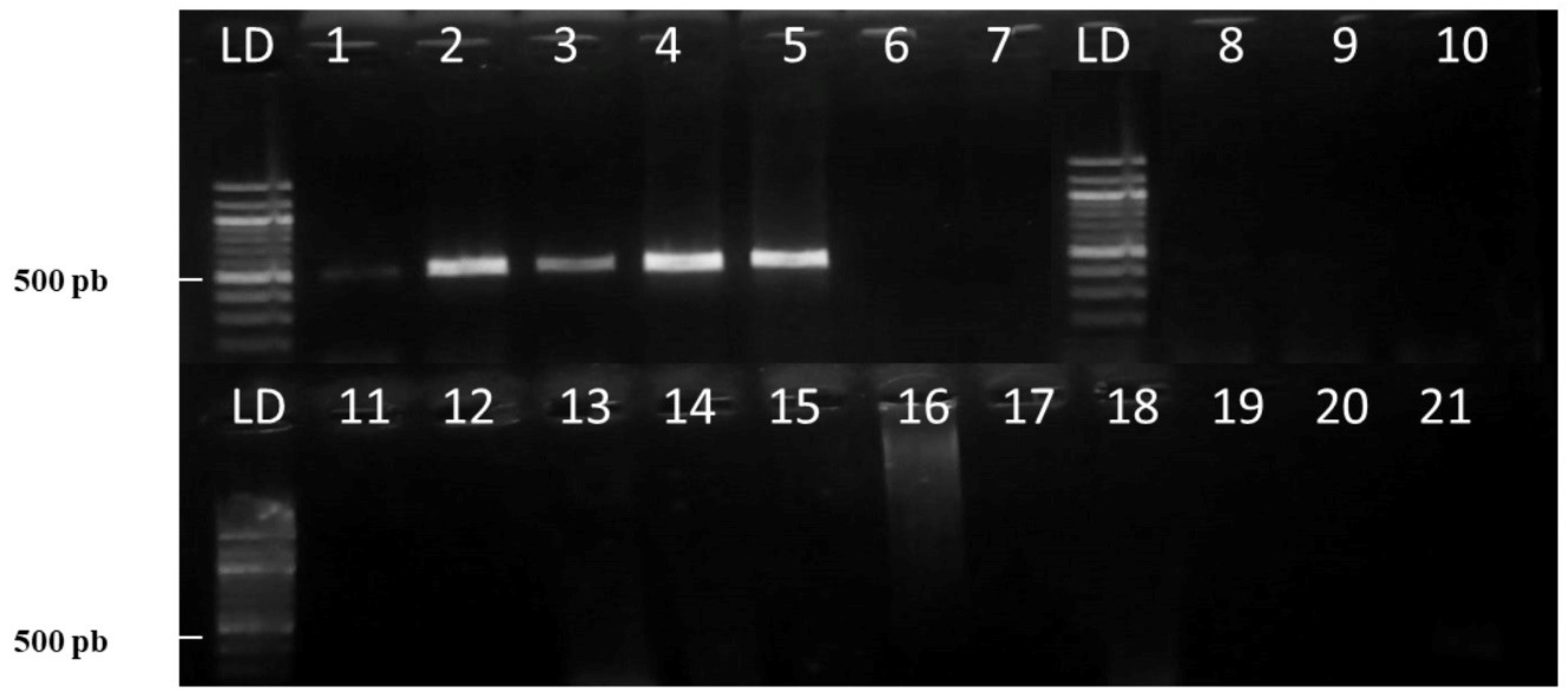
Figure 1. Conventional PCR-based specificity assays using Act-31F/Act-543R as molecular markers for the detection of the genus Corinectria spp. PCR amplification products of genomic DNA of Corinectria spp (lanes 1 to 5), negative controls with distilled water (lanes 6 and 7), and different fungal plant pathogenic species (lanes 8 to 19) using Act-31F/Act-543R molecular markers. Lanes 1 to 3—C. constricta (LASB 330, LASB 266, and LASB 352); lanes 4 to 5—C. fuckeliana (AR 239.69 and AR 3103.61); lane 8—Phomopsis tuberívora; lane 9—Diaporthe phoenicicola; lane 10—Diplodia sapinea; lane 11—Fusarium verticillioides; lane 12—Neonectria ditissima; lane—13 Clonostachys rosea; lanes 14 to 18—fungal endophytes isolated from P. radiata; lane 19—Phytophthora cinnamomic; lane 20—P. radiata; lane 21—negative control (H2Od).
Forests 16 00697 g001
Figure 2. Conventional PCR-based specificity assays using BtubF and BtubR as molecular markers for the detection of the genus Corinectria spp. PCR amplification products of genomic DNA of Corinectria spp (lanes 1 to 5) and different plant fungal pathogenic species (lanes 8 to 19) using BtubF/BtubR molecular markers. Lanes 1 to 3—C. constricta (LASB 330, LASB 266, and LASB 352); lanes 4 to 5—C. fuckeliana (AR 239.69 and AR 3103.61); lanes 6 and 7—negative controls with distilled water; lane 8—Phomopsis tuberivora; lane 9—Diaporthe phoenicicola; lane 10—Diplodia sapinea; lane 11—Fusarium verticillioides; lane 12—Neonectria ditissima; lane 13—Clonostachys rosea; lanes 14 to 18—fungal endophytes isolated from P. radiata; lane 19—Phytophthora cinnamomic; lane 20—P. radiata; lane 21—negative control (distilled water).
Forests 16 00697 g002
Figure 3. Sensitivity test of molecular markers. PCR amplification products of C. constricta genomic DNA at different dilutions using the following molecular markers: (A) Act-31F/Act-543R and (B) BtubF/BtubR. Lane 1—25 ng; lane 2—10 ng; lane 3—5 ng; lane 4—2 ng; lane 5—1 ng; lane 6—0.5 ng; lane 7—0.1 ng; lane 8—0.05 ng; lane 9—0.025 ng; lane 10—0.012 ng; lane 11 control (ddH2O).
Forests 16 00697 g003
Table 1. List of strains used to verify the specificity of the molecular markers designed in this study.
SpeciesIsolate/Voucher aGeographical OrigenGenbank Accession Number
Corinectria constrictaLASBE 330ChileKY636422.1
Corinectria constrictaLASBE 266ChileKY636417.1
Corinectria constrictaLASBE 352ChileKY636425.1
Corinectria fuckelianaCBS 239.29ScotlandDQ789871
Corinectria fuckelianaAR 3103.61AustriaHM352857
Phomopsis tuberívoraLSB 158Chile-
Diaporthe phoenicicolaLSB 160Chile-
Diplodia sapineaLSB 161Chile-
Fusarium verticillioidesLSB 162Chile-
Neonectria ditissimaLSB 163Chile-
Epicoccum nigrumLSB 277Chile-
Arthrinium sp.LSB 278Chile-
Trichoderma virideLSB 279Chile-
Oomycete
Phytophthora cinnamomiLSB 159Chile-
Plants
Pinus radiata-Chile-
a LASBE, LSB: Fungus Collection, Laboratorio de Salud de Bosques y Ecosistemas, Universidad Austral de Chile, Valdivia, Chile; AR: AR. Collection of A. Y. Rossman. CBS: Fungal Biodiversity Center, Utrecht, The Netherlands.
Table 2. Sequences of the ACT and Btub gene regions of Corinectria constricta and Neonectria spp. used to design molecular markers.
SpeciesIsolate/Voucher aGenbank Accession Number
ACTBtub
Corinectria constrictaLASBE 260_KY636416.1
Corinectria constrictaLASBE 266_KY636417.1
Corinectria constrictaLASBE 284_KY636418.1
Corinectria constrictaLASBE 301_KY636419.1
Corinectria constrictaLASBE 306B_KY636420.1
Corinectria constrictaLASBE 314KY636431.1KY636421.1
Corinectria constrictaLASBE 330KY636432.1KY636422.1
Corinectria constrictaLASBE 340KY636433.1KY636423.1
Corinectria constrictaLASBE 344KY636434.1KY636424.1
Corinectria constrictaLASBE 352KY636435.1KY636425.1
Neonectria fuckelianaGJS02-67HM352886.1_
Neonectria coccineaCBS 119158; MAFF 241561KC660426.1DQ789892.1
Neonectria faginataCBS 134246; CBS 119198KC660414.1KC660743.1
Neonectria microconidiaMAFF 241530; MAFF 241522KC660427.1KC660757.1
Neonectria ditissimaCBS 100.316; CBS 226; CBS 100316; CBS 100318HM352880.1_
Neonectria puniceaABT12-1; CBS 119527; ABT12-1; CBS 124262; voucher 135KC660403.1; MW538899.1MW538902.1
a LASBE: Fungus Collection, Laboratorio de Salud de Bosques y Ecosistemas, Universidad Austral de Chile, Valdivia, Chile; ABT: CBS: Fungal Biodiversity Center, Utrecht, The Netherlands; MAFF: MAFF Genebank, National Institute of Agrobiological Sciences, Ibaraki, Japan.
Table 3. Characteristics of designed molecular markers specific to the genus Corinectria.
NameDirectionSequense (5-3 Prima)Size%GC aTm b
Act 81 FforwardCGAGACTTTCAACGCCCC1861.158.4
Act 543 RreverseAGTGGTGACGTGAATGCC1855.657.6
BtubforwardCTGATTCTACCCCGCCGAAG206060.2
BtubRreverseGCCAGAGGCCTAAGGGTTTT205560
a %GC: guanine-cytosine validation of molecular marker content. b Tm: melting temperature.
Disclaimer/Publisher’s Note: The statements, opinions and data contained in all publications are solely those of the individual author(s) and contributor(s) and not of MDPI and/or the editor(s). MDPI and/or the editor(s) disclaim responsibility for any injury to people or property resulting from any ideas, methods, instructions or products referred to in the content.