Review

Adenovirus Biology, Recombinant Adenovirus, and Adenovirus Usage in Gene Therapy

by
Maki Watanabe
1,†,
Yuya Nishikawaji
1,†,
Hirotaka Kawakami
1 and
Ken-ichiro Kosai
1,2,3,4,*
1
Department of Gene Therapy and Regenerative Medicine, Kagoshima University Graduate School of Medical and Dental Sciences, Kagoshima 890-8544, Japan
2
South Kyushu Center for Innovative Medical Research and Application, Kagoshima University Graduate School of Medical and Dental Sciences, Kagoshima 890-8544, Japan
3
Center for Innovative Therapy Research and Application, Kagoshima University Graduate School of Medical and Dental Sciences, Kagoshima 890-8544, Japan
4
Center for Clinical and Translational Research, Kagoshima University Hospital, Kagoshima 890-8544, Japan
*
Author to whom correspondence should be addressed.
Equally contributed.
Viruses 2021, 13(12), 2502; https://doi.org/10.3390/v13122502
Submission received: 29 October 2021 / Revised: 8 December 2021 / Accepted: 10 December 2021 / Published: 14 December 2021
(This article belongs to the Special Issue Novel Developments and Perspectives in Viral Vector Technology)

Abstract

:
Gene therapy is currently in the public spotlight. Several gene therapy products, including oncolytic virus (OV), which predominantly replicates in and kills cancer cells, and COVID-19 vaccines have recently been commercialized. Recombinant adenoviruses, including replication-defective adenoviral vector and conditionally replicating adenovirus (CRA; oncolytic adenovirus), have been extensively studied and used in clinical trials for cancer and vaccines. Here, we review the biology of wild-type adenoviruses, the methodological principle for constructing recombinant adenoviruses, therapeutic applications of recombinant adenoviruses, and new technologies in pluripotent stem cell (PSC)-based regenerative medicine. Moreover, this article describes the technology platform for efficient construction of diverse “CRAs that can specifically target tumors with multiple factors” (m-CRAs). This technology allows for modification of four parts in the adenoviral E1 region and the subsequent insertion of a therapeutic gene and promoter to enhance cancer-specific viral replication (i.e., safety) as well as therapeutic effects. The screening study using the m-CRA technology successfully identified survivin-responsive m-CRA (Surv.m-CRA) as among the best m-CRAs, and clinical trials of Surv.m-CRA are underway for patients with cancer. This article also describes new recombinant adenovirus-based technologies for solving issues in PSC-based regenerative medicine.

1. Introduction

As of 26 October 2021, there were 22 cellular and gene therapy products (three products if cellular products are excluded) approved by the US Food and Drug Administration (FDA), including one oncolytic virus (OV) [1]. The last one and a half years (2020–2021) have proven to be a landmark in the history of pharmaceutical development due to worldwide demand for the rapid development of vaccines against COVID-19. Hence, the clinical development and approval of new modalities of gene therapy drugs, including mRNA and adenoviral vectors, have made remarkable progress worldwide. Consequently, the advantages of adenoviral vectors over conventional technologies, including efficacy, utility, and safety, as well as speed of development and commercial applications, have garnered widespread attention.
In this article, we review the biology of wild-type adenovirus, the construction of recombinant adenoviruses, including replication-defective adenoviral vectors and conditionally replicating adenoviruses (CRAs a.k.a. oncolytic adenoviruses). We also address the application of recombinant adenoviruses in cancer therapy and regenerative medicine.

2. Biology of Wild-Type Adenoviruses and the Principle behind Recombinant Adenoviruses

2.1. Biology of Adenoviruses

Adenoviruses are non-enveloped, icosahedral, 90–100 nm viruses with a linear, double-stranded DNA genome spanning 26–45 kb [2,3]. Adenoviruses are divided into seven species (A–G) and human adenoviruses are classified into more than 100 subtypes, including serotypes 1–52 and genotypes 53–103 [4,5]. In particular, serotypes 2 and 5 of species C, which often cause mild inflammation in cases of childhood infection of the upper respiratory tract, have been extensively studied and used in gene therapy technologies [6,7,8].
The cell surface receptors responsible for infections by individual adenoviruses vary among the different species. The coxsackie/adenovirus receptor (CAR) is the primary receptor for adenovirus species C, including serotypes 2 and 5 [9,10]. Attachment of adenovirus species C to the host cell surface is mediated by a high-affinity interaction between the knob domain of the adenovirus fiber and the CAR [11]. Subsequently, the interaction between cell surface integrins and an Arg–Gly–Asp motif, located in the viral capsid penton base, initiates virus internalization by receptor-mediated endocytosis [11,12]. Following entry, viral particles undergo a complex disassembly process to yield viral nucleocapsids that are transported along microtubules to the nuclear pore complex, where viral genomes are imported into the nucleus [13,14,15]. In contrast to retroviral infection, adenoviral infection does not depend on cell-cycle status and rarely involves the integration of viral genes into the host genome [16,17]. These features, which enable a broad range of infective cell types and reduced risk of chromosome mutagenesis, can provide great advantages in terms of utility and safety in in vivo gene therapy [18,19].
Serotype 5 adenoviral genome includes four early genes (E1–E4) and five late genes (L1–L5) that are transcribed before and after viral DNA replication, respectively (Figure 1) [6,20]. The early genes encode proteins that activate transcription of other viral genes and mediate viral DNA replication, while the late genes mostly encode viral structural proteins.
The immediate early gene (E1A) is first expressed after wild-type adenovirus infection and is the most important transcriptional activator for subsequent viral gene expression. Conserved region (CR) 2 of the E1A gene displaces retinoblastoma (Rb) proteins from E2F family transcription factors in infected cells, thereby inducing quiescent cells to progress into S-phase, which is essential for viral replication [21].
Two major proteins encoded by the E1B gene are the 55 kDa E1B55K and 19 kDa E1B19K [22]. E1B55K binds to the tumor suppressor p53 and promotes its degradation to prevent apoptosis induction, while E1B19K functions as an antiapoptotic factor. Together, the two proteins prevent the death of the infected host cell, thereby allowing viral replication to occur [6]. The late genes encode structural/capsid (e.g., penton, hexon, and fiber) and core (e.g., protein VII and protease) viral proteins [2,3,23]. Adenovirus also devotes many proteins, including E1B55K, E4orf3, E4orf4, E4orf6, and core protein VII, and utilizes a variety of mechanisms to inhibit the host’s DNA damage response (DDR); the DDR would otherwise restrict adenoviral replication [24].

2.2. Principle of Recombinant Adenoviruses

The E1A gene is the first gene expressed upon viral infection and is crucial for all subsequent viral gene expression. Hence, E1A deletion is commonly used to generate replication-defective adenoviral vectors [25]. In contrast, CRA have been generated by either mutating the E1A gene or by replacing the native E1A promoter with a cancer-specific promoter to alter E1A expression [26]. Since the adenoviral virion can package up to 105% of the wild-type genome length, some portions of viral genes that are not essential for virion formation, e.g., the E3 region, are often deleted to insert a therapeutic gene into a recombinant adenoviral genome [27,28]. The technology for constructing a replication-defective adenoviral vector was established in the 1990s. Currently, several commercial kits enable its experimental use without special expertise [29,30,31,32,33]. Therefore, this review does not describe detailed protocols for constructing replication-defective adenoviral vectors. Instead, it focuses on the construction and applications of CRA, which is currently an attractive tool for innovative cancer therapy. The characteristic features of a replication-defective adenoviral vector, CRA and CRA that can specifically target tumors with multiple factors (m-CRA) were described in Table 1.

2.3. CRA Construction

Research on OVs, which are recombinant viruses, including CRAs, that replicate in and kill cancer cells specifically, began in the late 1990s. As mentioned above, either mutation or altered expression of the E1 gene in adenoviral DNA is necessary to confer the property of cancer-specific viral replication on an adenovirus.
There are two strategies for generating CRAs [53,73]. The first involves deleting specific portions of the E1 genes [54,55,56,74]. The binding between the CR2 region of the adenoviral E1A protein and cellular Rb protein results in the transcription factor E2F being released from a preexisting cellular E2F-Rb complex. Consequently, E2F activates cell cycle-regulatory genes and induces quiescent cells to progress into S-phase, thereby providing a suitable environment for viral DNA synthesis [55]. This process is essential for adenovirus to replicate in quiescent normal cells, but is dispensable for replication in proliferating cancer cells, which have sufficient free E2F [75]. Accordingly, CRA that express mutant E1A will replicate in cancer cells that express free E2F but lack Rb; however, this replication is largely attenuated in normal cells with functional Rb. In contrast, early studies have suggested that the E1B55K protein, which binds to and inactivates p53 protein, protects infected cells from E1A-induced apoptosis and therefore permits efficient viral replication [24,76].
According to these ideas, a deletion in CR2 of the E1A gene and in the E1B55K gene within E1B would lead to selective adenoviral replication in cancer cells deficient in Rb and p53, respectively. Such mutant adenoviruses would not, however, replicate efficiently in quiescent normal cells with normal Rb and p53 proteins [57,58,77,78]. However, clinical trials involving E1B55K-deleted CRA have shown limited therapeutic effects for local and metastatic cancers. More importantly, recent studies have suggested that replication of E1B55K-deleted CRA is biologically more complex and not merely p53 status–dependent [76].
The second CRA generation strategy involves the transcriptional control of viral replication [59,60]. Since the E1A gene is essential for efficient adenoviral replication, replacing the native E1A promoter with a cancer-specific promoter leads to selective E1A expression, resulting in much higher levels of viral replication in cancer cells than in normal cells [61,79,80,81,82,83]. This strategy appears to depend on a simpler biological mechanism than the E1 mutation strategy. Hence, this transcription control strategy is promising if an ideal promoter, i.e., one that is both highly active and strictly cancer cell-specific, could be identified and used to regulate E1A and E1B transcription.
Given the above strategies, conventional CRAs have crucial technical limitations. First, no CRA has so far been developed that replicates efficiently in cancer cells yet is completely attenuated in normal cells. Conventional CRAs, particularly E1-deletion types, suffer from insufficient cancer cell specificity. Some CRAs may replicate in normal cells and cause cytopathic effects to some degree, even though their toxicity towards normal cells is attenuated compared with cancer cells [62,63,64]. Some studies have shown that the cancer specificity of CRAs is enhanced by two or three cancer-specific factors e.g., any combination of mutant E1A, mutant E1B, and upstream cancer-specific promoters, if not all of the above [65,84,85,86]. However, extensive and comparative studies on CRAs that are regulated with multiple cancer-specific factors were hampered by the lack of standardized methods to efficiently construct CRAs. The production of diverse types of highly modified CRAs in large numbers remains time-consuming and labor-intensive [84,85,86,87,88,89]. This fundamental issue must be solved for other type of OVs as well to maximize their potential.

2.4. CRAs That Can Specifically Target Tumors with Multiple Factors (m-CRA)

In order to solve this problem, we first developed a method for efficient construction of “CRAs that can specifically target tumors with multiple factors (m-CRA)”; the replication of these viruses can be simultaneously regulated by up to four independent factors of the E1 region: mutant E1A, mutant E1B, their transcriptional control by cancer-specific promoters, or all of the above [67]. In addition, our m-CRA technology makes the insertion of a therapeutic gene with a suitable upstream promoter into the m-CRA backbone feasible and efficient. The resulting m-CRA may be armed with either a cytotoxic or immune gene to enhance its anticancer effects, thereby adding distinct cytotoxic effects to the oncolytic effects of adenoviral proteins [64,67]. Moreover, the characteristic features of adenovirus, including the fiber region, which is a major determinant of infectivity, can be modified by replacement with other adenovirus backbones.
The experimental protocol was previously described in detail [67]. In brief, gene elements involving viral replication, therapeutic genes, and adenoviral backbones were introduced separately into three plasmids named P1, P2, and P3, respectively, which allows unrestricted construction and efficient fusion of individual elements (Figure 2). More than seven cancer-specific factors can be easily introduced into the m-CRA.

3. Application of Recombinant Adenoviruses

3.1. Replication-Defective Adenovirus-Based Cancer Gene Therapy and Vaccines

In vivo gene therapy for cancer using replication-defective adenoviral vectors has been studied extensively using both basic research and clinical trials, resulting in the conclusion that it is safe but ineffective. A common caveat in in vivo cancer gene therapy using any replication-defective vector is that while it may work for in vitro experiments, such vectors cannot physically transduce and kill all cancer cells when administered as in vivo therapy. An intracellular p53 gene therapy using replication-defective adenoviral vector was not approved by the FDA because of insufficient therapeutic efficacy [34]. Hence, a therapeutic gene that can kill both non-transduced and transduced cancer cells should be used for replication-defective vector-based cancer gene therapy.
In order to kill both non-transduced and transduced cancer cells with a replication-defective vector, we developed a combination gene therapy using a suicide gene and various cytokine genes delivered via a replication-defective adenoviral vector in the 1990s [35,36,37]. A suicide gene converts a nontoxic pro-drug into a toxic drug e.g., herpes simplex virus thymidine kinase converts non-toxic ganciclovir (GCV) to GCV-triphosphate, which causes actively dividing cells to die with a strong bystander effect, but not quiescent normal cells [38,39,40,41]. This effect results not only in tumor volume reduction, but also in the local release of tumor antigen that induces a cellular immune response. Locally, long-lasting secretion of cytokine proteins from transduced cells activates immune cells safely and efficiently [42,43,44]. Finally, the induced systemic antitumor cellular immunity abolishes non-transduced cancer cells both locally and in distant regions in animal experiments [35,36]. Likewise, a large number of immune genes, such as cytokine genes, are included in replication-defective adenoviral vectors for treating cancer [36,42,43,44,45,46].
This sort of strategy may provide some therapeutic benefits against both local and distant metastatic cancer cells in patients with certain types of cancer; however, the trend in cancer gene therapy has shifted almost completely from replication-defective vector-based immune gene therapy to OV immunotherapy (OVs armed with therapeutic genes). Nevertheless, early extensive studies on immune gene therapy have paved the way for current OV immunotherapies (see below).
In contrast, attempts at in vivo gene therapy using replication-defective adenoviral vectors for treating genetic disorders have failed due to the strong immunogenicity of adenovirus vectors. The representative case is a grievous incident, i.e., the death of the patient with ornithine transcarbamylase deficiency after an in vivo gene therapy clinical trial using replication-defective adenoviral vectors [47]. This is because adenoviral vectors exhibit multiple complex vector-host interactions and induce innate and adaptive immune responses [48]. To modulate adenoviral vector-induced immune responses, extensive genomic and chemical capsid modifications have been studied [48]. Helper-dependent adenoviral vector (HD-Ad), of which all viral genes except for cis-acting elements needed for genome replication and packaging are deleted, exhibited prolonged transgene expression and reduced toxicity [49]. Nevertheless, the acute inflammatory responses could be also induced after HD-Ad injection because the viral capsid of HD-Ad is identical to the first generation E1-deleted adenoviral vector. In this regard, methods to prevent viral vectors from being by recognized by innate and adaptive immune cells and to shield virions from the undesirable interactions with preexisting humoral immunity are potentially ideal [48]. Coupling capsid proteins to polymers and shielding attempts engineered protein coats showed promising results in vitro, but were ineffective in vivo [50,51]. Recently, a stealth layer based on hexon-binding single-chain antibody variable fragment was successfully used to shield vector particles from neutralizing antibodies [50]. In addition, the shield particles equipped with adaptor proteins achieved the predominant gene transduction to tumor cells in vitro and in vivo [50].
In contrast, such strong immunogenicity is useful not only for cancer gene therapy but also as a vaccination tool against infectious diseases. In fact, both adenoviral vector-based and mRNA-based gene therapy vaccines have just revealed not only more rapid development but also improved efficacy compared with conventional inactivated virus (protein)-based vaccines against COVID-19 [52].

3.2. History and Current Status of CRA-Based Oncolytic Virotherapy and Immunotherapy

As briefly described in Section 2.3, both extensive basic research and clinical trials involving OVs have been performed since the mid-1990s and early 2000s, respectively. However, early clinical trials on “OV, including CRA, without immune transgene” therapy, did not demonstrate significant efficacy for patients with intractable cancers, particularly those with metastatic cancers [66,90,91]. In order to enhance their anticancer effects by adding anticancer immunity to oncolysis, the development of OVs armed with therapeutic genes (OV immunotherapy) is being aggressively pursued worldwide [92]. The therapeutic mechanism has much in common with the underlying principle of our combination gene therapy using replication-defective adenoviral vectors (Section 3.1). It is as if the cancer-specific cytotoxicity of suicide gene therapy has been replaced by that of OV.
Amgen’s Talimogene laherparepvec (IMLYGIC®), which is herpes simplex virus type 1 carrying the granulocyte-macrophage colony-stimulating factor (GM-CSF) gene, was approved by the US FDA and the European Medicines Agency as a First-In-Class drug in 2015 [93,94]. Nevertheless, no OV immunotherapies, including IMLYGIC®, have yet to be declared a Best-In-Class product; a Best-In-Class drug would be able to eradicate all disseminated metastatic cancers [93]. It should, however, be noted that OV immunotherapy improved the therapeutic effects of immune checkpoint inhibitors by promoting intratumor T-cell infiltration in clinical trials [95]. Thus, the combination of OV immunotherapy and immune checkpoint inhibitor may become a standard therapy for patients with metastatic cancer.

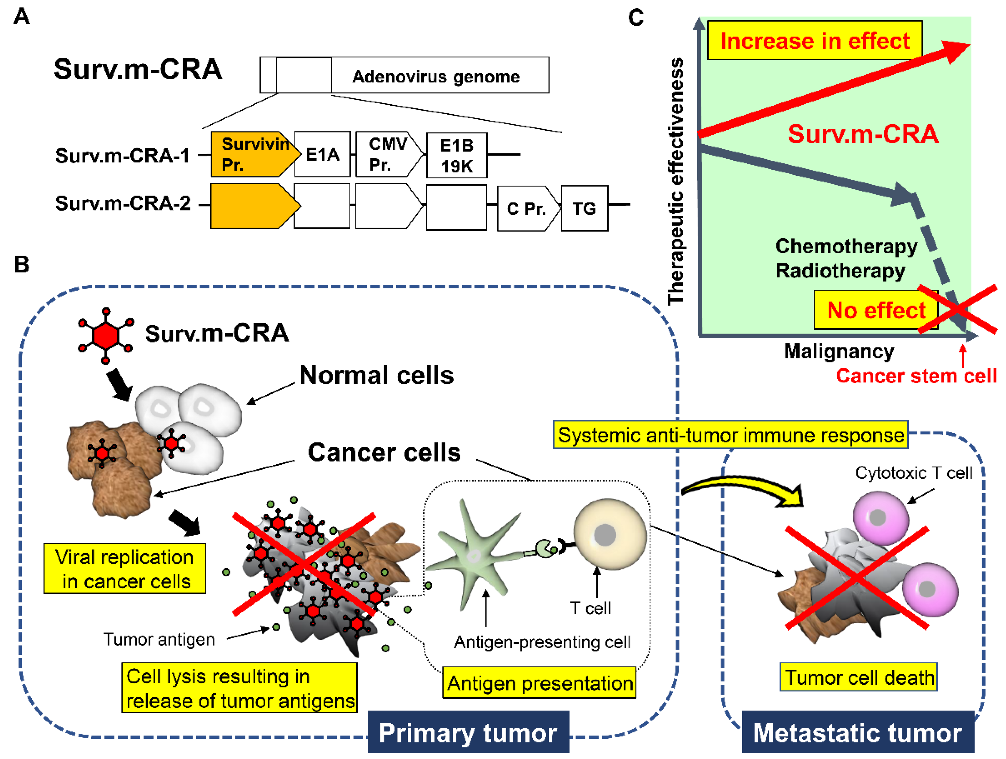
3.3. Survivin Responsive m-CRA

Using our m-CRA platform technology, we screened the cancer specificity and transcriptional activity of several cancer-specific promoters, constructed diverse m-CRAs using the candidate promoters identified by the screen, and determined their virological characteristics [67]. In doing so, we identified survivin-responsive m-CRAs (Surv.m-CRA), in which wild-type E1A and mutant E1B (E1B55K-deletion) genes are regulated by the best region of the survivin promoter and the cytomegalovirus enhancer/promoter, respectively, as one of the best m-CRAs [68,69] (Figure 3). Survivin, a member of the inhibitor of apoptosis protein family, is expressed at high levels in cancer cells, but either very low or undetectable in normal cells [69,96]. Another group employed a telomerase reverse transcriptase (Tert)-responsive CRA in a human cancer trial although a partial response was observed in only one out of 15 patients [66].
Prior to this report, we identified the best region of the Tert promoter, which showed both the most strictly cancer-specific and the strongest activities, and which has been used to generate the best Tert-responsive m-CRA (Tert.m-CRA) [69,97]. Nevertheless, comparisons of viral properties and therapeutic potentials between Surv.m-CRA and Tert.m-CRA revealed that Surv.m-CRA is safer and more effective [69,97]. Another advantage of Surv.m-CRA is its high effectiveness towards cancer stem cells (CSCs), which are resistant to conventional chemo-radiotherapy [70,98,99]. Interestingly, past clinical studies have indicated a positive correlation between high survivin expression levels and poor prognosis, accelerated rate of recurrence, and increased resistance to therapy in patients with cancer [100]. Accordingly, Surv.m-CRAs killed all populations efficiently while also demonstrating increased therapeutic efficacy against CSCs [70].
Moreover, Surv.m-CRA-OCp, in which mutant E1B (E1B55K-deletion) expression is regulated by the osteosarcoma/prostate cancer-specific but weakly active osteocalcin promoter, significantly increased cancer specificity without decreasing anticancer effects against osteosarcoma and prostate cancer [68]. This finding suggests that cancer-specific or tissue-specific promoters, many of which have some cancer specificity but are relatively weak at driving transcription, are widely useful for also regulating mutant E1B expression, at least on the condition that E1A expression is regulated by a strictly cancer-specific and strongly active promoter. In contrast, replacing wild-type E1A with mutant E1A (24 amino acid deletion in the C2 region) downstream of the survivin promoter does not further increase the cancer specificity of Surv.m-CRA [69]. This finding suggests that producing the ideal m-CRA will depend more on altering the regulation of E1A expression than on mutating E1A. Likewise, platform technologies, including our m-CRA, that allow rapid generation of diverse next-generation OVs armed with transgenes downstream of suitable promoters, will be important [71].
We have conducted ICH (International Council for Harmonisation of Technical Requirements for Pharmaceuticals for Human Use) compliant GMP (Good Manufacturing Practice) drug production and GLP (Good Laboratory Practice) non-clinical studies and completed the First-In-Human investigator-initiated Phase I clinical trial of Surv.m-CRA-1 (Surv.m-CRA without a therapeutic gene) for patients with malignant tumors in bones and soft tissue. Both safety and the efficacy were good (results to be reported in the future), indicating that the results of basic research and preclinical studies were reproduced in this clinical application. Two clinical trials involving Surv.m-CRA-1 for patients with pancreatic cancer (Phase I/II) and malignant bone sarcoma (Phase II) are underway.

3.4. Application of Recombinant Adenoviruses to Pluripotent Stem Cell-Based Regenerative Medicine

Regenerative medicine based on human pluripotent stem cells (PSCs) is a potential, innovative therapy for intractable diseases. The application of recombinant adenoviruses has been studied by very few researchers, including the authors of this review, in comparison with recombinant retroviruses and lentiviruses, which integrate transgenes into the host genome [72,101,102,103,104]. In somatic cells, integration enables long-term expression of the inserted transgenes, but in undifferentiated PSCs, exogenous genes are silenced at a high frequency. The most critical issue is that integration poses significant safety risks as retrovirus-mediated ex vivo stem cell gene therapy causes a high incidence of leukemia in treated patients [105]. In contrast, adenoviral DNA and its transgenes very rarely integrate into the host chromosome, but the transgene persists as an episome in the nucleus, which confers a significant advantage in terms of clinical safety. In this respect, accurate knowledge of the properties of recombinant adenoviruses may facilitate the development of new biotechnologies to overcome the obstacle to human PSC-based regenerative medicine.
We first used a replication-defective adenoviral vector against PSCs, and developed an “adenoviral conditional targeting in stem cells” (ACT-SC) method that specifically separates any target cells from other types of differentiated cells and the remnant undifferentiated PSCs during and after in vitro differentiation [104]. In addition, we recently reported an optimized method for efficient adenovirus-based gene delivery to and expression in human PSCs [103]. Comparative studies on the activities of three representative ubiquitous promoters indicated that only the cytomegalovirus enhancer and beta-actin promoter allow robust transgene expression in human PSCs. Moreover, adenoviral vector infection of PSCs in single-cell suspension culture yields high gene transduction efficiency with low cytotoxicity, without causing loss of the undifferentiated state of the PSCs.
In contrast, despite their enormous potential, human PSC-derived cell transplantation therapies pose a risk of malignant transformation of undifferentiated remnants [102]. Conventional methods that establish safe human PSC master cell banks through gene expression analysis and genome-wide sequencing merely suppress and do not completely abolish tumorigenesis [102,106]. To eradicate tumorigenesis directly, we used gene therapy vector technologies, including recombinant adenoviruses for human PSC-based regenerative medicine. We developed three innovative strategies such as the ACT-SC method, a novel method for generating tumorigenic cell-targeting lentiviral vectors efficiently, and another novel method for applying m-CRA technology to kill tumorigenic PSCs specifically [30,72,101,102,103,104]. We found that the survivin promoter almost completely lost its activity in differentiated normal cells but was highly active in undifferentiated human PSCs as well as cancer cells [72,101]. Accordingly, in vitro infection experiments with human PSCs have indicated that Surv.m-CRA propagates vigorously and exerts a strong cytotoxic effect on undifferentiated and tumorigenic human PSCs; however, Surv.m-CRA did not damage normal cells that were differentiated from the same human PSCs. Moreover, in vivo experiments showed no tumor formation from the human PSCs that had been infected with Surv.m-CRA before being transplanted into animals. Because Surv.m-CRA safety was already verified in human clinical trials for patients with cancer as described in Section 3.3, obstacles to the usage of this technology on human patients may be largely mitigated.

4. Conclusions

More than thirty years have passed since the world’s first authorized gene therapy clinical trial in 1990, and data accumulated since from both basic and clinical studies are now blossoming into a host of practical applications. One important factor for success is the technology of recombinant viruses. In this respect, recombinant adenoviruses contribute to the development of innovative technologies, which will not only advance clinical cancer immunotherapy and vaccine development against infectious diseases, but also facilitate research on developmental biology and regenerative medicine using PSCs.

Author Contributions

Conceptualization (M.W., Y.N., H.K., K.-i.K.), writing and editing (M.W., Y.N., K.-i.K.) and funding acquisition (K.-i.K.). All authors have read and agreed to the published version of the manuscript.

Funding

Some research articles and/or summarized conclusions of the author (K.-i.Kosai) that were described in this review article were supported by several public grants, including AMED under grant number JP21ck0106660 and JP20ck0106415 and JSPS KAKENHI grant number JP21H03645.

Institutional Review Board Statement

Not applicable.

Informed Consent Statement

Not applicable.

Data Availability Statement

Not applicable.

Conflicts of Interest

K.-i.Kosai is the founder of WyK BiotechPharma Inc., but does not earn a salary from the company. All other authors declare no conflict of interest.

References

  1. FDA. Approved Cellular and Gene Therapy Products. 2020. Available online: https://www.fda.gov/vaccines-blood-biologics/cellular-gene-therapy-products/approved-cellular-and-gene-therapy-products (accessed on 12 December 2021).
  2. Davison, A.J.; Benkő, M.; Harrach, B. Genetic content and evolution of adenoviruses. J. Gen. Virol. 2003, 84, 2895–2908. [Google Scholar] [CrossRef]
  3. Saha, B.; Parks, R.J. Recent Advances in Novel Antiviral Therapies against Human Adenovirus. Microorganisms 2020, 8, 1284. [Google Scholar] [CrossRef] [PubMed]
  4. Bányai, K.; Martella, V.; Meleg, E.; Kisfali, P.; Péterfi, Z.; Benkö, M.; Melegh, B.; Szucs, G. Searching for HAdV-52, the putative gastroenteritis-associated human adenovirus serotype in Southern Hungary. New Microbiol. 2009, 32, 185–188. [Google Scholar]
  5. Yang, C.; Zhu, C.; Qian, Y.; Deng, J.; Zhang, B.; Zhu, R.; Wang, F.; Sun, Y.; Chen, D.; Guo, Q.; et al. Application of Human Adenovirus Genotyping by Phylogenetic Analysis in an Outbreak to Identify Nosocomial Infection. Virol. Sin. 2020, 36, 393–401. [Google Scholar] [CrossRef]
  6. Yang, Z.R.; Wang, H.F.; Zhao, J.; Peng, Y.Y.; Wang, J.; Guinn, B.; Huang, L.Q. Recent developments in the use of adenoviruses and immunotoxins in cancer gene therapy. Cancer Gene Ther. 2007, 14, 599–615. [Google Scholar] [CrossRef] [Green Version]
  7. Saha, B.; Wong, C.M.; Parks, R.J. The Adenovirus Genome Contributes to the Structural Stability of the Virion. Viruses 2014, 6, 3563–3583. [Google Scholar] [CrossRef] [PubMed] [Green Version]
  8. Rowe, W.P.; Huebner, R.J.; Gilmore, L.K.; Parrott, R.H.; Ward, T.G. Isolation of a Cytopathogenic Agent from Human Adenoids Undergoing Spontaneous Degeneration in Tissue Culture. Exp. Biol. Med. 1953, 84, 570–573. [Google Scholar] [CrossRef]
  9. Bergelson, J.M.; Cunningham, J.A.; Droguett, G.; Kurt-Jones, E.A.; Krithivas, A.; Hong, J.S.; Horwitz, M.S.; Crowell, R.L.; Finberg, R.W. Isolation of a Common Receptor for Coxsackie B Viruses and Adenoviruses 2 and 5. Science 1997, 275, 1320–1323. [Google Scholar] [CrossRef]
  10. Tomko, R.P.; Xu, R.; Philipson, L. HCAR and MCAR: The human and mouse cellular receptors for subgroup C adenoviruses and group B coxsackieviruses. Proc. Natl. Acad. Sci. USA 1997, 94, 3352–3356. [Google Scholar] [CrossRef] [Green Version]
  11. Gaden, F.; Franqueville, L.; Magnusson, M.; Hong, S.S.; Merten, M.D.; Lindholm, L.; Boulanger, P. Gene Transduction and Cell Entry Pathway of Fiber-Modified Adenovirus Type 5 Vectors Carrying Novel Endocytic Peptide Ligands Selected on Human Tracheal Glandular Cells. J. Virol. 2004, 78, 7227–7247. [Google Scholar] [CrossRef] [Green Version]
  12. Wiethoff, C.M.; Wodrich, H.; Gerace, L.; Nemerow, G.R. Adenovirus Protein VI Mediates Membrane Disruption following Capsid Disassembly. J. Virol. 2005, 79, 1992–2000. [Google Scholar] [CrossRef] [PubMed] [Green Version]
  13. Bremner, K.H.; Scherer, J.; Yi, J.; Vershinin, M.; Gross, S.P.; Vallee, R.B. Adenovirus Transport via Direct Interaction of Cytoplasmic Dynein with the Viral Capsid Hexon Subunit. Cell Host Microbe 2009, 6, 523–535. [Google Scholar] [CrossRef] [PubMed] [Green Version]
  14. Wiethoff, C.M.; Nemerow, G.R. Adenovirus membrane penetration: Tickling the tail of a sleeping dragon. Virology 2015, 479-480, 591–599. [Google Scholar] [CrossRef] [PubMed] [Green Version]
  15. Charman, M.; Herrmann, C.; Weitzman, M.D. Viral and cellular interactions during adenovirus DNA replication. FEBS Lett. 2019, 593, 3531–3550. [Google Scholar] [CrossRef] [PubMed] [Green Version]
  16. Ikoma, T.; Takahashi, T.; Nagano, S.; Li, Y.-M.; Ohno, Y.; Ando, K.; Fujiwara, T.; Fujiwara, H.; Kosai, K.-I. A Definitive Role of RhoC in Metastasis of Orthotopic Lung Cancer in Mice. Clin. Cancer Res. 2004, 10, 1192–1200. [Google Scholar] [CrossRef] [Green Version]
  17. Kosai, K.I.; Finegold, M.J.; Thi-Huynh, B.T.; Tewson, M.; Ou, C.N.; Bowles, N.; Woo, S.L.; Schwall, R.H.; Darlington, G.J. Ret-rovirus-mediated in vivo gene transfer in the replicating liver using recombinant hepatocyte growth factor without liver injury or partial hepatectomy. Hum. Gene Ther. 1998, 9, 1293–1301. [Google Scholar] [CrossRef]
  18. Zhong, L.; Granelli-Piperno, A.; Choi, Y.; Steinman, R.M. Recombinant adenovirus is an efficient and non-perturbing genetic vector for human dendritic cells. Eur. J. Immunol. 1999, 29, 964–972. [Google Scholar] [CrossRef]
  19. Humphries, E.H.; Glover, C.; Reichmann, M.E. Rous sarcoma virus infection of synchronized cells establishes provirus integration during S-phase DNA synthesis prior to cellular division. Proc. Natl. Acad. Sci. USA 1981, 78, 2601–2605. [Google Scholar] [CrossRef] [Green Version]
  20. Gingeras, T.; Sciaky, D.; Gelinas, R.E.; Bing-Dong, J.; Yen, C.E.; Kelly, M.M.; Bullock, P.A.; Parsons, B.; O’Neill, K.E.; Roberts, R. Nucleotide sequences from the adenovirus-2 genome. J. Biol. Chem. 1982, 257, 13475–13491. [Google Scholar] [CrossRef]
  21. Abudoureyimu, M.; Lai, Y.; Tian, C.; Wang, T.; Wang, R.; Chu, X. Oncolytic Adenovirus—A nova for gene-targeted oncolytic viral therapy in HCC. Front. Oncol. 2019, 9, 1182. [Google Scholar] [CrossRef]
  22. Rao, L.; Debbas, M.; Sabbatini, P.; Hockenbery, D.; Korsmeyer, S.; White, E. The adenovirus E1A proteins induce apoptosis, which is inhibited by the E1B 19-kDa and Bcl-2 proteins. Proc. Natl. Acad. Sci. USA 1992, 89, 7742–7746. [Google Scholar] [CrossRef] [PubMed] [Green Version]
  23. Russell, W.C. Adenoviruses: Update on structure and function. J. Gen. Virol. 2009, 90, 1–20. [Google Scholar] [CrossRef] [PubMed]
  24. Kleinberger, T. En Guard! The Interactions between Adenoviruses and the DNA Damage Response. Viruses 2020, 12, 996. [Google Scholar] [CrossRef]
  25. Li, S.; Ou, M.; Wang, G.; Tang, L. Application of conditionally replicating adenoviruses in tumor early diagnosis technology, gene-radiation therapy and chemotherapy. Appl. Microbiol. Biotechnol. 2016, 100, 8325–8335. [Google Scholar] [CrossRef]
  26. Peter, M.; Kühnel, F. Oncolytic Adenovirus in Cancer Immunotherapy. Cancers 2020, 12, 3354. [Google Scholar] [CrossRef] [PubMed]
  27. Bett, A.J.; Prevec, L.; Graham, F.L. Packaging capacity and stability of human adenovirus type 5 vectors. J. Virol. 1993, 67, 5911–5921. [Google Scholar] [CrossRef] [Green Version]
  28. Bett, A.J.; Haddara, W.; Prevec, L.; Graham, F.L. An efficient and flexible system for construction of adenovirus vectors with insertions or deletions in early regions 1 and 3. Proc. Natl. Acad. Sci. USA 1994, 91, 8802–8806. [Google Scholar] [CrossRef] [Green Version]
  29. Khai, N.C.; Takahashi, T.; Ushikoshi, H.; Nagano, S.; Yuge, K.; Esaki, M.; Kawai, T.; Goto, K.; Murofushi, Y.; Fujiwara, T.; et al. In vivo hepatic HB-EGF gene transduction inhibits Fas-induced liver injury and induces liver regeneration in mice: A comparative study to HGF. J. Hepatol. 2006, 44, 1046–1054. [Google Scholar] [CrossRef]
  30. Matsuda, E.; Obama, Y.; Kosai, K.-I. Safe and low-dose but therapeutically effective adenovirus-mediated hepatocyte growth factor gene therapy for type 1 diabetes in mice. Life Sci. 2021, 268, 119014. [Google Scholar] [CrossRef] [PubMed]
  31. Ushikoshi, H.; Takahashi, T.; Chen, X.; Khai, N.C.; Esaki, M.; Goto, K.; Takemura, G.; Maruyama, R.; Minatoguchi, S.; Fujiwara, T.; et al. Local overexpression of HB-EGF exacerbates remodeling following myocardial infarction by activating noncardiomyocytes. Lab. Investig. 2005, 85, 862–873. [Google Scholar] [CrossRef] [Green Version]
  32. Yuge, K.; Takahashi, T.; Khai, N.C.; Goto, K.; Fujiwara, T.; Fujiwara, H.; Kosai, K.-I. Intramuscular injection of adenoviral hepatocyte growth factor at a distal site ameliorates dextran sodium sulfate-induced colitis in mice. Int. J. Mol. Med. 2014, 33, 1064–1074. [Google Scholar] [CrossRef] [Green Version]
  33. Sakamoto, K.; Khai, N.C.; Wang, Y.; Irie, R.; Takamatsu, H.; Matsufuji, H.; Kosai, K.-I. Heparin-binding epidermal growth factor-like growth factor and hepatocyte growth factor inhibit cholestatic liver injury in mice through different mechanisms. Int. J. Mol. Med. 2016, 38, 1673–1682. [Google Scholar] [CrossRef] [Green Version]
  34. Bressy, C.; Hastie, E.; Grdzelishvili, V.Z. Combining Oncolytic Virotherapy with p53 Tumor Suppressor Gene Therapy. Mol. Ther. Oncolytics 2017, 5, 20–40. [Google Scholar] [CrossRef] [PubMed] [Green Version]
  35. Chen, S.H.; Wang, Y.; Kosai, K.; Finegold, M.J.; Rich, S.S.; Woo, S.L. Combination gene therapy for liver metastasis of colon carcinoma in vivo. Proc. Natl. Acad. Sci. USA 1995, 92, 2577–2581. [Google Scholar] [CrossRef] [PubMed] [Green Version]
  36. Chen, S.H.; Kosai, K.; Xu, B.; Pham-Nguyen, K.; Contant, C.; Finegold, M.J.; Woo, S.L. Combination suicide and cytokine gene therapy for hepatic metastases of colon carcinoma: Sustained antitumor immunity prolongs animal survival. Cancer Res. 1996, 56, 3758–3762. [Google Scholar] [PubMed]
  37. Kwong, Y.-L.; Chen, S.-H.; Kosai, K.; Finegold, M.; Woo, S.L. Combination therapy with suicide and cytokine genes for hepatic metastases of lung cancer. Chest 1997, 112, 1332–1337. [Google Scholar] [CrossRef]
  38. Block, A.; Chen, S.H.; Kosai, K.; Finegold, M.; Woo, S.L. Adenoviral-mediated herpes simplex virus thymidine kinase gene transfer: Regression of hepatic metastasis of pancreatic tumors. Pancreas 1997, 15, 25–34. [Google Scholar] [CrossRef]
  39. Fukunaga, M.; Takamori, S.; Hayashi, A.; Shirouzu, K.; Kosai, K.-I. Adenoviral herpes simplex virus thymidine kinase gene therapy in an orthotopic lung cancer model. Ann. Thorac. Surg. 2002, 73, 1740–1746. [Google Scholar] [CrossRef]
  40. Kwong, Y.L.; Chen, S.H.; Kosai, K.; Finegold, M.J.; Woo, S.L. Adenoviral-mediated suicide gene therapy for hepatic metastases of breast cancer. Cancer Gene Ther. 1996, 3, 339–344. [Google Scholar] [PubMed]
  41. Terazaki, Y.; Yano, S.; Nagano, S.; Fukunaga, M.; Guo, Z.S.; Yuge, K.; Komiya, S.; Shirouzu, K.; Kosai, K. An optimal therapeutic expression level is crucial for suicide gene therapy for hepatic metastatic cancer in mice. Hepatology 2003, 37, 155–163. [Google Scholar] [CrossRef]
  42. Huang, H.-P.; Chen, S.H.; Kosai, K.; Finegold, M.J.; Woo, S.L. Gene therapy for hepatocellular carcinoma: Long-term remission of primary and metastatic tumors in mice by interleukin-2 gene therapy in vivo. Gene Ther. 1996, 3, 980–987. [Google Scholar]
  43. Nagano, S.; Yuge, K.; Fukunaga, M.; Terazaki, Y.; Fujiwara, H.; Komiya, S.; Kosai, K. Gene therapy eradicating distant dissemi-nated micro-metastases by optimal cytokine expression in the primary lesion only: Novel concepts for successful cytokine gene therapy. Int. J. Oncol. 2004, 24, 549–558. [Google Scholar] [PubMed]
  44. O’Malley, B.W., Jr.; Sewell, D.A.; Li, D.; Kosai, K.-I.; Chen, S.-H.; Woo, S.L.; Duan, L. The Role of Interleukin-2 in Combination Adenovirus Gene Therapy for Head and Neck Cancer. Mol. Endocrinol. 1997, 11, 667–673. [Google Scholar] [CrossRef] [PubMed]
  45. Caruso, M.; Pham-Nguyen, K.; Kwong, Y.L.; Xu, B.; Kosai, K.I.; Finegold, M.; Woo, S.L.; Chen, S.H. Adenovirus-mediated interleukin-12 gene therapy for metastatic colon carcinoma. Proc. Natl. Acad. Sci. USA 1996, 93, 11302–11306. [Google Scholar] [CrossRef] [PubMed] [Green Version]
  46. Li, C.-Y.; Huang, Q.; Kung, H.-F. Cytokine and immuno-gene therapy for solid tumors. Cell. Mol. Immunol. 2005, 2, 81–91. [Google Scholar]
  47. Raper, S.E.; Chirmule, N.; Lee, F.; Wivel, N.A.; Bagg, A.; Gao, G.-P.; Wilson, J.; Batshaw, M.L. Fatal systemic inflammatory response syndrome in a ornithine transcarbamylase deficient patient following adenoviral gene transfer. Mol. Genet. Metab. 2003, 80, 148–158. [Google Scholar] [CrossRef] [PubMed]
  48. Kreppel, F.; Hagedorn, C. Capsid and Genome Modification Strategies to Reduce the Immunogenicity of Adenoviral Vectors. Int. J. Mol. Sci. 2021, 22, 2417. [Google Scholar] [CrossRef]
  49. Parks, R.J.; Graham, F.L. A helper-dependent system for adenovirus vector production helps define a lower limit for efficient DNA packaging. J. Virol. 1997, 71, 3293–3298. [Google Scholar] [CrossRef] [Green Version]
  50. Schmid, M.; Ernst, P.; Honegger, A.; Suomalainen, M.; Zimmermann, M.; Braun, L.; Stauffer, S.; Thom, C.; Dreier, B.; Eibauer, M.; et al. Adenoviral vector with shield and adapter increases tumor specificity and escapes liver and immune control. Nat. Commun. 2018, 9, 450. [Google Scholar] [CrossRef]
  51. Chen, C.Y.; May, S.M.; Barry, M.A. Targeting Adenoviruses with Factor X–Single-Chain Antibody Fusion Proteins. Hum. Gene Ther. 2010, 21, 739–749. [Google Scholar] [CrossRef] [Green Version]
  52. Ling, Y.; Zhong, J.; Luo, J. Safety and effectiveness of SARS-CoV-2 vaccines: A systematic review and meta-analysis. J. Med. Virol. 2021, 93, 6486–6495. [Google Scholar] [CrossRef] [PubMed]
  53. Goradel, N.H.; Mohajel, N.; Malekshahi, Z.V.; Jahangiri, S.; Najafi, M.; Farhood, B.; Mortezaee, K.; Negahdari, B.; Arashkia, A. Oncolytic adenovirus: A tool for cancer therapy in combination with other therapeutic approaches. J. Cell. Physiol. 2019, 234, 8636–8646. [Google Scholar] [CrossRef] [PubMed]
  54. Lang, F.F.; Conrad, C.; Gomez-Manzano, C.; Alfred Yung, W.K.; Sawaya, R.; Weinberg, J.S.; Prabhu, S.S.; Rao, G.; Fuller, G.N.; Aldape, K.D.; et al. Phase I Study of DNX-2401 (Delta-24-RGD) Oncolytic Adenovirus: Replication and Immunotherapeutic Effects in Recurrent Malignant Glioma. J. Clin. Oncol. 2018, 36, 1419–1427. [Google Scholar] [CrossRef] [PubMed]
  55. Fueyo, J.; Gomez-Manzano, C.; Alemany, R.; Lee, P.S.; McDonnell, T.J.; Mitliagka, P.; Shi, Y.-X.; Levin, V.A.; Yung, W.K.A.; Kyritsis, A.P. A mutant oncolytic adenovirus targeting the Rb pathway produces anti-glioma effect in vivo. Oncogene 2000, 19, 2–12. [Google Scholar] [CrossRef] [Green Version]
  56. Heise, C.; Hermiston, T.; Johnson, L.; Brooks, G.A.; Sampson-Johannes, A.; Williams, A.; Hawkins, L.; Kirn, D.H. An adenovirus E1A mutant that demonstrates potent and selective systemic anti-tumoral efficacy. Nat. Med. 2000, 6, 1134–1139. [Google Scholar] [CrossRef]
  57. Garcia-Moure, M.; Martinez-Vélez, N.; Patiño-García, A.; Alonso, M.M. Oncolytic adenoviruses as a therapeutic approach for osteosarcoma: A new hope. J. Bone Oncol. 2016, 9, 41–47. [Google Scholar] [CrossRef]
  58. Bischoff, J.R.; Kirn, D.H.; Williams, A.; Heise, C.; Horn, S.; Muna, M.; Ng, L.; Nye, J.A.; Sampson-Johannes, A.; Fattaey, A.; et al. An Adenovirus Mutant That Replicates Selectively in p53- Deficient Human Tumor Cells. Science 1996, 274, 373–376. [Google Scholar] [CrossRef]
  59. Zhang, K.-J.; Zhang, J.; Wu, Y.-M.; Qian, J.; Liu, X.-J.; Yan, L.-C.; Zhou, X.-M.; Xiao, R.-J.; Wang, Y.-G.; Cao, X.; et al. Complete eradication of hepatomas using an oncolytic adenovirus containing AFP promoter controlling E1A and an E1B deletion to drive IL-24 expression. Cancer Gene Ther. 2012, 19, 619–629. [Google Scholar] [CrossRef]
  60. Irving, J.; Wang, Z.; Powell, S.; O’Sullivan, C.; Mok, M.; Murphy, B.; Cardoza, L.; Lebkowski, J.S.; Majumdar, A.S. Conditionally replicative adenovirus driven by the human telomerase promoter provides broad-spectrum antitumor activity without liver toxicity. Cancer Gene Ther. 2004, 11, 174–185. [Google Scholar] [CrossRef] [Green Version]
  61. Wirth, T.; Zender, L.; Schulte, B.; Mundt, B.; Plentz, R.; Rudolph, K.L.; Manns, M.; Kubicka, S.; Kühnel, F. A telomerase-dependent conditionally replicating adenovirus for selective treatment of cancer. Cancer Res. 2003, 63, 3181–3188. [Google Scholar]
  62. Harada, J.N.; Berk, A.J. p53-Independent and -Dependent Requirements for E1B-55K in Adenovirus Type 5 Replication. J. Virol. 1999, 73, 5333–5344. [Google Scholar] [CrossRef] [Green Version]
  63. Rothmann, T.; Hengstermann, A.; Whitaker, N.J.; Scheffner, M.; Hausen, H.Z. Replication of ONYX-015, a Potential Anticancer Adenovirus, Is Independent of p53 Status in Tumor Cells. J. Virol. 1998, 72, 9470–9478. [Google Scholar] [CrossRef] [Green Version]
  64. Geoerger, B.; Vassal, G.; Opolon, P.; Dirven, C.M.; Morizet, J.; Laudani, L.; Grill, J.; Giaccone, G.; Vandertop, W.P.; Gerritsen, W.R.; et al. Oncolytic Activity of p53-Expressing Conditionally Replicative Adenovirus AdΔ24-p53 against Human Malignant Glioma. Cancer Res. 2004, 64, 5753–5759. [Google Scholar] [CrossRef] [Green Version]
  65. Nettelbeck, D.M.; Rivera, A.A.; Balagué, C.; Alemany, R.; Curiel, D.T. Novel oncolytic adenoviruses targeted to melanoma: Specific viral replication and cytolysis by expression of E1A mutants from the tyrosinase enhancer/promoter. Cancer Res. 2002, 62, 4663–4670. [Google Scholar]
  66. Nemunaitis, J.; Tong, A.W.; Nemunaitis, M.; Senzer, N.; Phadke, A.P.; Bedell, C.; Adams, N.; Zhang, Y.-A.; Maples, P.B.; Chen, S.; et al. A Phase I Study of Telomerase-specific Replication Competent Oncolytic Adenovirus (Telomelysin) for Various Solid Tumors. Mol. Ther. 2010, 18, 429–434. [Google Scholar] [CrossRef] [PubMed]
  67. Nagano, S.; Oshika, H.; Fujiwara, H.; Komiya, S.; Kosai, K. An efficient construction of conditionally replicating adenoviruses that target tumor cells with multiple factors. Gene Ther. 2005, 12, 1385–1393. [Google Scholar] [CrossRef] [PubMed]
  68. Horikawa, Y.; Wang, Y.; Nagano, S.; Kamizono, J.; Ikeda, M.; Komiya, S.; Kosai, K.-I. Assessment of an altered E1B promoter on the specificity and potency of triple-regulated conditionally replicating adenoviruses: Implications for the generation of ideal m-CRAs. Cancer Gene Ther. 2011, 18, 724–733. [Google Scholar] [CrossRef] [Green Version]
  69. Kamizono, J.; Nagano, S.; Murofushi, Y.; Komiya, S.; Fujiwara, H.; Matsuishi, T.; Kosai, K.-I. Survivin-Responsive Conditionally Replicating Adenovirus Exhibits Cancer-Specific and Efficient Viral Replication. Cancer Res. 2005, 65, 5284–5291. [Google Scholar] [CrossRef] [Green Version]
  70. Tanoue, K.; Wang, Y.; Ikeda, M.; Mitsui, K.; Irie, R.; Setoguchi, T.; Komiya, S.; Natsugoe, S.; Kosai, K.-I. Survivin-responsive conditionally replicating adenovirus kills rhabdomyosarcoma stem cells more efficiently than their progeny. J. Transl. Med. 2014, 12, 27. [Google Scholar] [CrossRef] [PubMed] [Green Version]
  71. Suzuki, S.; Kofune, H.; Uozumi, K.; Yoshimitsu, M.; Arima, N.; Ishitsuka, K.; Ueno, S.-I.; Kosai, K.-I. A survivin-responsive, conditionally replicating adenovirus induces potent cytocidal effects in adult T-cell leukemia/lymphoma. BMC Cancer 2019, 19, 516. [Google Scholar] [CrossRef]
  72. Mitsui, K.; Ide, K.; Takayama, A.; Wada, T.; Irie, R.; Kosai, K.-I. Conditionally replicating adenovirus prevents pluripotent stem cell–derived teratoma by specifically eliminating undifferentiated cells. Mol. Ther. Methods Clin. Dev. 2015, 2, 15026. [Google Scholar] [CrossRef]
  73. Wold, W.S.M.; Toth, K. Adenovirus Vectors for Gene Therapy, Vaccination and Cancer Gene Therapy. Curr. Gene Ther. 2013, 13, 421–433. [Google Scholar] [CrossRef] [PubMed]
  74. Flinterman, M.; Gäken, J.; Farzaneh, F.; Tavassoli, M. E1A-mediated suppression of EGFR expression and induction of apoptosis in head and neck squamous carcinoma cell lines. Oncogene 2003, 22, 1965–1977. [Google Scholar] [CrossRef] [PubMed]
  75. Berk, A.J. Recent lessons in gene expression, cell cycle control, and cell biology from adenovirus. Oncogene 2005, 24, 7673–7685. [Google Scholar] [CrossRef] [Green Version]
  76. Cheng, P.-H.; Wechman, S.L.; McMasters, K.M.; Zhou, H.S. Oncolytic Replication of E1b-Deleted Adenoviruses. Viruses 2015, 7, 5767–5779. [Google Scholar] [CrossRef] [PubMed] [Green Version]
  77. Gürlevik, E.; Woller, N.; Schache, P.; Malek, N.P.; Wirth, T.C.; Zender, L.; Manns, M.P.; Kubicka, S.; Kühnel, F. p53-dependent antiviral RNA-interference facilitates tumor-selective viral replication. Nucleic Acids Res. 2009, 37, e84. [Google Scholar] [CrossRef] [Green Version]
  78. Ramachandra, M.; Rahman, A.; Zou, A.; Vaillancourt, M.; Howe, J.A.; Antelman, D.; Sugarman, B.J.; Demers, G.W.; Engler, H.; Johnson, D.E.; et al. Re-engineering adenovirus regulatory pathways to enhance oncolytic specificity and efficacy. Nat. Biotechnol. 2001, 19, 1035–1041. [Google Scholar] [CrossRef]
  79. Kawashima, T.; Kagawa, S.; Kobayashi, N.; Shirakiya, Y.; Umeoka, T.; Teraishi, F.; Taki, M.; Kyo, S.; Tanaka, N.; Fujiwara, T. Telomerase-specific replication-selective virotherapy for human cancer. Clin. Cancer Res. 2004, 10, 285–292. [Google Scholar] [CrossRef] [Green Version]
  80. Lanson, N.A.; Friedlander, P.L.; Schwarzenberger, P.; Kolls, J.K.; Wang, G. Replication of an adenoviral vector controlled by the human telomerase reverse transcriptase promoter causes tumor-selective tumor lysis. Cancer Res. 2003, 63, 7936–7941. [Google Scholar]
  81. Kurihara, T.; Brough, D.E.; Kovesdi, I.; Kufe, D.W. Selectivity of a replication-competent adenovirus for human breast carcinoma cells expressing the MUC1 antigen. J. Clin. Investig. 2000, 106, 763–771. [Google Scholar] [CrossRef] [Green Version]
  82. Hallenbeck, P.L.; Chang, Y.-N.; Hay, C.; Golightly, D.; Stewart, D.; Lin, J.; Phipps, S.; Chiang, Y.L. A Novel Tumor-Specific Replication-Restricted Adenoviral Vector for Gene Therapy of Hepatocellular Carcinoma. Hum. Gene Ther. 1999, 10, 1721–1733. [Google Scholar] [CrossRef]
  83. Rodriguez, R.; Schuur, E.R.; Lim, H.Y.; Henderson, G.A.; Simons, J.W.; Henderson, D.R. Prostate attenuated replication competent adenovirus (ARCA) CN706: A selective cytotoxic for prostate-specific antigen-positive prostate cancer cells. Cancer Res. 1997, 57, 2559–2563. [Google Scholar]
  84. Gomez-Manzano, C.; Balague, C.; Alemany, R.; Lemoine, M.G.; Mitliagka, P.; Jiang, H.; Khan, A.; Alonso, M.; Lang, F.F.; Conrad, C.A.; et al. A novel E1A–E1B mutant adenovirus induces glioma regression in vivo. Oncogene 2004, 23, 1821–1828. [Google Scholar] [CrossRef] [PubMed] [Green Version]
  85. Johnson, L.; Shen, A.; Boyle, L.; Kunich, J.; Pandey, K.; Lemmon, M.; Hermiston, T.; Giedlin, M.; McCormick, F.; Fattaey, A. Selectively replicating adenoviruses targeting deregulated E2F activity are potent, systemic antitumor agents. Cancer Cell 2002, 1, 325–337. [Google Scholar] [CrossRef] [Green Version]
  86. Yu, D.M.; Sakamoto, G.T.; Henderson, D.R. Identification of the transcriptional regulatory sequences of human kallikrein 2 and their use in the construction of calydon virus 764, an attenuated replication competent adenovirus for prostate cancer therapy. Cancer Res. 1999, 59, 1498–1504. [Google Scholar] [PubMed]
  87. Chartier, C.; Degryse, E.; Gantzer, M.; Dieterle, A.; Pavirani, A.; Mehtali, M. Efficient generation of recombinant adenovirus vectors by homologous recombination in Escherichia coli. J. Virol. 1996, 70, 4805–4810. [Google Scholar] [CrossRef] [Green Version]
  88. Miyake, S.; Makimura, M.; Kanegae, Y.; Harada, S.; Sato, Y.; Takamori, K.; Tokuda, C.; Saito, I. Efficient generation of recombinant adenoviruses using adenovirus DNA-terminal protein complex and a cosmid bearing the full-length virus genome. Proc. Natl. Acad. Sci. USA 1996, 93, 1320–1324. [Google Scholar] [CrossRef] [Green Version]
  89. Mizuguchi, H.; Kay, M.A. Efficient Construction of a Recombinant Adenovirus Vector by an Improved In Vitro Ligation Method. Hum. Gene Ther. 1998, 9, 2577–2583. [Google Scholar] [CrossRef] [Green Version]
  90. Makower, D.; Rozenblit, A.; Kaufman, H.; Edelman, M.; Lane, M.E.; Zwiebel, J.; Haynes, H.; Wadler, S. Phase II clinical trial of intralesional administration of the oncolytic adenovirus ONYX-015 in patients with hepatobiliary tumors with correlative p53 studies. Clin. Cancer Res. 2003, 9, 693–702. [Google Scholar]
  91. Chiocca, E.A.; Abbed, K.M.; Tatter, S.; Louis, D.N.; Hochberg, F.H.; Barker, F.; Kracher, J.; Grossman, S.A.; Fisher, J.D.; Carson, K.; et al. A Phase I Open-Label, Dose-Escalation, Multi-Institutional Trial of Injection with an E1B-Attenuated Adenovirus, ONYX-015, into the Peritumoral Region of Recurrent Malignant Gliomas, in the Adjuvant Setting. Mol. Ther. 2004, 10, 958–966. [Google Scholar] [CrossRef]
  92. Guo, Z.S.; Lotze, M.T.; Zhu, Z.; Storkus, W.J.; Song, X.-T. Bi- and Tri-Specific T Cell Engager-Armed Oncolytic Viruses: Next-Generation Cancer Immunotherapy. Biomedicines 2020, 8, 204. [Google Scholar] [CrossRef]
  93. Goradel, N.H.; Baker, A.T.; Arashkia, A.; Ebrahimi, N.; Ghorghanlu, S.; Negahdari, B. Oncolytic virotherapy: Challenges and solutions. Curr. Probl. Cancer 2021, 45, 100639. [Google Scholar] [CrossRef]
  94. Harrington, K.; Puzanov, I.; Hecht, J.R.; Hodi, F.S.; Szabo, Z.; Murugappan, S.; Kaufman, H.L. Clinical development of talimogene laherparepvec (T-VEC): A modified herpes simplex virus type-1–derived oncolytic immunotherapy. Expert Rev. Anticancer. Ther. 2015, 15, 1389–1403. [Google Scholar] [CrossRef]
  95. Ribas, A.; Dummer, R.; Puzanov, I.; VanderWalde, A.; Andtbacka, R.H.I.; Michielin, O.; Olszanski, A.J.; Malvehy, J.; Cebon, J.; Fernandez, E.; et al. Oncolytic Virotherapy Promotes Intratumoral T Cell Infiltration and Improves Anti-PD-1 Immunotherapy. Cell 2017, 170, 1109–1119.e10. [Google Scholar] [CrossRef] [Green Version]
  96. Ambrosini, G.; Adida, C.; Altieri, D.C. A novel anti-apoptosis gene, survivin, expressed in cancer and lymphoma. Nat. Med. 1997, 3, 917–921. [Google Scholar] [CrossRef] [PubMed]
  97. Murofushi, Y.; Nagano, S.; Kamizono, J.; Takahashi, T.; Fujiwara, H.; Komiya, S.; Matsuishi, T.; Kosai, K. Cell cycle-specific changes in hTERT promoter activity in normal and cancerous cells in adenoviral gene therapy: A promising implication of te-lomerase-dependent targeted cancer gene therapy. Int. J. Oncol. 2006, 29, 681–688. [Google Scholar] [CrossRef] [PubMed] [Green Version]
  98. Yu, Z.; Pestell, T.G.; Lisanti, M.P.; Pestell, R.G. Cancer stem cells. Int. J. Biochem. Cell Biol. 2012, 44, 2144–2151. [Google Scholar] [CrossRef] [Green Version]
  99. Samadani, A.A.; Keymoradzdeh, A.; Shams, S.; Soleymanpour, A.; Norollahi, S.E.; Vahidi, S.; Rashidy-Pour, A.; Ashraf, A.; Mirzajani, E.; Khanaki, K.; et al. Mechanisms of cancer stem cell therapy. Clin. Chim. Acta 2020, 510, 581–592. [Google Scholar] [CrossRef] [PubMed]
  100. Altieri, D.C. The molecular basis and potential role of survivin in cancer diagnosis and therapy. Trends Mol. Med. 2001, 7, 542–547. [Google Scholar] [CrossRef]
  101. Ide, K.; Mitsui, K.; Irie, R.; Matsushita, Y.; Ijichi, N.; Toyodome, S.; Kosai, K. A novel construction of lentiviral vectors for eliminating tumorigenic cells from pluripotent stem cells. Stem Cells 2018, 36, 230–239. [Google Scholar] [CrossRef] [Green Version]
  102. Mitsui, K.; Ide, K.; Takahashi, T.; Kosai, K.-I. Viral Vector-Based Innovative Approaches to Directly Abolishing Tumorigenic Pluripotent Stem Cells for Safer Regenerative Medicine. Mol. Ther. Methods Clin. Dev. 2017, 5, 51–58. [Google Scholar] [CrossRef] [PubMed] [Green Version]
  103. Mitsui, K.; Takahashi, T.; Ide, K.; Matsuda, E.; Kosai, K.-I. Optimization of adenoviral gene transfer in human pluripotent stem cells. Biochem. Biophys. Res. Commun. 2021, 541, 78–83. [Google Scholar] [CrossRef]
  104. Takahashi, T.; Kawai, T.; Ushikoshi, H.; Nagano, S.; Oshika, H.; Inoue, M.; Kunisada, T.; Takemura, G.; Fujiwara, H.; Kosai, K.-I. Identification and Isolation of Embryonic Stem Cell-Derived Target Cells by Adenoviral Conditional Targeting. Mol. Ther. 2006, 14, 673–683. [Google Scholar] [CrossRef] [PubMed]
  105. McCormack, M.P.; Rabbitts, T.H. Activation of the T-Cell OncogeneLMO2after Gene Therapy for X-Linked Severe Combined Immunodeficiency. N. Engl. J. Med. 2004, 350, 913–922. [Google Scholar] [CrossRef]
  106. Lee, A.S.; Tang, C.; Rao, M.S.; Weissman, I.L.; Wu, J.C. Tumorigenicity as a clinical hurdle for pluripotent stem cell therapies. Nat. Med. 2013, 19, 998–1004. [Google Scholar] [CrossRef] [PubMed] [Green Version]
Figure 1. Schematic representation of the serotype 5 adenoviral genome. DNA strands are shown as a pair of lines. The number below the lines indicates the 0–100 map unit (mu) of the adenoviral genome.
Viruses 13 02502 g001
Figure 2. Constitution of m-CRA plasmids. Potentially, more than seven cancer-specific factors can be introduced into m-CRA. (1) Promoter A, which drives wild-type or mutant E1A expression. (2) Mutant E1A, which lacks an Rb-binding site (E1A∆24). (3) Promoter B, which drives wild-type or mutant E1B. (4) Mutant E1B, which lacks a p53-binding protein that is encoded by E1B55K (E1B∆55K). (5) Promoter C, which controls a therapeutic gene. (6) A therapeutic gene (seven or more can be incorporated). Modification of the adenoviral backbone is also possible, such as fiber modification to alter infectivity. A, antibiotic resistance gene; O, origin of replication.
Viruses 13 02502 g002
Figure 3. Therapeutic mechanism and characteristic features of survivin-responsive m-CRA (Surv.m-CRA). (A) The basic constitution of Surv.m-CRA. (B) Therapeutic mechanism of OV therapy, including Surv.m-CRA. (C) The putative therapeutic advantage of Surv.m-CRA against cancer stem cells in comparison to conventional chemo-radiotherapy.
Viruses 13 02502 g003
Table 1. The characteristic features of recombinant adenoviruses.
Type of Recombinant AdenovirusModification of AdenovirusAdvantageDisadvantageApplicable Diseases and TherapiesReferences
Replication-defective adenoviral vector
  • E1A deletion
  • Robust but transient transgene expression and high immunogenicity applicable to cancer gene therapy and vaccines
  • No indication for genetic diseases because of short-term effect and safety concerns
  • Gene medicine for solid tumor
  • Vaccination to prevent infectious diseases
[28,34,35,36,37,38,39,40,41,42,43,44,45,46,47,48,49,50,51,52]
Conditionally replicating adenovirus (CRA; oncolytic adenovirus)
  • A deletion in CR2 of the E1A gene and in the E1B55K gene within E1B
  • Replacement of the native E1A promoter with a cancer-specific promoter
  • Predominantly replicating in and killing cancer cells
  • Armed transgene enhancing therapeutic effects
  • Insufficient cancer-specific viral replication resulting in insufficient therapeutic effect of virotherapy alone
  • Virotherapy and immunotherapy for solid tumor
[21,25,53,54,55,56,57,58,59,60,61,62,63,64,65,66]
CRA that can specifically target tumors with multiple factors (m-CRA)
  • Simultaneously regulated by up to four independent factors of the E1 region
  • Feasible to add a therapeutic gene with a suitable promoter and to modify adenoviral backbone in the step of viral constructions
  • Strictly cancer-specific and effective viral replication producing more potent anticancer effects and higher safety than the conventional CRAs
  • More potently and safely enhancing therapeutic effects by a therapeutic gene with a suitable promoter and to alter infectivity
  • More potent and safer virotherapy and immunotherapy for solid tumor
  • A new strategy to prevent stem cell-derived tumorigenesis in regenerative medicine
[67,68,69,70,71,72]
Publisher’s Note: MDPI stays neutral with regard to jurisdictional claims in published maps and institutional affiliations.

