Systematic Review

Clinical Landscape and Rate of Exposure to Ilheus Virus: Insights from Systematic Review and Meta-Analysis

by
Vivaldo Gomes da Costa
1,*,†,
Marielena Vogel Saivish
2,*,†,
Nikolas Alexander Borsato Lino
1,
Cíntia Bittar
1,
Marília de Freitas Calmon
1,
Maurício Lacerda Nogueira
2,* and
Paula Rahal
1,*
1
Laboratório de Estudos Genômicos, Departamento de Biologia, Instituto de Biociências Letras e Ciências Exatas (IBILCE), Universidade Estadual Paulista (UNESP), São José do Rio Preto 15054-000, Brazil
2
Laboratório de Pesquisas em Virologia, Departamento de Doenças Dermatológicas, Infecciosas e Parasitárias, Faculdade de Medicina de São José do Rio Preto (FAMERP), São José do Rio Preto 15090-000, Brazil
*
Authors to whom correspondence should be addressed.
These authors contributed equally to this work.
Viruses 2023, 15(1), 92; https://doi.org/10.3390/v15010092
Submission received: 17 November 2022 / Revised: 14 December 2022 / Accepted: 24 December 2022 / Published: 29 December 2022
(This article belongs to the Special Issue Sero-Epidemiology of Viral Infection)

Abstract

:
Ilheus fever is a mosquito-borne, poorly known tropical disease. We aimed to report the pooled rate of exposure to the Ilheus virus (ILHV) and clinical outcomes of infection to determine the epidemiological patterns of ILHV. We conducted a meta-analysis of 37 studies (n = 17,722 individuals) from Latin America. The common clinical characteristics of ILHV infection were fever (82.3%), headache (52.9%), and myalgia (52.9%). Encephalitis complicated the course of the infection in 29.4% cases. Monotypic serological reactions detected a pooled rate of exposure of 2% to ILHV (95% CI: 1–2). Studies were mainly conducted in Brazil, with a pooled proportion of ILHV positivity of 8% (95% CI: 3–14). Males (12%) had higher rates of seropositivity than females (7%) and had high chances of ILHV infection (OR: 1.7, 95% CI: 1.2–2.5). Seropositivity increased with age, from 2% (95% CI: 2–3) among people aged 0–14 years to 8% (95% CI: 6–10) among people aged 15–64 years. Our analysis indicated a low and relatively constant burden of ILHV in Latin America. More research is needed to evaluate and innovate serological assays for ILHV to better estimate the burden and dynamics of epidemiological changes in ILHV infection in different regions.

1. Introduction

Ilheus virus (ILHV), a neglected human pathogen, is believed to be maintained in nature by an enzootic cycle between mosquito vectors and birds [1]. Structurally, ILHV has a spherical morphology with enveloped positive-sense single-stranded RNA and belongs to the genus Flavivirus in the Ntaya antigenic serocomplex [2,3]. It is phylogenetically associated with the Rocio virus (ROCV) with 73.37–74.5% amino acid identity, and ILHV and ROCV are considered members of the same species [4]. ILHV infection in humans can lead to Ilheus fever, an acute undifferentiated febrile disease with non-outbreak cases. However, the clinical spectrum can vary in severity, and the fever can progress to severe life-threatening meningoencephalitis [5,6,7,8,9,10]. In 2017, the first documented case of a fatality following ILHV detection via nucleic acid amplification in an elderly patient with encephalitis in Brazil drew public attention to the need to better understand the clinical–epidemiological scenario [11]. It is important to mention that in this case, it was not determined whether the virus caused encephalitis in the patient, who had co-morbidity (diabetes and hypertension).
Historically, ILHV was isolated in 1944 from a pool of Psorophora and Aedes mosquito species captured near Ilhéus city in Bahia state, Brazil, during an entomological surveillance of the yellow fever virus vector [1]. Viral infection in humans occurs via mosquito bites, especially from mosquitoes of the genera Psorophora and Ochlerotatus. ILHV was isolated from humans in 1959 in Belém city (Pará, Brazil). This was followed by viral isolation in Trinidad, West Indies (1962), Colombia (1966), Panama (1967), French Guiana (1973), Brazil (1995), and Ecuador (2007) [5,7,8,9,12,13,14]. Currently, several arboviral surveillance studies, including those on ILHV isolates, molecular identification, and serosurvey to detect antibodies against ILHV, have been carried out in the last few decades in South and Central America and in the Caribbean islands. Thus, according to the results of these studies, ILHV infections have been observed in several vertebrate species, such as monkeys, birds, rodents, bats, horses, coatis, and buffaloes (Supplementary Files S1 and S2) [15,16,17,18,19,20,21,22,23,24,25,26,27,28,29,30,31,32,33,34,35,36].
However, several barriers related to the difficulty in ILHV detection remain. The main factors include diverse endemic mosquito-borne flavivirus populations, a short viremic period, lack of specific clinical features, and the possibility of serological tests exhibiting cross-reactivity to shared epitopes on other endemic flaviviruses. Overall, these factors contribute to the gap in estimating the true ILHV burden on the population.
A clear and comprehensive understanding of the overall exposure rates to ILHV and its clinical manifestations is necessary to provide useful information for healthcare workers and policymakers and to underpin effective prevention and control strategies. However, these studies are lacking in scientific literature. The aims of this study were to compute the pooled rate of exposure to laboratory-confirmed cases of ILHV infection and describe the clinical characteristics of individuals following ILHV infection.

2. Materials and Methods

2.1. Systematic Review and Meta-Analysis

We performed a systematic review and meta-analysis according to the Preferred Reporting Items for Systematic reviews and Meta-Analysis (PRISMA) protocol (Supplementary File S3) [37]. The study protocol is available in PROSPERO with registration number CRD42022343010 (see https://www.crd.york.ac.uk/prospero/display_record.php?ID=CRD42022343010, accessed on 26 August 2022).

2.1.1. Search Strategy

After defining the research protocol, we performed a systematic search in the PubMed, SciELO, and ScienceDirect databases. Articles in English, Spanish, and Portuguese were screened until July 2022, with no restrictions in terms of publication date. To refine the studies of interest, a combination of descriptors was used. We also identified additional studies by screening the reference lists of the selected articles and highly cited reviews of the topic of interest.
The following were the inclusion criteria: (1) original articles published in scientific journals containing information on serological and molecular surveys for the detection of ILHV in humans; (2) studies containing data related to the proportion and rate of viral infection by laboratory tests; (3) seroepidemiological surveys for the detection of anti-ILHV antibodies that included data concerning subgroups; and (4) demographic data from ILHV-positive patients for analyzing other factors, such as sex and age. The following exclusion criteria were adopted in our search strategy: (1) absence or confusing specification of the outcome of interest regarding ILHV positivity in laboratory tests; (2) revisions, book chapters, and seroprevalence studies not involving humans; and (3) small-scale seroepidemiological studies with sample sizes <15.

2.1.2. Data Analysis

For all selected studies, we extracted the following data: first author, year of publication, place of study, baseline characteristics of study participants, including mean age, sex, method of diagnosis, monotypic or heterotypic serological reaction, number of humans investigated for ILHV infection, proportion of positive humans, and clinical signs and symptoms. The main outcomes of interest in the data analysis were: (1) the proportion of ILHV cases (laboratory-confirmed compared to clinically suspected ILHV cases); (2) the proportion of cases with recent and/or previous laboratory-confirmed ILHV infections among apparently healthy humans; and (3) the signs and symptoms reported in ILHV cases. The Newcastle–Ottawa Scale (NOS) was used to evaluate the risk of bias of the methodological quality of the included studies [38]. NOS evaluates group comparability, studies group selection, and verifies the exposure or outcome of interest in observational studies. NOS components were summed, and final scores were classified as: 0–2 low quality, 3 moderate quality, and 4–5 high quality [38].

2.1.3. Statistical Analyses

Data were extracted using Microsoft Excel. Several tables containing dichotomous data (presence or absence of an event of interest) were generated for the relative and cumulative calculation of the frequencies of the outcomes of interest, with 95% CI. We performed a predefined subgroup analysis according to the origin and year of publication of the studies, sex and age of the participants, and serological assays used. The dichotomous data of the selected studies were extracted and plotted in a 2 × 2 table to obtain individual and combined odds ratios, with the purpose of analyzing risk factors (i.e., demographic characteristics: age, sex, and professional occupation) associated with ILHV positivity. Meta-analysis was performed using STATA IC/64 with the metaprop, metafunnel, and metaninf commands. The relative frequency of ILHV was determined by dividing the number of confirmed cases (ILHV positivity) by the pooled denominator (i.e., population), with results expressed as percentages. The variance of each frequency estimate (known as effect size (ES)) was calculated as pq/n, where p is the frequency, q is 1 − p, and n is the total number of humans screened [39]. Confidence intervals for the average ES were calculated using the following formula: 95% CI = ES ± 1.96 × SE, where SE is the standard error (SE = √(pq/n)). To ensure proportionate weight distribution to studies presenting extreme frequencies (near 0 or 1), we applied the Freeman–Tukey arcsine methodology [40,41,42]. Heterogeneity, that is, any kind of variability among the results of the studies, was assessed using the I2 index and Cochran’s Q test. I2 values of 25%, 50%, and >75% indicated low, moderate, and high heterogeneities among studies, respectively. Cochran’s Q test p-value < 0.05 was consistent with significant heterogeneity (I2 > 75%) [42]. Due to the nature of the studies, the existence of heterogeneity was expected; therefore, we chose to use the random-effects model for the meta-analysis, as proposed by DerSimonian-Laird [43]. We performed sensitivity analysis to test the effect of the influence of each study on the overall estimate. Furthermore, we performed subgroup analysis to reduce the possibility of heterogeneity. Publication bias was determined by the visual inspection of Begg’s funnel plot and Egger’s test calculations. For all tests, p values < 0.05 were considered statistically significant [44,45].

3. Results

3.1. Characteristics of the Included Studies and Quality Assessment

We identified 308 studies using our initial search strategy (Supplementary File S4). After applying the inclusion and exclusion criteria, we were left with 37 studies that contributed to the systematic review and meta-analysis, all of which were conducted between 1962 and 2021 in the Americas (Supplementary Files S5 and S6) [5,6,7,8,9,10,11,12,13,14,36,46,47,48,49,50,51,52,53,54,55,56,57,58,59,60,61,62,63,64,65,66,67,68,69,70,71]. The median year of publication was 1975 (interquartile range [IQR] 1967–1997). The included studies reported 17,722 individuals, of which the majority came from the Amazon Region in Brazil (65.3%) [46,50,51,53,56,57,59,60,61,62,63,64,65,66,67,68,69,70,71], followed by Curaçao and Aruba (17%) [58], Peru (6.4%) [54,55], Panama (5.5%) [8,36], Colombia (3.8%) [12,52], Mexico (1.2%) [48], and the USA (0.1%) [47] (Figure 1). The sample sizes ranged from 18 to 3044 individuals. Regarding the study quality, evaluated by NOS, it was considered unlikely, as the mean score attained was moderate (3.6 score: Supplementary File S7).
There was a slight male predominance when stratified by sex (1.19:1) [9,36,50,53,55,60,61,62,63,65,66,68,70]. The general age profile of the individuals included in the studies was detailed in 9 (24.3%) of the 37 studies [7,9,36,46,53,58,62,63,70]. According to age category, there was representation in a ratio of 3:1:0.2 by age groups 15–64 years, <15 years, and >64 years, respectively.
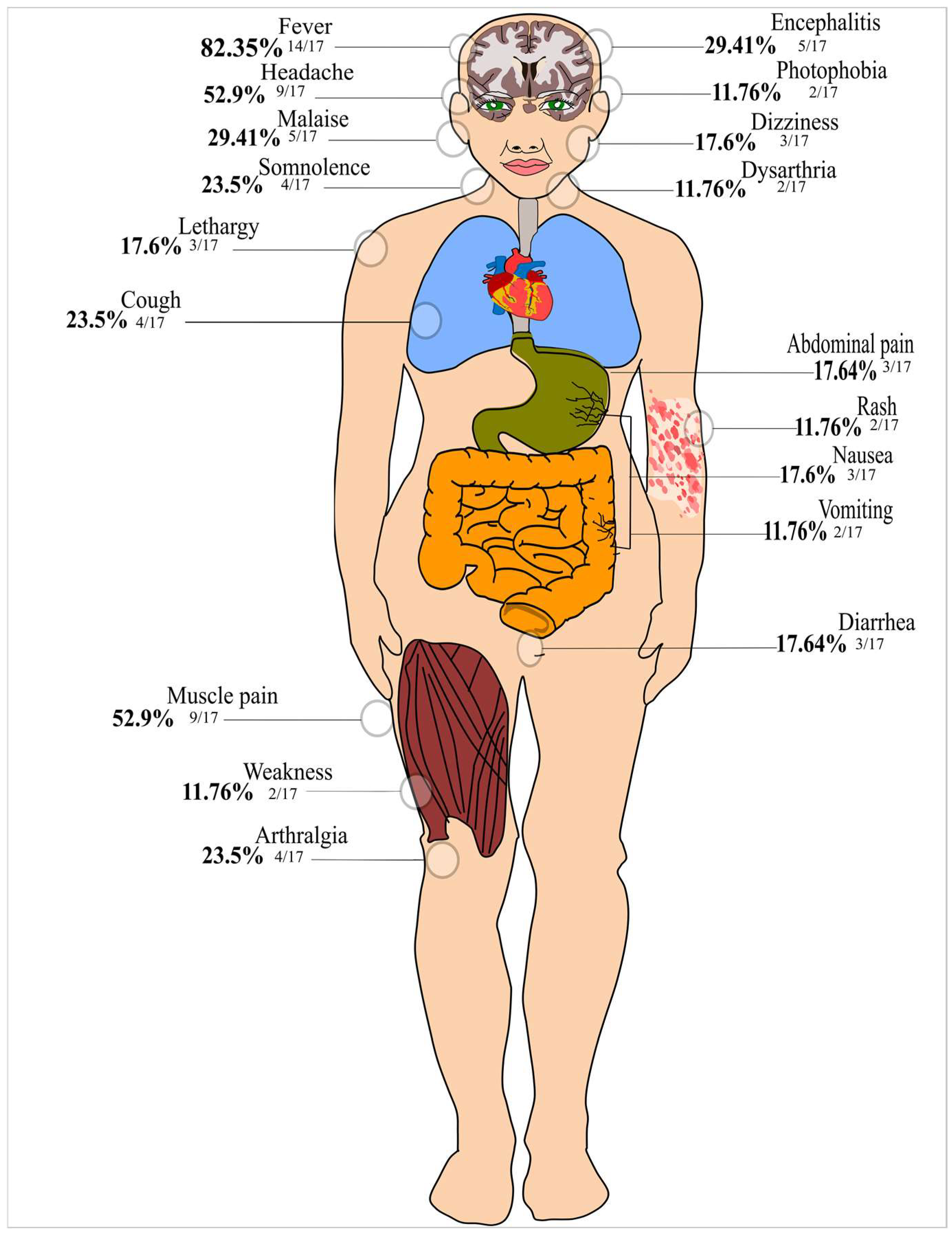
Figure 2 depicts the clinical presentation of the individuals with ILHV infection. The most prevalent clinical signs and symptoms were fever, headache, muscle pain, malaise, and encephalitis.

3.2. Sensitivity Analysis and Publication Bias in the Meta-Analysis

When performing a sensitivity analysis to assess the weight of each individual study on the combined exposure rate to ILHV, through the removal of individual studies, no studies were found to significantly affect the combined positivity (S08 File). Additionally, subgroup analyses were performed; however, for most results, high heterogeneity was observed (I2 > 75%). It should be emphasized that these results were expected, given that in observational epidemiological studies, there is considerable diversity due to variations in study design, detection methodology, and epidemiological variations.
In the analysis of publication bias, asymmetry of the funnel plot was noted for ILHV positivity following subgroups: serological surveys and heterotypic vs. monotypic reactions. However, when analyzing the asymmetry by Egger’s test, the result was not considered statistically significant (t = 1.24, p = 0.22 [serological surveys], t = 1.13 and p = 0.27 [heterotypic], t = −0.2, and p = 0.8 [monotypic reaction]), indicating no small-study effects.

3.3. A Meta-Analysis to Estimate the Pooled Frequency of ILHV Infection

Hemagglutination inhibition (HI) and neutralization (NT) tests were used for the laboratory screening of anti-ILHV antibodies to confirm previous ILHV infections. The combined estimation of ILHV infection as detected through serological assays was 9% (95% CI: 5–13), with considerable evidence for regional epidemiological variation (Figure 3). The term monotypic serological reaction refers to a sample that was only positive for ILHV (i.e., antibody against ILHV) and negative for at least one pair of tested flaviviruses (i.e., Saint Louis encephalitis, yellow fever, dengue, Bussuquara, Rocio viruses). Thus, the results of heterotypic serological reactions (i.e., unspecified serological reaction if unique to ILHV) were higher than those of monotypic serological reactions (14%, 95% CI: 8–22 versus 2%, 95% CI: 1–2).

3.3.1. Subgroup Analysis by Origin and Year of Publication of the Studies

In the analysis that evaluated seroprevalence stratified by region, the highest estimated pooled frequency (20%, 95% CI: 18–23, n = 1152 individuals) was observed in Peru. Panama (19%, 95% CI: 16–21, n = 991), Colombia (12%, 95% CI: 9–14, n = 689), Brazil (8%, 95% CI: 3–14, n = 11,587), and Curaçao and Aruba (4%, 95% CI: 3–5, n = 3044) contributed to the “top 5” regions in terms of ILHV seropositivity (Supplementary File S9).
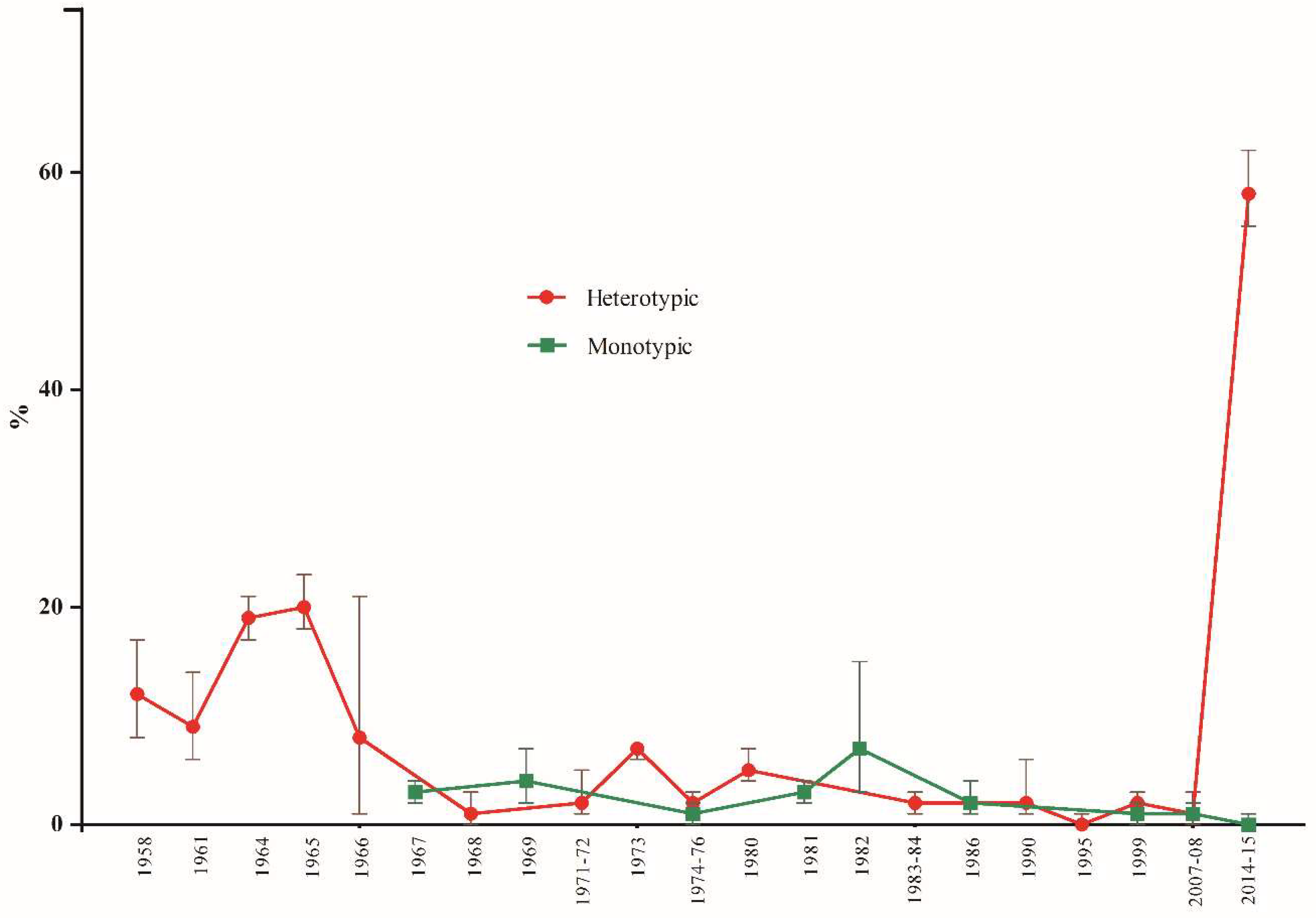
Seropositivity rates were observed to increase in studies performed in 2014–2015, although the general trend over the last few decades remains stable (Figure 4).

3.3.2. Subgroup Analysis by Sex and Age of Participants, and Serological Assays Used

Children and adolescents (aged 0–14 years) had a lower frequency of ILHV infection (2%, 95% CI: 2–3, n = 3347 individuals) than individuals aged 15–64 years (8%, 95% CI: 6–10, n = 1374) (Supplementary File S10). Males had a higher pooled seroprevalence (12%, 95% CI = 10–14, n = 904) than females (7%, 95% CI = 5–9, n = 736).
Regarding the serological assays used, NT and HI had similar results, with overall ILHV positivities of 7% (95% CI: 2–15) and 9% (95% CI: 5–14), respectively.

3.3.3. A Meta-Analysis to Evaluate Factors Associated with ILHV Positivity

Demographic characteristics (i.e., age, sex, and professional occupation) associated with ILHV occurrence were analyzed. Nonetheless, the only covariate available for testing in the analysis of risk factors for ILHV infection was the sex of infected participants. Males had significantly higher odds of seropositivity than females (OR = 1.78, 95% CI: 1.27–2.51) (Supplementary File S11).

4. Discussion

To the best of our knowledge, this is the first meta-analysis of ILHV infection. The main finding of the rates of ILHV seropositivity (9%, 95% CI: 5–13) included 17,722 individuals, with serological samples collected over many decades (1958–2015). Even when considering only the monotypic reactions, positivity remained at a considerable rate of 2% (95% CI: 1–2), with an indication of ILHV prevalence beyond the Amazon region (including parts of Brazil, Colombia, and Peru), as there was also positivity in the Brazilian states of Bahia and São Paulo. The moderate prevalence of ILHV-neutralizing antibodies, coupled with the low number of clinical cases of ILHV infection, suggests that most infections are inapparent or mild, which may be an important vehicle for viral spread to non-endemic regions [2,72,73].
Through our meta-analysis, we were able to elucidate the clinical characteristics of ILHV infection. Fever, headache, myalgia, encephalitis, malaise, arthralgia, gastrointestinal distress, and cough were the most frequently described clinical features. Since the infection is indistinguishable from other febrile syndromes and has a wide spectrum of clinical severity, ranging from subclinical to severe, a portion of symptomatic ILHV infections could be masked by clinical manifestations associated with multiple human pathogenic arboviruses [74], which may contribute to the underestimation of the burden of ILHV. Notably, no case of long-term sequelae or death has been linked to ILHV infection. This fact demands continuous efforts to establish effective public health arbovirus surveillance programs because although the number of confirmed cases of the disease is low, there is a high probability of underreporting due to difficulties in accurate laboratory diagnosis.
Regarding Figure 4, for the two studies from 2014–2015 [70,71], it is difficult to explain this high rate of seropositivity (mean = 58%, 95% CI: 55–62), but it is interesting to mention that these studies involved populations at high risk for arboviruses: male military personnel of working age in the Amazon rainforest or rural population. A high rate of heterotypic serological reactions were observed. These variables were also present in most of the other studies included in the meta-analysis; however, except for Buckley et al. [54], all other studies had lower ILHV positivity.
In South America, Central America, and the Caribbean, different geographic regions have multiple endemic mosquito-borne flaviviruses in co-circulation [75,76,77]. Twenty-nine (78.3%) of the 37 studies included in our meta-analysis investigated the antibody ratios against multiple arbovirus antigens. Flavivirus species have been linked to sporadic cases of human infection, including Bussuquara (BUSV), Cacipacore (CPCV), and ILHV [78,79]. All these viral pathogens may cause undifferentiated febrile illness in humans, and laboratory diagnosis is difficult due to the substantial cross-reactivity of antibodies [75]. Additionally, as occurs for ILHV, BUSV, CPCV, and other native flaviviruses are neglected, with misdiagnosis of cases compared to other medical arboviruses associated with outbreaks or epidemics that attract the most public attention, such as DENV, ZIKV, and YFV.
ILHV shares antigenic similarities with Bagaza, Ntaya, and Tembusu viruses in cross-neutralization tests [80]. These antigenic similarities are attributed to their genetic similarities. Interestingly, serocomplex cross-reactive memory can elicit viral cross-protection against infections with different serocomplexes [81,82]. In other words, polyclonal antisera (post-immune) against a specific virus can neutralize other viruses. As a result, Ilheus and Saint Louis encephalitis (SLE) flavivirus infections engender cross-protection against a lethal ROCV challenge in mice [83]. Similar cross-protection has also been demonstrated between Chikungunya and Mayaro alphaviruses and between DENV and West Nile and SLE flaviviruses [84,85]. New insights into protective ILHV infection responses are needed to elucidate its effects on previous flavivirus exposure and subsequent ILHV exposure. In addition, it is also important to establish whether ILHV immunity provides certain types of cross-protection against other co-circulating flaviviruses in certain geographic locations. Clarification of these factors is essential for a better understanding of the epidemiology of ILHV infection and for improving diagnostic methods.
Although seroepidemiological surveys are useful, this method could overestimate the frequency of ILHV positivity owing to cross-reactivity with other flavivirus infections. Since the included studies used HI/NT tests to capture total antibodies in the target serum, it was not possible to determine the dominant ILHV antigen (protein) involved. There is marked variation in the amino acid identity of the polyprotein of mosquito-borne flaviviruses; for example, in the polyprotein of Japanese encephalitis, dengue, yellow fever, Zika, and West Nile viruses, the amino acid similarity ranges between 45–77% [75]. As HI and serum neutralization tests target neutralizing antibodies, virus E (envelope) protein may be a promising target. The E protein shares 43–79% amino acid similarity between flaviviruses. The most cross-reactive ILHV antigen is the NS3 protein, which shares amino acid homology with the JEV complex [34]. In our meta-analysis, the cross-reactivity of serodiagnostic tests was likely modulated because we were able to demonstrate data on monotypic-type reactions [51,52,56,59,60,61,63,67,69,71].
The HI assay has been a mainstay serodiagnostic test for a long period of time, with the first assay described in the 1940s used to measure the hemagglutinating activity of the influenza virus [86]. Toward the end of the 1950s, antigens prepared from the Laemmert strain of ILHV were used in the HI assay [46]. The HI assay is highly applicable for the detection of anti-ILHV antibodies because no licensed kits are available for this purpose, and in-house HI presents low equipment and laboratory supply costs. A potential drawback is that this method is not commonly standardized, as is the plate-reduction neutralization test (PRNT) [87]. Thus, it is necessary to empirically determine how to weigh antibody cross-reactivity when interpreting the results. Therefore, new insights into ILHV serology assays that aid in determining the specificity (antibody cross-reactivity rate) and sensitivity across current and future laboratory techniques are of paramount importance.
A key factor to consider is the lack of detailed analyses of cross-neutralization studies involving ILHV cases; such studies may contribute to the level of uncertainty in ILHV diagnoses. This potential uncertainty may be mitigated by the studies included in our analysis (more than one-third) that used the viral neutralization assay, that is, NT, and showed good specificity [36,46,48,51,54,57,63,65,66,88,89]. Herein, in direct comparisons of NT and HI, similar results were noted, with overall ILHV positivities of 7% and 9%, respectively. Certainly, the search for neutralizing antibodies by PRNT, considered the “gold standard” in discriminating serodiagnosis of flavivirus infections, will contribute in reducing false-positive cases. However, PRNT may require handling live pathogens in a biosafety level 3–4 laboratory and is a labor-intensive and costly technique for routine laboratory use, making it difficult to be employed on a large scale [88,89].
Our study has important strengths, including the meta-analytic approach and the inclusion of 37 studies describing the serological and clinical characteristics of a large number of individuals. Although studies with small sample sizes were included, they did not impact the estimated pooled rate of exposure, likely because only three studies had sample sizes <100 individuals. The potential limitations of our study include the inconsistent availability of data in some of the selected studies. Several studies failed to report basic demographic details, such as sex, age, and professional activities, and were therefore excluded from subgroup analyses. Demographic variables are important as they enable the assessment of potential risk factors associated with higher seroprevalence. Further studies with complete data are required. Second, because most studies were not designed to determine the prevalence and incidence rates of ILHV infection, it was only possible to estimate the individual and combined proportions regarding the frequency of positivity of the pathogen. Therefore, future studies that are better distributed in the Americas and have data on monotypic serological reactions are necessary. A third potential limitation was the heterogeneity observed in the majority of the analyses, which was possibly due to differences in epidemiological settings, such as the endemicity of place linked with transmission intensity, differences in population groups with greater or lesser probability of exposure to the mosquito vector, laboratory accuracy of the techniques, and varied period of time.

5. Conclusions

In summary, our evidence suggests that due to the low and relatively constant positivity rate found for ILHV, research focused on the diagnosis of this virus should be a priority, especially for the detection of infrequent regional flaviviruses in undifferentiated febrile cases. As the majority of the studies included in our meta-analysis were carried out several decades ago and in concentrated locations, it is essential to encourage novel research, mainly comprising data on monotypic reactions, to trace a more accurate epidemiological pattern of ILHV infections. These studies are essential to understand the varying trends in ILHV epidemiology.

Supplementary Materials

The supporting information can be downloaded at: https://www.mdpi.com/article/10.3390/v15010092/s1.

Author Contributions

Conceptualization, V.G.d.C. and M.V.S.; methodology, V.G.d.C., N.A.B.L. and M.V.S.; formal analysis and data curation, V.G.d.C., M.V.S., N.A.B.L., M.d.F.C., M.L.N., P.R.; writing—review and editing, V.G.d.C., M.V.S., N.A.B.L., C.B.; M.d.F.C., M.L.N., P.R.; supervision, V.G.d.C., P.R.; funding acquisition, V.G.d.C. and P.R. All authors have read and agreed to the published version of the manuscript.

Funding

This research was funded by grants 2021/00603-3 (to P.R.) and 2013/21719-3 (to M.L.N.) from FAPESP (Fundação de Amparo à Pesquisa do Estado de São Paulo). M.L.N. is partially supported by the Centers for Research in Emerging Infectious Disease via The Coordinating Research on Emerging Arboviral Threats Encompassing the Neotropics (CREATE-NEO) grant U01AI151807 awarded by the National Institutes of Health (NIH/USA). M.L.N., M.d.F.C. and P.R. are CNPq (Conselho Nacional de Desenvolvimento Científico e Tecnológico) Research Fellow. V.G.d.C. was supported by a Unesp Post-doctoral Scholarship, with project n. 4320 (Edital PROPe 13/2022). V.G.d.C, CNPq Post-doctoral Scholarship, process n. 381840/2022-0, July-October/2022. M.V.S. was supported by a FAPESP PhD Scholarship, process 2020/12875-5.

Institutional Review Board Statement

Not applicable.

Informed Consent Statement

Not applicable.

Data Availability Statement

Not applicable.

Conflicts of Interest

The authors declare no conflict of interest. The funders had no role in the design of the study; in the collection, analyses, or interpretation of data; in the writing of the manuscript; or in the decision to publish the results.

References

  1. Laemmert, J.R.; Hugo, W.; Hughes; Thomas, P. The virus of Ilheus encephalitis: Isolation, serological specificity and trans-mission. J. Immunol. 1947, 55, 61–67. [Google Scholar] [PubMed]
  2. Cruz, A.C.; da Rosa, A.P.; Ferreira, I.I.; Albuquerque, M.M.; Galler, R. Ilheus virus (Flaviviridae, Flavivirus) is closely related to Japanese encephalitis virus complex. Intervirology 1997, 40, 220–225. [Google Scholar] [CrossRef] [PubMed]
  3. van den Elsen, K.; Quek, J.P.; Luo, D. Molecular Insights into the Flavivirus Replication Complex. Viruses 2021, 13, 956. [Google Scholar] [CrossRef] [PubMed]
  4. Saivish, M.V.; Menezes, G.L.; Costa, V.G.D.; Silva, G.C.D.D.; Marques, R.E.; Nogueira, M.L.; Silva, R.A.D. Predict-ing Antigenic Peptides from Rocio Virus NS1 Protein for Immunodiagnostic Testing Using Immunoinformatics and Molecular Dynamics Simulation. Int. J. Mol. Sci. 2022, 23, 7681. [Google Scholar] [CrossRef] [PubMed]
  5. Causey, O.R.; Causey, C.E.; Maroja, O.M.; Macedo, D.G. The isolation of arthropod-borne viruses, including members of two hitherto undescribed serological groups, in the Amazon region of Brazil. Am. J. Trop. Med. Hyg. 1961, 10, 227–249. [Google Scholar] [CrossRef]
  6. Southam, C.M.; Moore, A.E. West Nile, Ilheus, and Bunyamwera Virus Infections in Man 1,2,3. Am. J. Trop. Med. Hyg. 1951, 31, 724–741. [Google Scholar] [CrossRef] [PubMed]
  7. Spence, L.; Anderson, C.R.; Downs, W.G. Isolation of Ilhéus virus from human beings in Trinidad, West Indies. Trans. R. Soc. Trop. Med. Hyg. 1962, 56, 504–509. [Google Scholar] [CrossRef]
  8. Srihongse, S.; Johnson, C.M. The Isolation of Ilhéus Virus from Man in Panamá. Am. J. Trop. Med. Hyg. 1967, 16, 516–518. [Google Scholar] [CrossRef]
  9. Nassar, E.; Coimbra, T.; Rocco, I.; Pereira, L.; Ferreira, I.; De Souza, L.; De Souza, D.; Ueda-Ito, M.; Moura, J.; Bergo, R. Human Disease Caused by an Arbovirus Closely Related to Ilheus Virus: Report of five cases. Intervirology 1997, 40, 247–252. [Google Scholar] [CrossRef]
  10. Venegas, E.A.; Aguilar, P.V.; Cruz, C.; Guevara, C.; Kochel, T.J.; Vargas, J.; Halsey, E.S. Ilheus Virus Infection in Human, Bolivia. Emerg. Infect. Dis. 2012, 18, 516–518. [Google Scholar] [CrossRef]
  11. Milhim, B.H.G.A.; Estofolete, C.F.; da Rocha, L.C.; Liso, E.; Brienze, V.M.S.; Vasilakis, N.; Terzian, A.C.B.; Nogueira, M.L. Fatal Outcome of Ilheus Virus in the Cerebrospinal Fluid of a Patient Diagnosed with Encephalitis. Viruses 2020, 12, 957. [Google Scholar] [CrossRef] [PubMed]
  12. Prías-Landínez, E.; Bernal-Cubides, C.; Morales-Alarcón, A. Isolation of Ilhéus Virus from Man in Colombia*. Am. J. Trop. Med. Hyg. 1968, 17, 112–114. [Google Scholar] [CrossRef] [PubMed]
  13. Panon, G.; Fauran, P.; Digoutte, J.P. Isolation of Ilheus virus in french Guyana. Bull. Soc. Pathol. Exot. Fil. 1979, 72, 315–318. [Google Scholar]
  14. Johnson, B.W.; Cruz, C.; Felices, V.; Espinoza, W.R.; Manock, S.R.; Guevara, C.; Olson, J.G.; Kochel, T.J. Ilheus Virus Isolate from a Human, Ecuador. Emerg. Infect. Dis. 2007, 13, 956–958. [Google Scholar] [CrossRef] [PubMed]
  15. Ferreira, I.B.; Pereira, L.E.; Rocco, I.M.; Marti, A.T.; de Souza, L.T.; Iversson, L.B. Surveillance of arbovirus infections in the Atlantic forest region, state of São Paulo, Brazil. Rev. Inst. Med. Trop. São Paulo 1994, 36, 265–274. [Google Scholar] [CrossRef] [Green Version]
  16. Pereira, L.E.; Suzuki, A.; Coimbra, T.L.; de Souza, R.P.; Chamelet, E.L. Arbovírus Ilheus em aves silvestres (Sporophila caerulescens e Molothrus bonariensis) [Ilheus arbovirus in wild birds (Sporophila caerulescens and Molothrus bonariensis)]. Rev. Saúde Pública 2001, 35, 119–123. [Google Scholar] [CrossRef] [Green Version]
  17. Casseb, A.R.; Cruz, A.V.; Jesus, I.S.; Chiang, J.O.; Martins, L.C.; Silva, S.P.; Henriques, D.F.; Casseb, L.M.; Vasconcelos, P.F. Seroprevalence of flaviviruses antibodies in water buffaloes (Bubalus bubalis) in Brazilian Amazon. J. Venom. Anim. Toxins Incl. Trop. Dis. 2014, 20, 9. [Google Scholar] [CrossRef] [Green Version]
  18. Iversson, L.B.; Silva, R.A.; da Rosa, A.P.; Barros, V.L. Circulation of eastern equine encephalitis, western equine encephalitis, Ilhéus, Maguari and Tacaiuma viruses in equines of the Brazilian Pantanal, South America. Rev. Inst. Med. Trop. Sao Paulo 1993, 35, 355–359. [Google Scholar] [CrossRef]
  19. Pauvolid-Corrêa, A.; Campos, Z.; Juliano, R.; Velez, J.; Nogueira, R.M.; Komar, N. Serological evidence of widespread circulation of West Nile virus and other flaviviruses in equines of the Pantanal, Brazil. PLoS. Negl. Trop. Dis. 2014, 8, e2706. [Google Scholar] [CrossRef] [Green Version]
  20. Medlin, S.; Deardorff, E.R.; Hanley, C.S.; Vergneau-Grosset, C.; Siudak-Campfield, A.; Dallwig, R.; da Rosa, A.T.; Tesh, R.B.; Martin, M.P.; Weaver, S.C.; et al. Serosurvey of selected arboviral pathogens in free-ranging, two-toed sloths (Choloepus hoffmanni) and three-toed sloths (Bradypus variegatus) in Costa Rica, 2005–2007. J. Wildl. Dis. 2016, 52, 883–892. [Google Scholar] [CrossRef] [Green Version]
  21. Morales, M.A.; Fabbri, C.M.; Zunino, G.E.; Kowalewski, M.M.; Luppo, V.C.; Enría, D.A.; Levis, S.C.; Calderón, G.E. Detection of the mosquito-borne flaviviruses, West Nile, Dengue, Saint Louis Encephalitis, Ilheus, Bussuquara, and Yellow Fever in free-ranging black howlers (Alouatta caraya) of Northeastern Argentina. PLoS. Negl. Trop. Dis. 2017, 10, e0005351. [Google Scholar] [CrossRef] [PubMed]
  22. Catenacci, L.S.; Ferreira, M.; Martins, L.C.; De Vleeschouwer, K.M.; Cassano, C.R.; Oliveira, L.C.; Canale, G.; Deem, S.L.; Tello, J.S.; Parker, P.; et al. Surveillance of Arboviruses in Primates and Sloths in the Atlantic Forest, Bahia, Brazil. EcoHealth 2018, 15, 777–791. [Google Scholar] [CrossRef] [PubMed]
  23. Thompson, N.N.; Auguste, A.J.; Coombs, D.; Blitvich, B.J.; Carrington, C.V.; da Rosa, A.P.; Wang, E.; Chadee, D.D.; Drebot, M.A.; Tesh, R.B.; et al. Serological evidence of flaviviruses and alphaviruses in livestock and wildlife in Trinidad. Vector Borne Zoonotic Dis. 2012, 12, 969–978. [Google Scholar] [CrossRef] [Green Version]
  24. Vieira, C.J.D.S.P.; Andrade, C.D.; Kubiszeski, J.R.; Silva, D.J.F.D.; Barreto, E.S.; Massey, A.L.; Canale, G.R.; Bernardo, C.S.S.; Levi, T.; Peres, C.A.; et al. Detection of Ilheus virus in mosquitoes from southeast Amazon, Brazil. Trans. R. Soc. Trop. Med. Hyg. 2019, 113, 424–427. [Google Scholar] [CrossRef] [PubMed]
  25. Almeida, M.A.B.; Santos, E.D.; Cardoso, J.D.C.; Noll, C.A.; Lima, M.M.; Silva, F.A.E.; Ferreira, M.S.; Martins, L.C.; Vasconcelos, P.F.D.C.; Bicca-Marques, J.C. Detection of antibodies against Icoaraci, Ilhéus, and Saint Louis Encephalitis arboviruses during yellow fever monitoring surveillance in non-human primates (Alouatta caraya) in southern Brazil. J. Med. Primatol. 2019, 48, 211–217. [Google Scholar] [CrossRef]
  26. Pauvolid-Corrêa, A.; Kenney, J.L.; Couto-Lima, D.; Campos, Z.M.; Schatzmayr, H.G.; Nogueira, R.M.; Brault, A.C.; Komar, N. Ilheus virus isolation in the Pantanal, west-central Brazil. PLoS Negl. Trop. Dis. 2013, 18, e2318. [Google Scholar] [CrossRef] [Green Version]
  27. da Silva Ferreira, R.; de Toni Aquino da Cruz, L.C.; de Souza, V.J.; da Silva Neves, N.A.; de Souza, V.C.; Filho, L.C.F.; da Silva Lemos, P.; de Lima, C.P.S.; Naveca, F.G.; Atanaka, M.; et al. Insect-specific viruses and arboviruses in adult male culicids from Midwestern Brazil. Infect. Genet. Evol. 2020, 85, 104561. [Google Scholar] [CrossRef]
  28. Cunha, M.S.; Luchs, A.; Dos Santos, F.C.P.; Caleiro, G.S.; Nogueira, M.L.; Maiorka, P.C. Applying a pan-flavivirus RT-qPCR assay in Brazilian public health surveillance. Arch. Virol. 2020, 165, 1863–1868. [Google Scholar] [CrossRef]
  29. Araújo, P.A.; Freitas, M.O.; Chiang, J.O.; Silva, F.A.; Chagas, L.L.; Casseb, S.M.; Silva, S.P.; Nunes-Neto, J.P.; Rosa-Júnior, J.W.; Nascimento, B.S.; et al. Investigation about the Occurrence of Transmission Cycles of Arbovirus in the Tropical Forest, Amazon Region. Viruses 2019, 11, 774. [Google Scholar] [CrossRef] [Green Version]
  30. Cunha, M.S.; Luchs, A.; da Costa, A.C.; Ribeiro, G.O.; Dos Santos, F.C.P.; Nogueira, J.S.; Komninakis, S.V.; Marinho, R.D.S.S.; Witkin, S.S.; Villanova, F.; et al. Detection and characterization of Ilheus and Iguape virus genomes in historical mosquito samples from Southern Brazil. Acta Trop. 2020, 205, 105401. [Google Scholar] [CrossRef]
  31. De Rodaniche, E.; Galindo, P. Isolation of Ilhéus virus from Sabethes chloropterus captured in Guatemala in 1956. Am. J. Trop. Med. Hyg. 1957, 6, 686–687. [Google Scholar] [CrossRef] [PubMed]
  32. De Rodaniche, E.; Galindo, P. Isolation of the virus of IIheus encephalitis from mosquitoes captured in Panama. Am. J. Trop. Med. Hyg. 1961, 10, 393–394. [Google Scholar] [CrossRef] [PubMed]
  33. De Rodaniche, E. Isolation of the virus of IIhéus encephalitis from mosquitoes of the genus Psorophora captured in Honduras. Am. J. Trop. Med. Hyg. 1956, 5, 797–801. [Google Scholar] [CrossRef] [PubMed]
  34. Aitken, T.H.; Anderson, C.R.; Downs, W.G. The isolation of Ilhéus virus from wild caught forest mosquitoes in Trinidad. Am. J. Trop. Med. Hyg. 1956, 5, 621–625. [Google Scholar] [CrossRef]
  35. Galindo, P.; de Rodaniche, E. Birds as hosts of Ilheus encephalitis virus in Panama. Am. J. Trop. Med. Hyg. 1961, 10, 395–396. [Google Scholar] [CrossRef]
  36. de Rodaniche, E.; Galindo, P. Ecological Observations on Ilhéus Virus in the Vicinity of Almirante, Republic of Panama*. Am. J. Trop. Med. Hyg. 1963, 12, 924–928. [Google Scholar] [CrossRef]
  37. Moher, D.; Shamseer, L.; Clarke, M.; Ghersi, D.; Liberati, A.; Petticrew, M.; Shekelle, P.; Stewart, L.A. Preferred reporting items for systematic review and meta-analysis protocols (PRISMA-P) 2015 statement. Syst. Rev. 2015, 4, 1. [Google Scholar] [CrossRef] [Green Version]
  38. Wells, G.A.; Shea, B.; O’Connell, D.; Peterson, J.; Welch, V.; Losos, M. The Newcastle-Ottawa Scale (NOS) for Assessing the Quality of Nonrandomized Studies in Meta-Analyses; Ottawa Health Research Institute: Ottawa, ON, USA, 2021; Available online: http://www.ohri.ca/programs/clinical_epidemiology/oxford.asp (accessed on 26 July 2022).
  39. Freeman, M.F.; Tukey, J.W. Transformations related to the angular and the square root. Ann. Math. Stat. 1950, 21, 607–611. [Google Scholar] [CrossRef]
  40. Barendregt, J.J.; Doi, S.A.; Lee, Y.Y.; Norman, R.E.; Vos, T. Meta-analysis of prevalence. J. Epidemiol. Community Health 2013, 67, 974–978. [Google Scholar] [CrossRef]
  41. Nyaga, V.N.; Arbyn, M.; Aerts, M. Metaprop: A Stata command to perform meta-analysis of binomial data. Arch. Public Health 2014, 72, 39. [Google Scholar] [CrossRef] [Green Version]
  42. Higgins, J.P.; Thompson, S.G. Quantifying heterogeneity in a meta-analysis. Stat. Med. 2002, 21, 1539–1558. [Google Scholar] [CrossRef] [PubMed]
  43. DerSimonian, R.; Laird, N. Meta-analysis in clinical trials. Control Clin. Trials 1986, 7, 177–188. [Google Scholar] [CrossRef] [PubMed]
  44. Egger, M.; Davey Smith, G.; Schneider, M.; Minder, C. Bias in meta-analysis detected by a simple, graphical test. BMJ 1997, 315, 629–634. [Google Scholar] [CrossRef] [PubMed] [Green Version]
  45. Begg, C.B.; Mazumdar, M. Operating characteristics of a rank correlation test for publication bias. Biometrics 1994, 50, 1088–1101. [Google Scholar] [CrossRef]
  46. Groot, H.; Riberiro, R.B. Neutralizing and haemagglutination-inhibiting antibodies to yellow fever 17 years after vaccination with 17D vaccine. Bull. World Health Organ. 1962, 27, 699–707. [Google Scholar] [PubMed]
  47. Ehrenkranz, N.J.; Pond, W.L.; Pennington, R.M.; Carter, M.J. Arthropod-borne virus disease in Florida: Report of a 1958 outbreak in Miami and a serologic survey of Miami residents. Am. J. Med. 1963, 35, 673–681. [Google Scholar] [CrossRef] [PubMed]
  48. Macias, M.J. Estudios Epidemiológicos Sobre Virus Arbor en el Sureste de México; Salud Pública de México: Cuernavaca, Mexico, 1963; Volume 5, pp. 523–527. Available online: File:///F:/Perfil%20Vivaldo/Downloads/3881-Texto%20del%20art%C3%ADculo-3828-1-10-20141111.pdf (accessed on 25 July 2022).
  49. Príans-landínez, E.; Bernal-Cúbides, C.; Torres, S.V.; Romero-León, M. Encuesta Serologica de Virus Transmitidos Por Artropodos; Boletín de la Oficina Sanitaria Panamericana: Bogotá, Colombia, 1966; Available online: https://iris.paho.org/bitstream/handle/10665.2/14510/v68n2p134.pdf?sequence=1&isAllowed=y (accessed on 22 July 2022).
  50. Neel, J.V.; Andrade, A.H.P.; Stollerman, G.H.; Weinstein, E.D.; Wheeler, A.H.; Brown, G.E.; Goobar, J.; Eveland, W.E.; Sodeman, W.A. Further Studies of the Xavante Indians. Am. J. Trop. Med. Hyg. 1968, 17, 486–498. [Google Scholar] [CrossRef]
  51. Niederman, J.C.; Henderson, J.R.; Opton, E.M.; Black, F.L.; Skvrnova, K. A nationwide serum survey of brazilian military recruits, 1964: Ii. antibody patterns with arboviruses, polioviruses, measles and mumps. Am. J. Epidemiol. 1967, 86, 319–329. [Google Scholar] [CrossRef]
  52. Evans, A.S.; Casals, J.; Opton, E.M.; Borman, E.K.; Levine, L.; Cuadrado, R.R. A nationwide serum survey of colombian military recruits, 1966. Am. J. Trop. Med. Hyg. 1969, 90, 292–303. [Google Scholar] [CrossRef]
  53. Black, F.L.; Woodall, J.P.; Evans, A.S.; Liebhaber, H.; Henle, G. Prevalence of antibody against viruses in the Tiriyo, an isolated amazon tribe. Am. J. Epidemiol. 1970, 91, 430–438. [Google Scholar] [CrossRef]
  54. Buckley, S.M.; Davis, J.L.; Madalengoitia, J.; Flores, W.; Casals, J. Arbovirus neutralization tests with Peruvian sera in Vero cell cultures. Bull. World Health Organ. 1972, 46, 451–455. [Google Scholar] [PubMed]
  55. Madalengoitia, J.; Flores, W.; Casals, J. Arbovirus Antibody Survey of Sera from Residents of Eastern Peru; English Edition; Boletín de la Oficina Sanitaria Panamericana (OSP): Lima, Peru, 1973; p. 7. Available online: https://iris.paho.org/handle/10665.2/11677 (accessed on 14 July 2022).
  56. Pinheiro, F.P.; Bensabath, G.; Andrade, A.H.; Lins, Z.C.; Fraiha, H.; Tang, A.T.; Lainson, R.; Shaw, J.J.; Azevedo, M.C. Infectious Diseases along Brazil’s Trans-Amazon Highway: Surveillance and Research; Pan American Health Organization (PAHO): Washington, DC, USA, 1974; Volume 8, Available online: https://iris.paho.org/handle/10665.2/27094 (accessed on 8 July 2022).
  57. Pinheiro, F.P.; Schatzmayr, H.; Rosa, A.P.A.T.; Homma, A.; Bensabath, G. Arbovirus Antibodies in Children of Rural Guanabara, Brazil. Intervirology 1975, 5, 93–96. [Google Scholar] [CrossRef] [PubMed] [Green Version]
  58. Weiland, H.T.; Williams, M.C.; Hull, B. Serologic survey of dengue and other arboviruses in Curaçao and Aruba, 1973. Bull. Pan Am. Health Organ. 1978, 12, 134–142. [Google Scholar] [PubMed]
  59. Dixon, K.E.; Llewellyn, C.H.; da Rosa, A.P.T.; da Rosa, J.F.T. A multidisciplinary program of infectious disease surveillance along the Transamazon highway in Brazil: Epidemiology of arbovirus infections. Bull. Pan Am. Health Organ. 1981, 15, 11–25. [Google Scholar] [PubMed]
  60. Iversson, L.B.; Rosa; Amélia, P.A.T.; Rosa, J.T. Estudos sorológicos para pesquisa de anticorpos de arbovírus em população humana da região do Vale do Ribeira: Ii-inquérito em pacientes do hospital regional de pariquera-açú, 1980. Rev. Saúde Pública 1981, 15, 587–602. [Google Scholar] [CrossRef] [Green Version]
  61. Iversson, L.B.; da Rosa, A.P.A.T.; da Rosa, J.T.; Costa, C.S. Estudos sorológicos para pesquisa de anticorpos de arbovírus em população humana da região do Vale do Ribeira: III-inquérito em coabitantes com casos de encefalite por Flavivirus Rocio. Rev. Saúde Pública 1982, 16, 160–170. [Google Scholar] [CrossRef]
  62. Figueiredo, L.T.M.; de Rosa, A.P.A.T.; Fiorillo, A.M. Níveis de anticorpos para arbovírus em indivíduos da região de Ribeirão Preto, SP (Brasil). Rev. Saúde Pública 1986, 20, 204–211. [Google Scholar] [CrossRef] [Green Version]
  63. Tavares-Neto, J.; Rosa, A.P.A.T.d.; Vasconcelos, P.F.C.; Costa, J.M.L.; de Rosa, J.F.S.T.; Marsden, P.D. Pesquisa de anticorpos para arbovírus no soro de residentes no povoado de Corte de Pedra, Valença, Bahia. Mem. Inst. Oswaldo Cruz 1986, 81, 351–358. [Google Scholar] [CrossRef] [Green Version]
  64. Degallier, N.; da Rosa, A.M.P.T.; Vasconcelos, P.F.C.; Hervé, J.P.; Filho, G.C.S.; da Rosa, J.F.S.T.; da Rosa, E.S.T.; Rodrigues, S.G. Modifications of arbovirus transmission in relation to construction of dams in Brazilian Amazonia. Sci. Cult 1992, 44, 124–135. Available online: https://horizon.documentation.ird.fr/exl-doc/pleins_textes/pleins_textes_6/b_fdi_33-34/38274.pdf (accessed on 5 July 2022).
  65. Straatmann, A.; Santos-Torres, S.; Vasconcelos, P.F.; Da Rosa, A.P.T.; Rodrigues, S.G.; Tavares-Neto, J. Evidências sorológicas da circulação do arbovírus Rocio (Flaviviridae) na Bahia. Rev. Soc. Bras. Med. Trop. 1997, 30, 511–515. [Google Scholar] [CrossRef] [Green Version]
  66. Romano-Lieber, N.S.; Iversson, L.B. Inquérito soroepidemiológico para pesquisa de infecções por arbovírus em moradores de reserva ecológica. Rev Saúde Pública 2000, 34, 236–242. [Google Scholar] [CrossRef] [PubMed]
  67. De Araújo, T.P.; Rodrigues, S.G.; Costa, M.I.W.D.A.; Vasconcelos, P.F.D.C.; Da Rosa, A.P.T. Diagnóstico sorológico de infecções por dengue e febre amarela em casos suspeitos no Estado do Pará, Brasil, 1999. Rev. Soc. Bras. Med. Trop. 2002, 35, 579–584. [Google Scholar] [CrossRef] [PubMed]
  68. Tavares-Neto, J.; Freitas-Carvalho, J.; Nunes, M.R.T.; Rocha, G.; Rodrigues, S.G.; Damasceno, E.; Darub, R.; Viana, S.; Vasconcelos, P.F.C. Pesquisa de anticorpos contra arbovírus e o vírus vacinal da febre amarela em uma amostra da população de Rio Branco, antes e três meses após a vacina 17D. Rev. Soc. Bras. Med. Trop. 2004, 37, 1–6. [Google Scholar] [CrossRef] [PubMed]
  69. Cruz, A.C.; Prazeres, A.S.C.; Gama, E.C.; Lima, M.F.; Azevedo, R.S.S.; Casseb, L.M.N.; Neto, J.P.N.; Martins, L.C.; Chiang, J.O.; Rodrigues, S.G.; et al. Serological survey for arboviruses in Juruti, para state, brazil. Cad Saude Publica 2009, 25, 2517–2523. [Google Scholar] [CrossRef] [PubMed] [Green Version]
  70. Salgado, B.B.; de Jesus, M.F.C.; Pereira, R.L.; Chiang, J.O.; de Oliveira, F.M.N.; Ferreira, M.S.; Martins, L.C.; da Costa Vasconcelos, P.F.; Ganoza, C.; Lalwani, P. Prevalence of arbovirus antibodies in young healthy adult population in Brazil. Parasites Vectors 2021, 14, 403. [Google Scholar] [CrossRef] [PubMed]
  71. Catenacci, L.S.; Ferreira, M.S.; Fernandes, D.; Padda, H.; Travassos-da-Rosa, E.S.; Deem, S.L.; Vasconcelos, P.F.C.; Martins, L.C. Individual, household and environmental factors associated with arboviruses in rural human populations, Brazil. Zoonoses Public Health 2021, 68, 203–212. [Google Scholar] [CrossRef]
  72. Pinheiro, F.P.; Travassos da Rosa, A.P.A.; Freitas, R.B.; Travasos da Rosa, J.F.S.; Vasconcelos, P.F.C. Aspectos Clínicos-Epidemiológicos: 50 anos do Instituto Evandro Chagas. Belem. Fundação Servidos Saude Publica 1986, 1, 375–407. [Google Scholar]
  73. Chastel, C. Asymptomatic infections in man: A Trojan horse for the introduction and spread of mosquito-borne arboviruses in non-endemic areas? Bull. Soc. Pathol. Exot. 2011, 104, 213–219. [Google Scholar] [CrossRef] [Green Version]
  74. Wilder-Smith, A.; Gubler, D.J.; Weaver, S.C.; Monath, T.P.; Heymann, D.L.; Scott, T.W. Epidemic arboviral diseases: Priorities for research and public health. Lancet Infect. Dis. 2017, 17, e101–e106. [Google Scholar] [CrossRef] [Green Version]
  75. Hou, B.; Chen, H.; Gao, N.; An, J. Cross-Reactive Immunity among Five Medically Important Mosquito-Borne Flaviviruses Related to Human Diseases. Viruses 2022, 14, 1213. [Google Scholar] [CrossRef]
  76. Qian, X.; Qi, Z. Mosquito-Borne Flaviviruses and Current Therapeutic Advances. Viruses 2022, 14, 1226. [Google Scholar] [CrossRef] [PubMed]
  77. Rufalco-Moutinho, P.; de Noronha, L.A.G.; de Souza Cardoso Quintão, T.; Nobre, T.F.; Cardoso, A.P.S.; Cilião-Alves, D.C.; Bellocchio Júnior, M.A.; von Glehn, M.P.; Haddad, R.; Romero, G.A.S.; et al. Evidence of co-circulation of multiple arboviruses transmitted by Aedes species based on laboratory syndromic surveillance at a health unit in a slum of the Federal District, Brazil. Parasites Vectors 2021, 19, 610. [Google Scholar] [CrossRef] [PubMed]
  78. Srihongse, S.; Johnson, C.M. The first isolation of Bussuquara virus from man. Trans. R. Soc. Trop. Med. Hyg. 1971, 65, 541–542. [Google Scholar] [CrossRef] [PubMed]
  79. Batista, W.C.; Tavares, G.D.S.B.; Vieira, D.S.; Honda, E.R.; Pereira, S.S.; Tada, M.S. Notification of the first isolation of Cacipacore virus in a human in the State of Rondônia, Brazil. Rev. Soc. Bras. Med. Trop. 2011, 44, 528–530. [Google Scholar] [CrossRef] [Green Version]
  80. Shope, R.E. Epidemiology of Other Arthropod-borne Flaviviruses infecting humans. Adv. Virus Res. 2003, 61, 373–391. [Google Scholar] [CrossRef] [PubMed]
  81. Rathore, A.P.S.; John, A.L.S. Cross-Reactive Immunity Among Flaviviruses. Front. Immunol. 2020, 11, 1–9. [Google Scholar] [CrossRef] [Green Version]
  82. John, A.L.S.; Rathore, A.P.S. Adaptive immune responses to primary and secondary dengue virus infections. Nat. Rev. Immunol. 2019, 19, 218–230. [Google Scholar] [CrossRef]
  83. Amarilla, A.A.; Fumagalli, M.J.; Figueiredo, M.L.; Lima-Junior, D.S.; Santos-Junior, N.N.; Alfonso, H.L.; Lippi, V.; Trabuco, A.C.; Lauretti, F.; Muller, V.D.; et al. Ilheus and Saint Louis encephalitis viruses elicit cross-protection against a lethal Rocio virus challenge in mice. PLoS ONE 2018, 13, e0199071. [Google Scholar] [CrossRef]
  84. Fumagalli, M.J.; de Souza, W.M.; de Castro-Jorge, L.A.; de Carvalho, R.V.H.; Castro, Í.A.; de Almeida, L.G.N.; Consonni, S.R.; Zamboni, D.S.; Figueiredo, L.T.M. Chikungunya Virus Exposure Partially Cross-Protects against Mayaro Virus Infection in Mice. J. Virol. 2021, 95, e0112221. [Google Scholar] [CrossRef]
  85. Sather, G.E.; Hammon, W.M. Protection against St. Louis encephalitis and West Nile arboviruses by previous dengue virus (types 1–4) infection. Proc. Soc. Exp. Biol. Med. 1970, 135, 573–578. [Google Scholar] [CrossRef]
  86. Hirst, G.K. The quantitative determination of influenza virus and antibodies by means of red cell agglutination. J. Exp. Med. 1942, 75, 49. [Google Scholar] [CrossRef] [PubMed]
  87. Maeda, A.; Maeda, J. Review of diagnostic plaque reduction neutralization tests for flavivirus infection. Vet. J. 2013, 195, 33–40. [Google Scholar] [CrossRef] [PubMed]
  88. Roehrig, J.T.; Hombach, J.; Barrett, A.D.T. Guidelines for Plaque-Reduction Neutralization Testing of Human Antibodies to Dengue Viruses. Viral Immunol. 2008, 21, 123–132. [Google Scholar] [CrossRef]
  89. Musso, D.; Despres, P. Serological Diagnosis of Flavivirus-Associated Human Infections. Diagnostics 2020, 10, 302. [Google Scholar] [CrossRef] [PubMed]
Figure 1. Map illustrating the geographical distribution of the included studies. (a) Color intensity represents the number of studies from each region. (b) Pooled estimated frequency of ILHV seropositivity. At the upper right of (b), the monotypic reaction data, which are samples that were only positive for ILHV (antibody against ILHV) and negative for at least one pair of tested flaviviruses, are shown.
Viruses 15 00092 g001
Figure 2. Clinical manifestations of ILHV infection [5,6,7,8,9,10,11,12,13,14].
Viruses 15 00092 g002
Figure 3. Forest plot of the proportion of laboratory confirmed ILHV infection according to the type of serological reaction (heterotypic vs monotypic) [8,36,46,47,48,49,50,51,52,53,54,55,56,57,58,59,60,61,62,63,64,65,66,67,68,69,70,71]. The black diamonds in gray squares indicate the mean of the ratio of ILHV positivity, while the size of the grey square represents the weight (population size) contributed by each study in the meta-analysis. The horizontal lines in blue represent their 95% confidence intervals (CI). The red diamond represents the pooled ratio of positives and its 95% CI, while dashed purple line represents the mean of the pooled estimate. id = identification of the study; Events = ILHV infection; CI = confidence interval; ES = effect size.
Viruses 15 00092 g003
Figure 4. Distribution of ILHV seropositivity according to the year in which the samples were obtained. The values represent the mean with their respective 95% CI.
Viruses 15 00092 g004
Disclaimer/Publisher’s Note: The statements, opinions and data contained in all publications are solely those of the individual author(s) and contributor(s) and not of MDPI and/or the editor(s). MDPI and/or the editor(s) disclaim responsibility for any injury to people or property resulting from any ideas, methods, instructions or products referred to in the content.