Article

Identification of Adenovirus E1B-55K Interaction Partners through a Common Binding Motif

1
Institute for Cancer and Genomic Sciences, The Medical School, University of Birmingham, Birmingham B15 2TT, UK
2
Leibniz Institute of Virology, Department of Viral Transformation, 20251 Hamburg, Germany
3
Department of Medical Microbiology and Immunology, Taibah University, P.O. Box 344, Madinah 41477, Saudi Arabia
4
Department of Biochemistry and Molecular Biology, School of Basic Medical Science, Chongqing Medical University, Chongqing 400016, China
*
Author to whom correspondence should be addressed.
These authors contributed equally to this work.
Current address: Institute of Cancer Research, London SW7 3RP, UK.
Viruses 2023, 15(12), 2356; https://doi.org/10.3390/v15122356
Submission received: 8 November 2023 / Revised: 22 November 2023 / Accepted: 28 November 2023 / Published: 30 November 2023
(This article belongs to the Special Issue 15th International Adenovirus Meeting)

Abstract

:
The adenovirus C5 E1B-55K protein is crucial for viral replication and is expressed early during infection. It can interact with E4orf6 to form a complex that functions as a ubiquitin E3 ligase. This complex targets specific cellular proteins and marks them for ubiquitination and, predominantly, subsequent proteasomal degradation. E1B-55K interacts with various proteins, with p53 being the most extensively studied, although identifying binding sites has been challenging. To explain the diverse range of proteins associated with E1B-55K, we hypothesized that other binding partners might recognize the simple p53 binding motif (xWxxxPx). In silico analyses showed that many known E1B-55K binding proteins possess this amino acid sequence; therefore, we investigated whether other xWxxxPx-containing proteins also bind to E1B-55K. Our findings revealed that many cellular proteins, including ATR, CHK1, USP9, and USP34, co-immunoprecipitate with E1B-55K. During adenovirus infection, several well-characterized E1B-55K binding proteins and newly identified interactors, including CSB, CHK1, and USP9, are degraded in a cullin-dependent manner. Notably, certain binding proteins, such as ATR and USP34, remain undegraded during infection. Structural predictions indicate no conservation of structure around the proposed binding motif, suggesting that the interaction relies on the correct arrangement of tryptophan and proline residues.

1. Introduction

Human adenoviruses (HAdVs) constitute a large family of more than 100 different types, divided into seven species (A to G) [1]. Generally, they are responsible for relatively mild infections of the respiratory tract, the gastro-intestine, and the eye. However, in immunocompromised patients, such as those undergoing transplantation, adenoviruses pose a serious health risk, often resulting in high mortality rates [2,3].
Adenoviruses have a double-stranded linear DNA genome of about 35 kb, which is transcribed in both directions and encodes early, intermediate, and late proteins. Adenovirus early region 1 (E1) comprises two transcription units—E1A and E1B—and both are translated to give two major proteins [4,5]. The two E1A proteins, translated from 12S and 13S mRNAs, are identical over most of their sequence. Simplistically, through a multitude of interactions with cellular targets, they cause cell cycle progression into a ‘pseudo-S phase’, which facilitates the expression of other adenovirus genes, making use of the host cell transcription/translation machinery [4,6]. Adenovirus E1B proteins are transcribed from a 22S mRNA. The two major proteins, E1B-19K and -55K, are translated in different reading frames, so they have no sequence homology. E1B-19K has limited homology to Bcl2 and similarly adopts an anti-apoptotic role both during viral infection and in E1-transformed cells [5,7].
E1B-55K also appears to protect infected cells against apoptosis, for example, by causing the rapid degradation of p53 in the case of the species A and group C viruses or by direct interaction, resulting in the inhibition of transcriptional activity [8,9,10,11,12,13]. Adenovirus E1B-55K-mediated protein degradation is accomplished by association with E4orf6, recruitment of a cellular E3 ubiquitin ligase comprising a cullin (cullin 5 in the case of HAdV-C5 and Cul2 in the case of HAdV-A12), elongins B and C and RING Box 1 (Rbx1), ubiquitination of the substrate, and degradation by the proteasome [9,14,15]. E1B-55K serves as the substrate recognition component whereas E4orf6 binds to elongin C.
Group A (HAdV-A12, for example) and C (HAdV-C2 and HAdV-C5, for example) adenoviruses also target an appreciable number of other cellular proteins for degradation [14,16,17]. Reports over the last two decades have shown that many of these are associated directly, or indirectly, with the DNA damage response (DDR); for example, BLM, MRE11, DNA ligase IV, TNK1BP1, TOPBP1 and p53 have all been shown to associate with HAdV-C5 E1B-55K and, in most cases HAdV-A12 E1B-55K, and are subsequently degraded [14,18,19,20,21,22]. A number of cellular proteins involved in chromatin remodeling, such as Daxx, ATRX, and SPOC1, are also degraded in an E1B-55K-dependent manner [23,24,25]. It is interesting to note that there is no requirement for E4orf6 for the degradation of Daxx. The degradation of many other proteins not associated with the DDR or chromatin remodeling—for example, integrin α3, Fas, and EPHA7—has also been reported [26,27].
As well as determining the level of cellular proteins, HAdV-C5 E1B-55K also plays an important role in the preferential export of viral RNA from the nucleus and accumulation in the cytoplasm late in infection at the expense of host cell RNA [28,29]. In addition, this phenomenon is attributed to increased viral RNA levels during the later stages of infection, where they evidently outcompete the export of cellular RNA [30].
Notably, certain proteins undergo ubiquitination without being subjected to degradation. It has been shown that the RNA binding proteins, RALY and hnRNP-C, are targets for HAdV-C5 E1B-55K/E4orf6-mediated ubiquitination but not degradation, and this contributes to increased levels of viral RNA splicing and progeny production [27]. It is considered that E1B-55K has a role in the release of mRNA from viral replication centers, when the mRNAs can interact with the Nxf1/Tap export pathway [31,32]. Whether the interaction with RALY, hnRNP-C, or other RNA binding proteins associated with HAdV-C5 E1B-55K is involved in this process is not clear at present. The binding of E1B-55K to hnRNPUL1 (E1B-AP5) and hnRNPUL2 is likely also required for the localization of mRNAs [33].
It is generally accepted that knowledge of a protein’s secondary structure plays a crucial role in unraveling its mechanism of action. HAdV-C5 E1B-55K likely has a complex secondary and tertiary structure. Structural predictions have suggested that the N-terminal and C-terminal regions are largely random coils whereas the central region, from about amino acids 200 to 350, forms a hydrophobic core. This region has been modeled as a β-solenoid fold, based on the known structure of the snake adenovirus LH3 protein [34,35,36,37]. Although the sequence similarity between LH3 and E1B-55K is only slight, it does seem likely that much of the E1B-55K protein is highly structured since relatively minor substitutions, insertions, and/or deletions have major effects on its biological properties [38,39,40,41,42,43]. Considering these observations, it is not surprising that it has been particularly difficult to pinpoint binding sites on E1B-55K for the known cellular interactors. It has been suggested that the binding site for p53 is towards the central region of HAdV-C2/5 E1B-55K (amino acids 224–354) although most studies fail to map this precisely; for example, deletion or insertion mutants at H180, R240, H260, A262, R309, and H326 disrupt the interaction [33,39,41,42,44,45]. However, a binding site for an additional E1B-55K binding protein, USP7, was localized to the N-terminal 71 residues of HAdV-C5 E1B-55K [20].
While binding domains/interaction regions on cellular proteins have been identified [46,47], exact binding sites for E1B-55K on its target proteins have generally not been mapped, apart from USP7 and p53 [20,48]. A detailed investigation of the binding site on p53 for HAdV-C5 E1B-55K was performed using a series of proteins with point mutations. A short hydrophobic region close to the N-terminus of p53 has been shown to comprise the HAdV-C5 E1B-55K binding site. with W23 and P27 considered to be of particular importance [48]. There is evidence suggesting the participation of a comparable region in the interaction between p53 and MDM2 [48]. In view of the multiplicity and diversity of the reported HAdV-C5 E1B-55K binding proteins (summarized in [34], but also see [27]) and the observation that some of the proposed interactions have only limited effect on viral replication [19,21,26], we considered the possibility that an identical motif could be present in some of the binding proteins and the interactions might be fortuitous. In this manuscript, we have identified novel HAdV-C5 E1B-55K binding partners based on the presence of the p53 xWxxxPx binding motif [48]. A number of these have also been shown to be degraded during adenovirus infection in a cullin-dependent manner. Recognition by HAdV-C5 E1B-55K appears to be largely if not entirely through the correctly spaced tryptophan and proline residues in the xWxxxPx sequence. It is important to note, however, that other sequences in the E1B-55K binding proteins could be involved in the interactions. In this study, our focus is specifically on substrates published for HAdV-C5 and -A12, unless stated otherwise. However, it is important to acknowledge that substrates for additional HAdVs have also been identified.

2. Materials and Methods

2.1. Cell Lines, Virus Infection and DNA Transfection

HeLa and U2OS cells were obtained from ATCC. HAdV-C5 E1-expressing HEK293 cells [49] were a generous gift from Frank Graham. NBS1-negative U2OS cells [50] were a generous gift from Manuel Stucki (University of Zurich). HAdV-A12 E1HER2 (HER2) cells have been described previously [51]. All cells were grown in Dulbecco’s modified Eagle’s medium (DMEM) (Sigma-Aldrich, St. Louis, MO, USA) supplemented with 8% fetal calf serum. Cells were infected with wt HAdV-C5 at an infectivity of 5 plaque-forming units (pfu) per cell, HAdV-C5 dl1520 [52] and H5pm4155 [19] with 10 pfu per cell, or HAdV-A12 dl703 [53] at an infectivity of 20 pfu per cell. In some experiments, 5 µM MLN4924 (MedChemExpress, Monmouth Junction, NJ, USA) was added to cells during viral infection to inhibit cullin activity, as described [19], or 6 µM or 20 µM PR619 (Tocris, Bristol, UK) was added during viral infection to inhibit USP activity. In all cases, cells were pre-incubated with the appropriate drug for 1 h before viral infection. HeLa cells were transfected using Lipofectamine 2000 (Invitrogen, Waltham, MA, USA), with constructs expressing N-terminally HA-tagged E1B-55K from HAdV-E4, D9, B34, and F40, and incubated for 48 h before harvesting.

2.2. SDS-PAGE, Immunoblotting and Antibodies

Infected cells were harvested in ice-cold PBS, and proteins were solubilized in 9 M urea, 40 mM Tris HCl pH 7.4, and 0.15 M β-mercaptoethanol. Prior to immunoblotting, lysates were fractionated on 10% or 8% SDS polyacrylamide gels. Proteins were electrophoretically transferred to nitrocellulose membranes before incubation with antibodies at 4 °C overnight. The following antibodies were used in this study: HAdV-C5 E1A, M73 [54]; HAdV-C5 E1B-55K, 2A6 [55] or a rabbit antibody raised against GST-HAdV-C5 E1B-55K (produced in house); and p53 (DO-1; generous gift from David Lane). Commercial antibodies were obtained as follows: SQSTM1 (p62) (Abcam, Cambridge, UK); ATR, CSB, LETM1, MRE11, NBS1, TNKS 1 and 2, USP6, USP7, USP9, USP15, USP33, and USP34 (all from Santa Cruz Biotechnology, Dallas, TX, USA); and BLM, DNA ligase IV, and DNMT1 (all from Bethyl Laboratories, Montgomery, TX, USA).

2.3. Immunoprecipitation and Peptide Pull-Down Assays

Cells were solubilized in NETN buffer (0.15 M NaCl, 40 mM Tris HCl pH 7.4, 5 mM EDTA, and 1% NP40) and clarified by centrifugation at 40,000× g for 30 min. Lysates were then incubated at 4 °C overnight with appropriate antibodies (co-immunoprecipitation). We used the 2A6 E1B-55K antibody or the rabbit antibody produced in-house for co-immunoprecipitation experiments, as appropriate. Antigen–antibody conjugates were captured with protein G-agarose beads (2 h incubation). After washing with NETN buffer, protein complexes were released in the SDS sample buffer. A synthetic peptide identical to an N-terminal region of human p53 (17ETFSDLWKLLPENNVLS33-Ahx-K-(Biotin)-amide) or a ‘mutated’ form (17ETFSDLAKLLAENNVLS33-Ahx-K-(Biotin)-amide) linked to biotin was used for pull-down assays. In this case, HEK293 and HER2 cell lysates and lysates prepared from HeLa cells transfected with HA-E1B-55K constructs from other adenovirus species, prepared as described, were incubated with peptide for 3 h. Peptides were captured using streptavidin-agarose beads (1 h incubation) and then released, together with bound proteins, for electrophoresis with an SDS sample buffer.

2.4. Immunofluorescence Microscopy

Microscopy was performed essentially as described previously [19]. Briefly, HeLa cells were grown on glass coverslips and then infected with HAdV-C5 (5 pfu per cell). After 24 h, cells were fixed in 3.6% paraformaldehyde for 10 min and then extracted in 0.5% Triton X-100 in PBS for 5 min. Fixed cells were stained with primary antibodies overnight at 4 °C, washed three times in PBS, and stained with secondary antibodies for 1 h. DNA was stained with 4′,6-diamidino-2-phenylindole (DAPI). Fluorescence images were taken by using a Nikon E600 Eclipse 333 microscope equipped with a 60× oil lens, and images were acquired and analyzed by using Volocity software 334 v4.1 (Improvision, Mountain View, CA, USA). Antibodies used were the same as those listed for Western blotting.

2.5. Structural Predictions

Sequences for all xWxxxPx-containing candidate proteins were obtained from NCBI (http://ncbi.nlm.nih.gov accessed between 5 February 2021 and 3 June 2022) and UniProtKB (https://www.uniprot.org accessed between 5 February 2021 and 3 June 2022). Amino acid sequence alignments were derived from comparisons between the MUSCLE and Clustal Omega (https://www.ebi.ac.uk accessed between 5 February 2021 and 3 June 2022 [56]) algorithms with MAFFT (https://mafft.cbrc.jp accessed between 5 February 2021 and 3 June 2022 [57]), set to default parameters and constrained to the input reference sequence for human TP53 protein (UniProtKB accession number P04637). Protein logos were computed for xWxxxPx motifs using Weblogo3 (http://weblogo.threeplusone.com accessed between 5 February 2021 and 3 June 2022 [58]) referenced to a full scale of 4.3 bits of information within the full-length p53 protein. Structure predictions for proteins containing xWxxxPx-motifs were derived for short sequences (23 amino acids) from within each protein, commencing between 1 and 19 amino acids for the N-terminal (NBS1 and ERCC61, respectively) and terminating between 3 and 21 residues for the C-terminal (ERCC6 and NBS1), for the motif in each instance using PSI-PRED (bioinf.cs.ucl.ac.uk/psipred; accessed between 5 February 2021 and 3 June 2022 [59]), Jpred4 (www.compbio.dundee.ac.uk/jpred accessed between 5 February 2021 and 3 June 2022 [60]), and PREDATOR (https://npsa-prabi.ibcp.fr accessed between 5 February 2021 and 3 June 2022 [61]) fold recognition algorithms. From these, weighted averages of structural preference for each amino acid within every sequence were generated.

3. Results

As mentioned, the binding site on p53 for the HAdV-C5 E1B-55K protein was closely mapped to a short N-terminal hydrophobic region between amino acids 23 and 27, with the tryptophan (W; amino acid 23) and proline (P; amino acid 27) residues considered to be of particular importance [48]. Because of the multiplicity and diversity of the identified E1B-55K binding proteins, we reasoned that this might be explained by the recognition, by the viral protein, of a comparable sequence (xWxxxPx) present in cellular target proteins. Some support for this idea comes from the most extensive study of E1B-55K co-immunoprecipitating proteins in which an appreciable proportion (37%) of those identified contained the amino acid sequence (Table 1, groups I and II) [62]. Similarly, in a study to identify cellular proteins ubiquitinated in the presence of HAdV-C5 E1B-55K and HAdV-C5 E4orf6 that were subsequently degraded, 44% contained an xWxxxPx motif [27] (Table 1, group IV).

3.1. Identification of Novel HAdV-C5 E1B-55K Binding Proteins

We first looked to see if other proteins, containing the xWxxxPx sequence, would also bind to HAdV-C5 E1B-55K. The nine candidates examined in this study (ATR, CHK1, CSB (ERCC6), DNMT1, LETM1 tankyrase (TNKS), USP9, USP33, and XPF) tended to be proteins primarily, although not exclusively, related to the DDR, but they were otherwise chosen at random.
Proteins were immunoprecipitated from HEK293 cells using either mouse or rabbit antibodies against HAdV-C5 E1B-55K and immunoblotted for the potential binding partner (Figure 1). It can be seen that ATR, CHK1, CSB (ERCC6), DNMT1, LETM1, TNKS 1 and/or 2 (the antibody used recognized both TNKS 1 and 2), USP9, USP33, and XPF all co-immunoprecipitated with the viral protein. Co-immunoprecipitations of the well-characterized binding partners hnRNPUL1, MRE11, and NBS1 are included for comparison, as well as SQSTM1 (p62), USP15, and USP34, which had previously been identified in a mass spectrometry screen [62]. Confirmatory co-immunoprecipitations, carried out with specific antibodies and immunoblotted for HAdV-C5 E1B-55K, are shown in Figure S1. The amino acid sequences around the potential binding sites of all the interacting proteins are shown in Table 1, and those identified in this study are in Table 1, group VI.

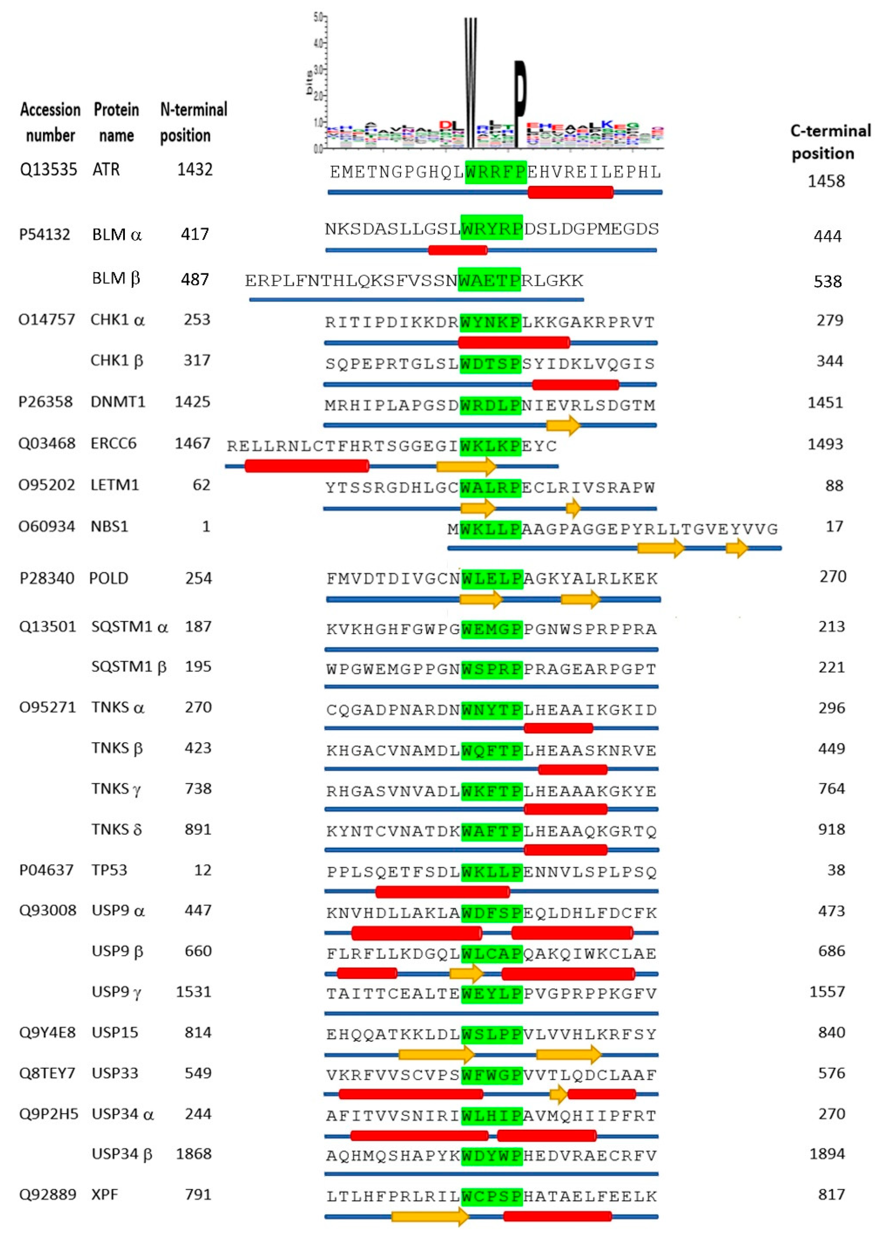
3.2. Interaction of Adenovirus E1B-55K Proteins with the xWxxxPx Sequence

To confirm that the HAdV-C5 E1B-55K interaction only requires a short sequence encompassing the xWxxxPx sequence, a lysate from HEK293 cells was incubated with a biotin-linked peptide identical to the N-terminal sequence of p53 (17ETFSDLWKLLPENNVLS33). After a pull-down with streptavidin beads, interacting proteins were fractionated by SDS-PAGE and HAdV-C5 E1B-55K identified by immunoblotting (Figure 2A). Pull-down with a similar peptide in which the tryptophan and proline residues were substituted with alanines (17ETFSDLAKLLAENNVLS33) showed no interaction (Figure 2A). Increasing amounts of peptide resulted in increased ‘pulled down’ HAdV-C5 E1B-55K (Figure 2A). In a similar experiment, the two peptides were incubated with lysates from HER2 cells. HAdV-A12 E1B-55K bound to the ETFSDLWKLLPENNVLS peptide but not to that containing alanines (Figure 2B).
To determine whether the same p53 amino acid sequence was recognized by E1B-55K proteins from other HAdV species, plasmids encoding HA-tagged versions of HAdV-E4 (species E), HAdV-D9 (species D), HAdV-B34 (species B) and HAdV-F40 (species F) E1B-55K proteins were transfected into HeLa cells. After 48 h, cell lysates were incubated with the biotin-tagged ETFSDLWKLLPENNVLS peptide. Interacting proteins were isolated using streptavidin beads and subjected to immunoblotting with an anti-HA antibody (Figure 2C). No interaction could be seen with the HAdV-E4, HAdV-D9, HAdV-B34, or HAdV-F40 E1B proteins, suggesting that the binding through xWxxxPx might differ between different HAdV species. This was somewhat unexpected as it has been previously reported by us and others that p53 binds to E1B-55K proteins from other HAdVs and in some cases is stabilized [8,12,67]. It should be noted, however, that these experiments are not directly comparable to those presented in Figure 1 as conditions are significantly different.

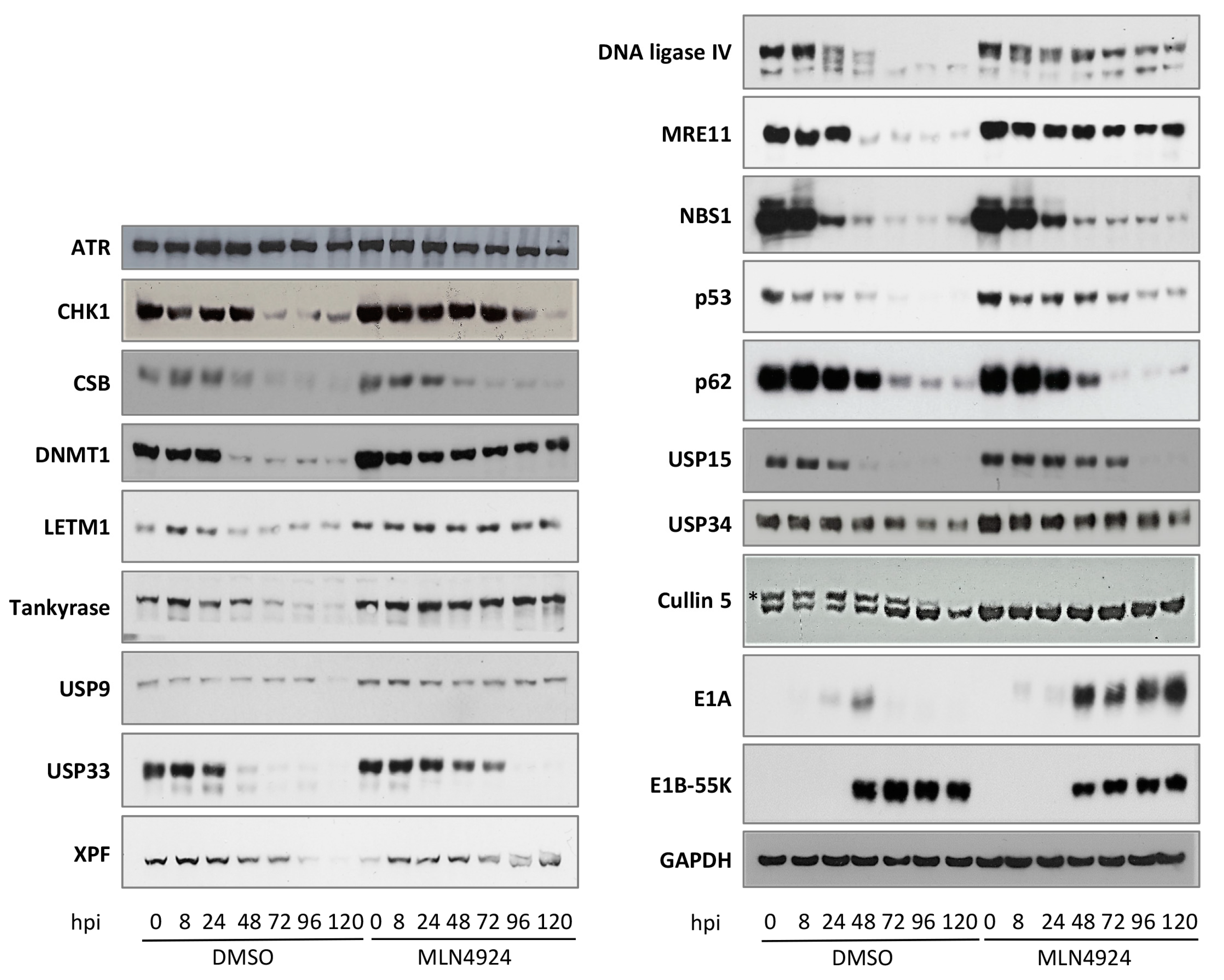
3.3. Adenovirus-Mediated Degradation of Novel Cellular Targets

To date, the major consequence of HAdV-C5 E1B-55K binding during viral infection has been shown to be rapid proteasome-mediated protein degradation [9,16]. With this in mind, the expression of the proteins (shown in Figure 1 and Figure S1 to bind HAdV-C5 E1B-55K) was assessed over an extended time course of HAdV-C5 infection. A similar set of infections was carried out in the presence of the NEDD8 (NAE)/cullin inhibitor, MLN4924. It can be seen from Figure 3 that CHK1, CSB, DNMT1, LETM1, SQSTM1 (p62), TNKS, USP15, USP33, and XPF are all degraded during HAdV-C5 infection (Figure 3). Well-characterized HAdV-C5 targets such as DNA ligase IV, p53, MRE11, or NBS1 are included for comparison (Figure 3). The rates of protein degradation vary, with some proteins being very rapidly degraded (for example, p53, DNA ligase IV, USP33, and CSB), whereas others are more stable, such as SQSTM1 (p62) and TNKS. We have previously noted that TAB182 (TNKS1BP1) is also degraded relatively slowly during HAdV infection [19]. It is interesting to mention that some HAdV-C5 E1B-55K binding proteins containing the xWxxxPx sequence are not degraded at all during HAdV-C5 infection (Figure 3). Thus, there appears to be little or no reduction in the level of USP9, USP34, or ATR. This is consistent with observations of Herrmann and colleagues [27], who found that many proteins ubiquitinated by HAdV-C5 E1B-55K/E4orf6 were also stable. To confirm that the reduction in protein expression was due to cullin-based E3 ligase activity, viral infections were carried out in the presence and absence of MLN4924. MLN4924 is an inhibitor of cullin NEDDylation and has been shown to inhibit protein degradation during adenovirus infection [9].
It can be seen from Figure 3 that HAdV-induced protein degradation is generally reduced in the presence of the inhibitor, although it is not completely negated (for example, TNKS, LETM1, USP15, and USP33). In the case of CSB, NBS1, and SQSTM1 (p62), however, it appears that the inhibition of the cullins does not result in protein stabilization; whether this indicates degradation by a different pathway is unclear at present. The immunoblot of cullin 5 shows that the higher-molecular-weight NEDDylated (active) band is absent in the cells treated with MLN4924 (Figure 3). At the latest times in the experiment, there is a marked reduction in the proportion of the NEDDylated cullin 5. The reasons for this are not clear, but there appears to be sufficient active protein present to continue with the degradation. Alternatively, it is possible that substrates could have been ubiquitinated by about 80 hpi and subsequently degraded, when there was no further need for an active cullin.
In the experiments shown in Figure 3, no account has been taken of the possibility that host cell shut-off could have contributed to the reduction in the level of particular proteins examined. However, the fact that proteins were generally stabilized by MLN4924 suggests that this was not a significant factor but cannot be discounted.

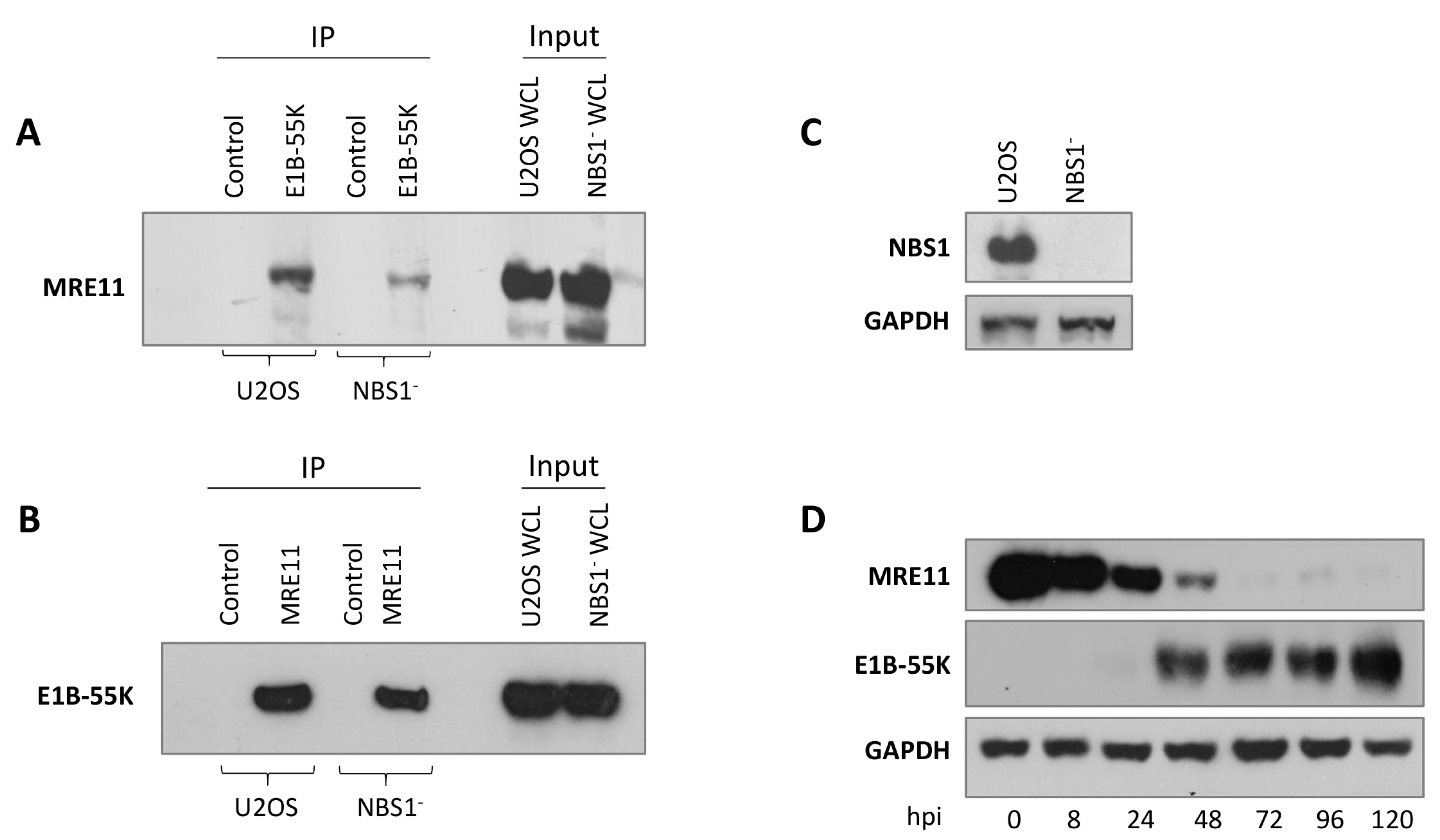
3.4. Interaction of MRE11 with HAdV-C5 E1B-55K

It is clear from several studies that proteins that do not contain the xWxxxPx motif interact with HAdV-C5 E1B-55K and are degraded during adenovirus infection (for example, [25,27,62]). Notably, MRE11 does not have the sequence but NBS1 does (MW2KLLP6AAGP). We considered the possibility that NBS1 is the principal target for HAdV-C5, and MRE11 associates with E1B-55K as an integral component of the MRN complex. The reduction in the level of MRE11 could then be attributed to the instability of the complex resulting from the degradation of NBS1. To address this possibility, U2OS cells that do not express NBS1 [50] were infected with H5pm4155 (HAdV-C5 E4orf3E4orf6), which expresses E1B-55K but does not lead to the degradation of cellular targets [19]. MRE11 is expressed to similar levels in the NBS1 and U2OS lines and is co-immunoprecipitated with E1B-55K (Figure 4A). In the complementary experiment, E1B-55K was immunoprecipitated with an MRE11 antibody (Figure 4B). The blot in Figure 4C illustrates the absence of NBS1 in the U2OS NBS1 cell line. MRE11 is degraded as expected following the infection of NBS1 cells with wild-type (wt) HAdV-C5 (Figure 4D). It is clear from these observations that MRE11 is a target for HAdV-C5 E1B-55K and interacts with it directly, with no requirement for the xWxxxPx motif.

3.5. Sub-Cellular Localization of Novel Adenovirus E1B-55K Binding Proteins during Infection

E1B-55K localizes to viral replication centers (VRCs) during infection. An appreciable number of DNA repair proteins have been also shown to localize to VRCs in adenovirus-infected cells, possibly relocalized by E1B-55K (for example, [68,69]; reviewed in [70]). We considered the possibility that proteins identified as novel interactors in Figure 1 and Figure S1 could also be present in VRCs. Therefore, we infected HeLa cells for 24 h with HAdV-C5. We fixed, permeabilized, and incubated the cells with antibodies for 18 h. The VRCs were visualized by staining with antibodies against the HAdV-C5 DNA binding protein (DBP) or RPA32, which has previously been shown to localize to VRCs [68]. The location of most of the cellular E1B-55K binding proteins examined was unaffected by HAdV infection (Figures S2–S4). Thus, CSB, DNMT1, LETM1, TNKS, USP9, and USP15 staining was very similar in the mock and infected cells, being pan-nuclear and/or cytoplasmic (Figures S2–S4). However, USP34 is clearly localized to VRCs. CHK1 staining was pan-cellular in uninfected cells, although it appeared to be predominantly cytoplasmic. Following infection, however, it was mainly observed in the nucleus, although not in VRCs. ATR was nuclear in uninfected cells but tended to be pan-cellular after HAdV-C5 infection; it also formed foci, although these did not correspond to VRCs (Figures S2–S4).

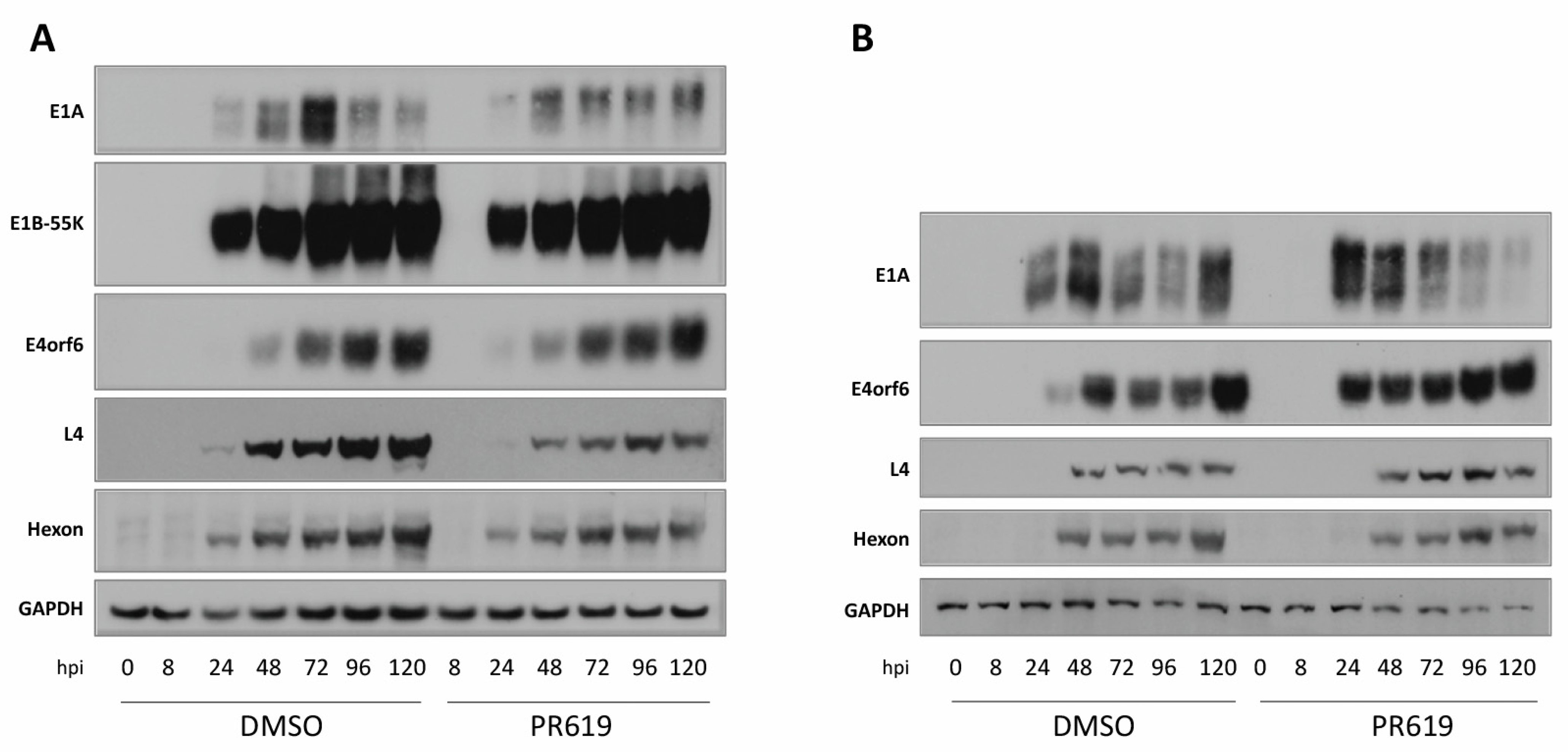
3.6. Effect of Deubiquitinase (DUB) Inhibition on HAdV-C5 Infection

Several proteins containing the xWxxxPx motif and interacting with HAdV-C5 E1B-55K are USPs and USP7 has been shown to bind to and interact with E1B-55K, although it does not contain the xWxxxPx motif [20]. Indeed, about half of the USPs contain the xWxxxPx sequence. In an overall study such as this, it is not feasible to deplete each USP in turn to determine which, if any, affects viral replication. Therefore, cells were treated with the non-selective broad-range inhibitor of deubiquitinating enzymes (DUBs), PR619 (for example, [71,72]), and then infected with HAdV-C5 or the E1B-55K-negative virus, dl1520. HeLa cells were pre-incubated with 20 µM PR619 for 1 h, infected with the virus for 2 h, and then incubated with medium containing the drug. Immunoblotting of an HAdV-C5-infected time course, in the presence and absence of 20 µM PR619, showed only minor differences in early viral protein expression, although E1A was at a higher level at early times in the absence of PR619 (Figure 5A).
Hexon levels were somewhat reduced in the presence of PR619, but the effect on L4-100K was much more marked, with appreciably reduced levels. The reasons for this are not clear at present. To determine whether the inhibition of DUBs is likely to affect adenovirus replication per se, infection was carried out in the absence of E1B-55K using the dl1520 mutant virus (Figure 5B). This removes any effects of interaction and/or USP degradation attributable to E1B-55K and looks at the effects of the DUB in isolation. However, again, differences in the presence of PR619 were not marked, although in this case there was no significant effect on L4-100K expression (Figure 5B).

3.7. Structural Prediction for the xWxxxPx Motif

It is clear that the proteins identified as E1B-55K binding proteins do not have a conserved amino acid sequence except for the tryptophan and proline residues spaced three amino acids apart, except for a slight preponderance of hydrophobic amino acids (Table 1 and Figure 6).
We considered the possibility that the binding site could have a conserved secondary structure, however. Using the programs listed in the Materials and Methods section, structural predictions were carried out on a 27 amino acid region encompassing the xWxxxPx motif in the binding proteins shown in Figure 1. It can be seen that there is no conservation of predicted structure; some regions are unstructured (for example SQSTM1 (p62)) and some have adjacent α-helices either N-terminal to the tryptophan (p53), C-terminal to the tryptophan (TNKS1) or both upstream and downstream of the binding site (USP34 and USP9). The site in other proteins contains β-strands, such as in USP15 and LETM1 (Figure 6). We conclude that the two amino acids, W and P, are the sole determining factors for interaction with HAdV-C5 E1B-55K.

4. Discussion

The adenovirus early region 1 proteins exert their effect on target cells, both during infection and cellular transformation, through a complex series of protein–protein interactions, as neither E1A nor E1B-55K have enzymatic activity nor bind to DNA. A large number of interacting proteins have been identified for both early region proteins from HAdV-C5 and, to a lesser extent, from HAdV-A12 [5,6,34,73,74]. Whilst all the sites of interaction have been closely mapped on E1A, they remain largely elusive for E1B-55K. This seems to be because E1A is highly modular and contains distinct regions that exist in a dynamic, conformationally unstructured state, giving rise to interactions with short discrete amino acid sequences [6,74]. In this way, it has been suggested that adenovirus E1A forms a molecular hub with dozens of primary interactions and hundreds and possibly thousands of secondary interactions [6,74]. HAdV-C5 E1B-55K, on the other hand, probably has a complex secondary structure. This suggestion is based on the observations that certain point mutations and small deletions destabilize the HAdV-C5 E1B-55K protein; similarly, point mutations at disparate sites, particularly in the central region, interfere with substrate binding and other phenotypes [33,38,39,40,41,42,45]. Several laboratories have attempted the large-scale purification of E1B-55K, and, to our current understanding, none were successful. It is the complex secondary structure of the protein that appears to pose significant challenges in the purification process. Interestingly, the LH3 protein purified from a snake adenovirus, which has been considered to have functional similarities to E1B-55K, has been shown to be highly structured [35]. The modeling of HAdV-C5 E1B-55K suggests unstructured N- and C-terminal regions with a hydrophobic core [34,35,36,37]. Probably because of this structural integrity, binding sites for partner proteins have been difficult to pinpoint. In complementary studies, relatively little progress has been made in the determination of binding sites for E1B-55K on target proteins except for p53 and USP7 [20,48].
By virtue of the wide disparity in the identified E1B-55K interacting proteins and the observations that some, such as integrin α3, BLM, and TAB182 [19,21,26], appear to have relatively little effect on viral replication, we considered the possibility that these diverse interactions were due to the chance presence of a conserved motif rather than direct, ‘intentional,’ targeting by the virus. As far as we are aware, the only binding site for which detailed information is available is that on p53, where a short hydrophobic sequence has been identified [48], and within that sequence, the tryptophan and proline residues, separated by three amino acids, are of particular importance. The sequence is common in previously identified HAdV-C5 E1B-55K binding proteins (for example, DNA ligase IV, BLM, TAB182, and NBS1); nevertheless, it is certainly not essential for interaction (Table 1). Notably, it is absent from MRE11 and Daxx and many of the binding proteins identified in a mass spectroscopy screen by Hung and Flint [62]; it is also absent from many of the ubiquitinated proteins identified by Herrmann and colleagues [27] in their HAdV-C5 E1B-55K/E4orf6 screen. However, it occurred frequently enough to encourage us to investigate whether other xWxxxPx motif-containing proteins would also bind to HAdV-C5 E1B-55K. In fact, this sequence is relatively common in the mammalian proteome, such that it is unlikely to be the only determining factor for the interaction between HAdV-E1B-55K and its cellular targets. We chose to examine nine novel proteins picked largely at random but predominantly on the basis that they are involved in the DDR. HAdV-C5 E1B-55K co-immunoprecipitates from HEK293 cells with the proteins listed in Table 1 (group VI) and with three other proteins (USP15, USP34, and SQSTM1 (p62)), previously identified by Hung and Flint [62], which had not been characterized in the context of adenovirus biology. Obviously, our study represents only a very small proportion of proteins containing xWxxxPx but we did not encounter any that could not be immunoprecipitated with HAdV-C5 E1B-55K. Notably, both RALY and hnRNP-C lack the xWxxxPx motif. It is tempting to speculate that the interaction between these proteins and E1B-55K occurs through RNA rather than the “xWxxxPx” motif, considering that RALY, hnRNP-C, and E1B-55K are all recognized as RNA-binding proteins [37].
It has been observed that interaction with HAdV-C5 E1B-55K/E4orf6 during viral infection would mostly result in proteasome-mediated degradation, as has been observed for p53, DNA ligase IV, and BLM. We, therefore, investigated whether the novel HAdV-C5 E1B-55K interactors were also targeted to the proteasome. Most were degraded, although at varying rates (Figure 3)—for example, CSB and USP33 were degraded at a similar rate to p53 and MRE11, whereas TNKS and LETM1 were much more stable (Figure 3). Interestingly, however, ATR, USP9, and USP34 were largely unaffected over a prolonged time course of infection. The E1B-55K/E4orf6 complex may regulate its targets in a highly diverse manner. This regulation does not seem to be exclusively dependent on uniform degradation pathways for all targets; instead, it suggests the presence of a nuanced fine-tuning process. It would be interesting to examine whether these proteins were ubiquitinated (without degradation), as has been reported for an appreciable number of other proteins that are not degraded in the presence of HAdV-C5 E1B-55K/E4orf6 [27]. Considering the known interaction between cullin-5 and E1B-55K, however, it is probable that the viral protein may disrupt host cullin-RING ubiquitin ligases, a phenomenon observed in many other viral infections as well [62,75]. Remarkably, USP9 and USP34 deviate from the norm with three and two xWxxxPx motifs, respectively, distinguishing them from the majority of other E1B-55K targets. It is noteworthy that both proteins harbor D (aspartic acid) or L (leucine) in close proximity to W (tryptophan). This is particularly significant since the presence of a double/triple xWL/DxxPx motif may induce a conformation in E1B-55K that hinders its interaction with E4orf6. Moreover, the identification of a unique xWRRFPx motif in ATR (and in CEP170) raises intriguing questions. The striking presence of this distinct “arginine-rich” motif suggests that it may induce a conformational state in E1B-55K, potentially impeding its binding to E4orf6. This could be a plausible explanation for the observed resistance of ATR to degradation despite its interaction with E1B-55K. Future research will elucidate the functional implications of these interactions. Numerous studies have now shown the presence of large numbers of DNA repair proteins, as well as proteins involved in the synthesis of viral DNA and RNA, at adenovirus replication centers [12,68,76,77,78]. Only USP34 out of the E1B-55K binding proteins identified here localized to VRCs. USP34 is a deubiquitinating enzyme (DUB), which appears to have multiple roles in development. It has also been suggested to stabilize RNF168, facilitating the cellular response to ionizing radiation [79]. Whether USP34 plays a role in the synthesis of viral DNA and/or DNA repair at VRCs requires further investigation.
As many USPs contain the xWxxxPx motif and we have confirmed the binding of at least four such proteins to HAdV-C5 E1B-55K, we have contemplated the potential influence, whether beneficial or detrimental, of these proteins on viral infection. To address this, we examined the effect of the DUB inhibitor PR619 on viral infection. Overall, there was little difference between the time courses in the presence or absence of the drug. Although this was a relatively superficial investigation, it points to a further set of HAdV-C5 E1B-55K interactions that have a minor role in adenovirus replication. It is interesting to note that several USPs have been implicated in viral infections. For example, USP15 participates in hepatitis C virus propagation through the regulation of viral RNA translation [80], whereas USP14 inhibition prevents alphaherpesvirus infection [81]. It is possible that the use of more specific USP inhibitors would demonstrate an effect on adenovirus replication.
Large numbers of cellular proteins have been shown to interact with HAdV-C5 E1B-55K, and many more are ubiquitinated by an HAdV-C5 E1B-55K/E4orf6-dependent mechanism [27,62]. However, a role for most of these associations has not been established. We suggest that many of these interactions with proteins containing the xWxxxPx motif are fortuitous and may not be of biological significance. This does not, of course, mean that none are required for viral replication. The observation that E1B-55K proteins from species other than HAdV-C5 and HAdV-A12 do not bind to the motif supports the idea that this interaction is not essential, although it must be borne in mind that the relationship of different adenovirus species with DDR proteins, such as p53 and MRE11, varies appreciably [8,12]. Notably, the degradation of p53 and MRE11 does not occur during infection with species B, D, E, and F viruses, although a marked reduction in the level of DNA ligase IV has been observed with all species examined [8,12]. The E1B-55K protein from most adenovirus species has previously been reported to interact with p53, MRE11, and DNA ligase IV as well as other proteins, suggesting that the xWxxxPx site is not important in these cases [67].
The binding sites on HAdV-C5 E1B-55K have not been closely mapped although several point mutations in the central structured region of the protein reduce or negate binding to p53. These include point mutations or insertions at residues 180, 240, 260, 262, 309, 326, 361, and 380 (summarized in [73]). Using a different approach, it has been suggested that a binding site for p53 was located between amino acids 216 and 235 [44]. If this latter region is, indeed, the primary site of interaction, it is feasible that mutations at an appreciable distance from it must have an impact on the E1B-55K structure sufficiently to disrupt binding. Furthermore, evidence is not available to establish whether other xWxxxPx motif-containing proteins have similar binding sites. However, studies with E1B-55K mutant proteins R240A, H354in, and H373A have established a separation of function in that R240A does not bind or degrade p53 but does bind to the MRN complex or cause its degradation or the degradation of DNA ligase IV. H354in and H373A fail to interact with the MRN complex or cause its degradation but do bind to p53, causing its degradation and that of DNA ligase IV [41,82,83,84]. Such observations are consistent with the p53 and DNA ligase IV sites of interaction being in the same region of E1B-55K and the MRE11 binding site being elsewhere. Several C-terminal HAdV-C5 E1B-55K mutants have been also characterized, and the differential binding and degradation of p53, MRN, and DNA ligase IV have been demonstrated [83]. These amino acids are not part of the ‘structured core’ of the proteins yet affect protein binding. Furthermore, the separation of activity against all three substrates was observed [83]. Simplistically, however, we suggest that there is a common binding site for xWxxxPx-containing proteins and alternative binding sites for others; however, further investigation would be required to establish this.

Supplementary Materials

The following supporting information can be downloaded at: https://www.mdpi.com/article/10.3390/v15122356/s1; Figure S1: Confirmation of the Interaction of HAdV-C5 E1B-55K with Novel Cellular Binding Partners; Figures S2–S4: Localization of Selected HAdV-C5 E1B-55K Binding Proteins during Infection.

Author Contributions

Conceptualization, N.C.H., W.-H.I., T.D., D.P.M., G.S.S. and R.J.G.; methodology, N.C.H., D.P.M. and R.J.G.; formal analysis, N.C.H., W.-H.I., L.D.B. and R.J.G.; investigation, N.C.H., W.-H.I., T.A., D.P.M. and R.J.G.; resources, N.C.H., W.-H.I., T.D. and R.J.G.; writing—original draft preparation, L.D.B. and R.J.G.; writing—review and editing, W.-H.I., L.D.B. and R.J.G.; visualization, L.D.B. and R.J.G.; supervision, T.D., D.P.M., G.S.S. and R.J.G.; project administration, R.J.G.; funding acquisition, T.D., D.P.M., G.S.S. and R.J.G. All authors have read and agreed to the published version of the manuscript.

Funding

We are grateful for funding from CR-UK (Programme Grant C17183/A23303 to G.S.S.) and the High-End Foreign Experts Program of the Ministry of Science and Technology of China, grant number G2021035005L (to D.P.M.). We are also grateful to Katie Grand for a charitable donation through the University of Birmingham JustGiving scheme. The Leibniz Institute of Virology (LIV) is supported by the Freie und Hansestadt Hamburg and the German Bundesministerium für Gesundheit (Federal Ministry of Health).

Institutional Review Board Statement

Not applicable.

Informed Consent Statement

Not applicable.

Data Availability Statement

The authors confirm that the data supporting the findings of this study are available within the article and its Supplementary Materials.

Conflicts of Interest

The authors declare no conflict of interest.

References

  1. Benko, M.; Aoki, K.; Arnberg, N.; Davison, A.J.; Echavarria, M.; Hess, M.; Jones, M.S.; Kajan, G.L.; Kajon, A.E.; Mittal, S.K.; et al. ICTV Virus Taxonomy Profile: Adenoviridae 2022. J. Gen. Virol. 2022, 103, 001721. [Google Scholar] [CrossRef]
  2. Khanal, S.; Ghimire, P.; Dhamoon, A.S. The Repertoire of Adenovirus in Human Disease: The Innocuous to the Deadly. Biomedicines 2018, 6, 30. [Google Scholar] [CrossRef] [PubMed]
  3. Lynch, J.P., 3rd; Kajon, A.E. Adenovirus: Epidemiology, Global Spread of Novel Serotypes, and Advances in Treatment and Prevention. Semin. Respir. Crit. Care Med. 2016, 37, 586–602. [Google Scholar] [CrossRef] [PubMed]
  4. Berk, A.J. Recent lessons in gene expression, cell cycle control, and cell biology from adenovirus. Oncogene 2005, 24, 7673–7685. [Google Scholar] [CrossRef] [PubMed]
  5. Ip, W.H.; Dobner, T. Cell transformation by the adenovirus oncogenes E1 and E4. FEBS Lett. 2020, 594, 1848–1860. [Google Scholar] [CrossRef] [PubMed]
  6. King, C.R.; Zhang, A.; Tessier, T.M.; Gameiro, S.F.; Mymryk, J.S. Hacking the Cell: Network Intrusion and Exploitation by Adenovirus E1A. mBio 2018, 9, e00390-18. [Google Scholar] [CrossRef] [PubMed]
  7. Cuconati, A.; White, E. Viral homologs of BCL-2: Role of apoptosis in the regulation of virus infection. Genes Dev. 2002, 16, 2465–2478. [Google Scholar] [CrossRef]
  8. Cheng, C.Y.; Gilson, T.; Dallaire, F.; Ketner, G.; Branton, P.E.; Blanchette, P. The E4orf6/E1B55K E3 ubiquitin ligase complexes of human adenoviruses exhibit heterogeneity in composition and substrate specificity. J. Virol. 2011, 85, 765–775. [Google Scholar] [CrossRef]
  9. Querido, E.; Blanchette, P.; Yan, Q.; Kamura, T.; Morrison, M.; Boivin, D.; Kaelin, W.G.; Conaway, R.C.; Conaway, J.W.; Branton, P.E. Degradation of p53 by adenovirus E4orf6 and E1B55K proteins occurs via a novel mechanism involving a Cullin-containing complex. Genes Dev. 2001, 15, 3104–3117. [Google Scholar] [CrossRef]
  10. Querido, E.; Marcellus, R.C.; Lai, A.; Charbonneau, R.; Teodoro, J.G.; Ketner, G.; Branton, P.E. Regulation of p53 levels by the E1B 55-kilodalton protein and E4orf6 in adenovirus-infected cells. J. Virol. 1997, 71, 3788–3798. [Google Scholar] [CrossRef]
  11. Steegenga, W.T.; Riteco, N.; Jochemsen, A.G.; Fallaux, F.J.; Bos, J.L. The large E1B protein together with the E4orf6 protein target p53 for active degradation in adenovirus infected cells. Oncogene 1998, 16, 349–357. [Google Scholar] [CrossRef] [PubMed]
  12. Forrester, N.A.; Sedgwick, G.G.; Thomas, A.; Blackford, A.N.; Speiseder, T.; Dobner, T.; Byrd, P.J.; Stewart, G.S.; Turnell, A.S.; Grand, R.J. Serotype-specific inactivation of the cellular DNA damage response during adenovirus infection. J. Virol. 2011, 85, 2201–2211. [Google Scholar] [CrossRef] [PubMed]
  13. von Stromberg, K.; Seddar, L.; Ip, W.H.; Günther, T.; Gornott, B.; Weinert, S.C.; Hüppner, M.; Bertzbach, L.D.; Dobner, T. The human adenovirus E1B-55K oncoprotein coordinates cell transformation through regulation of DNA-bound host transcription factors. Proc. Natl. Acad. Sci. USA 2023, 120, e2310770120. [Google Scholar] [CrossRef] [PubMed]
  14. Blackford, A.N.; Patel, R.N.; Forrester, N.A.; Theil, K.; Groitl, P.; Stewart, G.S.; Taylor, A.M.; Morgan, I.M.; Dobner, T.; Grand, R.J.; et al. Adenovirus 12 E4orf6 inhibits ATR activation by promoting TOPBP1 degradation. Proc. Natl. Acad. Sci. USA 2010, 107, 12251–12256. [Google Scholar] [CrossRef] [PubMed]
  15. Blanchette, P.; Cheng, C.Y.; Yan, Q.; Ketner, G.; Ornelles, D.A.; Dobner, T.; Conaway, R.C.; Conaway, J.W.; Branton, P.E. Both BC-box motifs of adenovirus protein E4orf6 are required to efficiently assemble an E3 ligase complex that degrades p53. Mol. Cell. Biol. 2004, 24, 9619–9629. [Google Scholar] [CrossRef] [PubMed]
  16. Schreiner, S.; Wimmer, P.; Dobner, T. Adenovirus degradation of cellular proteins. Future Microbiol. 2012, 7, 211–225. [Google Scholar] [CrossRef] [PubMed]
  17. Ip, W.H.; Tatham, M.H.; Krohne, S.; Gruhne, J.; Melling, M.; Meyer, T.; Gornott, B.; Bertzbach, L.D.; Hay, R.T.; Rodriguez, E.; et al. Adenovirus E1B-55K controls SUMO-dependent degradation of antiviral cellular restriction factors. J. Virol. 2023, e0079123. [Google Scholar] [CrossRef] [PubMed]
  18. Baker, A.; Rohleder, K.J.; Hanakahi, L.A.; Ketner, G. Adenovirus E4 34k and E1b 55k oncoproteins target host DNA ligase IV for proteasomal degradation. J. Virol. 2007, 81, 7034–7040. [Google Scholar] [CrossRef]
  19. Chalabi Hagkarim, N.; Ryan, E.L.; Byrd, P.J.; Hollingworth, R.; Shimwell, N.J.; Agathanggelou, A.; Vavasseur, M.; Kolbe, V.; Speiseder, T.; Dobner, T.; et al. Degradation of a Novel DNA Damage Response Protein, Tankyrase 1 Binding Protein 1, following Adenovirus Infection. J. Virol. 2018, 92, e02034-17. [Google Scholar] [CrossRef]
  20. Ching, W.; Koyuncu, E.; Singh, S.; Arbelo-Roman, C.; Härtl, B.; Kremmer, E.; Speiseder, T.; Meier, C.; Dobner, T. A ubiquitin-specific protease possesses a decisive role for adenovirus replication and oncogene-mediated transformation. PLoS Pathog. 2013, 9, e1003273. [Google Scholar] [CrossRef]
  21. Orazio, N.I.; Naeger, C.M.; Karlseder, J.; Weitzman, M.D. The adenovirus E1b55K/E4orf6 complex induces degradation of the Bloom helicase during infection. J. Virol. 2011, 85, 1887–1892. [Google Scholar] [CrossRef]
  22. Stracker, T.H.; Carson, C.T.; Weitzman, M.D. Adenovirus oncoproteins inactivate the Mre11-Rad50-NBS1 DNA repair complex. Nature 2002, 418, 348–352. [Google Scholar] [CrossRef] [PubMed]
  23. Schreiner, S.; Bürck, C.; Glass, M.; Groitl, P.; Wimmer, P.; Kinkley, S.; Mund, A.; Everett, R.D.; Dobner, T. Control of human adenovirus type 5 gene expression by cellular Daxx/ATRX chromatin-associated complexes. Nucleic Acids Res. 2013, 41, 3532–3550. [Google Scholar] [CrossRef] [PubMed]
  24. Schreiner, S.; Kinkley, S.; Bürck, C.; Mund, A.; Wimmer, P.; Schubert, T.; Groitl, P.; Will, H.; Dobner, T. SPOC1-mediated antiviral host cell response is antagonized early in human adenovirus type 5 infection. PLoS Pathog. 2013, 9, e1003775. [Google Scholar] [CrossRef] [PubMed]
  25. Schreiner, S.; Wimmer, P.; Sirma, H.; Everett, R.D.; Blanchette, P.; Groitl, P.; Dobner, T. Proteasome-dependent degradation of Daxx by the viral E1B-55K protein in human adenovirus-infected cells. J. Virol. 2010, 84, 7029–7038. [Google Scholar] [CrossRef] [PubMed]
  26. Dallaire, F.; Blanchette, P.; Groitl, P.; Dobner, T.; Branton, P.E. Identification of integrin alpha3 as a new substrate of the adenovirus E4orf6/E1B 55-kilodalton E3 ubiquitin ligase complex. J. Virol. 2009, 83, 5329–5338. [Google Scholar] [CrossRef]
  27. Herrmann, C.; Dybas, J.M.; Liddle, J.C.; Price, A.M.; Hayer, K.E.; Lauman, R.; Purman, C.E.; Charman, M.; Kim, E.T.; Garcia, B.A.; et al. Adenovirus-mediated ubiquitination alters protein-RNA binding and aids viral RNA processing. Nat. Microbiol. 2020, 5, 1217–1231. [Google Scholar] [CrossRef] [PubMed]
  28. Flint, S.J.; Gonzalez, R.A. Regulation of mRNA production by the adenoviral E1B 55-kDa and E4 Orf6 proteins. Curr. Top. Microbiol. Immunol. 2003, 272, 287–330. [Google Scholar] [CrossRef]
  29. Gales, J.P.; Kubina, J.; Geldreich, A.; Dimitrova, M. Strength in Diversity: Nuclear Export of Viral RNAs. Viruses 2020, 12, 1014. [Google Scholar] [CrossRef]
  30. Valdés Alemán, M.; Bertzbach, L.D.; Speiseder, T.; Ip, W.H.; González, R.A.; Dobner, T. Global Transcriptome Analyses of Cellular and Viral mRNAs During HAdV-C5 Infection Highlight New Aspects of Viral mRNA Biogenesis and Cytoplasmic Viral mRNA Accumulations. Viruses 2022, 14, 2428. [Google Scholar] [CrossRef]
  31. Yatherajam, G.; Huang, W.; Flint, S.J. Export of adenoviral late mRNA from the nucleus requires the Nxf1/Tap export receptor. J. Virol. 2011, 85, 1429–1438. [Google Scholar] [CrossRef] [PubMed]
  32. Hidalgo, P.; Garces, Y.; Mundo, E.; Lopez, R.E.; Bertzbach, L.D.; Dobner, T.; Gonzalez, R.A. E1B-55K is a phosphorylation-dependent transcriptional and post-transcriptional regulator of viral gene expression in HAdV-C5 infection. J. Virol. 2022, 96, JVI0206221. [Google Scholar] [CrossRef] [PubMed]
  33. Gabler, S.; Schutt, H.; Groitl, P.; Wolf, H.; Shenk, T.; Dobner, T. E1B 55-kilodalton-associated protein: A cellular protein with RNA-binding activity implicated in nucleocytoplasmic transport of adenovirus and cellular mRNAs. J. Virol. 1998, 72, 7960–7971. [Google Scholar] [CrossRef] [PubMed]
  34. Hidalgo, P.; Ip, W.H.; Dobner, T.; Gonzalez, R.A. The biology of the adenovirus E1B 55K protein. FEBS Lett. 2019, 593, 3504–3517. [Google Scholar] [CrossRef] [PubMed]
  35. Menendez-Conejero, R.; Nguyen, T.H.; Singh, A.K.; Condezo, G.N.; Marschang, R.E.; van Raaij, M.J.; San Martin, C. Structure of a Reptilian Adenovirus Reveals a Phage Tailspike Fold Stabilizing a Vertebrate Virus Capsid. Structure 2017, 25, 1562–1573.e5. [Google Scholar] [CrossRef] [PubMed]
  36. Sieber, T.; Scholz, R.; Spoerner, M.; Schumann, F.; Kalbitzer, H.R.; Dobner, T. Intrinsic disorder in the common N-terminus of human adenovirus 5 E1B-55K and its related E1BN proteins indicated by studies on E1B-93R. Virology 2011, 418, 133–143. [Google Scholar] [CrossRef]
  37. Tejera, B.; Lopez, R.E.; Hidalgo, P.; Cardenas, R.; Ballesteros, G.; Rivillas, L.; French, L.; Amero, C.; Pastor, N.; Santiago, A.; et al. The human adenovirus type 5 E1B 55kDa protein interacts with RNA promoting timely DNA replication and viral late mRNA metabolism. PLoS ONE 2019, 14, e0214882. [Google Scholar] [CrossRef]
  38. Gonzalez, R.A.; Flint, S.J. Effects of mutations in the adenoviral E1B 55-kilodalton protein coding sequence on viral late mRNA metabolism. J. Virol. 2002, 76, 4507–4519. [Google Scholar] [CrossRef]
  39. Kao, C.C.; Yew, P.R.; Berk, A.J. Domains required for in vitro association between the cellular p53 and the adenovirus 2 E1B 55k proteins. Virology 1990, 179, 806–814. [Google Scholar] [CrossRef]
  40. Rubenwolf, S.; Schutt, H.; Nevels, M.; Wolf, H.; Dobner, T. Structural analysis of the adenovirus type 5 E1B 55-kilodalton-E4orf6 protein complex. J. Virol. 1997, 71, 1115–1123. [Google Scholar] [CrossRef]
  41. Shen, Y.; Kitzes, G.; Nye, J.A.; Fattaey, A.; Hermiston, T. Analyses of single-amino-acid substitution mutants of adenovirus type 5 E1B-55K protein. J. Virol. 2001, 75, 4297–4307. [Google Scholar] [CrossRef] [PubMed]
  42. Yew, P.R.; Kao, C.C.; Berk, A.J. Dissection of functional domains in the adenovirus 2 early 1B 55K polypeptide by suppressor-linker insertional mutagenesis. Virology 1990, 179, 795–805. [Google Scholar] [CrossRef] [PubMed]
  43. Fiedler, M.; Ip, W.H.; Hofmann-Sieber, H.; Wilkens, B.; Nkrumah, F.K.; Zhang, W.; Ehrhardt, A.; Bertzbach, L.D.; Dobner, T. Protein–Protein Interactions Facilitate E4orf6-Dependent Regulation of E1B-55K SUMOylation in HAdV-C5 Infection. Viruses 2022, 14, 463. [Google Scholar] [CrossRef] [PubMed]
  44. Grand, R.J.; Parkhill, J.; Szestak, T.; Rookes, S.M.; Roberts, S.; Gallimore, P.H. Definition of a major p53 binding site on Ad2E1B58K protein and a possible nuclear localization signal on the Ad12E1B54K protein. Oncogene 1999, 18, 955–965. [Google Scholar] [CrossRef] [PubMed]
  45. Yew, P.R.; Berk, A.J. Inhibition of p53 transactivation required for transformation by adenovirus early 1B protein. Nature 1992, 357, 82–85. [Google Scholar] [CrossRef] [PubMed]
  46. Wimmer, P.; Berscheminski, J.; Blanchette, P.; Groitl, P.; Branton, P.E.; Hay, R.T.; Dobner, T.; Schreiner, S. PML isoforms IV and V contribute to adenovirus-mediated oncogenic transformation by functionally inhibiting the tumor-suppressor p53. Oncogene 2016, 35, 69–82. [Google Scholar] [CrossRef] [PubMed]
  47. Bürck, C.; Mund, A.; Berscheminski, J.; Kieweg, L.; Müncheberg, S.; Dobner, T.; Schreiner, S. KAP1 Is a Host Restriction Factor That Promotes Human Adenovirus E1B-55K SUMO Modification. J. Virol. 2016, 90, 930–946. [Google Scholar] [CrossRef]
  48. Lin, J.; Chen, J.; Elenbaas, B.; Levine, A.J. Several hydrophobic amino acids in the p53 amino-terminal domain are required for transcriptional activation, binding to mdm-2 and the adenovirus 5 E1B 55-kD protein. Genes Dev. 1994, 8, 1235–1246. [Google Scholar] [CrossRef]
  49. Graham, F.L.; Smiley, J.; Russell, W.C.; Nairn, R. Characteristics of a human cell line transformed by DNA from human adenovirus type 5. J. Gen. Virol. 1977, 36, 59–74. [Google Scholar] [CrossRef]
  50. Mooser, C.; Symeonidou, I.E.; Leimbacher, P.A.; Ribeiro, A.; Shorrocks, A.K.; Jungmichel, S.; Larsen, S.C.; Knechtle, K.; Jasrotia, A.; Zurbriggen, D.; et al. Treacle controls the nucleolar response to rDNA breaks via TOPBP1 recruitment and ATR activation. Nat. Commun. 2020, 11, 123. [Google Scholar] [CrossRef]
  51. Byrd, P.; Brown, K.W.; Gallimore, P.H. Malignant transformation of human embryo retinoblasts by cloned adenovirus 12 DNA. Nature 1982, 298, 69–71. [Google Scholar] [CrossRef] [PubMed]
  52. Barker, D.D.; Berk, A.J. Adenovirus proteins from both E1B reading frames are required for transformation of rodent cells by viral infection and DNA transfection. Virology 1987, 156, 107–121. [Google Scholar] [CrossRef] [PubMed]
  53. Byrd, P.J.; Grand, R.J.; Breiding, D.; Williams, J.F.; Gallimore, P.H. Host range mutants of adenovirus type 12 E1 defective for lytic infection, transformation, and oncogenicity. Virology 1988, 163, 155–165. [Google Scholar] [CrossRef] [PubMed]
  54. Harlow, E.; Franza, B.R., Jr.; Schley, C. Monoclonal antibodies specific for adenovirus early region 1A proteins: Extensive heterogeneity in early region 1A products. J. Virol. 1985, 55, 533–546. [Google Scholar] [CrossRef]
  55. Sarnow, P.; Ho, Y.S.; Williams, J.; Levine, A.J. Adenovirus E1b-58kd tumor antigen and SV40 large tumor antigen are physically associated with the same 54 kd cellular protein in transformed cells. Cell 1982, 28, 387–394. [Google Scholar] [CrossRef]
  56. Madeira, F.; Park, Y.M.; Lee, J.; Buso, N.; Gur, T.; Madhusoodanan, N.; Basutkar, P.; Tivey, A.R.N.; Potter, S.C.; Finn, R.D.; et al. The EMBL-EBI search and sequence analysis tools APIs in 2019. Nucleic Acids Res. 2019, 47, W636–W641. [Google Scholar] [CrossRef] [PubMed]
  57. Katoh, K.; Rozewicki, J.; Yamada, K.D. MAFFT online service: Multiple sequence alignment, interactive sequence choice and visualization. Brief. Bioinform. 2019, 20, 1160–1166. [Google Scholar] [CrossRef]
  58. Crooks, G.E.; Hon, G.; Chandonia, J.M.; Brenner, S.E. WebLogo: A sequence logo generator. Genome Res. 2004, 14, 1188–1190. [Google Scholar] [CrossRef]
  59. Jones, D.T. Protein secondary structure prediction based on position-specific scoring matrices. J. Mol. Biol. 1999, 292, 195–202. [Google Scholar] [CrossRef]
  60. Drozdetskiy, A.; Cole, C.; Procter, J.; Barton, G.J. JPred4: A protein secondary structure prediction server. Nucleic Acids Res. 2015, 43, W389–W394. [Google Scholar] [CrossRef]
  61. Frishman, D.; Argos, P. Incorporation of non-local interactions in protein secondary structure prediction from the amino acid sequence. Protein Eng. 1996, 9, 133–142. [Google Scholar] [CrossRef] [PubMed]
  62. Hung, G.; Flint, S.J. Normal human cell proteins that interact with the adenovirus type 5 E1B 55kDa protein. Virology 2017, 504, 12–24. [Google Scholar] [CrossRef] [PubMed]
  63. Punga, T.; Akusjärvi, G. The adenovirus-2 E1B-55K protein interacts with a mSin3A/histone deacetylase 1 complex. FEBS Lett. 2000, 476, 248–252. [Google Scholar] [CrossRef] [PubMed]
  64. Gupta, A.; Jha, S.; Engel, D.A.; Ornelles, D.A.; Dutta, A. Tip60 degradation by adenovirus relieves transcriptional repression of viral transcriptional activator EIA. Oncogene 2013, 32, 5017–5025. [Google Scholar] [CrossRef] [PubMed]
  65. Nayak, R.; Farris, K.D.; Pintel, D.J. E4Orf6-E1B-55k-dependent degradation of de novo-generated adeno-associated virus type 5 Rep52 and capsid proteins employs a cullin 5-containing E3 ligase complex. J. Virol. 2008, 82, 3803–3808. [Google Scholar] [CrossRef] [PubMed]
  66. Sukhu, L.; Pintel, D. The large Rep protein of adeno-associated virus type 2 is polyubiquitinated. J. Gen. Virol. 2011, 92 Pt 12, 2792–2796. [Google Scholar] [CrossRef] [PubMed]
  67. Cheng, C.Y.; Gilson, T.; Wimmer, P.; Schreiner, S.; Ketner, G.; Dobner, T.; Branton, P.E.; Blanchette, P. Role of E1B55K in E4orf6/E1B55K E3 ligase complexes formed by different human adenovirus serotypes. J. Virol. 2013, 87, 6232–6245. [Google Scholar] [CrossRef]
  68. Blackford, A.N.; Bruton, R.K.; Dirlik, O.; Stewart, G.S.; Taylor, A.M.; Dobner, T.; Grand, R.J.; Turnell, A.S. A role for E1B-AP5 in ATR signaling pathways during adenovirus infection. J. Virol. 2008, 82, 7640–7652. [Google Scholar] [CrossRef]
  69. Ornelles, D.A.; Shenk, T. Localization of the adenovirus early region 1B 55-kilodalton protein during lytic infection: Association with nuclear viral inclusions requires the early region 4 34-kilodalton protein. J. Virol. 1991, 65, 424–429. [Google Scholar] [CrossRef]
  70. Kleinberger, T. En Guard! The Interactions between Adenoviruses and the DNA Damage Response. Viruses 2020, 12, 996. [Google Scholar] [CrossRef]
  71. Cowell, I.G.; Ling, E.M.; Swan, R.L.; Brooks, M.L.W.; Austin, C.A. The Deubiquitinating Enzyme Inhibitor PR-619 is a Potent DNA Topoisomerase II Poison. Mol. Pharmacol. 2019, 96, 562–572. [Google Scholar] [CrossRef] [PubMed]
  72. Seiberlich, V.; Goldbaum, O.; Zhukareva, V.; Richter-Landsberg, C. The small molecule inhibitor PR-619 of deubiquitinating enzymes affects the microtubule network and causes protein aggregate formation in neural cells: Implications for neurodegenerative diseases. Biochim. Biophys. Acta 2012, 1823, 2057–2068. [Google Scholar] [CrossRef] [PubMed]
  73. Blackford, A.N.; Grand, R.J. Adenovirus E1B 55-kilodalton protein: Multiple roles in viral infection and cell transformation. J. Virol. 2009, 83, 4000–4012. [Google Scholar] [CrossRef] [PubMed]
  74. Pelka, P.; Ablack, J.N.; Fonseca, G.J.; Yousef, A.F.; Mymryk, J.S. Intrinsic structural disorder in adenovirus E1A: A viral molecular hub linking multiple diverse processes. J. Virol. 2008, 82, 7252–7263. [Google Scholar] [CrossRef] [PubMed]
  75. Liu, Y.; Tan, X. Viral Manipulations of the Cullin-RING Ubiquitin Ligases. Adv. Exp. Med. Biol. 2020, 1217, 99–110. [Google Scholar] [CrossRef] [PubMed]
  76. Charman, M.; Herrmann, C.; Weitzman, M.D. Viral and cellular interactions during adenovirus DNA replication. FEBS Lett. 2019, 593, 3531–3550. [Google Scholar] [CrossRef] [PubMed]
  77. Hidalgo, P.; Anzures, L.; Hernandez-Mendoza, A.; Guerrero, A.; Wood, C.D.; Valdes, M.; Dobner, T.; Gonzalez, R.A. Morphological, Biochemical, and Functional Study of Viral Replication Compartments Isolated from Adenovirus-Infected Cells. J. Virol. 2016, 90, 3411–3427. [Google Scholar] [CrossRef]
  78. Charman, M.; Weitzman, M.D. Replication Compartments of DNA Viruses in the Nucleus: Location, Location, Location. Viruses 2020, 12, 151. [Google Scholar] [CrossRef]
  79. Sy, S.M.; Jiang, J.; O, W.S.; Deng, Y.; Huen, M.S. The ubiquitin specific protease USP34 promotes ubiquitin signaling at DNA double-strand breaks. Nucleic Acids Res. 2013, 41, 8572–8580. [Google Scholar] [CrossRef]
  80. Kusakabe, S.; Suzuki, T.; Sugiyama, Y.; Haga, S.; Horike, K.; Tokunaga, M.; Hirano, J.; Zhang, H.; Chen, D.V.; Ishiga, H.; et al. USP15 Participates in Hepatitis C Virus Propagation through Regulation of Viral RNA Translation and Lipid Droplet Formation. J. Virol. 2019, 93, e01708-18. [Google Scholar] [CrossRef]
  81. Ming, S.L.; Zhang, S.; Wang, Q.; Zeng, L.; Zhou, L.Y.; Wang, M.D.; Ma, Y.X.; Han, L.Q.; Zhong, K.; Zhu, H.S.; et al. Inhibition of USP14 influences alphaherpesvirus proliferation by degrading viral VP16 protein via ER stress-triggered selective autophagy. Autophagy 2022, 18, 1801–1821. [Google Scholar] [CrossRef] [PubMed]
  82. Carson, C.T.; Schwartz, R.A.; Stracker, T.H.; Lilley, C.E.; Lee, D.V.; Weitzman, M.D. The Mre11 complex is required for ATM activation and the G2/M checkpoint. EMBO J. 2003, 22, 6610–6620. [Google Scholar] [CrossRef] [PubMed]
  83. Schwartz, R.A.; Lakdawala, S.S.; Eshleman, H.D.; Russell, M.R.; Carson, C.T.; Weitzman, M.D. Distinct requirements of adenovirus E1b55K protein for degradation of cellular substrates. J. Virol. 2008, 82, 9043–9055. [Google Scholar] [CrossRef] [PubMed]
  84. Lakdawala, S.S.; Schwartz, R.A.; Ferenchak, K.; Carson, C.T.; McSharry, B.P.; Wilkinson, G.W.; Weitzman, M.D. Differential requirements of the C terminus of Nbs1 in suppressing adenovirus DNA replication and promoting concatemer formation. J. Virol. 2008, 82, 8362–8372. [Google Scholar] [CrossRef]
Figure 1. The interaction of HAdV-C5 E1B-55K with novel cellular binding partners. Lysates from HEK293 cells were immunoprecipitated with antibodies (either mouse or rabbit as appropriate) against the HAdV-C5 E1B-55K protein. After immunoblotting, interacting proteins were detected with the antibodies shown. Known binding proteins such as NBS1, MRE11, and hnRNPUL1 are included for comparison (left column). “Control” is an irrelevant antibody included as a negative control, raised against either collagen type IV (rabbit) or vimentin (mouse). Images represent the results of three repeated experiments. WCL, whole cell lysate.
Viruses 15 02356 g001
Figure 2. The interaction of adenovirus E1B-55K proteins with the xWxxxPx motif. (A) HEK293 cell lysates were mock incubated or incubated with either biotin-linked ETFSDLWKLLPENNVLS peptide (WP peptide) or ETFSDLAKLLAENNVLS peptide (AA peptide), (i) 1 µg/mL, (ii) 5 µg/mL and (iii) 20 µg/mL for 2 h. Streptavidin (SA) beads were added for a further 90 min. After washing bound proteins were released with SDS sample buffer and fractionated by PAGE. Bound HAdV-C5 E1B-55K was detected by immunoblotting. (B) HER2 cell lysate was incubated with WP peptide or AA peptide (10 µg/mL) as in (A). Bound HAdV-A12 E1B-55K was detected by immunoblotting. (C) HeLa cells were transfected with pcDNA3 constructs expressing HA-tagged E1B-55K proteins from Ads 4, 34, 40, and 9. After 48 h, cell lysates were incubated or mock incubated with WP peptide (10 µg/mL) for 2 h. After incubation with streptavidin beads samples were processed as in (A) and then immunoblotted for the HA tag. Images represent the results of three repeated experiments. WCL, whole cell lysate.
Viruses 15 02356 g002
Figure 3. Degradation of novel binding partners during HAdV-C5 infection is dependent on cullin function. The left-hand blots display novel E1B-55K binding partners, while the right-hand blots serve as controls. HeLa cells were infected with HAdV-C5 at an infectivity of 4 pfu/cell, either in the presence or absence of 5 mM MLN4924. Cells were harvested at the times shown, post-infection, fractionated by PAGE, and immunoblotted with the antibodies as indicated. For the cullin 5 blots, * indicates the NEDDylated form. Images represent the results of three repeated experiments.
Viruses 15 02356 g003
Figure 4. HAdV-C5 E1B-55K binds to MRE11 in the absence of NBS1. (A,B) U2OS or NBS1-negative cells were infected with H5pm4155 (E4orf3, E4orf6) at 10 pfu/cell for 48 h. Cells were harvested and immunoprecipitated with antibodies against either HAdV-C5 E1B-55K (A) or MRE11 (B). After PAGE and immunoblotting, co-immunoprecipitated MRE11 (A) or HAdV-C5 E1B-55K (B) was detected. Control, immunoprecipitated with an irrelevant antibody as in Figure 1. (C) Immunoblot showing no expression of NBS1 in NBS1 cells. (D) NBS1 cells were infected with HAdV-C5 (5 pfu/cell) and harvested at the times shown. Lysates were immunoblotted for MRE11, HAdV-C5 E1B-55K, and GAPDH as a loading control. Images represent the results of three repeated experiments. WCL, whole cell lysate.
Viruses 15 02356 g004
Figure 5. The effect of the DUB inhibitor PR619 on adenovirus infection. HeLa cells were treated for 1 h with 20 µM PR619 and infected with (A) HAdV-C5 (5 pfu/cell) or (B) dl1520 (10 pfu/cell). Cultures were then incubated in the presence of 20 µM PR619 and harvested at the times shown. Lysates were subjected to immunoblotting as shown. Images represent the results of three repeated experiments. L4, L4-100K.
Viruses 15 02356 g005
Figure 6. Structural prediction of xWxxxPx-containing motifs. The accession numbers of candidate proteins were obtained from the UniProtKB databank. Proteins containing xWxxxPx motifs (green background highlighted typeset) are listed alphabetically. The N- and C-terminal positions of each protein primary sequence are shown. Where a protein contains more than one xWxxxPx motif they are designated by Greek letters (α, β, γ, etc.). A weblogo cartoon of amino acids in the sequence of the proteins listed is shown at the top of the figure. Average predicted structural propensities for each amino acid in each protein are illustrated as either α-helix (red cylinder), β-strand (yellow arrow), or random coil (thin blue cylinder) below the primary sequence.
Viruses 15 02356 g006
Table 1. Ad5 E1B-55K binding proteins.
Group 1ProteinAmino Acid SequenceResidues (WxxxP)Reference
ICEP170NSRWRRFPTDYA1230–1234[62]
ICNOT3EAAWHHMPHPSD622–626[19]
ICullin 5LFAWNQRPREKI506–510[62]
IhnRNPRQQNWGSQPIAQQ597–601[62]
IIntegrin α3 (ITGA3)NGKWLLYPTEIT
NGSWPCRPPGDL
HCVWLECPIPDA
829–833
843–847
913–917
[26]
ImSin3aNDTWVSFPSWSE585–589[63]
IMYCB2AGKWVELPITKS
VPYWNAKPAPMP
DVIWRFRPNTRE
571–575
1034–1038
1138–1142
[62]
ISQSTM1 (p62)WPGWEMGPPGNW
PGNWSPRPPRAG
198–202
206–210
[62]
ISRSF3PPSWGRRPRDDY96–100[62]
IUBR5LYWWGVVPFSQR
DPDWLDLPPISS
PPSWVPDPPAMD
494–498
835–839
1027–1031
[62]
IUSP8IEIWKLPPVLLV1001–1005[62]
IUSP15LDLWSLPPVLVV825–829[62]
IUSP34IRIWLHIPAVMQ
PYKWDYWPHEDV
255–259
1879–1883
[62]
IZNF638GSRWDDEPHISA104–108[62]
IIBLMGSLWRYRPDSLD428–432[21]
IIDNA ligase IVRYSWDCSPLSMF805–809[18]
IINBS1 (NBN)MWKLLPAAGP2–6[22]
IIp53SDLWKLLPENNV23–27[55]
IITab182 (TNKS1BP1)PPSWRPQPDGEA1507–1511[19]
IITIP60LKPWYFSPYPQE245–249[64]
IITOPBP1NLQWPSCPTQYS163–1167[14]
IIIAAV Rep52NTIWLFGPATTG108–112[65]
IIIAAV Rep68EKEWELPPDSDM
NTIWLFGPATTG
35–39
331–335
[66]
IIIAAV CapsidYKNWFPGPMGRT
GPIWAKIPETGA
464–468
608–612
[65]
IVTNFRSF10ATQQWEHSPLGEL123–127[27]
IVRP2ELNWSLLPEDAV186–190[27]
IVCLPTM1YLSWILFPLLGC481–485[27]
IVPDGKRBIMLWQKKPRYEI556–560[27]
IVFASLGIWTLLPLVLT4–8[27]
IVCXADRDIEWLISPADNQ57–61[27]
IVEPHA7DIEWLISPADNQ
ELEWISSPPNGW
57–61
397–401
[27]
IVSTK11IPHGSWSLSPPPER774–778[27]
IVTRPC4APWGGWGGRPRPGN25–29[27]
IVCLCC1NPIWLVPPTKAL242–246[27]
IVBABAM1PKSWQVPPPAPE73–77[27]
VHAX1DDVWPMDPHPRT173–177[27]
VSCAMP3QNNWPPLPSFCP135–139[27]
VSPTLC1IEEWQPEPLVPP64–68[27]
VEGFREGCWGPEPRDCV471–475[27]
VRPN2ASTWALTPTHYL21–25[27]
VITGB4GSFWWLIPLLLL712–716[27]
VTARSAESWKTTPYQIA
PRSWRELPLRLA
98–102
423–427
[27]
VCANXPEDWDERPKIPD341–345[27]
VEPHA2ELGWLTHPYGKG
SVSWSIPPPQQS
42–46
456–460
[27]
VEPHB4DLKWVTFPQVDG32–36[27]
VRPL15DTQWITKPVHKH147–151[27]
VGTF2ISPSWYGIPRLEK518–522[27]
SPTWFGIPRLER623–627
VCDK6VTLWYRAPEVLL184–188[27]
VhnRNPUGQFWGQKPWSQH811–815[27]
VSLC7A5GVWWKNKPKWLL478–482[27]
VTAP1LLHWGSHPTAFV193–197[27]
VDYNC1H1KMVWRINPAHRK
TPSWLGLPNNAE
450–454
4320–4324
[27]
VIATRHQLWRRFPEHVR1443–1447This work
VICHK1KDRWYNKPLKKG
LSLWDTSPSYID
264–268
328–332
This work
VICSB (ERCC6)EGIWKLKPEYC1486–1490This work
VIDNMT1GSDWRDLPNIEV1436–1440This work
VILETM1LGCWALRPECLR73–77This work
VISQSTM1 (p62)WPGWEMGPPGNW
PGNWSPRPPRAG
198–202
206–210
[62]
VITNKS1RDNWNYTPLHEA
MDLWQFTPLHEA
ADLWKFTPLHEA
TDKWAFTPLHEA
281–285
434–438
748–752
902–906
This work
VITNKS2RDNWNYTPLHEA
MDLWQFTPLHEA
ADLWKFTPLHEA
TDKWAFTPLHEA
123–127
276–280
591–595
744–748
This work
VIUSP9KLAWDFSPEQLD
GQLWLCAPQAKQ
LTEWEYLPPVGP
458–462
671–675
1542–1546
This work
VIUSP15LDLWSLPPVLVV825–829[62]
VIUSP33VPSWFWGPVVTL561–565This work
VIUSP34IRIWLHIPAVMQ
PYKWDYWPHEDV
255–259
1879–1883
[62]
VIXPFRILWCPSPHATA802–806This work
1 Table listing HAdV-C5 E1B-55K binding proteins containing the xWxxxPx motif. (I) and (II), previously identified mammalian HAdV-C5 E1B-55K binding proteins with those proteins directly linked to the DDR listed in (II); (III), adenovirus-associated virus proteins previously shown to bind to HAdV-C5 E1B-55K; (IV), proteins previously shown to be ubiquitinated by HAdV-C5 E1B-55K/E4orf6 and to be degraded; (V), proteins shown to be ubiquitinated by HAdV-C5 E1B-55K/E4orf6 and not to be degraded; (VI), novel HAdV-C5 E1B-55K binding proteins identified in the present study or previously identified and examined in more detail in this study.
Disclaimer/Publisher’s Note: The statements, opinions and data contained in all publications are solely those of the individual author(s) and contributor(s) and not of MDPI and/or the editor(s). MDPI and/or the editor(s) disclaim responsibility for any injury to people or property resulting from any ideas, methods, instructions or products referred to in the content.

