Article

Dimerization of Rabies Virus Phosphoprotein and Phosphorylation of Its Nucleoprotein Enhance Their Binding Affinity

by
Euripedes de Almeida Ribeiro, Jr.
,
Cédric Leyrat
,
Francine C. A. Gérard
§ and
Marc Jamin
*
Institut de Biologie Structurale, Université Grenoble Alpes, CEA, CNRS, 38000 Grenoble, France
*
Author to whom correspondence should be addressed.
Current address: Baxalta Innovations GmbH (now part of Takeda), Industriestraße 72, A-1220 Vienna, Austria.
Current address: Institut de Génomique Fonctionnelle, Université de Montpellier, CNRS, INSERM, 34094 Montpellier, France.
§
Current address: Laboratory of Molecular Microbiology and Structural Biochemistry, CNRS, Université de Lyon, 69367 Lyon, France.
Viruses 2024, 16(11), 1735; https://doi.org/10.3390/v16111735
Submission received: 4 September 2024 / Revised: 30 October 2024 / Accepted: 31 October 2024 / Published: 4 November 2024
(This article belongs to the Section Human Virology and Viral Diseases)

Abstract

:
The dynamic interplay between a multimeric phosphoprotein (P) and polymeric nucleoprotein (N) in complex with the viral RNA is at the heart of the functioning of the RNA-synthesizing machine of negative-sense RNA viruses of the order Mononegavirales. P multimerization and N phosphorylation are often cited as key factors in regulating these interactions, but a detailed understanding of the molecular mechanisms is not yet available. Working with recombinant rabies virus (RABV) N and P proteins and using mainly surface plasmon resonance, we measured the binding interactions of full-length P dimers and of two monomeric fragments of either circular or linear N-RNA complexes, and we analyzed the equilibrium binding isotherms using different models. We found that RABV P binds with nanomolar affinity to both circular and linear N-RNA complexes and that the dimerization of P protein enhances the binding affinity by 15–30-fold as compared to the monomeric fragments, but less than expected for a bivalent ligand, in which the binding domains are connected by a flexible linker. We also showed that the phosphorylation of N at Ser389 creates high-affinity sites on the polymeric N-RNA complex that enhance the binding affinity of P by a factor of about 360.

1. Introduction

Rabies is a fearsome zoonotic disease that has been known to humans since prehistoric times [1]. The availability of vaccine and post-exposure treatment together with vaccination campaigns of dogs and wild animals successfully brought the disease under control in Europe and North America [2,3]. Over the years, rabies has become a neglected tropical disease that has an estimated annual economic burden of nearly USD 9 billion and continues to kill more than 50,000 people every year, 40% of whom are young people under the age of 15, mainly in rural areas of Africa and Asia [4,5,6].
The rabies virus (RABV) and other lysaviruses are the etiological agents of this disease. RABV is a neurotropic virus that causes an incurable human brain disease characterized by a complex pathogenesis [7,8,9] and by mechanisms of enzootic maintenance, spread, and evolution that remain poorly understood [10]. RABV is a prototypic member of the Rhabdoviridae, a large family of nonsegmented negative-sense RNA viruses (NNVs) (phylum: Negarnaviricota—sub-phylum: Haploviricotina) that includes the vesicular stomatitis virus (VSV) [11], Chandipura virus [12], and a great number of underexplored viruses infecting various animals and plants [13]. Because they share with many other NNV similar organizations of their genome and virion as well as similar strategies for the transcription, replication, and encapsidation of their genome, the Rhabdoviridae are classified in the order Mononegavirales with the families Paramyxoviridae (including parainfluenza viruses, measles virus, mumps virus, Nipah and Hendra viruses), Pneumoviridae (including respiratory syncytial virus and human metapneumovirus), Filoviridae (including Ebola and Marburg viruses), and Bornaviridae (Borna disease virus) [14,15].
For RABV, as for most NNV members, the infectious ribonucleoprotein complex is composed of the viral RNA genome and three viral proteins: the nucleoprotein (N), the phosphoprotein (P), and the large RNA-dependent RNA polymerase (L) (Figure 1A). More than 1300 N molecules, each one coating nine nucleotides, assemble into a linear homo-polymer that encapsidates the genomic RNA, forming a ribonucleoprotein complex named as a nucleocapsid (NC) [16]. The L polymerase uses the NC as a template for both transcription and replication and carries out RNA synthesis as well as 3′ and 5′ processing of messenger RNAs [17]. P is a multifunctional protein with a modular architecture [18], which with its different naturally truncated forms [19] plays essential roles in viral RNA transcription and replication but also in hijacking cellular machineries [20] and escaping innate immune responses [21,22,23]. P forms dimers [24,25], and each protomer (297 aa) consists of a long N-terminal intrinsically disordered region (aa 1–88) and a C-terminal region made of two folded domains, the dimerization domain, MD (aa 89–132), and the C-terminal domain (aa 194–295), CTD, which are connected by a flexible linker (aa 133–193) [18,20,25,26] (Figure 1B,C). As part of its role in viral replication, P works as a hub, in which structural and functional modules interact with their different partners (Figure 1C): (i) by a module (aa 1–39) located at its N-terminus, and P acts as a chaperone of unassembled RNA-free nucleoprotein (N0) [27,28,29,30,31] (Figure 1D); (ii) by a module encompassing the last part of the N-terminal region (aa 51–88), and P binds to L [32], and by its CTD, it binds to the NC [26,33,34], tethering the polymerase to its template and ensuring its processivity during RNA synthesis [17] (Figure 1E,F); (iii) by being dimeric and forming weak multivalent interactions with itself and with N, and P acts as a scaffold protein for the formation of membrane-less viral factory compartments (MLO) by liquid–liquid phase separation [35,36]. In our previous study, we generated a model of P CTD bound to a circular polymeric N-RNA complex, where the binding site for P CTD involved two adjacent N protomers (Figure 1F) [33]. P CTD lies on the top of the NCTD of the Ni protomer, while by an induced-fit mechanism, the NCT-ARM of the same protomer (Ni) and that of the adjacent one (Ni−1) mold around the P CTD, making extensive protein–protein contacts.
The dynamic interplay between P, N, and L is at the heart of the functioning of the RNA polymerase machine. In NC, the N proteins sequester the genomic RNA, suggesting that, during both the transcription and replication of the genome, a short RNA segment must transiently extract from the N homo-polymer, forming a bubble that allows the polymerase access to the bases [16] (Figure 1A). Also, the L polymerase and its cofactor P must move along the RNA template, displacing the transcription/replication bubble, while during replication, the chaperone P must deliver its N0 cargo to the site of RNA synthesis and transfer N0 to the nascent RNA molecule (Figure 1A). Understanding the mechanisms of this machine thus requires a quantitative characterization of the stability and dynamics of these different molecular interactions and the identification of the structural features that control them. In particular, two features are regularly put forward as key regulators of these interactions, namely the mutimerization of P and the phosphorylation of P or N.
In all NNVs, the P protein is multimeric, forming dimers in Rhabdoviridae [24,25,37] and tetramers in all other families [38,39,40,41]. It has been proposed that multimerization is essential for allowing P, in association with L, to cartwheel along polymeric N during RNA synthesis [42]. In such a model, each PCTD would dissociate from the template one at a time and, following the displacement of the polymerase complex, re-associate downstream of their initial position. The alternate binding and release of the different PCTD would allow the polymerase complex to almost permanently remain attached to the template NC [43].
Similarly, the phosphorylation of P and/or N is considered as an important regulation mechanism of the RNA polymerase complex in different NNVs [44,45,46], although the establishment of a molecular mechanism clearly explaining the role of phosphorylation is still lacking for any of them. In the case of RABV, N is the most phosphorylated protein among the proteins present in the virion [44] or when expressed in insect cells as a recombinant protein [47]. The major phosphorylation site is Ser389, which is localized within a flexible loop of the C-terminal domain of N, named NCT-ARM [48]. This phosphorylation, catalyzed by casein kinase II [49], is essential for both viral transcription and replication [45,46] and for regulating the encapsidation process and the assembly of NCs [45,50]. It has been demonstrated that the phosphorylation of Ser389 in N is not required for the formation of the N0-P complex [50,51] and that the phosphorylation of N occurs only after the formation of NC [50], enhancing the stability of the complex between NC and the C-terminal domain of P [51].
Here, we set out to test how the dimerization of P and the phosphorylation of N affect the affinity of RABV P for the NC. To these ends, we used equilibrium surface plasmon resonance (SPR) experiments and measured binding affinities between recombinant full-length dimeric RABV P (P2) and circular or linear N-RNA complexes. We found that the P dimer binds with an affinity that is 15–30-fold larger than that of the monomers, but with no evidence of strong cooperativity. We also showed that the phosphorylation of N enhances the affinity for P by a factor of about 360. We discuss the importance of these results for the operation of the RABV RNA synthesis machine.

2. Materials and Methods

2.1. Sample Preparation

Recombinant N-RNA rings, full-length P, PΔ90–133, and the C-terminal domain of P (PCTD) were produced and purified as previously described [34,35,36]. RABV nucleocapsids were produced and purified from infected BSR cells as described previously [52]. P protein concentrations were measured by absorbance spectroscopy using Edelhoch’s method [53]. N-RNA concentrations were measured by integrating the elution peak of the SEC profile monitored by refractometry and using a dn/dc value of 0.185 mL·g1. Each sample used was tested by SEC-MALLS-RI for the homogeneity of the preparation.

2.2. Native Gel Electrophoresis

A native gel of 4% acrylamide with a 19:1 acrylamide/bisacrylamide ratio was pre-run for 30 min at 200 V in a cold room. Typically, 10 µL of the protein mixture was loaded, and the gel was run for 5 h at 200 V and 4 °C in Tris-Borate-EDTA (0.2×), pH 8.0. Proteins were identified by Coomassie blue staining.

2.3. Western Blotting

The protein samples were separated by SDS polyacrylamide gel electrophoresis (SDS-PAGE), using a 10% acrylamide slab gel with a 5% acrylamide stacking gel, in a Mini-Protean II Dual Slab Cell (Bio-Rad, Hercules, CA, USA). The current was set at 25 mA, and the gels were run for 2 h. After electrophoresis, the gels were blotted onto 0.45 µm nitrocellulose membranes (Schleicher and Schuell, Keene, NH, USA) for 1 h, at 20 V, using a Trans-Blot semi-dry transfer cell. The membranes were then blocked by incubation for 45 min with 5% BSA in Tris-buffered saline (TBS: 20 mM Tris-HCl, pH 7.6, 150 mM NaCl) containing 0.1% Tween 20 (TBS-T), and then washed twice with TBS-Tween. The protein blots were probed with an IgG Rabbit polyclonal anti-phosphoserine antibody conjugated to biotin at 0.5 µg·mL−1 (abcam®, ab9335, Cambridge, UK) in TBS-T 5% BSA. After overnight incubation at 4 °C, the blots were washed three times with TBS-T for 10 min at room temperature followed by treatment with a streptavidin–horseradish peroxidase conjugate (Invitrogen, Waltham, MA, USA) at 0.05 µg·mL1 in TBS-T 5% BSA for 1 h at room temperature. The blots were then washed four times with TBS-T for 15 min at room temperature and the band detection was made using a chemiluminescence procedure (SuperSignal West Pico Chemiluminescent substrate, Thermo Scientific, Waltham, MA, USA). Blots were exposed to an imaging plate for 2 min.

2.4. Surface Plasmon Resonance

Analyses were carried out on a Biacore X (GE Healthcare, Chicago, IL, USA). The RABV N10-RNA complex was immobilized on a CM5 sensor chip using an amine coupling kit (GE Healthcare) to final resonance values of about 4000 response units (RU). RABV P or P fragments were in a 20 mM Tris-HCl buffer at pH 7.5 containing 150 mM NaCl, 1 mM DTT, and 0.005% (v/v) Tween20. Data were collected at 20 °C at a flow rate of 20 µL·min1. Binding curves were corrected for background and bulk refractive index contribution by subtracting the reference flow cell. The regeneration of the surface was achieved by the injection of 20 µL of a 20 mM Tris-HCl buffer at pH 7.5 containing 1 M NaCl at a flow rate of 50 µL·min−1. Over the course of the experiment, the amount of immobilized N10-RNA decreased by 10–20% between the first and last PCTD injections. The binding at each ligand (δmes) concentration was adjusted (δcorr) for the level of N10-RNA on the surface immediately preceding that injection (RUN10-RNA)initial using the equation
δ c o r r = δ m e s R U N 10 R N A i n i t i a l R U N 10 R N A i n j e c t i o n
where (RUN10-RNA)initial is the level of immobilized N10-RNA immediately preceding the first injection of P2. The equilibrium resonance measurements were fitted to a simple Langmuir binding isotherm,
δ c o r r = R U m a x L K d , a p p + L
where δ c o r r is the adjusted signal measured in RUs, (RU)max is the maximum response, L is the concentration of the ligand, and Kd,app is the apparent dissociation constant, or, two, a two-term Langmuir binding isotherm,
δ c o r r = R U m a x 1 L K d , a p p 1 + L + R U m a x 2 L K d , a p p 2 + L
where (RU)max1 and (RU)max2 are the maximum responses and Kd,app1 and Kd,app2 are the apparent dissociation constants. The stoichiometry of the complexes was calculated from the R U N 10 R N A i n i t i a l and (RU)max values using
n = R U m a x R U N 10 R N A i n i t i a l . M M N 10 R N A M M L
where M M N 10 R N A is the molecular mass of the N10-RNA complex (530 kDa) and M M L is the molecular mass of the ligand, either PCTD, PΔ90–133, or P2.

2.5. Modeling Equilibrium Binding Isotherms

2.5.1. Binding of Monomeric Ligand L to Independent Sites (Binding Model 1)

If we consider a system in which two sites (denoted by the subscripts a and b) of the same homo-polymeric N-RNA complex (named M) bind independently of each other, the following applies to monomeric ligand L :
    K a M + L ML a       K b M + L ML b
The dissociation constants for each site are given by
K a = M L M L a K b = M L M L b
The total degree of binding ν ¯ (i.e., moles of L bound per moles of M) is given by the sum of the amounts bound to each site:
ν ¯ = L K a 1 + L K a + L K b 1 + L K b
If each site binds L with the same affinity, the dissociation constants are the same:
Ka = Kb = K
and
ν ¯ = 2 L K 1 + L K
The fraction of sites filled by L is given by ν ¯ and the fraction of empty sites is given by 2 ν ¯ . The ratio of filled to empty sites is then given by
ν ¯ 2 ν ¯ = 2 L K 2 + 2 L K 2 L K = L K  
The Hill plot is obtained by plotting l o g ν ¯ 2 ν ¯ against log L .

2.5.2. Binding of One Dimeric Ligand L to a Homo-Polymeric Complex (Binding Model 2)

If we consider a system in which two sites of the same homo-polymeric N-RNA complex bind independently, the two binding domains of a dimeric ligand L, the first binding reaction is bimolecular, whereas the second is intramolecular (denoted by superscript i):
      K a     K b i M + L ML a ML ab
The dissociation constants are given by
K a = M L M L a K b i = M L a M L b
The total degree of binding ν ¯ is given by
ν ¯ = M L a + M L b   M + M L a + M L b = L K a + L K a K b i 1 + L K a + L K a K b i
The ratio of filled to empty sites is then given by
ν ¯ 1 ν ¯ = L K a + L K a K b i 1 + L K a + L K a K b i L K a L K a K b i = L K a + L K a K b i = 1 + K b i K a K b i L

2.5.3. Binding of Two or More of Dimeric Ligand L to a Homo-Polymeric Complex (Binding Model 3)

If we consider a system in which two dimeric ligands bind independently to a homo-polymeric N-RNA complex,
      K a 1     K b 1 i M + L ML a 1 ML ab 1         K a 2     K b 2 i M + L ML a 2 ML ab 2
The dissociation constants are given by
K a 1 = M L M L a 1 K b 1 i = M L a 1 M L b 1
K a 2 = M L M L a 2 K b 2 i = M L a 1 M L b 2
The total degree of binding ν ¯ is given by the sum of the amounts of each ligand bound:
ν ¯ = L K a 1 + L K a 1 K b 1 i 1 + L K a 1 + L K a 1 K b 1 i + L K a 2 + L K a 2 K b 2 i 1 + L K a 2 + L K a 2 K b 2 i
The ratio of filled to empty sites is then given by
ν ¯ 2 ν ¯ = L K a 1 + L K a 2 + L K a 1 K b 1 i + L K a 2 K b 2 i + 2   L 2 K a 1 K a 2 + 2   L 2 K a 1 K a 2 K b 1 i + 2   L 2 K a 1 K a 2 K b 2 i + 2   L 2 K a 1 K a 2 K b 1 i K b 2 i 2 + L K a 1 + L K a 2 + L K a 1 K b 1 i + L K a 2 K b 2 i
Equations (6) and (10) can be combined to any number of independent binding sites. To reproduce the isotherm for P2 binding to N10-RNA, we considered the binding of two P2 via two CTD to high-affinity phosphorylated sites with different dissociation constants and two P2 via only one CTD to low-affinity non-phosphorylated sites (using a dissociation constant K a 3 :
ν ¯ = L K a 1 + L K a 1 K b 1 i 1 + L K a 1 + L K a 1 K b 1 i + L K a 2 + L K a 2 K b 2 i 1 + L K a 2 + L K a 2 K b 2 i + 2 L K a 3 1 + L K a 3

2.5.4. Calculation of SPR Signal

In each case, if the binding of one ligand L generates an SPR signal Δ R U L , the SPR signal for the entire binding isotherm is given by
δ R U = Δ R U L · ν ¯

3. Results

3.1. Binding of P Dimers to Phosphorylated Circular N10-RNA Complex

We expressed recombinant RABV N in insect cells yielding linear N-RNA complexes resembling viral nucleocapsids and different circular N-RNA complexes containing varying numbers of N protomers [54] (Figure 2A, lane 1). We purified the circular complex containing 10 N protomers (N10-RNA) by preparative electrophoresis (Figure 2A, lane 3) and confirmed its identity by negative staining electron microscopy as previously described [16,54]. We showed by SDS-PAGE followed by a Western blot using anti-phosphoserine antibodies that the N protein in the recombinant N10-RNA complex was phosphorylated in agreement with previous reports [47,50] (Figure 2B,C). By incubating the complex with calf intestinal alkaline phosphatase (CIAP) for 2 h, we almost completely dephosphorylated the protein as shown by immunolabeling (Figure 2C), while we confirmed the presence of similar amounts of N in both samples by Coomassie staining (Figure 2B). We could subsequently re-phosphorylate the N protein with casein kinase II (CKII).
To obtain a semi-quantitative estimate of the level of the phosphorylation of N in the N10-RNA complex, we used peptide mass spectrometry fingerprinting. We analyzed samples of N10-RNA treated or non-treated with thermo-sensitive alkaline phosphatase (FastAP) by SDS PAGE with staining by Coomassie blue. Subsequently, the protein was treated with trypsin by in-gel digestion, and the peptides were analyzed by electrospray Q-TOF mass spectrometry with 95% sequence coverage. In both samples, we found the peptide corresponding to residues 377 to 400 of N (SDVALADDGTVNSDDEDYFSGETR) and containing Ser389 in both its unphosphorylated (m/z 860.0 [M+H]2+; m/z 1289.5 [M+H]3+) and phosphorylated forms (m/z 886.6 [M+H]2+; m/z 1329.5 [M+H]3+) (Figure 2D). In the non-treated sample, peak area ratios of 2.7 and 3.1 were found between the phosphorylated and unphosphorylated peptides for the doubly and triply charged ions, respectively. No other peptide containing a phosphorylated residue was detected in agreement with previous data [48]. After treatment with FastAP, the same unphosphorylated and phosphorylated forms were found, with intensity ratios 0.005 and 0.030 for the doubly and triply charged ions, respectively (Figure 2D).
These results demonstrated that about 7 out of 10 N molecules on average were phosphorylated on Ser389 in the N10-RNA complex produced in insect cells. It also showed that only a low fraction of the N molecules (less than 5%) remained phosphorylated after phosphatase treatment.
Full-length RABV P was produced and purified as previously described [24] (Figure 2A, lane 2). The protein forms dimers (P2) [24,25], and our previous results showed (i) that the complexes formed between P2 and circular N10-RNA sustained separation by size exclusion chromatography (SEC), suggesting a submicromolar affinity, and (ii) that at least two P2 bind to the N10-RNA complex in successive steps [33]. Here, we monitored binding of full-length dimers to the circular N10-RNA complex by native gel electrophoresis (Figure 2A). The N10-RNA complex appeared as a single band in the gel. At sub-stoichiometric concentrations of P2 (using a N10-RNA concentration of 2 μM), a first complex (complex I) was detected as a distinct up-shifted band. At increasing [P2]/[N10-RNA] ratios, the amount of free N10-RNA decreased whereas the amount of complex I increased. A second complex was detected as an upper-shifted band (complex II) in agreement with the binding of a second dimer of P [33]. No excess of P2 was detected, indicating that the concentrations used in these experiments were above the dissociation constants of the complexes. At [P2]/[N10-RNA] ratios up to 1.0, only the bands corresponding to complexes I and II and to N10-RNA alone were visible, but at higher ratios, a third complex of even larger size and lower affinity (complex III) appeared, which had not been detected previously by SEC [33]. SEC experiments result in large sample dilution, and because of the low stability of complex III, it is likely that it dissociated during elution. These results confirmed that complexes with one (complex I) or two P dimers (complex II) bound per N10-RNA ring form in a concentration-dependent process and that these two complexes are the major species present at [P2] ≤ 2 µM.

3.2. Phosphorylation of N Creates High-Affinity Binding Sites for P

The binding of P2 to the recombinant circular N10-RNA complex was then monitored by surface plasmon resonance (SPR). The N10-RNA complex was immobilized on a CM5 sensor chip, while P2 was injected through the flow cell. The sensorgrams (Figure 3A) revealed (i) that the association of P2 with the N10-RNA complex was slow and that equilibrium was not reached after 200 s at P2 concentrations lower than 100 nM, and (ii) that the dissociation behavior, when the buffer flowed through the chip, depended on P2 concentration in the association step. At low P2 concentrations, almost no dissociation process was measurable on a typical SPR experiment timescale, whereas at increasing P2 concentrations, one part of the complex dissociated with measurable kinetics, while the remaining part still dissociated too slowly to be monitored (Figure 3A). The proportion of the complex that dissociated with measurable kinetics increased with P2 concentration. Thus, at 500 nM P2, the observable dissociation process represented about 60% of the total signal and followed bi-exponential kinetics with off-rates of 0.122 ± 0.006 s−1 (A1 = 30%) and of 0.015 ± 0.001 s−1 (A2 = 30%), suggesting a multi-step process or the presence of different species in the sample (Figure 3B). For 40% of the apparently non-dissociating complex, an upper limit of 0.0001 s−1 was estimated for the off-rate constant. Similar kinetics were obtained when P2 was immobilized on the sensor chip, while the N10-RNA complex was injected through the flow cell.
After treatment of the N10-RNA complex with alkaline phosphatase in situ on the sensor chip for 2 h at 20 °C, the association process remained similar but 95% of the complex population dissociated with measurable bi-exponential kinetics (koff = 0.25 ± 0.01 s−1 (A1 = 71%) and 0.021 ± 0.001 s−1 (A2 = 25%)) (Figure 3C). Only about 5% of the population of bound P2 still dissociated too slowly to be seen. The reduction in the proportion of the slowly dissociating complex clearly indicated that the dephosphorylation of N eliminated high-affinity binding sites for P2. A subsequent in situ treatment of the dephosphorylated N10-RNA complex with CKII restored the dissociation kinetics observed with the initial phosphorylated material, nearly the same proportions of bound P2 dissociating in the measurable kinetic phases (66%) and dissociating too slowly to be measured (34%) (Figure 2D) compared to in the initial sample (Figure 3B).
The results of these SPR experiments, in agreement with the native gel experiment, supported the existence of different populations of bound P2 molecules. At all P2 concentrations, both the association and dissociation processes were multiphasic and could not be fitted with a simple binding isotherm, and no attempt was made to derive a complex mathematical model that could reproduce these kinetic data. P2 molecules that dissociated with measurable kinetics corresponded to molecules bound with lower affinities than those that dissociated with kinetics too slow to be measured. These populations increased from 60% to 100% upon the dephosphorylation of N, suggesting that these kinetic phases corresponded to the dissociation of P2 from non-phosphorylated sites of N10-RNA and corresponded to complex III on native gel. Given their fast dissociation, it is likely that a large part of complex III dissociated during electrophoresis, possibly explaining the presence of complex III only at the larger [P2]/[N10-RNA] ratios and the existence of the smear between the three complexes seen on the gel (Figure 2A). The population of P2 molecules that dissociated with a very slow off-rate indicated binding with high affinity. The size of this population increased upon the phosphorylation of N and must include complexes I and II.

3.3. Equilibrium SPR Analysis

Because both association and dissociation processes exhibited complex kinetic behaviors that could not be fitted with simple binding models, and because both association and dissociation kinetics were slow, equilibrium measurements were obtained by performing multiple sequential injections of the ligand until equilibrium was reached. Figure 4 shows a typical sensorgram obtained for a series of sequential injections. In this example, the response signal reached equilibrium after six injections of the P2 solution. The response at equilibrium was measured at increasing P2 concentrations, and these values were used to draw binding isotherms and determine the stoichiometry and binding affinity.

3.4. Dimerization Increases the Affinity of P for the N-RNA Template

To test the effect of P dimerization on the binding affinity for the N10-RNA complexes, we established equilibrium binding isotherms for P2 and for the truncated PΔ90–133 and compared them with the isotherm previously determined under identical conditions for the C-terminal domain of P (PCTD) [33] (Figure 1C and Figure 5a).
For both P fragments (PCTD and PΔ90–133), which were shown to be monomeric [18,33], the binding isotherm reached saturation at concentrations above 1 µM (1000 nM). For full-length P2, a first plateau was reached near 1 µM, but additional binding events occurred at higher concentrations (Figure 5a). Saturation for this second process could not be reached in our experiments because P2 could not be prepared at higher concentrations. This additional binding process occurred in the same concentration range and thus likely corresponded to the formation of complex III detected by native gel electrophoresis (Figure 2A). Similar data for the interaction between P2 and N10-RNA were obtained in the presence of 500 mM NaCl (Figure 5a), ruling out non-specific binding to the chip.
Assuming that in the SPR experiments all bound N10-RNA complexes were accessible for ligand binding, the stoichiometry of the complexes was readily calculated from the amplitudes of the response change at saturation. In these experiments, the level of N10-RNA immobilized was 4600 ± 50 R.U. At saturation, binding of PCTD and of PΔ90–133 reached levels of 260 ± 10 R.U. and 430 ± 30 R.U. above the initial signal, respectively, indicating stoichiometries of 2.0 ± 0.2 and 1.7 ± 0.2 monomers of the P fragment bound per N10-RNA ring, respectively. At a P2 concentration of 1 μM, the signal reached a level of 1040 ± 30 R.U., indicating a stoichiometry of 3.5 ± 0.2 monomers of P bound per N10-RNA ring, close to 4 being expected for the binding of two dimers. Thus, monomeric RABV PCTD and PΔ90–133 bound to the N10-RNA ring with a stoichiometry of two monomers per ring, whereas full-length P2 bound with a stoichiometry of two dimers per ring (four P monomers per ring) at a concentration of 1 μM in agreement with the values obtained previously by SEC titration at analyte concentrations above saturation [33].
In our experimental conditions, the binding of individual PCTD, PΔ90–133, or P2 molecules was not resolved and, within experimental errors, the binding of two molecules could be reproduced up to 1 μM with a single Langmuir binding isotherm (Equation (2)), yielding an apparent dissociation constant, Kd,app, of 160 ± 20 nM for PCTD, 80 ± 10 nM for PΔ90–133, and 5.5 ± 1.0 nM for full-length P2 (Figure 5a). Figure 5b shows the normalized isotherms, and highlights that binding of P2 to the N10-RNA complex was 15- to 30-fold stronger than binding of monomeric fragments. We also note that the affinity of PΔ90–133 is twice that of PCTD, suggesting that somehow the long N-terminal disordered region contributes to P binding to the NC. The Hill plots calculated from the fitted curves (Figure 5c) yielded Hill coefficients of 1.09 ± 0.06, 0.92 ± 0.05, and 0.86 ± 0.05 for PCTD, PΔ90–133, and P2, respectively, indicating poor (for P2) or no binding cooperativity (for PCTD and PΔ90–133).
Accordingly, the binding isotherms for both P fragments could also be reproduced by assuming that two monomers bind independently of each other, each with the same single-site binding constant K (binding model 1). In line with this model, the amount of ligand bound is the sum of the amount of ligand bound to each site (Equation (7)), and the degree of binding (Hill coefficient) is given by L K   (Equation (8)) in good agreement with the data (Figure 5c).
To reproduce the binding isotherm for P2 and explain the native gel results (Figure 2A), we assumed that two P2 bound with high affinity (binding below 1 μM) to a single N10-RNA complex via their two CTDs (the name CTD refers to the C-terminal domain (aa 194–295), whether isolated or part of PΔ90–133 or full-length P2, whereas the name PCTD refers to the fragment of P containing only the C-terminal domain) and that additional P2 bound with lower affinity (binding above 1 μM). Within P2, the two CTD are connected by flexible linkers to the dimerization domain, and thereby are connected to each other. Thus, once the first CTD is bound to one N10-RNA complex, the binding of the second CTD is intramolecular instead of bimolecular, providing an entropic advantage [55]. Because the phosphorylation of N10-RNA significantly enhanced the binding affinity and that 7 out of 10 N were phosphorylated on average in the circular complex, we assumed that the first two P2 bound to phosphorylated sites. If we assumed that the first two P2 bound completely independently of each other (binding model 3), i.e., each with the same K a (80 μM) and K b i (0.03), the predicted Hill coefficient (Equation (11)) slightly diverged from the data (Figure 5c) possibly because the second CTD had less binding sites available. A better fit could be obtained by using the model (binding model 3) in which the first P2 bound with K a 1 = 80 μM and K b 1 i = 0.03 (which yielded a macroscopic dissociation constant of K a 1 · K b 1 i = 2.4 μM), while the second P2 bound with K a 2   = 560 μM and K b 2 i = 0.03 (which yielded a macroscopic dissociation constant of K a 2 · K b 2 i = 16.8 μM). This model reproduced the experimental binding isotherm (Equation (10)) and Hill plot (Equation (11)) up to 1 μM slightly better than the independent-site model (Figure 5a,c) and also provided an explanation for the appearance of complex I (one bound P2) at lower P2 concentrations compared to complex II (two bound P2) in the native gel (Figure 2A).
To explain the additional binding observed above 1 μM (Figure 5a), the two kinetic observable phases in the SPR experiments (Figure 3B), and the presence of complex III in the native gel (Figure 2A), we considered that one or two additional molecules of P2 were binding to the ring with lower affinities. Because two P2 molecules were already bound with high affinity to phosphorylated sites, and the apparent dissociation constant was not very different from that determined for P2 binding to the dephosphorylated N10-RNA complex (see below), we hypothesized that one or two additional P2 molecules bound independently to non-phosphorylated sites. However, it is also possible that they bound by another mechanism involving, for example, the N-terminus of P. By adding a third term to our binding equation (Equation (12)), we could reproduce the entire binding isotherm (the dashed line in Figure 5a was calculated assuming the binding of two additional P2 molecules).

3.5. Interactions with Linear NCs

Similarly, we studied the binding of full-length P2 or of the truncated forms of P to linear RABV NCs purified from insect cells. In contrast to the circular N10-RNA complex, samples of linear NC were heterogeneous in size, and therefore the amplitude of the response signal could not be used to estimate the stoichiometry of the complexes. We also did not determine the degree of the phosphorylation of N in these samples. As with the N10-RNA circular complexes, the association kinetics were slow at low P2 concentrations and the rate increased with increasing P2 concentration. At 1 µM P2, the equilibrium was reached in less than 200 s, and the dissociation of the complex occurred in two different phases. About 50% of the complex disappeared in a measurable reaction, while 50% dissociated too slowly to be monitored. The presence of two kinetic processes of similar amplitude occurring on two different timescales also indicated the presence on the linear NCs of binding sites of different affinities for P2. No evidence of additional binding, however, was found at P2 concentrations higher than 1 µM as found with the N10-RNA complex.
The binding isotherms for full-length P2 but also for PCTD and for P∆90-133 spread in a broader concentration range than those obtained with the N10-RNA rings (Figure 6a). For all protein, but mainly for P2 and PCTD, the binding isotherm clearly exhibited two successive and distinct saturation processes within the accessible concentration range, and they could be reproduced by an equation containing two binding terms (Equation (3)). The interactions with linear NCs thus appeared to follow similar binding schemes compared to the N10-RNA rings, although without knowing neither the number of ligand molecules that bind in each binding step or the stoichiometry of the different bound forms, it was difficult to further analyze the mechanism. However, the comparison of the normalized binding curves clearly revealed that about one half of dimeric full-length P molecules bound with a higher affinity than the other half, and compared to the monomeric fragments (Figure 6b). The macroscopic dissociation constants Kd,app1 = 0.9 ± 0.3 nM and Kd,app2 = 135 ± 60 nM for full-length P2 determined by non-linear regression for the high- and low-affinity sites indicated that P2 bound even stronger to the high-affinity sites of NCs than to those of the circular N10-RNA complex. This may reflect geometrical constraints and/or steric hindrance in the circular complex.

3.6. Phosphorylation Strongly Enhances the Binding Affinity of P Dimers

To quantify the effect of N phosphorylation on the binding affinity for full-length P2, we also measured the binding curves for the dephosphorylated N10-RNA complex and circular NC obtained by in situ treatment with CIAP. Loss of material during the treatment with CIAP prevented the direct comparison of the amplitudes, which are therefore reported in their normalized form.
The binding curve obtained with the dephosphorylated N10-RNA complex was rather well reproduced by considering that 5% of N molecules remained phosphorylated and bound P2 with a high affinity (using the same values for the parameters K a 1 , K b 1 i , K a 2 , and K b 2 i as for the fit with phosphorylated N10-RNA complexes) and fitting the remaining part with a single binding isotherm and an apparent value Kd,app of 2000 ± 300 nM. The binding affinity of P2 to the dephosphorylated N10-RNA complex is thus 360-fold weaker than that of P2 for the phosphorylated complex (Figure 7a). A similar value of Kd,app of 1600 ± 300 nM was also determined for binding to linear NCs, also by considering that 5% of N molecules remained phosphorylated and bound P2 with a high affinity (Figure 7b). These results confirmed that the phosphorylation of RABV N largely enhanced the affinity for P binding and that both high- and intermediate-affinity sites for P2 were affected by phosphorylation.

4. Discussion

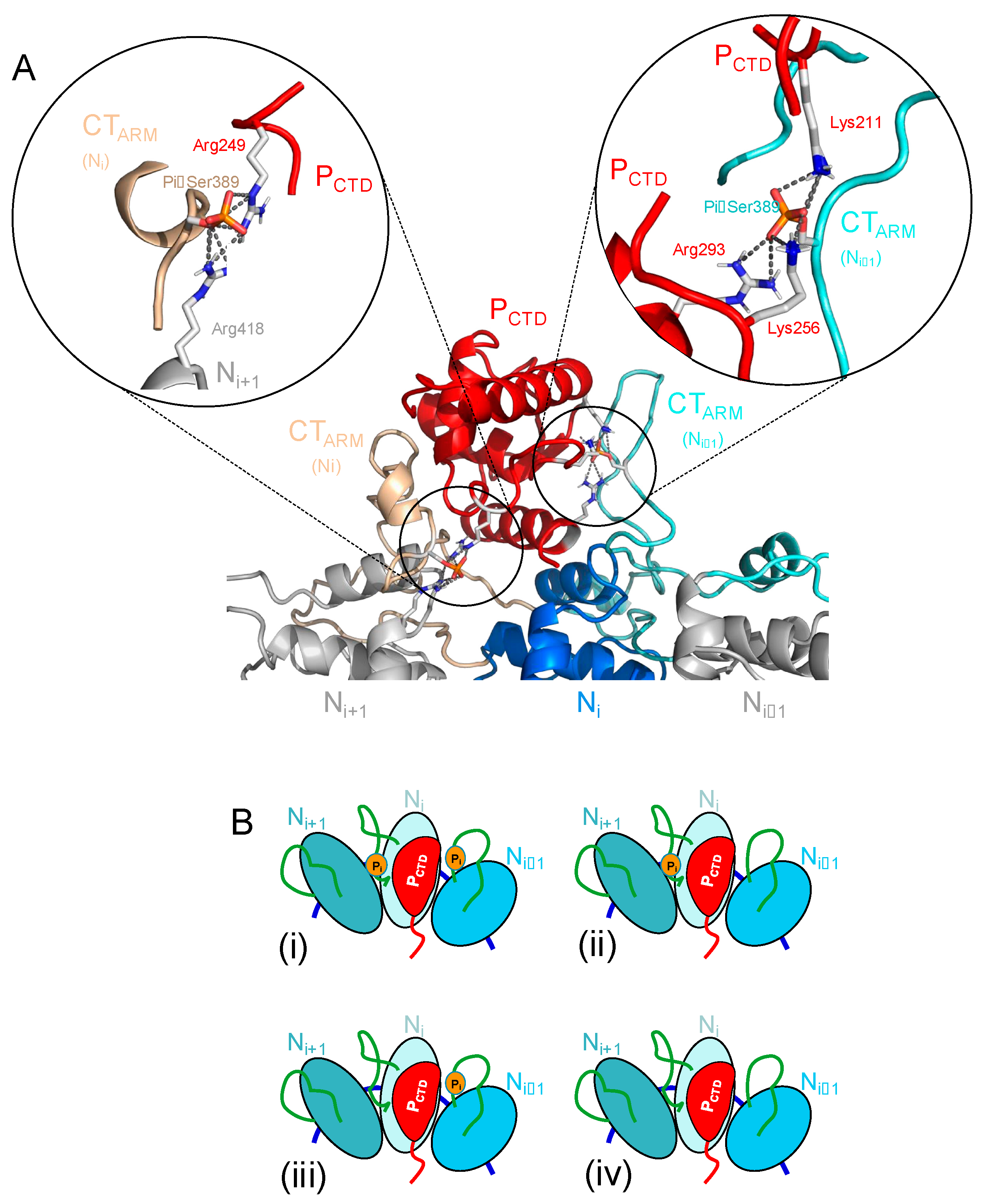
4.1. Phosphorylation of Ser389 in RABV N Enhances Binding Affinity for P CTD

We demonstrated here that the phosphorylation of Ser389 in RABV N significantly enhances the binding affinity of P, supporting previous observations [51] and rationalizing our previous structural model of P CTD bound to a polymeric N-RNA complex with phosphorylated N Ser389 [33]. In this model generated by flexible docking and MD simulations using SAXS constraints, the CTD was positioned on the top of the NCTD of the Ni protomer and was pinched between the flexible NCT-ARM of the same protomer and of the adjacent protomer (Ni−1) that molded around the CTD (Figure 8A). Several hydrophobic residues in the NCT-ARM of the Ni1 protomer filled a hydrophobic cavity (W-hole) on one face of P CTD, while acidic residues in the NCT-ARM of the Ni protomer interacted with the opposite basic face of P CTD [33]. In addition, when N Ser389 was phosphorylated in both Ni and Ni1 protomers, networks of salt bridges appeared in the MD simulations that suggested additional stability of the complex: Ni phosphoserine created new salt bridges with the side chains of P CTD Arg249 and of Ni+1 Arg418, while Ni−1 phosphoserine made a network of salt bridges with the side chains of P CTD Lys211, Lys256, and Arg293 (insets in Figure 8A). In agreement with this model, the partial phosphorylation of N leads us to consider four types of binding sites, with possibly different affinities depending on if one or two phosphoserines are involved in the binding site (Figure 8B): sites of high affinities with two adjacent N protomers phosphorylated on Ser389 (i), sites of intermediate stability with only the Ni (ii) or the Ni−1 (iii) protomer phosphorylated on Ser389, and sites of low affinity with two unphosphorylated N protomers (iv). These different types of binding sites could explain at least part of the complexity observed in the dissociation kinetics.
In RABV-infected cells, phosphorylated N was found in the polymeric N-RNA complex (NC), but not in the soluble, RNA-free N0 form associated with P [50]. The absence of the phosphorylation of N0 would thus prevent a strong intramolecular interaction between P CTD and N in the N0-P complex. Ser389 may not be accessible to casein kinase II in the N0-P complex [50], but in the polymeric N-RNA complex, the first half of this NCT-ARM (aa 350–375) becomes stabilized by docking onto the C-terminal domain of the adjacent Ni+1 subunit [16], possibly exposing the phosphorylation site and allowing kinase access to Ser389. The phosphorylation of N could thus play a role during the replicative cycle by controlling the interactions between P and the NC and therefore affect transcription and replication [45,46].

4.2. RABV P Dimerization Moderately Enhances the Affinity for Multimeric N-RNA Complexes

Our results show that the dimeric full-length RABV P protein (P2) binds to the purified recombinant circular N10-RNA complex and linear nucleocapsid about 15–30-fold better than the monomeric constructs (PΔ90–133 and PCTD), revealing an important effect of the dimerization for the association of P with the N-RNA complex (difference in binding energy of about 3 RT). The slight difference in affinity between PΔ90–133 and PCTD can result from the slow diffusion of PΔ90–133 (see above) or indicate the presence of a minor additional binding site in the N-terminal part of the protein (possibly the N-terminal region involved in the formation of the N0-P complex [27]). However, a two-fold difference in the dissociation constant contributes only a minor difference of 0.7 RT to the binding energy. The affinity enhancement with respect to monomeric fragments can rather be explained by an avidity effect.
The equilibrium binding isotherms support a model in which the monomeric fragments of P (PΔ90–133 and PCTD) bind to independent sites, but both SEC and SPR data show that only two CTD can bind to an N10-RNA complex [33]. The experimental methods (SPR, SEC) used here to monitor the association of P2 with the N-RNA arrays do not allow us to differentiate between dimers that are bound by one or two CTDs, and therefore only measure the total binding of individual P2 molecules. In contrast to a simple binding model where one monomeric ligand binds to one monomeric receptor, in our system, different CTD binding sites exist along the N-RNA array, and once one CTD is bound, there are still different sites available for binding the second CTD.
In such a model, if each CTD binds with the same microscopic affinity to any possible binding site of the N-RNA lattice, the enhancement of the apparent affinity for P2 can be explained if we consider that both CTDs of each P2 can bind to the N10-RNA circular complex [55]. If binding of the first CTD occurs with the same affinity (Ka = 80 nM) as isolated PΔ90–133 (or PCTD), the higher apparent affinity originates from the additional stability provided to the N10-RNA-P2 complex by the intramolecular association of the second CTD ( K b i = 0.03) (Figure 5). Although the isotherm could be reproduced using the same parameters for both P2 molecules (Kd,app = 5.5 ± 1.0 nM), a slightly better fit could be obtained by considering that the second P2 molecule binds with a lower affinity than the first (Figure 5). The effective concentration given by the product K a . K b i is 2.7 μM for the binding of the first P2 and 16.8 μM for the second. The slight reduction in apparent affinity (which we modeled by an increase in Ka, but could as well be modeled by changing K b i or both constants) could be explained by a reduction in the number of available binding sites once the first P2 molecule is bound as described for ligand binding on the lattice [56]. This reading of the data implies that four CTDs and possibly even more (we found that one or two additional P2 molecules bind to the same complex with micromolar affinities) bind to a single circular N10-RNA complex, as demonstrated by our SEC [33] and SPR data, but in contradiction with the results expected from the experiments with the monomeric fragments.
The magnitude of this affinity enhancement is, however, less than that expected for a bivalent ligand binding to multimeric independent sites. The connection by a flexible linker of two ligands that bind to independent sites has been exploited to increase the effective affinity, in particular of antibodies [57,58] or DNA binding proteins [59]. According to a model developed by Zhou, the effective concentration in such a system can be predicted by calculating the probability density of the end-to-end vector of the flexible linker (using a wormlike chain model) when the ligands are bound to their receptor [60,61]. In the complex formed with the N10-RNA circular complex, our structural model based on SAXS data predicted that the CTDs bind with their N-terminus oriented towards the inside of the ring and that the distance between the linker ends is of the order of 2.7 nm [33]. Considering distance and the length of linkers as the sum of the two flexible linkers connecting MD to CTD (aa 133–193) in P2, the effective concentration predicted by Zhou’s model is of the order of 1 mM [60,61]. According to this prediction, the intramolecular dissociation constant K b i should be 0.00008 and the first P2 molecule should bind with an apparent binding affinity ( K a · K b i ) of 6.4 pM. These predicted values contrast strikingly with those determined experimentally, probably reflecting steric hindrance and/or conformational constraints in P2 and indicating a strong energetic penalty for binding the second CTD. Thus, the dimerization of P only provides a moderate enhancement of apparent affinity for the N-RNA complexes as compared to the theoretical potential.
Why the P protein of MNV members is multimeric remains a matter of debate. It has been shown previously that a mutant of RABV P deleted from the central dimerization region was still active in transcription in a minireplicon assay [62], suggesting that the dimerization domain of RABV P is dispensable for RNA synthesis in vitro, at least for transcription. Similar results have been obtained for VSV [63,64]. In contrast, the RABV P dimerization domain was found to be essential for inducing liquid–liquid phase separation and for the formation of the membrane-less compartments where viral RNA synthesis takes place [35,65], indicating that this domain is essential for viral replication in a cellula.
There are stages in the viral cycle in which the formation of a stable complex between P and NC may be essential for the virus. First, for viral particle assembly, according to the cartwheeling model [42] and the structure of L [32], one P dimer is attached to each polymerase molecule, but a large number of P molecules are bound to the NC in the viral particles [66]. Although the exact role of these extra P molecules is unknown, the tight binding of CTD to phosphorylated NC would allow for recruiting P2 to the nascent NC and allow the assembly of a stable ribonucleoprotein complex, preceding its incorporation into a new viral particle. Second, for the initial stage of the viral cycle, when the ribonucleoprotein complex is released into the cytoplasm of infected cells, at that stage, the cellular concentration of soluble P is low or even non-existent. Tight binding of P2 to the viral NC because of P dimerization and N phosphorylation could ensure that P2 remains bound to the template during the early stages of the replication cycle in the infected cell and may also be used for initiating phase separation.

4.3. Interactions Between RABV P and Polymeric N-RNA Complexes

Interactions between multivalent ligands and linear lattices of multiple binding sites are important systems in biology but their binding behaviors can be complex, making a detailed analysis difficult [56]. In this case, we are studying the binding of bivalent RABV P either to a circular N-RNA lattice of 10 N subunits (N10-RNA complex), or to a mixture of a linear lattice (NC) containing an heterogeneous (and unknown) number of N subunits. From our previous model of the complex between P CTD and a circular N-RNA complex [33], we know that the binding site for P CTD straddled on two adjacent N protomers, suggesting that a 10 mer circular complex could accommodate up to five CTDs in this binding mode. Our RABV system is further complicated by the phosphorylation of only a fraction of the binding sites on N, which creates high-affinity sites for P CTD and potentially different types of sites of different affinities (Figure 8B). As previously observed for the binding of PCTD [33], both association and dissociation processes of P2 and PΔ90–133 to circular and linear N-RNA complexes exhibited complex kinetic behaviors when monitored by SPR that could not be fitted with simple models. This complexity may be explained by the “car parking” problem typical of multivalent ligand binding on a lattice of binding sites [56], and by the large dimensions of P due to its flexibility (see above), but also by the existence of binding sites with different affinities resulting from the partial phosphorylation of N (Figure 8B) or by mass transport effects.
The binding association occurs on a similar timescale for all forms of P, suggesting that the differences found in binding affinity are mainly explained by differences in off-rates. For all three proteins, the dissociation kinetics from the highest-affinity binding sites were too slow to be monitored, and koff values could not be determined. In the case of full-length P2, we hypothesized that two P dimers were bound to these high-affinity phosphorylated sites of the circular N10-RNA complex (complexes I and II in Figure 2A) and dissociated too slowly to be monitored (Figure 3). With CTD binding using two adjacent N protomers, the binding of two P2 occupies up to 8 N protomers, mainly protomers in which N Ser389 is phosphorylated. In contrast, the dissociation of one or two additional P2 molecules bound to low-affinity non-phosphorylated sites (complex III) gave rise to the observable part of the dissociation kinetics. The probably random distribution of phosphorylated N within circular and linear complexes (average: 70%) generates heterogeneity in the number and position of high-affinity binding sites, which must affect the shape of the dissociation kinetic curve.
The complexity of the binding association and dissociation kinetics may also reflect a mass transport limitation. In a situation where the association rate (kon) is of the same order of magnitude as the diffusion rate, a large amount of the dissociated ligand will rebind before diffusing away from the receptor, resulting in an apparent slow dissociation or an apparent absence of dissociation. The presence of long intrinsically disordered regions imposes a considerable viscous drag on the motion of the protein. The translational diffusion coefficient of RABV P2 (D value of 4.5 ± 0.1 × 10⁻⁷ cm2 s−1) previously measured by dynamic light scattering and analytical ultracentrifugation [24] was lower than expected for a protein of this molecular mass (a theoretical value of 6.3 × 10⁻⁷ cm2 s−1 can be calculated for a protein of 68 kDa from a plot of log D versus log MM of globular proteins) [67]. This suggests that the slow diffusion of P2 or PΔ90–133 molecules and their rebinding to high-affinity sites can slow down the dissociation from the complex formed with N-RNA and could, for example, explain the factor of two observed between the affinity of PCTD and PΔ90–133.
Although our results did not permit establishing a complete mechanism describing all of these interactions, they do shed new light on the effects of P dimerization and N phosphorylation on the interactions between P and polymeric N-RNA complexes.

4.4. Relevance of RABV P-N Interaction Dynamics for Polymerase Motion

The RNA-dependent RNA polymerase is a processive enzyme [68,69] that must remain attached to the N-RNA template while it progresses along it. As the P protein in all NNVs has been found to be multimeric and to bind both the polymerase and the N-RNA template, it is currently accepted on the basis of work primarily performed with the Sendai virus that the P multimer moves in a cartwheel fashion along the template [42,70]. In this process, the CTD of one P multimer continually makes and breaks contacts with successive N protomers, while the central multimeric domain undergoes a concerted rotational movement [42,70]. Although the exact mechanism has yet to be established, it is likely that the P multimers would cartwheel by being dragged along by the polymerase as the latter moves along its template, rather than being the motor moving the polymerase. P would then act as a processivity factor that keeps the polymerase attached to the template.
Every RABV N protomer binds nine nucleotides [16]; thereby, about 1300 copies of N are required to form an NC, which covers the entire genome (about 12,000 nt) [66]. NNV polymerases synthesize RNA at a rate that varies between one and five nucleotides per second, depending on the virus and the available results (VSV: 3.1–4.3 nt.s−1 [71], Sendai virus: 1.7 nt.s−1 [72], measles virus: 3 nt.s−1 [73]). In line with these data, we assume that it takes 2 to 5 s for the RABV polymerase to assemble 9 nt. In a model in which P cartwheels along the N-RNA template during RNA synthesis, each CTD should then dissociate from its binding site on the N-RNA complex and re-associate on the next binding site roughly every time that the polymerase copies 9 nt. This implies a koff value of the order of 0.2–0.5 s−1. The off-rate measured here for the dissociation of P from the dephosphorylated N-RNA template is of this order of magnitude (0.12 s−1), suggesting that the dynamics of interaction between P and low-affinity unphosphorylated sites is compatible with a mechanism in which P dimers progress with the L polymerase. In contrast, the off-rate for high-affinity sites in the phosphorylated N-RNA template was too low to be measured over the time of our experiments. We estimated an upper limit of 0.001 s−1 for the off-rate of CTD, which is more than two orders of magnitude slower than expected to keep pace with the polymerase.
This paradox raises questions about the mechanism of polymerase motion along the N-RNA template, as well as the function and timing of N phosphorylation. Based on similar arguments, we previously proposed an alternative model to the cartwheeling of P, in which P dimers would attach at regular intervals along the N-RNA template and the polymerase would progress by jumping from one P to the next [33]. Alternatively, P dimers could be tightly bound to the phosphorylated NC, and cartwheeling during RNA synthesis could be triggered and regulated by the dephosphorylation of the template on the passage of the polymerase. Although neither casein kinase nor any cellular phosphatase has been found in the RABV virion [74], different cellular kinases and phosphatases are involved in the regulation of the viral transcription of different NNVs [75,76,77], and this needs to be investigated for RABV. In conclusion, interactions between P and N-RNA polymeric complexes are a potential target for the development of new antiviral compounds, and we hope that our quantitative study of these interactions will pave the way for this approach.

Author Contributions

Conceptualization, E.d.A.R.J., F.C.A.G., C.L. and M.J.; methodology, E.d.A.R.J., F.C.A.G. and C.L.; writing—original draft preparation, E.d.A.R.J. and M.J.; writing—review and editing, E.d.A.R.J., F.C.A.G., C.L. and M.J.; supervision, M.J.; funding acquisition, M.J. All authors have read and agreed to the published version of the manuscript.

Funding

This work was supported by a grant from the French ANR [ANR-07-001-01(ANRAGE)]. E.d.A.R.J. was supported by postdoctoral fellowships from Joseph Fourier University and from the ANR program. F.C.A.G. and C.L. were supported by a fellowship from the French Ministère de l’Education Nationale, de la Recherche et de la Technologie.

Institutional Review Board Statement

Not applicable.

Informed Consent Statement

Not applicable.

Data Availability Statement

Data available upon request.

Acknowledgments

The authors thank Danièle Blondel (Gif-sur-Yvette, FR) for kindly providing samples of purified RABV NC.

Conflicts of Interest

The authors declare no conflicts of interest.

References

  1. Tarantola, A. Four Thousand Years of Concepts Relating to Rabies in Animals and Humans, Its Prevention and Its Cure. Trop. Med. Infect. Dis. 2017, 2, 5. [Google Scholar] [CrossRef] [PubMed]
  2. Muller, F.T.; Freuling, C.M. Rabies control in Europe: An overview of past, current and future strategies. Rev. Sci. Tech. 2018, 37, 409–419. [Google Scholar] [CrossRef] [PubMed]
  3. Fehlner-Gardiner, C. Rabies control in North America—Past, present and future. Rev. Sci. Tech. 2018, 37, 421–437. [Google Scholar] [CrossRef] [PubMed]
  4. Hampson, K.; Coudeville, L.; Lembo, T.; Sambo, M.; Kieffer, A.; Attlan, M.; Barrat, J.; Blanton, J.D.; Briggs, D.J.; Cleaveland, S.; et al. Estimating the global burden of endemic canine rabies. PLoS Negl. Trop. Dis. 2015, 9, e0003709. [Google Scholar]
  5. WHO Rabies—Fact Sheet. Available online: https://www.who.int/news-room/fact-sheets/detail/rabies (accessed on 15 July 2024).
  6. Knobel, D.L.; Cleaveland, S.; Coleman, P.G.; Fèvre, E.M.; Meltzer, M.I.; Miranda, M.E.; Shaw, A.; Zinnstag, J.; Meslin, F.X. Re-evaluating the burden of rabies in Africa and Asia. Bull. World Health Organ. 2005, 83, 360–368. [Google Scholar]
  7. Dietzschold, B.; Li, J.; Faber, M.; Schnell, M. Concepts in the pathogenesis of rabies. Future Virol. 2008, 3, 481–490. [Google Scholar] [CrossRef]
  8. Schnell, M.J.; McGettigan, J.P.; Wirblich, C.; Papaneri, A. The cell biology of rabies virus: Using stealth to reach the brain. Nat. Rev. Microbiol. 2010, 8, 51–61. [Google Scholar] [CrossRef]
  9. Fooks, A.R.; Cliquet, F.; Finke, S.; Freuling, C.; Hemachudha, T.; Mani, R.S.; Muller, T.; Nadin-Davis, S.; Picard-Meyer, E.; Wilde, H.; et al. Rabies. Nat. Rev. Dis. Primers 2017, 3, 17091. [Google Scholar] [CrossRef]
  10. Fisher, C.R.; Streicker, D.G.; Schnell, M.J. The spread and evolution of rabies virus: Conquering new frontiers. Nat. Rev. Microbiol. 2018, 16, 241–255. [Google Scholar] [CrossRef]
  11. Litchy, B.D.; Power, A.T.; Stojdl, D.F.; Bell, J.C. Vesicular stomatitis virus: Re-inventing the bullet. Trends Mol. Med. 2004, 10, 210–216. [Google Scholar]
  12. Basak, A.; Mondal, A.; Polley, S.; Mukhopadhyay, S.; Chattopadhyay, D. Reviewing Chandipura: A Vesiculovirus in Human Epidemics. Biosci. Rep. 2007, 27, 275–298. [Google Scholar] [CrossRef] [PubMed]
  13. Hogenhout, S.A.; Redinbaugh, M.G.; Ammar, E.-D. Plant and animal rhabdovirus host range: A bug’s view. Trends Microbiol. 2003, 11, 264–271. [Google Scholar] [CrossRef]
  14. Pringle, C.R. The order Mononegavirales. Arch. Virol. 1997, 142, 2321–2326. [Google Scholar]
  15. Kuhn, J.H.; Adkins, S.; Alioto, D.; Alkhovsky, S.V.; Amarasinghe, G.K.; Anthony, S.J.; Avsic-Zupanc, T.; Ayllon, M.A.; Bahl, J.; Balkema-Buschmann, A.; et al. 2020 taxonomic update for phylum Negarnaviricota (Riboviria: Orthornavirae), including the large orders Bunyavirales and Mononegavirales. Arch. Virol. 2020, 165, 3023–3072. [Google Scholar] [CrossRef]
  16. Albertini, A.A.; Wernimont, A.K.; Muziol, T.; Ravelli, R.B.; Clapier, C.R.; Schoehn, G.; Weissenhorn, W.; Ruigrok, R.W. Crystal structure of the rabies virus nucleoprotein-RNA complex. Science 2006, 313, 360–363. [Google Scholar] [CrossRef] [PubMed]
  17. Morin, B.; Liang, B.; Gardner, E.; Ross, R.A.; Whelan, S.P.J. An In Vitro RNA Synthesis Assay for Rabies Virus Defines Ribonucleoprotein Interactions Critical for Polymerase Activity. J. Virol. 2017, 91, e01508-16. [Google Scholar] [CrossRef]
  18. Gérard, F.C.A.; Ribeiro, E.A.J.; Leyrat, C.; Ivanov, I.; Blondel, D.; Longhi, S.; Ruigrok, R.W.; Jamin, M. Modular organization of rabies virus phosphoprotein. J. Mol. Biol. 2009, 388, 978–996. [Google Scholar] [CrossRef] [PubMed]
  19. Chenik, M.; Chebli, K.; Blondel, D. Translation initiation at alternate in-frame AUG codons in the rabies virus phosphoprotein mRNA Is mediated by a ribosomal leaky scanning mechanism. J. Virol. 1995, 69, 707–712. [Google Scholar] [CrossRef] [PubMed]
  20. Jespersen, N.E.; Leyrat, C.; Gerard, F.C.; Bourhis, J.M.; Blondel, D.; Jamin, M.; Barbar, E. The LC8-RavP ensemble Structure Evinces A Role for LC8 in Regulating Lyssavirus Polymerase Functionality. J. Mol. Biol. 2019, 431, 4959–4977. [Google Scholar] [CrossRef]
  21. Scrima, N.; Le Bars, R.; Nevers, Q.; Glon, D.; Chevreux, G.; Civas, A.; Blondel, D.; Lagaudriere-Gesbert, C.; Gaudin, Y. Rabies virus P protein binds to TBK1 and interferes with the formation of innate immunity-related liquid condensates. Cell Rep. 2023, 42, 111949. [Google Scholar] [CrossRef]
  22. Ito, N.; Moseley, G.W.; Sugiyama, M. The importance of immune evasion in the pathogenesis of rabies virus. J. Vet. Med. Sci. 2016, 78, 1089–1098. [Google Scholar] [CrossRef] [PubMed]
  23. Vidy, A.; Chelbi-Alix, M.; Blondel, D. Rabies virus P protein interacts with STAT1 and inhibits interferon signal transduction pathways. J. Virol. 2005, 79, 14411–14420. [Google Scholar] [CrossRef] [PubMed]
  24. Gérard, F.C.A.; Ribeiro, E.A.J.; Albertini, A.A.; Gutsche, I.; Zaccai, G.; Ruigrok, R.W.; Jamin, M. Unphosphorylated Rhabdoviridae phosphoproteins form elongated dimers in solution. Biochemistry 2007, 46, 10328–10338. [Google Scholar] [CrossRef]
  25. Ivanov, I.; Crepin, T.; Jamin, M.; Ruigrok, R.W. Structure of the dimerization domain of the rabies virus phosphoprotein. J. Virol. 2010, 84, 3707–3710. [Google Scholar] [CrossRef]
  26. Mavrakis, M.; McCarthy, A.A.; Roche, S.; Blondel, D.; Ruigrok, R.W. Structure and function of the C-terminal domain of the polymerase cofactor of rabies virus. J. Mol. Biol. 2004, 343, 819–831. [Google Scholar] [CrossRef]
  27. Gérard, F.C.A.; Bourhis, J.M.; Mas, C.; Branchard, A.; Vu, D.D.; Varhoshkova, S.; Leyrat, C.; Jamin, M. Structure and Dynamics of the Unassembled Nucleoprotein of Rabies Virus in Complex with Its Phosphoprotein Chaperone Module. Viruses 2022, 14, 2813. [Google Scholar] [CrossRef]
  28. Mavrakis, M.; Iseni, F.; Mazza, C.; Schoehn, G.; Ebel, C.; Gentzel, M.; Franz, T.; Ruigrok, R.W. Isolation and characterisation of the rabies virus N degrees-P complex produced in insect cells. Virology 2003, 305, 406–414. [Google Scholar] [CrossRef]
  29. Howard, M.; Wertz, G. Vesicular Stomatitis Virus RNA Replication: A Role for the NS Protein. J. Gen. Virol. 1989, 70, 2683–2694. [Google Scholar] [CrossRef] [PubMed]
  30. Masters, P.S.; Banerjee, A.K. Complex Formation with Vesicular Stomatitis Virus Phosphoprotein NS prevents Binding of Nucleocapsid Protein N to Nonspecific RNA. J. Virol. 1988, 62, 2658–2664. [Google Scholar] [CrossRef]
  31. Mavrakis, M.; Mehouas, S.; Real, E.; Iseni, F.; Blondel, D.; Tordo, N.; Ruigrok, R.W. Rabies virus chaperone: Identification of the phosphoprotein peptide that keeps nucleoprotein soluble and free from non-specific RNA. Virology 2006, 349, 422–429. [Google Scholar] [CrossRef]
  32. Horwitz, J.A.; Jenni, S.; Harrison, S.C.; Whelan, S.P.J. Structure of a rabies virus polymerase complex from electron cryo-microscopy. Proc. Natl. Acad. Sci. USA 2020, 117, 2099–2107. [Google Scholar] [CrossRef] [PubMed]
  33. Ribeiro, E.A.J.; Leyrat, C.; Gerard, F.C.; Albertini, A.A.; Falk, C.; Ruigrok, R.W.; Jamin, M. Binding of rabies virus polymerase cofactor to recombinant circular nucleoprotein-RNA complexes. J. Mol. Biol. 2009, 394, 558–575. [Google Scholar] [CrossRef] [PubMed]
  34. Zhan, J.; Watts, E.; Brice, A.M.; Metcalfe, R.D.; Rozario, A.M.; Sethi, A.; Yan, F.; Bell, T.D.M.; Griffin, M.D.W.; Moseley, G.W.; et al. Molecular Basis of Functional Effects of Phosphorylation of the C-Terminal Domain of the Rabies Virus P Protein. J. Virol. 2022, 96, e00111-22. [Google Scholar] [CrossRef]
  35. Nikolic, J.; Le Bars, R.; Lama, Z.; Scrima, N.; Lagaudriere-Gesbert, C.; Gaudin, Y.; Blondel, D. Negri bodies are viral factories with properties of liquid organelles. Nat. Commun. 2017, 8, 58. [Google Scholar] [CrossRef]
  36. Lahaye, X.; Vidy, A.; Pomier, C.; Obiang, L.; Harper, F.; Gaudin, Y.; Blondel, D. Functional Characterization of Negri Bodies (NBs) in Rabies Virus-Infected Cells: Evidence that NBs Are Sites of Viral Transcription and Replication. J. Virol. 2009, 83, 7948–7958. [Google Scholar] [CrossRef]
  37. Ding, H.; Green, T.J.; Lu, S.; Luo, M. Crystal structure of the oligomerization domain of the phosphoprotein of vesicular stomatitis virus. J. Virol. 2006, 80, 2808–2814. [Google Scholar] [CrossRef]
  38. Jensen, M.R.; Yabukarski, F.; Communie, G.; Condamine, E.; Mas, C.; Volchkova, V.; Tarbouriech, N.; Bourhis, J.M.; Volchkov, V.; Blackledge, M.; et al. Structural Description of the Nipah Virus Phosphoprotein and Its Interaction with STAT1. Biophys. J. 2020, 118, 2470–2488. [Google Scholar] [CrossRef] [PubMed]
  39. Tarbouriech, N.; Curran, J.; Ebel, C.; Ruigrok, R.W.; Burmeister, W.P. On the domain structure and the polymerization state of the sendai virus P protein. Virology 2000, 266, 99–109. [Google Scholar] [CrossRef]
  40. Bruhn, J.F.; Kirchdoerfer, R.N.; Urata, S.M.; Li, S.; Tickle, I.J.; Bricogne, G.; Saphire, E.O. Crystal Structure of the Marburg Virus VP35 Oligomerization Domain. J. Virol. 2017, 91, e01085-16. [Google Scholar] [CrossRef]
  41. Leyrat, C.; Renner, M.; Harlos, K.; Grimes, J.M. Solution and crystallographic structures of the central region of the phosphoprotein from human metapneumovirus. PLoS ONE 2013, 8, e80371. [Google Scholar] [CrossRef]
  42. Curran, J. Reexamination of the Sendai virus P protein domains required for RNA synthesis: A possible supplemental role for the P protein. Virology 1996, 221, 130–140. [Google Scholar] [CrossRef]
  43. Bourhis, J.M.; Yabukarski, F.; Communie, G.; Schneider, R.; Volchkova, V.A.; Freneat, M.; Gerard, F.C.; Ducournau, C.; Mas, C.; Tarbouriech, N.; et al. Structural Dynamics of the C-terminal X Domain of Nipah and Hendra Viruses Controls the Attachment to the C-terminal Tail of the Nucleocapsid Protein. J. Mol. Biol. 2022, 434, 167551. [Google Scholar] [CrossRef] [PubMed]
  44. Sokol, F.; Clark, H.F. Phosphoproteins, structural components of rhabdoviruses. Virology 1973, 52, 246–263. [Google Scholar] [CrossRef]
  45. Yang, J.; Koprowski, H.; Dietzschold, B.; Fu, Z.F. Phosphorylation of rabies virus nucleoprotein regulates viral RNA transcription and replication by modulating leader RNA encapsidation. J. Virol. 1999, 73, 1661–1664. [Google Scholar] [CrossRef]
  46. Wu, X.; Gong, X.; Foley, H.D.; Schnell, M.J.; Fu, Z.F. Both viral transcription and replication are reduced when the rabies virus nucleoprotein is not phosphorylated. J. Virol. 2002, 76, 4153–4161. [Google Scholar] [CrossRef] [PubMed]
  47. Préhaud, C.; Harris, R.D.; Fulop, V.; Koh, C.; Wong, J.; Flamand, A.; Bishop, D.H.L. Expression, characterization, and purification of a phosphorylated rabies nucleoprotein synthesized in insect cells by baculovirus vectors. Virology 1990, 178, 486–497. [Google Scholar] [CrossRef]
  48. Dietzschold, B.; Lafon, M.; Wang, H.; Otvos, L., Jr.; Celis, E.; Wunner, W.H.; Koprowski, H. Localization and immunological characterization of antigenic domains of the rabies virus internal N and NS proteins. Virus Res. 1987, 8, 103–125. [Google Scholar] [CrossRef]
  49. Wu, X.; Lei, X.; Fu, Z.F. Rabies virus nucleoprotein is phosphorylated by cellular casein kinase II. Biochem. Biophys. Res. Commun. 2003, 304, 333–338. [Google Scholar] [CrossRef]
  50. Kawai, A.; Toriumi, H.; Tochikura, T.S.; Takahashi, T.; Honda, Y.; Morimoto, K. Nucleocapsid formation and/or subsequent conformational change of rabies virus nucleoprotein (N) is a prerequisite step for acquiring the phosphatase-sensitive epitope of monoclonal antibody 5-2-26. Virology 1999, 263, 395–407. [Google Scholar] [CrossRef] [PubMed]
  51. Toriumi, H.; Kawai, A. Association of rabies virus nominal phosphoprotein (P) with viral nucleocapsid (NC) is enhanced by phosphorylation of the viral nucleoprotein (N). Microbiol. Immunol. 2004, 48, 399–409. [Google Scholar] [CrossRef]
  52. Leyrat, C.; Schneider, R.; Ribeiro, E.A., Jr.; Yabukarski, F.; Yao, M.; Gerard, F.C.; Jensen, M.R.; Ruigrok, R.W.; Blackledge, M.; Jamin, M. Ensemble structure of the modular and flexible full-length vesicular stomatitis virus phosphoprotein. J. Mol. Biol. 2012, 423, 182–197. [Google Scholar] [CrossRef]
  53. Edelhoch, H. Spectroscopic determination of tryptophan and tyrosine in proteins. Biochemistry 1967, 6, 1948–1954. [Google Scholar] [CrossRef] [PubMed]
  54. Albertini, A.A.; Clapier, C.R.; Wernimont, A.K.; Schoehn, G.; Weissenhorn, W.; Ruigrok, R.W. Isolation and crystallization of a unique size category of recombinant Rabies virus Nucleoprotein-RNA rings. J. Struct. Biol. 2007, 158, 129–133. [Google Scholar] [CrossRef] [PubMed]
  55. Jencks, W.P. On the attribution and additivity of binding energies. Proc. Natl. Acad. Sci. USA 1991, 78, 4046–4050. [Google Scholar] [CrossRef]
  56. McGhee, J.D.; von Hippel, P.H. Theoretical aspects of DNA-Protein Interactions: Co-operative and non-co-operative binding of large ligands to a one-dimensional homogeneous lattice. J. Mol. Biol. 1974, 86, 469–489. [Google Scholar] [CrossRef]
  57. Neri, D.; Momo, M.; Prospero, T.; Winter, G. High-affinity Antigen Binding by Chelating Recombinant Antibodies (CRAbs). J. Mol. Biol. 1995, 246, 367–373. [Google Scholar] [CrossRef]
  58. Crothers, D.M.; Metzger, H. The influence of polyvalency on the binding properties of antibodies. Immunochemistry 1972, 9, 341–357. [Google Scholar] [CrossRef]
  59. Klemm, J.D.; Pabo, C.O. Oct-1 POU domain-DNA interactions: Cooperative binding of isolated subdomams and effects of covalent linkage. Genes Dev. 1996, 10, 27–36. [Google Scholar] [CrossRef]
  60. Zhou, H.-X. Loops in Proteins Can Be Modeled as Worm-Like Chains. J. Phys. Chem. B 2001, 105, 6763–6766. [Google Scholar] [CrossRef]
  61. Zhou, H.-X. Quantitative Account of the Enhanced Affinity of Two Linked scFvs Specific for Different Epitopes on the Same Antigen. J. Mol. Biol. 2003, 329, 1–8. [Google Scholar] [CrossRef] [PubMed]
  62. Jacob, Y.; Real, E.; Tordo, N. Functional interaction map of lyssavirus phosphoprotein: Identification of the minimal transcription domains. J. Virol. 2001, 75, 9613–9622. [Google Scholar] [CrossRef] [PubMed]
  63. Gérard, F.C.A.; Jamin, M.; Blackledge, M.; Blondel, D.; Bourhis, J.M. Vesicular Stomatitis Virus Phosphoprotein Dimerization Domain Is Dispensable for Virus Growth. J. Virol. 2020, 94, e01789-19. [Google Scholar] [CrossRef] [PubMed]
  64. Bloyet, L.M.; Morin, B.; Brusic, V.; Gardner, E.; Ross, R.A.; Vadakkan, T.; Kirchhausen, T.; Whelan, S.P.J. Oligomerization of the Vesicular Stomatitis Virus Phosphoprotein Is Dispensable for mRNA Synthesis but Facilitates RNA Replication. J. Virol. 2020, 94, e00115-20. [Google Scholar] [CrossRef]
  65. Nevers, Q.; Scrima, N.; Glon, D.; Le Bars, R.; Decombe, A.; Garnier, N.; Ouldali, M.; Lagaudriere-Gesbert, C.; Blondel, D.; Albertini, A.; et al. Properties of rabies virus phosphoprotein and nucleoprotein biocondensates formed in vitro and in cellulo. PLoS Pathog. 2022, 18, e1011022. [Google Scholar] [CrossRef]
  66. Thomas, D.; Newcomb, W.W.; Brown, J.C.; Wall, J.S.; Hainfeld, J.F.; Trus, B.L.; Steven, A.C. Mass and molecular composition of vesicular stomatitis virus: A scanning transmission electron microscopy analysis. J. Virol. 1985, 54, 598–607. [Google Scholar] [CrossRef]
  67. Zaccai, N.; Serdyuk, I.N.; Zaccai, G. Methods in Molecular Biophysics—Structure, Dynamics, Function for Biology and Medicine, 2nd ed.; Cambridge University Press: Cambridge, UK, 2017. [Google Scholar]
  68. Stillman, E.A.; Whitt, M. Transcript initiation and 5’-end modifications are separable events during vesicular stomatitis virus transcription. J. Virol. 1999, 73, 7199–7209. [Google Scholar] [CrossRef]
  69. Morin, B.; Rahmeh, A.A.; Whelan, S.P. Mechanism of RNA synthesis initiation by the vesicular stomatitis virus polymerase. Embo J. 2012, 31, 1320–1329. [Google Scholar] [CrossRef]
  70. Kolakofsky, D.; Le Mercier, P.; Iseni, F.; Garcin, D. Viral RNA polymerase scanning and the gymnastics of Sendai virus RNA synthesis. Virology 2004, 318, 463–473. [Google Scholar] [CrossRef]
  71. Barr, J.N.; Whelan, S.P.; Wertz, G.W. Transcriptional control of the RNA-dependent RNA polymerase of vesicular stomatitis virus. Biochim. Biophys. Acta 2002, 1577, 337–353. [Google Scholar] [CrossRef]
  72. Gubbay, O.; Curran, J.; Kolakofsky, D. Sendai virus genome synthesis and assembly are coupled: A possible mechanism to promote viral RNA polymerase processivity. J. Gen. Virol. 2001, 82 Pt 12, 2895–2903. [Google Scholar] [CrossRef]
  73. Plumet, S.; Duprex, W.P.; Gerlier, D. Dynamics of viral RNA synthesis during measles virus infection. J. Virol. 2005, 79, 6900–6908. [Google Scholar] [CrossRef] [PubMed]
  74. Zhang, Y.; Wang, Y.; Feng, Y.; Tu, Z.; Lou, Z.; Tu, C. Proteomic Profiling of Purified Rabies Virus Particles. Virol. Sin. 2020, 35, 143–155. [Google Scholar] [CrossRef] [PubMed]
  75. Richard, C.A.; Rincheval, V.; Lassoued, S.; Fix, J.; Cardone, C.; Esneau, C.; Nekhai, S.; Galloux, M.; Rameix-Welti, M.A.; Sizun, C.; et al. RSV hijacks cellular protein phosphatase 1 to regulate M2-1 phosphorylation and viral transcription. PLoS Pathog. 2018, 14, e1006920. [Google Scholar] [CrossRef] [PubMed]
  76. Huang, M.; Sato, H.; Hagiwara, K.; Watanabe, A.; Sugai, A.; Ikeda, F.; Kozuka-Hata, H.; Oyama, M.; Yoneda, M.; Kai, C. Determination of a phosphorylation site in Nipah virus nucleoprotein and its involvement in virus transcription. J. Gen. Virol. 2011, 92 Pt 9, 2133–2141. [Google Scholar] [CrossRef]
  77. Kruse, T.; Biedenkopf, N.; Thrane Hertz, E.P.; Dietzel, E.; Stalmann, G.; Lopez-Mendez, B.; Davey, N.E.; Nilsson, J.; Becker, S. The Ebola Virus Nucleoprotein Recruits the Host PP2A-B56 Phosphatase to Activate Transcriptional Support Activity of VP30. Mol. Cell 2018, 69, 136–145. [Google Scholar] [CrossRef]
Figure 1. The RNA synthesis ribonucleoprotein complex of RABV. (A) A schematic representation of the RABV RNA synthesis ribonucleoprotein complex in replication mode. The nucleoprotein globular domains (NNTD and NCTD) are shown in two shades of blue. The NNT-ARM and NCT-ARM are shown in magenta and cyan, respectively. The RNA is shown in green. The L polymerase is shown in gray, while dimeric P is shown in gray with its PMD in orange, and PCTD in red. P is attached to N0 through the first part of its PNTR (shown in gold) and to NC through its C-terminal domain (PCTD). P is also attached to L through the second part of its PNTR (shown in pink). (B) A structural model of RABV P showing its folded domains (PMD in orange and PCTD in red in cartoon representation). The binding regions for N0, L, and LC8 in its intrinsically disordered regions are shown as lines. The model is a conformer taken from an ensemble model reproducing SAXS data [20]. (C) The architecture of RABV P constructs used in this study showing the long N-terminal intrinsically disordered region (PNTR), the multimerization domain (PMD, in orange), and the NC-binding domain (PCTD, in red) connected by a short flexible linker (PLINK). Boxes represent folded domains of known structure and lines represent intrinsically disordered regions. Functional modules are colored and indicated by arrows. (D) The structure of the N0-P complex. The cartoon representation of the crystal structure of RABV N0-P with N in blue and the N-terminal chaperone module of PNTR in gold [27]. (E) The architecture of RABV N. The cartoon representation and schematic representation of a protomer of N extracted from the crystal structure of the circular N11-RNA complex [16] showing the two globular domains, NNTD and NCTD, and the two subdomains, NNT-ARM (in magenta) and NCT-ARM (in cyan), which exchange with adjacent subunits in polymeric N. (F) The N-RNA-PCTD complex. The cartoon representation of the model of P CTD bound to the polymeric N-RNA complex showing the NCT-ARM of the adjacent Ni and Ni−1 protomer molding around P CTD [33].
Viruses 16 01735 g001
Figure 2. The existence of binding sites of different affinities revealed by the titration of the circular N10-RNA complex by full-length P2. (A) Electrophoresis in native gel. The different circular Nn-RNA species were separated by native gel electrophoresis (4%). Lane 1 is a mix of different Nn-RNA rings, where the number of protomers of N in each ring is indicated as a subscript. Lane 2 was loaded with 10 μM of P and lane 3 with 2 μM of purified N10-RNA. [P2] and [N10-RNA] at different ratios were mixed and incubated during 10 min at room temperature and loaded in lanes 4 to 11. In lane 4, [Pdimer]/[N10-RNA], is 0.16, in lane 5 is 0.33, in lane 6 is 0.66, in lane 7 is 0.83, in lane 8 is 1.00, in lane 9 is 1.3, in lane 10 is 1.66, and in lane 11 is 2.00. (B) SDS-PAGE of the nucleoprotein before and after phosphatase treatment. The RABV circular N10-RNA complex purified from insect cells was treated with calf intestine alkaline phosphatase (CIAP). The reaction mixtures were denatured and applied to 10% SDS-PAGE, followed by Coomassie staining. The first lane (MW) contains a molecular weight marker. (C) The Western blot. The proteins separated by SDS-PAGE were blotted onto a nitrocellulose filter. The membrane was incubated with the anti-phosphoserine antibody conjugated to biotin and revealed by a treatment with a streptavidin–horseradish peroxidase conjugate. (D) Mass spectrometry fingerprints. The chromatographic profiles show the intensity of the signal for the unphosphorylated (upper trace) and phosphorylated forms of the peptide (lower trace) after treatment with the alkaline phosphatase (+AP) or without treatment (−AP).
Viruses 16 01735 g002
Figure 3. Interactions between the full-length P dimer (P2) and circular N10-RNA complex by surface plasmon resonance. In each graph, the arrow indicates the initiation of dissociation by flowing the buffer over the surface. The concentration of P2 is shown above the lines and the double arrows on the right indicate the amplitude of dissociation processes whose kinetics may or may not be measurable. (A) The sensorgram (response in resonance units (R.U.) versus time) of the binding of RABV P2 to the immobilized N10-RNA complex. The N10-RNA complex was immobilized on a CM5 sensor chip as described in the Section 2. The full-length P dimer (P2) was injected over the immobilized N10-RNA complex at different concentrations in the 20 mM Tris-HCl buffer at pH 7.5 containing 150 mM NaCl, 1 mM DTT, and 0.005% (v/v) Tween20 at 20 °C and at a flow rate of 20 µL·min−1. The specific binding signals were obtained after the subtraction of a background signal measured in a reference channel with no bound N10-RNA complex. (B) The sensorgram of binding full-length P2 to the N10-RNA complex purified from insect cells. Full-length P2 was injected at a concentration of 500 nM. Response was normalized at a signal of 100 R.U. at equilibrium. (C) The sensorgram of binding full-length P2 to dephosphorylated N10-RNA after treatment with alkaline phosphatase (CIAP). (D) The sensorgram of binding the full-length P dimer to re-phosphorylated N10-RNA after treatment with casein kinase II (CKII).
Viruses 16 01735 g003
Figure 4. The equilibrium SPR analysis using multiple injections. The N10-RNA complex was immobilized on a CM5 sensor chip as described in the Section 2. Full-length P2 was injected at a concentration of 50 nM in the 20 mM Tris-HCl buffer at pH 7.5 containing 150 mM NaCl, 1 mM DTT, and 0.005% (v/v) Tween20 at 20 °C and at a flow rate of 20 µL·min−1. Successive injections of the ligand are shown by arrows. Equilibrium was reached after approximately 25 min and the signal remained unchanged during subsequent injections.
Viruses 16 01735 g004
Figure 5. Equilibrium binding interactions with circular N10-RNA complexes. (a) Binding isotherms of full-length P2 (circles), PCTD (diamonds), and PΔ90–133 (squares). The SPR signal at equilibrium is shown as a function of ligand (L) concentration. Open symbols are in the presence of 150 mM NaCl and closed symbols are in the presence of 500 mM NaCl. The dotted lines show the fit for independent-site binding. The solid line shows the fit to binding model 3 using two binding terms and the dashed lines show the fit using three binding terms, assuming that two additional P2 bind to low-affinity sites. (b) Normalized binding isotherms. The data of panel (a) below 1 μM were normalized to a saturation value of 100 R.U. The symbols are the same as in panel a. The dotted lines show the fits to an independent site, and the solid line shows the fit to model 3 for the first two P2 molecules. (c) Hill plots. The symbols are the same as in panel (a). The dotted lines show the fits to an independent site, and the solid line shows the fit to model 3 for the first two P2 molecules.
Viruses 16 01735 g005
Figure 6. Equilibrium binding interactions with linear NCs. (a) Binding isotherms of full-length P2 (circles), PCTD (diamonds), and PΔ91–131 (squares). The SPR signal at equilibrium is shown as a function of ligand (L) concentration. The dotted lines show the fit for binding two ligands with two independent-site bindings using the same dissociation constant for both. The solid lines show the fit for binding two ligands with two independent-site bindings using different dissociation constants. (b) Normalized binding isotherms. The data of panel (b) were normalized to a saturation value of 100 R.U. The symbols are the same as in panel (a). The lines show the fits to an independent site using different binding constants. (c) Hill plots. The symbols are the same as in panel (a). The lines show the fits to an independent site.
Viruses 16 01735 g006
Figure 7. Equilibrium binding interactions with the dephosphorylated circular N10-RNA complex and linear NCs. The N10-RNA complexes or the NC were first bound to the SPR chip and then were treated with CIAP. The SPR signal at equilibrium is shown as a function of ligand (L) concentration. (a) Normalized binding isotherms of full-length P2 to circular N10-RNA complexes. The empty circles show the data obtained with the phosphorylated N10-RNA complex taken from Figure 5b, together with their fit (shown as a line). The empty triangles show the data obtained with the dephosphorylated N10-RNA complex and the line shows the fit including 5% of P2 bound to the remaining high-affinity phosphorylated sites. (b) Normalized binding isotherms of full-length P2 to linear NC. The filled circles show the data obtained with the phosphorylated linear NC taken from Figure 5b, together with their fit (shown as a line). The filled triangles show the data obtained with dephosphorylated linear NC and the line shows the fit including 5% of P2 bound to the remaining high-affinity phosphorylated sites. The empty triangles and the dotted lines show the data and their fit obtained with dephosphorylated N10-RNA complexes taken from panel (a) as comparison.
Viruses 16 01735 g007
Figure 8. A structural model of the phosphorylated sites in RABV N-RNA. (A) A structural model of CTD in complex with the Ser389-phosphorylated N-RNA complex. The model was generated by computational modeling using SAXS data [33]. P CTD and the core of three adjacent N protomers are shown in the cartoon representation. Ser389 is phosphorylated in the Ni and Ni−1 protomers, and the residues that were found to form salt bridges (gray dotted lines) with the phosphate group during the MD simulations are shown in the insets and are labeled. (B) Schematic representations of one CTD bound to the Ni protomer in the N-RNA complex that show the four types of possible binding sites depending on the phosphorylation of Ser389 in the Ni and Ni−1 protomers. (i) Ser389 in the NCT-ARM of the Ni and Ni-1 protomers are phosphorylated. (ii) Only Ser389 the NCT-ARM of the Ni protomer is phosphorylated. (iii) Ser389 in the NCT-ARM of the Ni-1 protomer is phosphorylated. (iv) Ser389 in neither the Ni or Ni-1 protomer is phosphorylated.
Viruses 16 01735 g008
Disclaimer/Publisher’s Note: The statements, opinions and data contained in all publications are solely those of the individual author(s) and contributor(s) and not of MDPI and/or the editor(s). MDPI and/or the editor(s) disclaim responsibility for any injury to people or property resulting from any ideas, methods, instructions or products referred to in the content.

