Communication

Assessment of Survival Kinetics for Emergent Highly Pathogenic Clade 2.3.4.4 H5Nx Avian Influenza Viruses

1
Virology Department, Animal and Plant Health Agency (APHA-Weybridge), Woodham Lane, Addlestone, Surrey KT15 3NB, UK
2
Department of Epidemiological Sciences, Animal and Plant Health Agency, Sutton Bonington, Loughborough LE12 5RB, UK
3
Office of the Chief Veterinary Officer (OCVO), Welsh Government, Cathays Park, Cardiff CF10 3NQ, UK
4
Veterinary Advice Services, Animal and Plant Health Agency (APHA-Weybridge), Woodham Lane, Addlestone, Surrey KT15 3NB, UK
5
WOAH/FAO International Reference Laboratory for Avian Influenza, Animal and Plant Health Agency, (APHA-Weybridge), Woodham Lane, Addlestone, Surrey KT15 3NB, UK
*
Authors to whom correspondence should be addressed.
Viruses 2024, 16(6), 889; https://doi.org/10.3390/v16060889
Submission received: 5 April 2024 / Revised: 24 May 2024 / Accepted: 27 May 2024 / Published: 31 May 2024
(This article belongs to the Special Issue Evolution and Adaptation of Avian Viruses)

Abstract

:
High pathogenicity avian influenza viruses (HPAIVs) cause high morbidity and mortality in poultry species. HPAIV prevalence means high numbers of infected wild birds could lead to spill over events for farmed poultry. How these pathogens survive in the environment is important for disease maintenance and potential dissemination. We evaluated the temperature-associated survival kinetics for five clade 2.3.4.4 H5Nx HPAIVs (UK field strains between 2014 and 2021) incubated at up to three temperatures for up to ten weeks. The selected temperatures represented northern European winter (4 °C) and summer (20 °C); and a southern European summer temperature (30 °C). For each clade 2.3.4.4 HPAIV, the time in days to reduce the viral infectivity by 90% at temperature T was established (DT), showing that a lower incubation temperature prolonged virus survival (stability), where DT ranged from days to weeks. The fastest loss of viral infectivity was observed at 30 °C. Extrapolation of the graphical DT plots to the x-axis intercept provided the corresponding time to extinction for viral decay. Statistical tests of the difference between the DT values and extinction times of each clade 2.3.4.4 strain at each temperature indicated that the majority displayed different survival kinetics from the other strains at 4 °C and 20 °C.

1. Introduction

Avian influenza viruses (AIVs) are classified into subtypes according to the external viral glycoproteins, namely the haemagglutinin (HA; H1-H16) and neuraminidase (NA; N1-N9) [1]. The H5 and H7 AIV subtypes are notifiable avian disease agents [2,3], and can mutate from the low pathogenicity (LP)AIV to the corresponding high pathogenicity (HP)AIV, with the latter causing high morbidity and mortality in poultry. Wild birds are the natural reservoir for AIVs [4], with migration enabling the maintenance and dissemination of a diverse array of AIV subtypes, including clade 2.3.4.4 HPAIVs of the “goose/Guangdong” (GsGd) H5 HPAIV lineage that have been decimating avian populations globally in recent years. The GsGd lineage first emerged in 1996 with subsequent evolution through diverse clades [5,6,7,8], followed by intercontinental spread via wild birds [9,10]. Clade 2.3.4.4 epizootics have impacted poultry production systems, local economies, and international trade [11].
Since 2014, multiple H5Nx HPAIV clade 2.3.4.4 incursions have occurred in Europe, with outbreaks increasing in frequency since 2020 [12,13,14]. The H5N8 subtype dominated the winter 2020–2021 epizootic and persisted into summer 2021, before significant re-emergence during autumn 2021 as the unique H5N1 subtype, albeit as multiple genotypes, for the ensuing winter [14]. This clade 2.3.4.4b H5N1 HPAIV persisted through summer 2022 [15], followed by numerous poultry outbreaks and wild bird cases during winter 2022–2023 [16]. Since 2020, these European clade 2.3.4.4b H5Nx epizootics have demonstrated that viruses circulating in wild birds represent the greatest threat to the poultry sector [13,14].
AIV survival in different environments is a critical component of understanding incursion risk to different poultry sectors [17]. The survival of infectious AIVs has been demonstrated in water, soil, and faeces [18,19,20,21], and in different egg products [22]. Multiple factors contribute to AIV survival metrics [23], hence comparisons in distilled water and saline [24,25], plus the effects of pH and temperature have featured in assessments of viral decay [26,27]. In this study, we investigated the survival kinetics for five clade 2.3.4.4 H5Nx HPAIVs linked to UK incursions which were incubated at up to three temperatures.

2. Materials and Methods

2.1. Viruses and Safety Statement

Five H5Nx clade 2.3.4.4 HPAIV isolates derived from outbreak events in the United Kingdom (UK) were investigated in this study (Table 1) as representatives of viruses which caused disease events during five UK/European clade 2.3.4.4 epizootic seasons between 2014 and 2021. Viruses were propagated in 9- to 11-day-old specified pathogen-free embryonated fowls’ eggs (SPF EFEs) [28]. The H5Nx clade 2.3.4.4 HPAIVs are categorised in the UK by the Specified Animal Pathogens Order at level 4 and the Advisory Committee on Dangerous Pathogens at level 3, hence all laboratory work was carried out in licensed biosafety level 3 laboratories [29].

2.2. Cell Lines

Madin-Darby canine kidney (MDCK) cells, supplied originally from the European Collection of Cell Cultures (ECACC, Genova, Italy), were propagated at 37 °C using Eagle’s minimal essential medium (EMEM) with 10% (v/v) foetal bovine serum (FBS), which contained antibiotics (penicillin 100 IU/mL and streptomycin 100µg/mL; Life Technologies Ltd., Carlsbad, CA, USA), in a 5% (v/v) CO2 atmosphere.

2.3. Preparation of Replicate Samples for Infectivity Assessment at Different Temperatures

Each virus stock was diluted in serum-free medium to prepare a working concentration at a 50% tissue culture infectious dose (TCID50) of 1 × 10⁶ TCID50/mL (Table 1). At time zero, three replicate samples (1.2 mL) were prepared, using sterile safe lock Eppendorf tubes (Anachem, Clonakilty, Ireland), for each investigated timepoint at a given temperature (detailed below). An additional eight to 10 aliquots of the working concentration of each virus were prepared and frozen promptly, to serve as time zero positive controls representative of no incubation treatment.
All five H5Nx isolates were incubated at 4 °C (±2 °C), and 20 °C (±2 °C), with three incubated at a higher temperature 30 °C (±2 °C), using an LR902 refrigerator (LEC Refrigeration plc., Prescot, UK) or a 305NP heat-cooled incubator (LMS Ltd., Sevenoaks, UK). Both instruments featured independent temperature monitoring (Traceable Wireless Sensor, Tutela Medical.com (https://www.tutelamedical.com, accessed on 23 May 2024). At each timepoint and incubation temperature, three biological replicates were removed and promptly transferred to −85 °C storage. The timepoint collections were as follows: at 4 °C on days 0, 1, 2, 3, 4, 7, 9, 11, and 14, thereafter weekly to ten weeks; at 20 °C on days 0, 1, 2, 3, 4, 7, 9, 11, and 14, and weekly to seven weeks; and at 30 °C on days 0, 1, 2, 3, 4, 5, 6, and 7, and twice each week for up to five weeks.

2.4. Virus Quantification after Incubation at Different Temperatures

The infectivity of each timepoint replicate was quantified as a 50% TCID50 value of 96-well plates (Nunc, ThermoFisher, Waltham, MA, USA) which contained 80% confluent MDCK monolayers. Virus survival was determined as the time (days) for the viral infectivity to be reduced by 90% at the incubation temperature T (DT), or a one log10 reduction. Before titration, the 96-well MDCK plates were washed in 100 µL of serum-free EMEM and replaced with 100 µL of fresh media. Once each stored biological replicate had thawed, 100 µL of media was removed from all wells in the first column of a 96-well MDCK plate, with all remaining wells containing 100 µL of serum-free EMEM. Each biological replicate was divided between wells in column 1 and applied as eight 146 µL volumes (technical replicates). Forty-six µL volumes were removed from the first column and diluted into the second column with titration repeated sequentially from column 2 into column 3 and continued across to column 11 to produce a 0.5 log10 dilution series. As an internal negative control, no virus was titrated into column 12. For the positive virus control (no incubation treatment), one time zero frozen aliquot was thawed, and applied for quantification in a separate 96-well plate of MDCK cells. After incubation at 37 °C (±2 °C) for 60 min, microtiter plates were washed using 100 µL of serum-free EMEM, before being overlaid with 100 µL of fresh serum-free EMEM. Each plate was incubated at 37 °C (±2 °C) and 5% CO2 for up to four days, for the cytopathic effect to develop.
Following incubation, the medium was removed, and all wells were washed in 100 µL of serum-free EMEM and blotted before 50 µL of crystal violet solution (1% (w/v) in water: ethanol (1:5.3); Sigma-Aldrich, St. Louis, MI, USA) was added to each well. After 30 min incubation at room temperature, the crystal violet solution was discarded, the wells were washed using 100 µL of 0.1 M phosphate-buffered saline (pH 7.2), and the plates were blotted dry. All titrations of biological replicate samples were performed in triplicate and a TCID50/mL was determined for each from the mean of the eight technical replicates assessed [30]. The limit of detection for virus infectivity was a virus titre of 1.625 TCID50/mL, below which a zero titre was recorded.

2.5. Statistical Analyses

Univariate linear regression analyses [24], were undertaken for each biological sample at each temperature treatment. Values for the viral log10 TCID50/mL were plotted against time in days to determine viral decay kinetics at a chosen temperature. The slope of the corresponding line of best fit was used to calculate DT for a 90% reduction in viable viral population [31]. Since three biological replicates per timepoint and temperature profile were prepared from the same working stock, each 50% TCID50 calculation represented an independent test. To compare the best graphical fit for virus survival against time, the extra-sum-of-squares F test was used to determine how well these observed values fitted those expected from a regression analysis. Deviation of the slope from zero was considered statistically significant at the 5% level when p < 0.05. The R2 values reflected the fit of these data to the regression line. Extrapolation of the straight line to the x-axis intercept (y = 1 log10 TCID50/mL) gave a corresponding time to extrapolated extinction (days) for a given H5Nx HPAIV at the stated incubation temperature. The statistical significance of differences in the DT and extinction times between isolates was carried out using the analysis of covariance tool in Matlab v2019b (MathWorks, Natick, MA, USA), adjusted for multiple comparisons using a Tukey test. All other statistical analysis were performed using GraphPad Prism, version 8.4.2 (GraphPad Software, La Jolla, CA, USA).

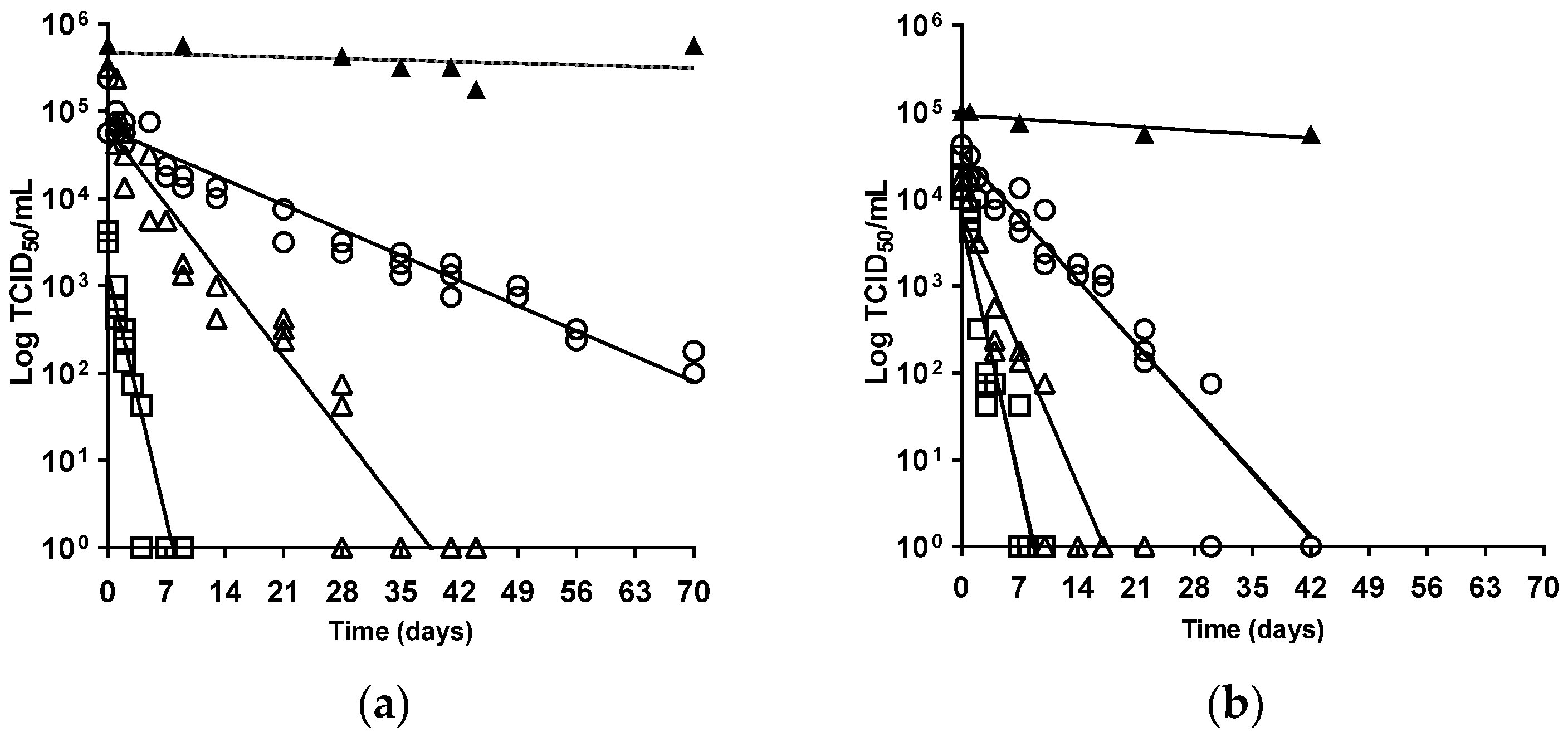
3. Results and Discussion

The ability of AIV infectivity to survive in the environment is a major contributing factor to viral persistence and spread [17], and merited investigation because of the extensive nature of the most recent H5N1-2021 clade 2.3.4.4b epizootic [16]. Experimental evidence has underlined the essential waterfowl-adapted tropism of H5Nx clade 2.3.4.4 HPAIVs, where the efficient acquisition and onward transmission of infection correlated with waterfowl interactions, within a strongly virus-contaminated environment [32,33,34,35,36]. This sustained viral maintenance within wild anseriformes produces the infection pressure for associated poultry outbreaks [7], with viral environmental contamination also recorded at outbreak premises [37,38]. We compared the temperature-associated virus stability (infectivity) of five UK-origin H5Nx clade 2.3.4.4 strains (Table 1) at three European outdoor temperatures, where 4 °C mirrored winter, while 20 °C and 30 °C corresponded to summer temperatures in northern and southern Europe, respectively. Viral stability was determined from the DT values at a given temperature [24], following incubation for up to 10 weeks. Extrapolation of the predicted straight line provided an intercept to also show the time to extinction of viral infectivity (Figure 1 and Figure 2, Table 2). Each clade 2.3.4.4 isolate showed greatest stability at 4 °C, whereas increased incubation temperatures, namely 20 °C and 30 °C, resulted in a faster reduction in viral infectivity (Figure 1 and Figure 2, Table 2), which was consistent with an inverse relationship for AIV survival kinetics and temperature [24,39].
For the five viruses assessed at 4 °C and 20 °C, pairwise comparisons of their DT values and extrapolated extinction times revealed that, in the majority of cases, the virus survival (stability) of each virus was significantly different from the others (p < 0.05; Table 3 and Table 4). The only exceptions among the 4 °C DT comparisons were those for H5N8-2014 versus H5N1-2021 (p = 0.33), and H5N8-2016 versus H5N6-2017 (p = 0.06), with comparison of H5N1-2021 versus H5N6-2017 being non-significant for both their DT values (p = 0.22) and extinction times (p = 0.58; Table 3). Exceptions among the 20 °C comparisons were noted for the DT values for H5N1-2021 versus H5N8-2014 (p = 0.55), for the extinction times for H5N1-2021 versus H5N6-2017 (p = 0.13), with comparison of H5N6-2017 versus H5N8-2014 being non-significant for both their DT values (p = 0.35) and extinction times (p = 0.97; Table 4). However, comparisons of both parameters obtained at 30 °C showed that only H5N8-2016 had a significantly longer extinction time than H5N8-2020 and H5N1-2021 (p < 0.001), with no significant difference between the extinction times of H5N8-2020 and H5N1-2021 (p = 0.33). There were no significant differences between the DT values among any of the three isolates at 30 °C (p > 0.28).
Overall, for a given H5Nx clade 2.3.4.4 isolate, these data showed the reduction in survival time at 20 °C was at least 2.5-fold faster than the DT values observed at the low incubation temperature (4 °C). Wild waterfowl cases, during the clade 2.3.4.4 epizootic waves, peaked in the European winter months when these heightened infection pressures would result in accompanying incursions in farmed poultry [12,16]. The two least stable viruses, at least when ranked by their extinction times, were H5N8-2014 and H5N6-2017 (Table 2), and their corresponding significance tests at 20 °C (Table 4) showed this virus pair was significantly different from the other viruses. Interestingly, the European winter incursions of H5N8-2014 and H5N6-2017 were the smallest of the five H5Nx clade 2.3.4.4 epizootics, which in the case of H5N6-2017 were essentially limited to wild birds with no accompanying commercial poultry outbreaks during winter 2017–2018 [12,40]. The relative instability of H5N8-2014 and H5N6-2017, reflected in low DT and extinction values, may have contributed to the limited scale of these incursions.
In field settings, there is the confounder of matrix effects upon virus deposited in the environment, which may unpredictably affect the complexity of virus survival. Therefore, it is important to apply uniform and consistent assessment of temperature-associated survival of any viral pathogen which is excreted into the environment, hence all five H5Nx HPAIVs were each isolated and propagated by using SPF EFEs. Progeny AIVs are produced during the orthomyxovirus replication cycle [8,41], during which they acquire an external lipid envelope of plasma membrane origin from the host [42]. Therefore, lipid envelope consistency was ensured by focusing the temperature stability investigations on viruses propagated in EFEs. This approach ensured the variation in infectivity survival was determined by mainly differences among the viral envelope proteins, namely the HA and NA [23,43]. However, potential contributions to viral stability by other internal structural proteins could not be entirely excluded [23,44], and may also extend to the stability of the encapsulated viral polymerase enzymes, so these could have contributed to the stability differences observed in our study for the three H5N8 clade 2.3.4.4 subtypes (Table 2).
Temperature-associated viral stability reflected in environmental survival is but one factor which may influence the scale and extent of epizootics. The recent H5N1 clade 2.3.4.4b HPAIV epizootic has included an expansion of the avian host range to include seabirds with many cases reported on a global scale, thereby enhancing viral spread and associated infection pressure [10,15,45]. However, the dynamics of spread within waterfowl may be modulated by host responses, including innate and/or H5-specific humoral immunity acquired during previous AIV incursions [46,47]. Other species–specific differences have been noted, where environmental contamination may not have such a prominent role in H5Nx clade 2.3.4.4 HPAIV spread among chickens [48,49].

4. Conclusions

These quantified virus survival data provide evidence to refine disease prevention strategies for poultry units and inform future veterinary risk assessments for outbreak management, particularly in view of the continuing H5N1 clade 2.3.4.4 HPAIV global epizootic [50,51]. Importantly, these virus survival outcomes contribute to the understanding of AIV persistence in the environment, thereby informing protection of poultry health and commercial production systems. These data highlight HPAIV persistence at low temperatures, so presenting a greater infection risk to avian species during the cooler months in temperate latitudes, especially in the case of clade 2.3.4.4 H5Nx HPAIVs to waterfowl with subsequent incursion risks for farmed poultry. Our statistical data, using pairwise comparisons of virus DT values and extrapolated extinction times at 4 °C and 20 °C, (p < 0.05; Table 3 and Table 4), revealed that, in many instances, the virus survival (stability) of each isolate was significantly different from the others. The relevance of these experimental findings has been underlined by the detection of H5N1 HPAIV in the immediate farm environment during UK clade 2.3.4.4 outbreaks in 2023 [38], affirming observations during earlier H5N1 GsGd HPAIV outbreaks [37]. These experimental and field-based studies are now identifying the consequences of environmental contamination, which have arisen from AIV incursions (either wild birds or poultry) and may influence onward spread. The temperature stability of isolates can also inform the likelihood of continuing infectivity, particularly during the vulnerable period prior to statutory cleansing and on-farm disinfection interventions [52,53]. Interestingly, heat treatment may provide an alternative to chemical disinfection, thereby giving additional importance to the outcomes of viral temperature stability investigations [54,55].

Author Contributions

Conceptualization, S.M.B., I.H.B. and R.M.I.; methodology, investigation, data curation, formal analysis, visualisation, original draft preparation, C.J.W.; statistical analyses, visualisation, M.E.A.; resources, S.M.B., M.J.S. and R.D.E.H.; supervision, M.J.S.; writing—review and editing, M.J.S. and A.C.B.; project administration, S.M.B., R.D.E.H. and A.C.B.; funding acquisition, I.H.B., S.M.B., A.C.B. and M.J.S. All authors have read and agreed to the published version of the manuscript.

Funding

This research was funded by the Department for Environment, Food and Rural Affairs (Defra), and the devolved administrations for Scotland and for Wales, through research projects (i) SE4011—Understanding infection dynamics and survival properties of avian influenza viruses and APMV-1, (ii) SE2211 FLU-FUTURES—Reducing Influenza A Virus (avian and mammalian) hazards in the UK, and (iii) SE2213 FLUFUTURES 2.0—Understanding the diverse spectrum of influenza virus based threats to the UK. Research using isolate H5N6-2017 was funded in part by the European Union’s Horizon 2020 research and innovation programme, Delta-Flu [grant agreement number 727922].

Institutional Review Board Statement

Not applicable.

Informed Consent Statement

Not applicable.

Data Availability Statement

All data are presented within this manuscript.

Acknowledgments

The authors would like to thank APHA Cell and Tissue Culture Unit for providing MDCK cell monolayers.

Conflicts of Interest

The authors declare no conflicts of interest. The funders had no role in the design of the study; in the collection, analyses, or interpretation of the data; in the writing of the manuscript; or in the decision to publish the results.

References

  1. Alexander, D.J. An overview of the epidemiology of avian influenza. Vaccine 2007, 25, 5637–5644. [Google Scholar] [CrossRef] [PubMed]
  2. WOAH. Infection with high pathogenicity avian influenza viruses. In Terrestrial Animal Health Code; Chapter 10.4; World Organisation for Animal Health: Paris, France, 2023; Available online: https://www.woah.org/en/what-we-do/standards/codes-and-manuals/terrestrial-code-online-access/ (accessed on 23 May 2024).
  3. Alexander, D.J.; Brown, I.H. History of highly pathogenic avian influenza. OIE Rev. Sci. Tech. 2009, 28, 19–38. [Google Scholar] [CrossRef] [PubMed]
  4. Van Dijk, J.G.B.; Hoyle, B.J.; Verhagen, J.H.; Nolet, B.A.; Fouchier, R.A.M.; Klaassen, M. Juveniles and migrants as drivers for seasonal epizootics of avian influenza virus. J. Anim. Ecol. 2014, 83, 266–275. [Google Scholar] [CrossRef] [PubMed]
  5. WHO. Evolution of H5N1 avian influenza viruses in Asia. Emerg. Infect. Dis. 2005, 11, 1515–1521. [Google Scholar] [CrossRef] [PubMed]
  6. Brown, I.H.; Banks, J.; Manvell, R.; Essen, S.C.; Shell, W.; Slomka, M.; Löndt, B.; Alexander, D.J. Recent epidemiology and ecology of influenza A viruses in avian species in Europe and the Middle East. Dev. Biol. 2006, 124, 45–50. [Google Scholar]
  7. Antigua, K.J.C.; Choi, W.S.; Baek, Y.H.; Song, M.S. The Emergence and Decennary Distribution of Clade 2.3.4.4 HPAI H5Nx. Microorganisms 2019, 7, 156. [Google Scholar] [CrossRef] [PubMed]
  8. Nunez, I.A.; Ross, T.M. A review of H5Nx avian influenza viruses. Ther. Adv. Vaccines Immunother. 2019, 7, 2515135518821625. [Google Scholar] [PubMed]
  9. Global Consortium for H5N8 and Related Influenza Viruses. Role for migratory wild birds in the global spread of avian influenza H5N8. Science 2016, 354, 213–217. [Google Scholar] [CrossRef] [PubMed]
  10. Caliendo, V.; Lewis, N.S.; Pohlmann, A.; Baillie, S.R.; Banyard, A.C.; Beer, M.; Brown, I.H.; Fouchier, R.A.M.; Hansen, R.D.E.; Lameris, T.K.; et al. Transatlantic spread of highly pathogenic avian influenza H5N1 by wild birds from Europe to North America in 2021. Sci. Rep. 2022, 12, 11729. [Google Scholar] [CrossRef]
  11. WOAH. Avian Influenza—Sitaution Reports; WOAH–World Organisation for Animal Health: Paris, France, 2024; Available online: https://www.woah.org/app/uploads/2024/01/hpai-situation-report-20240109.pdf (accessed on 23 May 2024).
  12. Alarcon, P.; Brouwer, A.; Venkatesh, D.; Duncan, D.; Dovas, C.I.; Georgiades, G.; Monne, I.; Fusaro, A.; Dan, A.; Śmietanka, K.; et al. Comparison of 2016–2017 and previous epizootics of highly pathogenic avian influenza H5 Guangdong lineage in Europe. Emerg. Infect. Dis. 2018, 24, 2270–2283. [Google Scholar] [CrossRef]
  13. Pohlmann, A.; King, J.; Fusaro, A.; Zecchin, B.; Banyard, A.C.; Brown, I.H.; Byrne, A.M.P.; Beerens, N.; Liang, Y.; Heutink, R.; et al. Has Epizootic Become Enzootic? Evidence for a Fundamental Change in the Infection Dynamics of Highly Pathogenic Avian Influenza in Europe, 2021. MBio 2022, 13, e0060922. [Google Scholar] [CrossRef] [PubMed]
  14. Byrne, A.M.P.; James, J.; Mollett, B.C.; Meyer, S.M.; Lewis, T.; Czepiel, M.; Seekings, A.H.; Mahmood, S.; Thomas, S.S.; Ross, C.R.; et al. Investigating the genetic diversity of H5 avian influenza in the UK 2020–2022. Microbiol. Spectr. 2023, 11, e04776-22. [Google Scholar] [CrossRef] [PubMed]
  15. Furness, R.W.; Gear, S.C.; Camphuysen, K.; Tyler, G.; de Silva, D.; Warren, C.J.; James, J.; Reid, S.M.; Banyard, A.C. Environmental Samples Test Negative for Avian Influenza Virus H5N1 Four Months after Mass Mortality at A Seabird Colony. Pathogens 2023, 12, 584. [Google Scholar] [CrossRef] [PubMed]
  16. Defra. Updated Outbreak Assessment #42. Highly pathogenic avian influenza (HPAI) in the UK and Europe, 4 May 2023. In Avian Influenza (Bird Flu) in Europe, Russia and the UK; Department for Environment, Food and Rural Affairs: London, UK, 2023. Available online: https://assets.publishing.service.gov.uk/government/uploads/system/uploads/attachment_data/file/1156065/4_May_2023_Highly_pathogenic_avian_influenza__HPAI__in_the_UK_and_Europe.pdf (accessed on 23 May 2024).
  17. Swayne, D.E. Epidemiology of avian influenza in agricultural and other man-made systems. In Avian Influenza, 1st ed.; Swayne, D.E., Ed.; John Wiley & Sons, Inc.: Hoboken, NJ, USA, 2008; Chapter 4; pp. 59–85. [Google Scholar]
  18. Hauck, R.; Crossley, B.M.; Rejmanek, D.; Zhou, H.; Gallardo, R.A. Persistence of highly pathogenic and low pathogenic avian influenza viruses in footbaths and poultry manure. Avian Dis. 2017, 61, 64–69. [Google Scholar] [CrossRef] [PubMed]
  19. Stallknecht, D.E.; Goekjian, V.H.; Wilcox, B.R.; Poulson, R.L.; Brown, J.D. Avian influenza virus in aquatic habitats: What do we need to learn? Avian Dis. 2010, 54, 461–465. [Google Scholar] [CrossRef] [PubMed]
  20. Wood, J.P.; Choi, Y.W.; Chappie, D.J.; Rogers, J.V.; Kaye, J.Z. Environmental persistence of a highly pathogenic avian influenza (H5N1) virus. Environ. Sci. Technol. 2010, 44, 7515–7520. [Google Scholar] [CrossRef] [PubMed]
  21. Gutiérrez, R.A.; Buchy, P. Contaminated soil and transmission of influenza virus (H5N1). Emerg. Infect. Dis. 2012, 18, 1530–1532. [Google Scholar] [CrossRef] [PubMed]
  22. Swayne, D.E.; Beck, J.R. Heat inactivation of avian influenza and Newcastle disease viruses in egg products. Avian Pathol. 2004, 33, 512–518. [Google Scholar] [CrossRef] [PubMed]
  23. Spackman, E.A. Review of the Stability of Avian Influenza Virus in Materials from Poultry Farms. Avian. Dis. 2023, 67, 229–236. [Google Scholar] [CrossRef]
  24. Stallknecht, D.E.; Kearney, M.T.; Shane, S.M.; Zwank, P.J. Effects of pH, temperature, and salinity on persistence of avian influenza viruses in water. Avian Dis. 1990, 34, 412–418. [Google Scholar] [CrossRef]
  25. Nazir, J.; Haumacher, R.; Ike, A.; Stumpf, P.; Boehm, R.; Marschang, R.E. Long-Term Study on Tenacity of Avian Influenza Viruses in Water (Distilled Water, Normal Saline, and Surface Water) at Different Temperatures. Avian Dis. 2010, 54, 720–724. [Google Scholar] [CrossRef]
  26. Rohani, P.; Breban, R.; Stallknecht, D.E.; Drake, J.M. Environmental transmission of low pathogenicity avian influenza viruses and its implications for pathogen invasion. Proc. Natl. Acad. Sci. USA 2009, 106, 10365–10369. [Google Scholar] [CrossRef]
  27. Guan, J.; Chan, M.; Grenier, C.; Wilkie, D.C.; Brooks, B.W.; Spencer, J.L. Survival of avian influenza and Newcastle disease viruses in compost and at ambient temperatures based on virus isolation and real-time reverse transcriptase PCR. Avian Dis. 2009, 53, 26–33. [Google Scholar] [CrossRef]
  28. WOAH. Avian Influenza (including infection with high pathogenicity avian influenza viruses). In Manual of Diagnostic Tests and Vaccines for Terrestrial Animals, 12th ed.; Chapter 3.3.4; World Organisation for Animal Health: Paris, France, 2023; Available online: https://www.woah.org/fileadmin/Home/eng/Health_standards/tahm/A_summry.htm (accessed on 23 May 2024).
  29. Slomka, M.J.; Seekings, A.H.; Mahmood, S.; Thomas, S.S.; Puranik, A.; Watson, S.; Byrne, A.M.P.; Hicks, D.; Nunez, A.; Brown, I.H.; et al. Unexpected infection outcomes of China-origin H7N9 low pathogenicity avian influenza virus in turkeys. Sci. Rep. 2018, 8, 7322. [Google Scholar] [CrossRef]
  30. Villegas, P. Titration of Biological Suspensions. In A Laboratory Manual for the Isolation and Identification of Avian Pathogens, 1st ed.; Swayne, D.E., Glisson, J.R., Jackwood, M.W., Pearson, J.E., Reed, W.M., Eds.; The American Association of Avian: Pathologists, PA, USA, 1998; pp. 248–254. [Google Scholar]
  31. Kamolsiripichaiporn, S.; Subharat, S.; Udon, R.; Thongtha, P.; Nuanualsuwan, S. Thermal inactivation of foot-and-mouth disease viruses in suspension. Appl. Environ. Microbiol. 2007, 73, 7177–7184. [Google Scholar] [CrossRef]
  32. Leyson, C.; Youk, S.S.; Smith, D.; Dimitrov, K.; Lee, D.H.; Larsen, L.E.; Swayne, D.E.; Pantin-Jackwood, M.J. Pathogenicity and genomic changes of a 2016 European H5N8 highly pathogenic avian influenza virus (clade 2.3.4.4) in experimentally infected mallards and chickens. Virology 2019, 537, 172–185. [Google Scholar] [CrossRef]
  33. Slomka, M.J.; Puranik, A.; Mahmood, S.; Thomas, S.S.; Seekings, A.H.; Byrne, A.M.P.; Nunez, A.; Bianco, C.; Mollett, B.C.; Watson, S.; et al. Ducks Are Susceptible to Infection with a Range of Doses of H5N8 Highly Pathogenic Avian Influenza Virus (2016, Clade 2.3.4.4b) and Are Largely Resistant to Virus-Specific Mortality, but Efficiently Transmit Infection to Contact Turkeys. Avian Dis. 2019, 63, 172–180. [Google Scholar] [CrossRef]
  34. Puranik, A.; Slomka, M.J.; Warren, C.J.; Thomas, S.S.; Mahmood, S.; Byrne, A.M.P.; Ramsay, A.M.; Skinner, P.; Watson, S.; Everett, H.E.; et al. Transmission dynamics between infected waterfowl and terrestrial poultry: Differences between the transmission and tropism of H5N8 highly pathogenic avian influenza virus (clade 2.3.4.4a) among ducks, chickens and turkeys. Virology 2020, 541, 113–123. [Google Scholar] [CrossRef]
  35. Seekings, A.H.; Warren, C.J.; Thomas, S.S.; Mahmood, S.; James, J.; Byrne, A.M.P.; Watson, S.; Bianco, C.; Nunez, A.; Brown, I.H.; et al. Highly pathogenic avian influenza virus H5N6 (clade 2.3.4.4b) has a preferable host tropism for waterfowl reflected in its inefficient transmission to terrestrial poultry. Virology. 2021, 559, 74–85. [Google Scholar] [CrossRef]
  36. James, J.; Billington, E.; Warren, C.J.; De Sliva, D.; Di Genova, C.; Airey, M.; Meyer, S.M.; Lewis, T.; Peers-Dent, J.; Thomas, S.S.; et al. Clade 2.3.4.4b H5N1 high pathogenicity avian influenza virus (HPAIV) from the 2021/22 epizootic is highly duck adapted and poorly adapted to chickens. J. Gen. Virol. 2023, 104, 001852. [Google Scholar] [CrossRef]
  37. Parker, C.D.; Irvine, R.M.; Slomka, M.J.; Pavlidis, T.; Hesterberg, U.; Essen, S.; Cox, B.; Ceeraz, V.; Alexander, D.J.; Manvell, R.; et al. Outbreak of Eurasian lineage H5N1 highly pathogenic avian influenza in turkeys in Great Britain in November 2007. Vet. Rec. 2014, 175, 282. [Google Scholar] [CrossRef]
  38. James, J.; Warren, C.J.; de Silva, D.; Lewis, T.; Grace, K.; Reid, S.M.; Falchieri, M.; Brown, I.H.; Banyard, A.C. The Role of Airborne Particles in the Epidemiology of Clade 2.3.4.4b H5N1 High Pathogenicity Avian Influenza Virus in Commercial Poultry Production Units. Viruses 2023, 15, 1002. [Google Scholar] [CrossRef]
  39. Brown, J.D.; Goekjian, G.; Poulson, R.; Valeika, S.; Stallknecht, D.E. Avian influenza virus in water: Infectivity is dependent on pH, salinity and temperature. Vet. Microbiol. 2009, 136, 20–26. [Google Scholar] [CrossRef]
  40. Poen, M.J.; Venkatesh, D.; Bestebroer, T.M.; Vuong, O.; Scheuer, R.D.; Oude Munnink, B.B.; de Meulder, D.; Richard, M.; Kuiken, T.; Koopmans, M.P.G.; et al. Co-circulation of genetically distinct highly pathogenic avian influenza A clade 2.3.4.4 (H5N6) viruses in wild waterfowl and poultry in Europe and East Asia, 2017–2018. Virus Evol. 2019, 5, vez004. [Google Scholar] [CrossRef]
  41. Dou, D.R.R.; Östbye, H.; Wang, H.; Daniels, R. Influenza A Virus Cell Entry, Replication, Virion Assembly and Movement. Front. Immunol. 2018, 9, 1581. [Google Scholar] [CrossRef]
  42. Lenard, J. Viral Membranes. In Encyclopedia of Virology; Elsevier: Amsterdam, The Netherlands, 2008; pp. 308–314. [Google Scholar]
  43. Burrell, C.J.; Howard, C.R.; Murphy, F.A. (Eds.) Virion Structure and Composition. In Fenner and White’s Medical Virology, 5th ed.; Elsevier: London, UK, 2017; Chapter 3, pp. 27–37. [Google Scholar]
  44. Gummersheimer, S.L.; Danthi, P. Reovirus Core Proteins λ1 and σ2 Promote Stability of Disassembly Intermediates and Influence Early Replication Events. J. Virol. 2020, 94, e00491-20. [Google Scholar] [CrossRef]
  45. Lane, J.V.; Jeglinski, J.W.E.; Avery-Gomm, S.; Ballstaedt, E.; Banyard, A.C.; Barychka, T.; Brown, I.H.; Brugger, B.; Burt, T.V.; Careen, N.; et al. High pathogenicity avian influenza (H5N1) in Northern Gannets (Morus bassanus): Global spread, clinical signs and demographic consequences. IBIS Av. Repro. Spec. Issue 2024, 166, 633–650. [Google Scholar] [CrossRef]
  46. Vigeveno, R.M.; Poen, M.J.; Parker, E.; Holwerda, M.; de Haan, K.; van Montfort, T.; Lewis, N.S.; Russell, C.A.; Fouchier, R.A.M.; de Jong, M.D.; et al. Outbreak severity of highly pathogenic avian influenza A(H5N8) viruses is inversely correlated to polymerase complex activity and interferon induction. J. Virol. 2020, 94, 11. [Google Scholar] [CrossRef]
  47. Hill, S.C.; Manvell, R.J.; Schulenburg, B.; Shell, W.; Wikramaratna, P.S.; Perrins, C.; Sheldon, B.C.; Brown, I.H.; Pybus, O.G. Antibody responses to avian influenza viruses in wild birds broaden with age. Proc. Biol. Sci. 2016, 283, 20162159. [Google Scholar] [CrossRef]
  48. Seekings, A.H.; Warren, C.J.; Thomas, S.S.; Lean, F.Z.X.; Selden, D.; Mollett, B.C.; van Diemen, P.M.; Banyard, A.C.; Slomka, M.J. Different Outcomes of Chicken Infection with UK-Origin H5N1-2020 and H5N8-2020 High-Pathogenicity Avian Influenza Viruses (Clade 2.3.4.4b). Viruses 2023, 15, 1909. [Google Scholar] [CrossRef]
  49. Seekings, A.; Liang, Y.; Warren, C.J.; Hjulsager, C.K.; Thomas, S.S.; Lean, F.Z.X.; Núñez, A.; Skinner, P.; Selden, D.; Falchieri, M.; et al. Transmission dynamics and pathogenesis differ between pheasants and partridges infected with clade 2.3.4.4b H5N8 and H5N1 high-pathogenicity avian influenza viruses. J. Gen. Virol. 2024, 105, 001946. [Google Scholar] [CrossRef]
  50. Pan American Health Organisation (PAHO). PAHO Issues Alert on Outbreaks of Avian Influenza in Birds in Ten Countries of the Americas; Pan American Health Organization: Washington, DC, USA, 2023. [Google Scholar]
  51. Bennison, A.; Byrne, A.M.P.; Reid, S.M.; Lynton-Jenkins, J.G.; Mollett, B.; de Sliva, D.; Peers-Dent, J.; Finlayson, K.; Hall, R.; Blockley, F.; et al. Detection and spread of high pathogenicity avian influenza virus H5N1 in the Antarctic Region. 2024. Available online: https://www.biorxiv.org/content/10.1101/2023.11.23.568045v1 (accessed on 23 May 2024).
  52. European Council. Council Directive 2005/94/EC of 20 December 2005 on Community measures for the control of avian influenza and repealing Directive 92/40/EEC. Off. J. Eur. Union L. 2005, 10, 16–65. Available online: https://www.legislation.gov.uk/eudr/2005/94/contents (accessed on 23 May 2024).
  53. Defra. Notifiable Avian Disease Control Strategy for Great Britain. Last updated September 2019. Derogations 46-48. 2019. Available online: https://www.gov.uk/government/publications/notifiable-avian-disease-control-strategy (accessed on 23 May 2024).
  54. Stephens, C.B.; Spackman, E. Thermal Inactivation of avian influenza virus in poultry litter as a method to decontaminate poultry houses. Prev. Vet. Med. 2017, 145, 73–77. [Google Scholar] [CrossRef]
  55. APHIS. HPAI Response. In Cleaning and Disinfection Basics (Virus Elimination); USDA Animal and Plant Health Inspection Service: Riverdale, MD, USA, 2022. Available online: https://www.aphis.usda.gov/sites/default/files/cleaning_disinfection.pdf (accessed on 23 May 2024).
Figure 1. Linear regression for virus kinetics profiles for (a) H5N8-2020; (b) H5N1-2021 survival on MDCK cell monolayers in serum-free EMEM with penicillin and streptomycin. Logarithm TCID50/mL was plotted versus the time in days at 4 °C (±2 °C), 20 °C (±2 °C), and 30 °C (±2 °C). Symbols: at 4 °C, ∆ at 20 °C, □ at 30 °C, ▲ positive controls (no incubation treatment) assessed per virus per batch of MDCK cell monolayers. For all isolates evaluated, mean R2 0.91, p < 0.0001.
Viruses 16 00889 g001
Figure 2. Linear regression for virus kinetics profiles for (a) H5N8-2016; (b) H5N8-2014; (c) H5N6-2017 survival, on MDCK cell monolayers in serum-free EMEM with penicillin and streptomycin. Logarithm TCID50/mL was plotted versus the time in days at 4 °C (±2 °C), 20 °C (±2 °C), and 30 °C (±2 °C). Symbols: at 4 °C, ∆ at 20 °C, □ at 30 °C, ▲ positive controls (no incubation treatment) assessed per virus per batch of MDCK cell monolayers. For all isolates evaluated, mean R2 0.91, p < 0.0001.
Viruses 16 00889 g002
Table 1. Summary of the five UK-origin H5Nx clade 2.3.4.4 HPAIVs assessed for temperature stability.
IsolateSubtype/CladeDescriptor in TextCollection Date 1
A/duck/England/1279/2014H5N8 2.3.4.4aH5N8-201416 November 2014
A/wigeon/Wales/52833/2016H5N8 2.3.4.4bH5N8-201614 December 2016
A/swan/England/001986/2017H5N6 2.3.4.4bH5N6-201731 December 2017
A/chicken/England/030786/2020H5N8 2.3.4.4bH5N8-20201 November 2020
A/chicken/Wales/053969/2021H5N1 2.3.4.4bH5N1-202131 October 2021
1 Collection dates of the five UK H5Nx clade 2.3.4.4 HPAIV isolates were sourced from the GISAID EpiFlu™ database (https://www.epicov.org/epi3/frontend accessed on 23 May 2024).
Table 2. Survival assessment of five H5Nx HPAIV isolates at up to three different temperatures. Results are expressed as DT time and the extrapolated time to extinction (both as days) and ranked according to the latter for a given temperature. Survival kinetics experiments at 4 °C (±2 °C) lasted to ten weeks, at 20 °C (±2 °C) to seven weeks (combined mean R2 0.92, p < 0.0001), and at 30 °C (±2 °C) to five weeks (mean R2 0.90, p < 0.0001).
Temperature4 °C20 °C 30 °C
StrainDTExtinction 1DTExtinction 1StrainDT (days)Extinction 1
H5N8-202024.2116.18.038.5H5N8-20162.713.2
H5N8-201614.069.86.128.8H5N1-20212.38.6
H5N1-20219.543.24.517.2H5N8-20202.57.8
H5N6-201711.240.02.810.4
H5N8-20147.822.93.08.7
1 Extinction (days) is the x axis intercept (y = 1 log10 TCID50/mL).
Table 3. Resulting p-values from significance tests of the difference between the DT values and extinction times at 4 °C for five different H5Nx HPAIV isolates.
HPAIVResult of Multiple Comparisons of DT Value/Extinction Time at 4 °C
HPAIV IsolateH5N8-2014H5N8-2016H5N6-2017H5N8-2020H5N1-2021
H5N8-2014-<0.05/<0.001<0.001<0.0010.33/<0.001
H5N8-2016--0.06/<0.001<0.001<0.001
H5N6-2017---<0.0010.22/0.58
H5N8-2020----<0.001
H5N1-2021-----
A single p-value cell means that the same p-value applies to both the DT value and extinction time. - indicates that either (i) table diagonals: result not applicable as it is for the virus strain versus itself, or (ii) lower half of table: result available in the top half of the table.
Table 4. Resulting p-values from significance tests of the difference between the DT values and extinction times at 20 °C for five different H5Nx HPAIV isolates.
HPAIVResult of Multiple Comparisons of DT Value/Extinction Time at 20 °C
HPAIV IsolateH5N8-2014H5N8-2016H5N6-2017H5N8-2020H5N1-2021
H5N8-2014-<0.01/<0.0010.35/0.97<0.0010.55/<0.05
H5N8-2016--<0.001<0.01/<0.001<0.001
H5N6-2017---<0.001<0.001/0.13
H5N8-2020----<0.001
H5N1-2021-----
A single p-value cell means that the same p-value applies to both the DT value and extinction time. - indicates that either (i) table diagonals: result not applicable as it is for the virus strain versus itself, or (ii) lower half of table: result available in the top-half of the table.
Disclaimer/Publisher’s Note: The statements, opinions and data contained in all publications are solely those of the individual author(s) and contributor(s) and not of MDPI and/or the editor(s). MDPI and/or the editor(s) disclaim responsibility for any injury to people or property resulting from any ideas, methods, instructions or products referred to in the content.

