Article

The MGF300-2R Protein of African Swine Fever Virus Promotes IKKβ Ubiquitination by Recruiting the E3 Ubiquitin Ligase TRIM21

1
State Key Laboratory for Animal Disease Control and Prevention, National African Swine Fever Para-Reference Laboratory, National High Containment Facilities for Animal Diseases Control and Prevention, Harbin Veterinary Research Institute, Chinese Academy of Agricultural Sciences, Harbin 150069, China
2
College of Animal Sciences, Yangtze University, Jingzhou 434000, China
3
College of Life Science and Engineering, Foshan University, Foshan 528000, China
*
Authors to whom correspondence should be addressed.
These authors contributed equally to this work.
Viruses 2024, 16(6), 949; https://doi.org/10.3390/v16060949
Submission received: 29 March 2024 / Revised: 6 June 2024 / Accepted: 8 June 2024 / Published: 12 June 2024
(This article belongs to the Special Issue African Swine Fever Virus 4.0)

Abstract

:
African swine fever (ASF) is an acute, hemorrhagic, highly contagious disease in pigs caused by African swine fever virus (ASFV). Our previous study identified that the ASFV MGF300-2R protein functions as a virulence factor and found that MGF300-2R degrades IKKβ via selective autophagy. However, the E3 ubiquitin ligase responsible for IKKβ ubiquitination during autophagic degradation still remains unknown. In order to solve this problem, we first pulled down 328 proteins interacting with MGF300-2R through immunoprecipitation-mass spectrometry. Next, we analyzed and confirmed the interaction between the E3 ubiquitin ligase TRIM21 and MGF300-2R and demonstrated the catalytic role of TRIM21 in IKKβ ubiquitination. Finally, we indicated that the degradation of IKKβ by MGF300-2R was dependent on TRIM21. In summary, our results indicate TRIM21 is the E3 ubiquitin ligase involved in the degradation of IKKβ by MGF300-2R, thereby augmenting our understanding of the functions of MGF300-2R and offering insights into the rational design of live attenuated vaccines and antiviral strategies against ASF.

1. Introduction

African swine fever (ASF) is an acute, hemorrhagic and severe infectious disease of pigs, which has become endemic or epidemic in nearly 50 countries in Africa, Europe, Asia and the Caribbean, causing huge economic losses that are difficult to estimate and posing a critical threat to the global pig industry and related industries [1,2,3]. The causative agent, African swine fever virus (ASFV), is a large, double-stranded DNA virus. The ASFV genome encodes 68 structural proteins and more than 100 non-structural proteins, with over 10 virulence factors discovered in the past five years, such as MGF300-2R, MGF300-4L, A137R, and H240R [2,4,5,6,7]. With the deletion of virulence-associated factors, attenuated strains that can induce protective immune responses against virulent parental virus challenge in pigs are promising live attenuated vaccines (LAVs) [8]. Compared with other types of ASF vaccines, LAVs can provide complete homologous and partial heterologous protection [8]. Hence, it is essential to discover and characterize more virulence-associated genes of ASFV for the future rational development of LAVs.
Selective autophagy degrades specific substrates mediated by selective autophagy receptors (SARs) [9]. The recognition of substrates by ubiquitin-dependent SARs depends on the ubiquitination of substrates by E3 ubiquitin ligases [10]. Accumulating evidence has demonstrated that viruses can regulate selective autophagy by recruiting SARs or E3 ubiquitin ligases to inhibit the host immune responses or promote viral replication. For example, the pseudorabies virus (PRV) tegument protein UL13 recruits the E3 ubiquitin ligase RNF5 to inhibit STING-mediated antiviral immunity [11]. Porcine reproductive and respiratory syndrome virus (PRRSV) degrades DDX10 via SAR p62-dependent selective autophagy to antagonize its antiviral activity [12]. The tegument protein UL21 of alpha-herpesviruses inhibits innate immunity by triggering cGAS degradation through the TOLLIP-mediated selective autophagy [13]. In our previous report, the highly virulent strain ASFV HLJ/18 (ASFV-WT) lacking the MGF300-2R gene (Del2R) showed a 1-log reduction in viral titer and induced higher IL-1β and TNF-α production in primary porcine alveolar macrophages than ASFV-WT. Mechanistically, the MGF300-2R protein interacts with and degrades IKKα and IKKβ via the selective autophagy pathway. Furthermore, we showed that MGF300-2R promoted the K27-linked polyubiquitination of IKKα and IKKβ, which subsequently served as a recognition signal for the selective autophagic degradation mediated by TOLLIP. Importantly, Del2R exhibited a significant reduction in both replication and virulence compared with ASFV-WT in pigs, likely due to increased IL-1β and TNF-α [6]. However, the E3 ubiquitin ligase responsible for IKKβ ubiquitination mediated by MGF300-2R remains unknown.
In this study, we conducted immunoprecipitation-mass spectrometry (IP-MS) and confirmed that TRIM21 acts as the E3 ubiquitin ligase responsible for the autophagic degradation of IKKβ by MGF300-2R, thereby facilitating the understanding of the mechanism underlying the MGF300-2R involvement in the pathogenicity of ASFV. These findings will provide valuable clues to the rational design of LAVs and antiviral strategies against ASF.

2. Materials and Methods

2.1. Cell Lines

Human embryonic kidney 293T (HEK293T) cells were grown in Dulbecco’s modified Eagle’s medium (DMEM) (C11995500CP; Thermo Fisher Scientific; Waltham, MA, USA) enriched with 10% fetal bovine serum (FBS) (F8687; Sigma-Aldrich; St. Louis, MO, USA) and incubated at 37 °C in a 5% CO2 environment. Using the CRISPR/Cas9 system, we generated TRIM21-knockout (KO) HEK293T cells by inserting the sgRNA (sgTRIM21: 5′-TCTCTCAGGTTGGGAAAGGT-3′) into the plasmid vector pX458.

2.2. Antibodies, Plasmids and Reagents

We purchased the following antibodies from Abclonal (Wuhan, China): rabbit anti β-tubulin (A12289), mouse anti HA-Tag (AE008), rabbit anti HA-Tag (AE105), mouse anti Flag-Tag (AE005) and mouse anti His-Tag (AE003). Anti-FLAG(R) M2 magnetic beads (M8823) and anti-HA agarose (A2095) were obtained from Sigma-Aldrich (USA). We procured Alexa Fluor 647-conjugated donkey anti-mouse IgG (H+L) (A31571) and Alexa Fluor 488-conjugated goat anti-rabbit IgG (H+L) (A11008) from Thermo Fisher Scientific. 4’, 6-Diamidino-2-phenylindole (DAPI) (C006) was procured from Solarbio (Beijing, China). The TRIM21 polyclonal antibody (12108-1-AP) was obtained from Proteintech (Chicago, IL, USA). The plasmids pHA-MGF300-2R, pFlag-MGF300-2R, pFlag-IKKβ, and pHis-Ub and the vectors pCMV-HA and pCMV-Flag have been described previously [6]. pHA-IKKβ was generated by mutating pFlag-IKKβ using polymerase chain reaction (PCR) site-directed mutagenesis. The pHA-TRIM21 plasmid (ZK3596) was procured from Beijing Zoman Biotechnology and subsequently modified through PCR site-directed mutagenesis to generate pFlag-TRIM21. The primers utilized in this study are summarized in Table 1.

2.3. Transfection

HEK293T cells were transfected with indicated plasmids using linear 25-kDa polyethyleneimine (PEI) (23966-2; Polysciences; Warrington, PA, USA). When the cells reached 80% confluence in 6-well cell culture plates, a transfection mixture containing 3 μg of plasmid, 9 μL of PEI (stock solution at 1 mg/mL) and 200 μL of Opti-MEM (31985070; Invitrogen; Carlsbad, CA, USA) was added to the cells and incubated for 24 to 36 h after mixing for 20 min at room temperature.

2.4. Immunoprecipitation-Mass Spectrometry (IP-MS) and Proteomics Functional Analysis

In order to analyze the proteins that interact with MGF300-2R, we first verified the MGF300-2R in the immunoprecipitates. Then, the IP-MS service was commissioned by the Beijing Genomics Institute. We conducted a Kyoto Encyclopedia of Genes and Genomes (KEGG) database search and Gene Ontology (GO) enrichment analysis encompassing the cellular component (CC), molecular function (MF) and biological process (BP) categories using clusterProfiler v4.3. The protein–protein interaction (PPI) network was constructed in the STRING database (https://string-db.org/ accessed on 22 December 2023) and visualized via Cytoscape software (version 3.9.1).

2.5. Western Blotting and Co-Immunoprecipitation (Co-IP)

HEK293T cells were collected and lysed in ice-cold RIPA lysis buffer (R0278; Sigma-Aldrich; St. Louis, MO, USA), which was supplemented with a protease inhibitor cocktail (4693116001; Roche; Basel, Switzerland). The cell lysates were denatured by boiling them in SDS-PAGE loading buffer (P1040; Solarbio; Beijing, China) and then separated using SDS-PAGE. The separated proteins were transferred onto polyvinylidene fluoride membranes, which were then blocked with 5% skim milk in Tris-buffered saline containing 0.05% Tween 20 (TBST) for 1 h at room temperature. The blocked membranes were subsequently probed using the appropriate primary and secondary antibodies. The signals were visualized using an Odyssey imaging system.
The transfected HEK293T cells were lysed in RIPA lysis buffer for 30 min on ice. After removing the nuclei through low-speed centrifugation and collecting 100 μL of the sample as input, the remaining 800 μL lysates were incubated with the appropriate antibody, after which anti-Flag M2 magnetic beads or anti-HA agarose were added and rotated at 4 °C overnight. After incubation, the beads were collected using a magnetic separator and washed three times with phosphate buffer saline (PBS) (PBS-100; Biocomma; Shenzhen, China). The bound proteins were then subjected to Western blotting using the appropriate antibodies.

2.6. Laser Confocal Microscopy

HEK293T cells were seeded onto a glass-bottom cell culture dish (801001; NEST; Wuxi, China) coated with poly-L-lysine. The plasmids were transfected into the cells using PEI. At 24 h post-transfection (hpt), the cells were fixed with 4% paraformaldehyde at room temperature for 20 min, followed by permeabilization with 0.1% Triton X-100 (V900502; Sigma-Aldrich; St. Louis, MO, USA) in PBS for 20 min and blocked with 5% bovine serum albumin (BSA). The primary antibodies were carefully added and allowed to incubate for 2 h. The cells were thoroughly washed with PBS five times, followed by staining with the Alexa Fluor-conjugated secondary antibodies (goat anti-mouse IgG or goat anti-rabbit IgG) for 1 h at room temperature. The cells were washed with PBS three times and then stained with DAPI for 5 min to visualize the nuclei. Following three PBS washing steps, the cells were visualized using confocal laser scanning microscopy with Airyscan (LSM800; Zeiss; Jena, Germany), and the most representative images were chosen for presentation. The scale bar represents 20 μm.

2.7. Cell Viability Assay

We utilized a cell counting kit-8 (CCK-8) (CK04; DOJINDO; Kumamoto, Japan) to evaluate the cell viability, following the manufacturer’s guidelines. In summary, the TRIM21-knockout (TRIM21−/−) or wild-type (WT) HEK293T cells were incubated in 96-well cell culture plates. The cell viability was assessed at 12, 24 and 36 h post-incubation (hpi). We added a volume of 10 μL of CCK-8 reagent to each well in the plates. The cells were incubated at 37 °C for 1 h, and subsequently, the absorbance at 450 nm was determined using a microplate reader.

2.8. Statistical Analysis

An unpaired two-tailed t-test was conducted using GraphPad Prism version 8.0.0 (GraphPad Software, San Diego, CA, USA) to assess statistical significance. The data are presented as the mean ± SD from three independent experiments. P ≥ 0.05 was considered statistically non-significant (ns); P < 0.05 was considered statistically significant (* P < 0.05; ** P < 0.01; *** P < 0.001).

3. Results

3.1. The Proteins Interacting with MGF300-2R Were Screened by IP-MS

To identify the E3 ubiquitin ligase responsible for the selective autophagic degradation of IKKβ by MGF300-2R, we initially transfected HEK293T cells with pFlag-MGF300-2R and subsequently performed immunoprecipitation using magnetic beads coupled with a Flag antibody. The proteins co-immunoprecipitated with MGF300-2R were subjected to IP-MS analysis to identify potential E3 ubiquitin ligases (Figure 1A). Our results indicated a total of 328 proteins pulled down by MGF300-2R, among which the key candidates exhibiting E3 ubiquitin ligase activity included HUWE1, ARIH2, TRIM21, PRPF19, UBR5 and OBI1 (Figure 1B and Table S1). Further Kyoto Encyclopedia of Genes and Genomes (KEGG) pathway analysis indicated the involvement of multiple signaling pathways, such as the proteasome system, the spliceosome system and the RNA transport system (Figure 1C).
In the Gene Ontology (GO) analysis of the IP-MS results, we observed a significant proportion of molecules associated with the ubiquitin–proteasome system (P < 0.05) across cellular components (CC), molecular function (MF) and biological processes (BP) (Figure 1D). While HUWE1, UBR5, PRPF19, ARIH2 and OBI1 are mainly related to tumor suppression, DNA damage response and DNA replication [14,15,16,17,18], TRIM21 is involved in regulating host innate immunity and viral immune evasion [19]. Moreover, the protein–protein interaction analysis revealed a direct interaction between TRIM21 and IKKβ (Figure 1E). Therefore, based on these findings, we selected the E3 ubiquitin ligase TRIM21 for further validation.

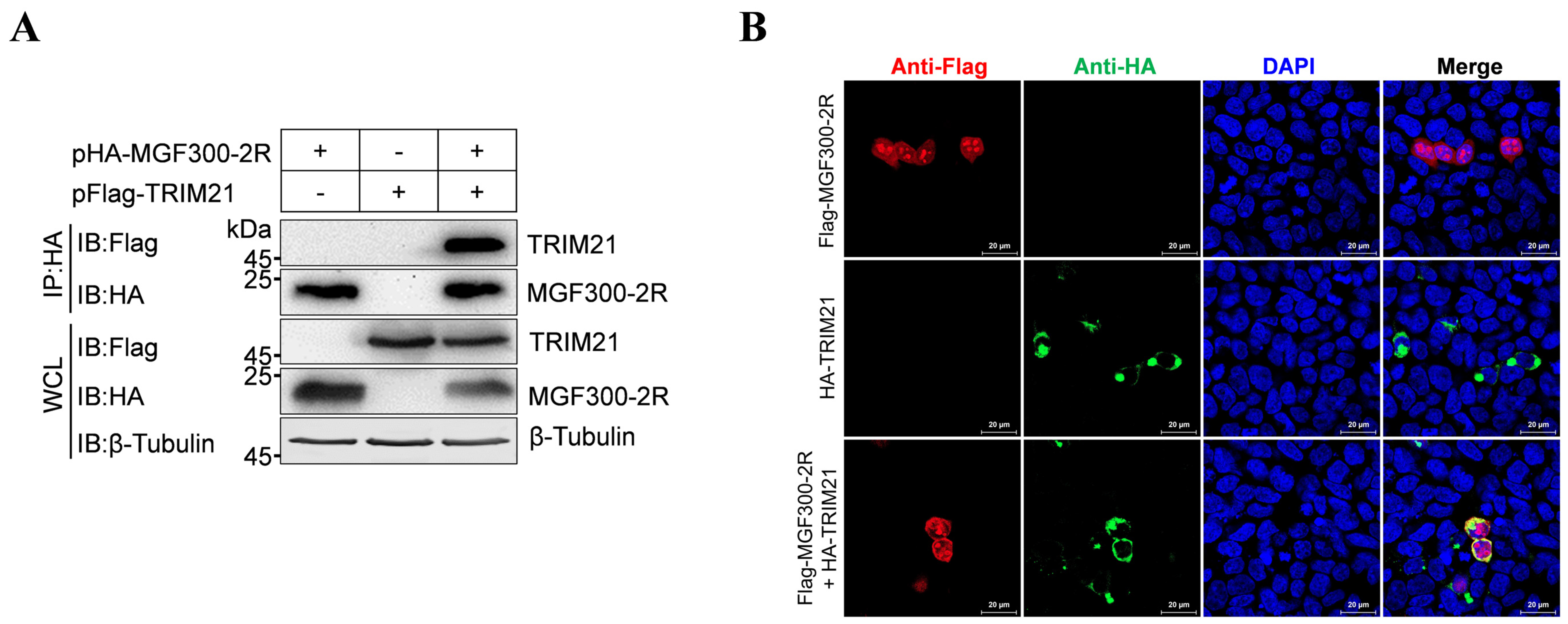
3.2. The E3 Ubiquitin Ligase TRIM21 Interacts with MGF300-2R

To validate the interaction between MGF300-2R and TRIM21, we performed a co-IP assay. The results showed that TRIM21 was precipitated by MGF300-2R in the lysates from the HEK293T cells co-transfected with pHA-MGF300-2R and pFlag-TRIM21 but not in those of the control group (Figure 2A). Moreover, after co-transfection of the HEK293T cells with pFlag-MGF300-2R and pHA-TRIM21, we observed the co-localization of MGF300-2R and TRIM21 in the cytoplasm of the HEK293T cells using laser confocal microscopy (Figure 2B). These data confirm that the E3 ubiquitin ligase TRIM21 interacts with MGF300-2R.

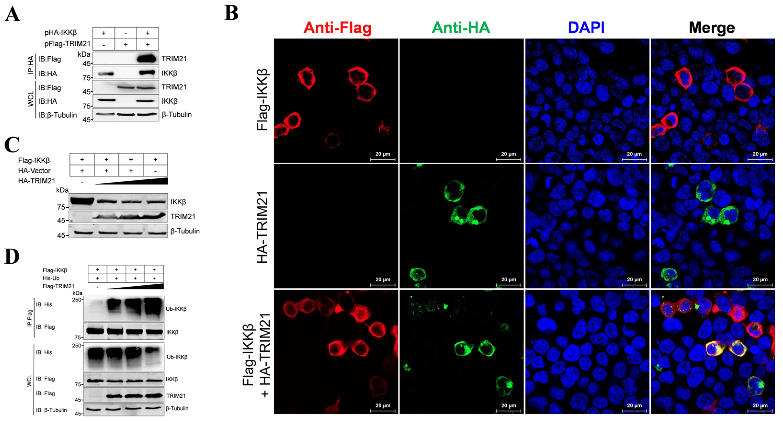
3.3. The E3 Ubiquitin Ligase TRIM21 Promotes the Ubiquitination and Degradation of IKKβ

To investigate the involvement of the E3 ubiquitin ligase TRIM21 in IKKβ degradation induced by MGF300-2R, we transfected HEK293T cells with pHA-IKKβ and pFlag-TRIM21 alone or co-transfected with them. As shown in Figure 3A, TRIM21 immunoprecipitated with IKKβ. Laser confocal microscopy analysis confirmed that TRIM21 and IKKβ were colocalized in the cytoplasm of HEK293T cells (Figure 3B), suggesting an interaction between IKKβ and TRIM21. To ascertain whether TRIM21 has a role in IKKβ degradation, we co-transfected the HEK293T cells with pFlag-IKKβ and varying amounts of pHA-TRIM21. The results showed a dose-dependent reduction in IKKβ protein levels (Figure 3C), indicating TRIM21 facilitates IKKβ degradation. Moreover, by overexpressing pFlag-IKKβ, along with pHis-Ub and increasing the doses of pFlag-TRIM21 in the HEK293T cells, followed by co-IP assay, we found the dose-dependent ubiquitination of IKKβ by TRIM21 (Figure 3D). These results indicate that TRIM21 functions as an E3 ubiquitin ligase, promoting the ubiquitination and subsequent degradation of IKKβ.

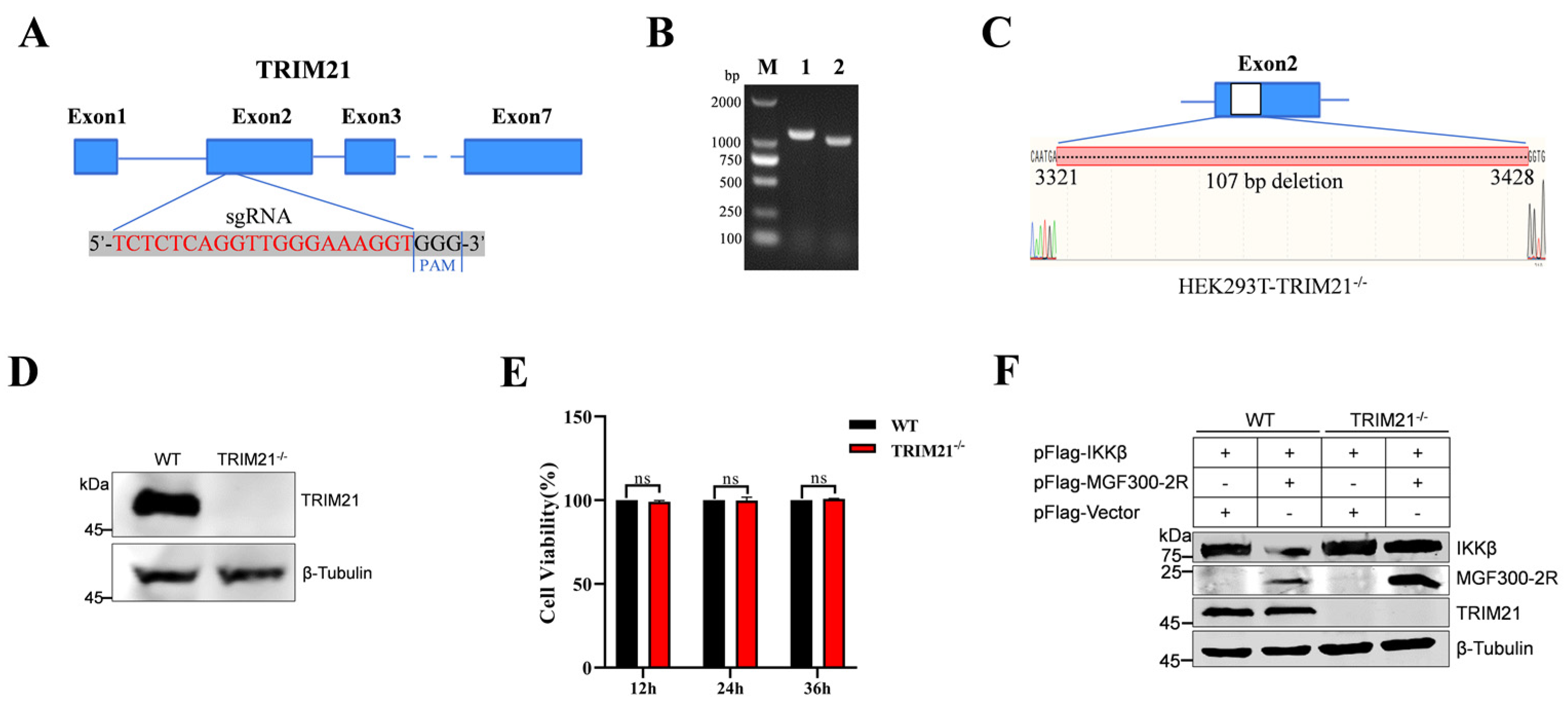
3.4. Degradation of IKKβ by MGF300-2R Depends on TRIM21
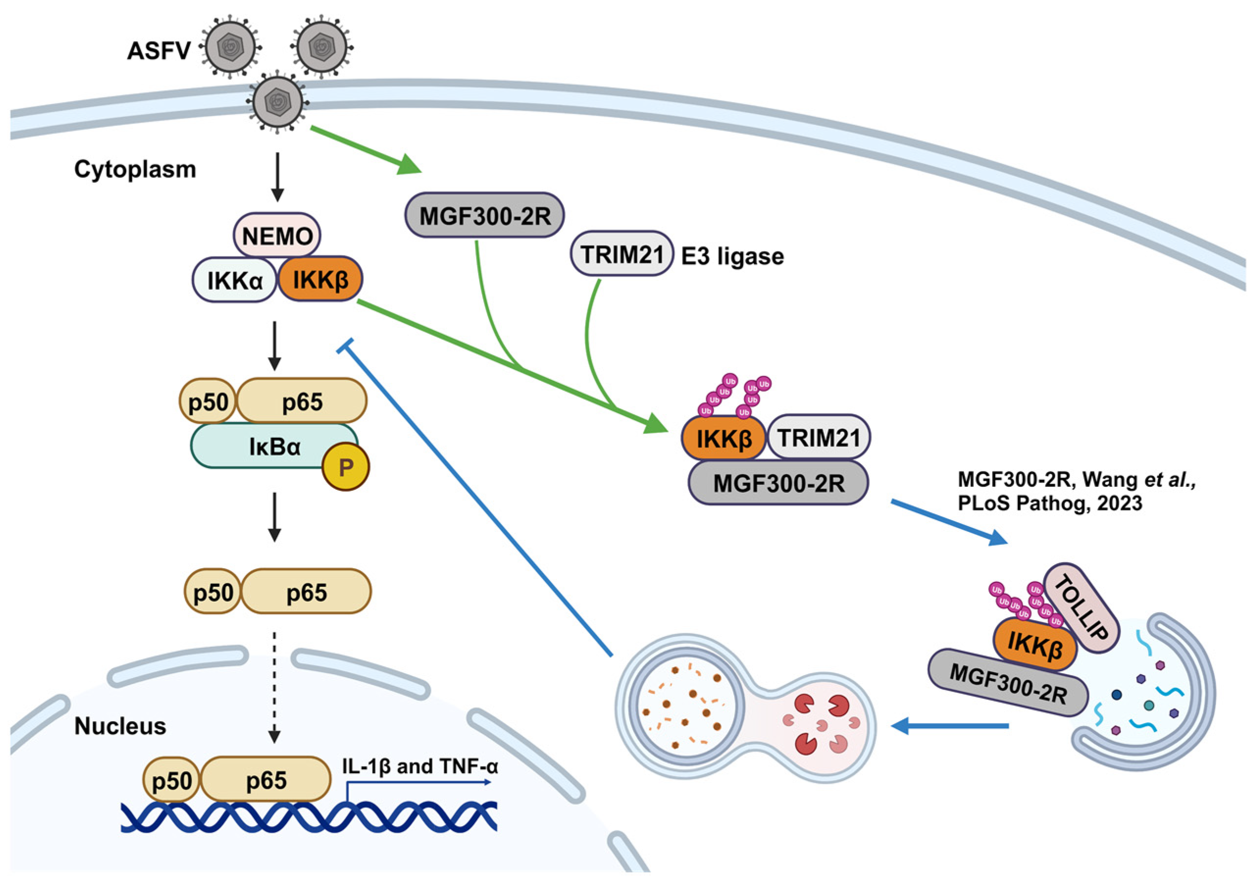
To study the role of TRIM21 in the degradation of IKKβ by MGF300-2R, we generated TRIM21-knockout (KO) HEK293T (HEK239T-TRIM21−/−) cells. Briefly, the targeting RNA (sgRNA) targeting exon 2 of the TRIM21 gene was designed using the CRISPR Design website (http://crispr.mit.edu, accessed on 24 February 2023) (Figure 4A). The knockout of TRIM21 was determined by PCR, Sanger sequencing and Western blotting. In comparison with the wild-type HEK239T (HEK293T-WT) cells, the HEK239T-TRIM21−/− cells exhibited a deletion of 107 base pairs in the exon 2 sequence of the TRIM21 gene (Figure 4B,C) and notably lacked TRIM21 protein expression (Figure 4D). Cell viability analysis revealed that knocking out TRIM21 in the HEK239T cells had no significant effect on the cell viability at 12, 24 and 36 h post-incubation (Figure 4E). Subsequently, to investigate the influence of TRIM21 deletion in the HEK239T cells on the degradation of IKKβ by MGF300-2R, both HEK293T-WT and HEK239T-TRIM21−/− cells were transfected either with pFlag-IKKβ alone or co-transfected with pFlag-MGF300-2R and pFlag-IKKβ. The results demonstrated that MGF300-2R promoted the degradation of IKKβ in the HEK293T-WT cells, whereas no such phenomenon was observed in the HEK239T-TRIM21−/− cells (Figure 4F). These findings conclusively establish TRIM21 as an E3 ubiquitin ligase crucially involved in MGF300-2R-mediated IKKβ degradation.

4. Discussion

ASF LAVs can provide complete homologous and partial heterologous protection [8]. Identifying new virulence factors and analyzing the pathogenic mechanisms involved in virulence factors will contribute to the development of LAVs [20]. Our previous study identified a new virulence factor MGF300-2R and found that it degrades IKKβ through selective autophagy. Here, we discovered that the E3 ubiquitin ligase TRIM21 is involved in the degradation of IKKβ by MGF300-2R. Through the IP-MS results, we pulled down 328 proteins that can interact with MGF300-2R (Figure 1). The screened E3 ubiquitin ligase TRIM21 can interact with MGF300-2R and IKKβ and promote IKKβ ubiquitination and degradation (Figure 2 and Figure 3). After the knockout of TRIM21, we demonstrated that the degradation of IKKβ by MGF300-2R is dependent on TRIM21 (Figure 4). Therefore, we conclude that ASFV MGF300-2R promotes IKKβ autophagic degradation by recruiting the E3 ubiquitin ligase TRIM21 (Figure 5).
The E3 ubiquitin ligase can promote the ubiquitination and degradation of viral proteins to resist viral infection [19,21]. On the contrary, the viruses antagonize antiviral responses by recruiting the E3 ubiquitin enzyme [11]. Our results show that ASFV MGF300-2R recruits TRIM21 to antagonize NF-κB. Additionally, studies have reported that ASFV has at least seven proteins that directly recruit E3 ubiquitin ligases in innate immunity to promote the degradation of regulatory factors by ubiquitination, indicating that the recruitment of E3 ubiquitin enzymes may be one important strategy for ASFV to antagonize the host immune defense [22,23,24,25,26,27,28]. TRIM21 plays essential roles in substrate binding and ubiquitination by two conservative domains, RING and PRY-SPRY [29]. The human papillomavirus (HPV) oncoprotein E7 can enhance the interaction between the PRY/SPRY domain of TRIM21 and the PYD domain of IFN-γ inducible protein 16 (IFI16), catalyzing the K33-linked ubiquitination degradation of IFI16, leading to cellular pyroptosis and viral immune escape [30]. Hence, identifying the key functional domains of TRIM21 involved in promoting the MGF300-2R-mediated degradation of IKKβ warrants further investigation. Studies have shown that the TRIM21 gene is a host restriction factor for PRRSV and porcine epidemic diarrhea virus (PEDV) infection in porcine alveolar macrophages and foot-and-mouth disease virus (FMDV) in porcine kidney 15 cells [31,32]. Given the important roles of TRIM21 in antiviral response, it is unsurprising that viruses have developed various strategies to counteract its activity.
Overall, our data indicate that TRIM21 functions as the E3 ubiquitin ligase responsible for degrading IKKβ through ASFV MGF300-2R. This finding highlights the negative regulatory role of TRIM21 recruited by MGF300-2R in the NF-κB signaling pathway. Furthermore, our study enriches the understanding of the mechanism underlying the MGF300-2R involvement in the pathogenicity of ASFV and identifies potential targets for the design of ASF vaccines and the development of antivirals.

Supplementary Materials

The following supporting information can be downloaded at https://www.mdpi.com/article/10.3390/v16060949/s1. Table S1: Identification of the proteins interacting with MGF300-2R.

Author Contributions

Z.L.: Conceptualization, data curation, formal analysis, investigation, methodology, visualization, writing—original draft and writing—review and editing. R.L.: Data curation, formal analysis, methodology, validation and visualization. J.L.: Data curation, formal analysis, validation and visualization. S.C.: Data curation and formal analysis. H.-J.Q.: Funding acquisition, methodology, supervision and writing—review and editing. T.W.: Funding acquisition, methodology, supervision and writing—review and editing. Y.S.: Funding acquisition, methodology, supervision and writing—review and editing. All authors have read and agreed to the published version of the manuscript.

Funding

This research was funded by the National Natural Science Foundation of China (32202774) (TW), the National Natural Science Foundation of China (32072854) (YS), the National Natural Science Foundation of China (U20A2060) (HJQ) and the Heilongjiang Provincial Natural Science Foundation of China (TD2023C007) (HJQ).

Institutional Review Board Statement

Not applicable.

Informed Consent Statement

Not applicable.

Data Availability Statement

The original contributions presented in the study are included in the article and Supplementary Materials, and further inquiries can be directed to the corresponding authors.

Conflicts of Interest

The authors declare no conflicts of interest.

References

  1. Zhou, X.; Li, N.; Luo, Y.; Liu, Y.; Miao, F.; Chen, T.; Zhang, S.; Cao, P.; Li, X.; Tian, K.; et al. Emergence of African swine fever in China, 2018. Transbound. Emerg. Dis. 2018, 65, 1482–1484. [Google Scholar] [CrossRef] [PubMed]
  2. Dixon, L.K.; Stahl, K.; Jori, F.; Vial, L.; Pfeiffer, D.U. African swine fever epidemiology and control. Annu. Rev. Anim. Biosci. 2020, 8, 221–246. [Google Scholar] [CrossRef] [PubMed]
  3. You, S.; Liu, T.; Zhang, M.; Zhao, X.; Dong, Y.; Wu, B.; Wang, Y.; Li, J.; Wei, X.; Shi, B. African swine fever outbreaks in China led to gross domestic product and economic losses. Nat. Food 2021, 2, 802–808. [Google Scholar] [CrossRef] [PubMed]
  4. Zhou, P.; Li, L.F.; Zhang, K.; Wang, B.; Tang, L.; Li, M.; Wang, T.; Sun, Y.; Li, S.; Qiu, H.J. Deletion of the H240R gene of African swine fever virus decreases infectious progeny virus production due to aberrant virion morphogenesis and enhances inflammatory cytokine expression in porcine macrophages. J. Virol. 2022, 96, e0166721. [Google Scholar] [CrossRef] [PubMed]
  5. Sun, M.; Yu, S.; Ge, H.; Wang, T.; Li, Y.; Zhou, P.; Pan, L.; Han, Y.; Yang, Y.; Sun, Y.; et al. The A137R protein of African swine fever virus inhibits type I interferon production via the autophagy-mediated lysosomal degradation of TBK1. J. Virol. 2022, 96, e0195721. [Google Scholar] [CrossRef] [PubMed]
  6. Wang, T.; Luo, R.; Zhang, J.; Lu, Z.; Li, L.F.; Zheng, Y.H.; Pan, L.; Lan, J.; Zhai, H.; Huang, S.; et al. The MGF300-2R protein of African swine fever virus is associated with viral pathogenicity by promoting the autophagic degradation of IKKα and IKKβ through the recruitment of TOLLIP. PLoS Pathog. 2023, 19, e1011580. [Google Scholar] [CrossRef]
  7. Wang, T.; Luo, R.; Zhang, J.; Lan, J.; Lu, Z.; Zhai, H.; Li, L.F.; Sun, Y.; Qiu, H.J. The African swine fever virus MGF300-4L protein is associated with viral pathogenicity by promoting the autophagic degradation of IKKβ and increasing the stability of IκBα. Emerg. Microbes Infect. 2024, 13, 2333381. [Google Scholar] [CrossRef] [PubMed]
  8. Wang, T.; Luo, R.; Sun, Y.; Qiu, H.J. Current efforts towards safe and effective live attenuated vaccines against African swine fever: Challenges and prospects. Infect. Dis. Poverty 2021, 10, 137. [Google Scholar] [CrossRef] [PubMed]
  9. Martens, S.; Behrends, C. Molecular mechanisms of selective autophagy. J. Mol. Biol. 2020, 432, 1–2. [Google Scholar] [CrossRef]
  10. Vargas, J.N.S.; Hamasaki, M.; Kawabata, T.; Youle, R.J.; Yoshimori, T. The mechanisms and roles of selective autophagy in mammals. Nat. Rev. Mol. Cell Biol. 2023, 24, 167–185. [Google Scholar] [CrossRef]
  11. Kong, Z.; Yin, H.; Wang, F.; Liu, Z.; Luan, X.; Sun, L.; Liu, W.; Shang, Y. Pseudorabies virus tegument protein UL13 recruits RNF5 to inhibit STING-mediated antiviral immunity. PLoS Pathog. 2022, 18, e1010544. [Google Scholar] [CrossRef] [PubMed]
  12. Li, J.; Zhou, Y.; Zhao, W.; Liu, J.; Ullah, R.; Fang, P.; Fang, L.; Xiao, S. Porcine reproductive and respiratory syndrome virus degrades DDX10 via SQSTM1/p62-dependent selective autophagy to antagonize its antiviral activity. Autophagy 2023, 19, 2257–2274. [Google Scholar] [CrossRef] [PubMed]
  13. Ma, Z.; Bai, J.; Jiang, C.; Zhu, H.; Liu, D.; Pan, M.; Wang, X.; Pi, J.; Jiang, P.; Liu, X. Tegument protein UL21 of alpha-herpesvirus inhibits the innate immunity by triggering CGAS degradation through TOLLIP-mediated selective autophagy. Autophagy 2023, 19, 1512–1532. [Google Scholar] [CrossRef] [PubMed]
  14. de Moura, T.R.; Mozaffari-Jovin, S.; Szabó, C.Z.K.; Schmitzová, J.; Dybkov, O.; Cretu, C.; Kachala, M.; Svergun, D.; Urlaub, H.; Lührmann, R.; et al. Prp19/Pso4 is an autoinhibited ubiquitin ligase activated by stepwise assembly of three splicing factors. Mol. Cell 2018, 69, 979–992. [Google Scholar] [CrossRef] [PubMed]
  15. Coulombe, P.; Nassar, J.; Peiffer, I.; Stanojcic, S.; Sterkers, Y.; Delamarre, A.; Bocquet, S.; Méchali, M. The ORC ubiquitin ligase OBI1 promotes DNA replication origin firing. Nat. Commun. 2019, 10, 2426. [Google Scholar] [CrossRef] [PubMed]
  16. Wang, P.; Dai, X.; Jiang, W.; Li, Y.; Wei, W. RBR E3 ubiquitin ligases in tumorigenesis. Semin. Cancer Biol. 2020, 67, 131–144. [Google Scholar] [CrossRef] [PubMed]
  17. Kim, J.G.; Shin, H.C.; Seo, T.; Nawale, L.; Han, G.; Kim, B.Y.; Kim, S.J.; Cha-Molstad, H. Signaling pathways regulated by UBR box-containing E3 ligases. Int. J. Mol. Sci. 2021, 22, 8323. [Google Scholar] [CrossRef]
  18. Singh, S.; Ng, J.; Sivaraman, J. Exploring the "Other" subfamily of HECT E3-ligases for therapeutic intervention. Pharmacol. Ther. 2021, 224, 107809. [Google Scholar] [CrossRef]
  19. Xue, B.; Li, H.; Guo, M.; Wang, J.; Xu, Y.; Zou, X.; Deng, R.; Li, G.; Zhu, H. TRIM21 promotes innate immune response to RNA viral infection through Lys27-linked polyubiquitination of MAVS. J. Virol. 2018, 92, e00321-18. [Google Scholar] [CrossRef]
  20. Wang, Y.; Kang, W.; Yang, W.; Zhang, J.; Li, D.; Zheng, H. Structure of African swine fever virus and associated molecular mechanisms underlying infection and immunosuppression: A review. Front. Immunol. 2021, 12, 715582. [Google Scholar] [CrossRef]
  21. Mao, S.; Cai, X.; Niu, S.; Wei, J.; Jiang, N.; Deng, H.; Wang, W.; Zhang, J.; Shen, S.; Ma, Y.; et al. TRIM21 promotes ubiquitination of SARS-CoV-2 nucleocapsid protein to regulate innate immunity. J. Med. Virol. 2023, 95, e28719. [Google Scholar] [CrossRef] [PubMed]
  22. Li, D.; Zhang, J.; Yang, W.; Li, P.; Ru, Y.; Kang, W.; Li, L.; Ran, Y.; Zheng, H. African swine fever virus protein MGF-505-7R promotes virulence and pathogenesis by inhibiting JAK1- and JAK2-mediated signaling. J. Biol. Chem. 2021, 297, 101190. [Google Scholar] [CrossRef] [PubMed]
  23. Ran, Y.; Li, D.; Xiong, M.G.; Liu, H.N.; Feng, T.; Shi, Z.W.; Li, Y.H.; Wu, H.N.; Wang, S.Y.; Zheng, H.X.; et al. African swine fever virus I267L acts as an important virulence factor by inhibiting RNA polymerase III-RIG-I-mediated innate immunity. PLoS Pathog. 2022, 18, e1010270. [Google Scholar] [CrossRef] [PubMed]
  24. Wang, Y.; Cui, S.; Xin, T.; Wang, X.; Yu, H.; Chen, S.; Jiang, Y.; Gao, X.; Jiang, Y.; Guo, X.; et al. African swine fever virus MGF360-14L negatively regulates type I interferon signaling by targeting IRF3. Front. Cell. Infect. Microbiol. 2022, 11, 818969. [Google Scholar] [CrossRef] [PubMed]
  25. Li, D.; Peng, J.; Wu, J.; Yi, J.; Wu, P.; Qi, X.; Ren, J.; Peng, G.; Duan, X.; Ru, Y.; et al. African swine fever virus MGF-360-10L is a novel and crucial virulence factor that mediates ubiquitination and degradation of JAK1 by recruiting the E3 ubiquitin ligase HERC5. mBio 2023, 14, e0060623. [Google Scholar] [CrossRef] [PubMed]
  26. Li, Y.H.; Peng, J.L.; Xu, Z.S.; Xiong, M.G.; Wu, H.N.; Wang, S.Y.; Li, D.; Zhu, G.Q.; Ran, Y.; Wang, Y.Y. African swine fever virus cysteine protease pS273R inhibits type I interferon signaling by mediating STAT2 degradation. J. Virol. 2023, 97, e0194222. [Google Scholar] [CrossRef] [PubMed]
  27. Li, Y.; Lou, H.; Zhao, Y.; Fan, W.; Jiao, P.; Sun, L.; Luo, T.; Liu, W. The I226R protein of African swine fever virus inhibits the cGAS-STING-mediated innate immune response. Sheng Wu Gong Cheng Xue Bao 2023, 39, 4796–4808. [Google Scholar] [PubMed]
  28. Li, Y.; Huang, L.; Li, H.; Zhu, Y.; Yu, Z.; Zheng, X.; Weng, C.; Feng, W.H. ASFV pA151R negatively regulates type I IFN production via degrading E3 ligase TRAF6. Front. Immunol. 2024, 15, 1339510. [Google Scholar] [CrossRef] [PubMed]
  29. Li, X.; Yang, L.; Chen, S.; Zheng, J.; Zhang, H.; Ren, L. Multiple roles of TRIM21 in virus infection. Int. J. Mol. Sci. 2023, 24, 1683. [Google Scholar] [CrossRef]
  30. Song, Y.; Wu, X.; Xu, Y.; Zhu, J.; Li, J.; Zou, Z.; Chen, L.; Zhang, B.; Hua, C.; Rui, H.; et al. HPV E7 inhibits cell pyroptosis by promoting TRIM21-mediated degradation and ubiquitination of the IFI16 inflammasome. Int. J. Biol. Sci. 2020, 16, 2924–2937. [Google Scholar] [CrossRef]
  31. Fan, W.; Zhang, D.; Qian, P.; Qian, S.; Wu, M.; Chen, H.; Li, X. Swine TRIM21 restricts FMDV infection via an intracellular neutralization mechanism. Antivir. Res. 2016, 127, 32–40. [Google Scholar] [CrossRef] [PubMed]
  32. Wei, Y.; Zou, C.; Zeng, S.; Xue, C.; Cao, Y. Characterization of porcine tripartite motif genes as host restriction factors against PRRSV and PEDV infection. Virus Res. 2019, 270, 197647. [Google Scholar] [CrossRef] [PubMed]
Figure 1. The E3 ubiquitin ligases pulled down by MGF300-2R were screened through Gene Ontology (GO), Kyoto Encyclopedia of Genes and Genomes (KEGG) pathway enrichment and the protein–protein interaction network. (A) Obtaining and analyzing the immunoprecipitation samples containing the proteins pulled down by MGF300-2R. (B) Venn diagram counted 328 different proteins pulled down in the HEK293T transfected with pFlag-MGF300-2R compared with the HEK293T transfected with vector pCMV-Flag. (C,D) KEGG pathway analysis and GO category functional enrichment according to three categories (cellular components, molecular functions and biological processes) were performed for 328 proteins. (E) The protein–protein interaction network of the proteins that interacted directly with TRIM21 was analyzed against the STRING database.
Viruses 16 00949 g001
Figure 2. The E3 ubiquitin ligase TRIM21 interacts with MGF300-2R. (A) TRIM21 interacts with MGF300-2R. HEK293T cells were transfected with pHA-MGF300-2R and pFlag-TRIM21 alone or co-transfected with them. The whole cell lysates (WCLs) were immunoprecipitated with anti-HA monoclonal antibody at 36 h post-transfection. The immunoprecipitates were examined by Western blotting with the indicated antibodies. (B) TRIM21 co-localizes with MGF300-2R. HEK293T cells were transfected with pFlag-MGF300-2R or pHA-TRIM21 alone or co-transfected with pHA-TRIM21 in combination with pFlag-MGF300-2R. TRIM21 and MGF300-2R were analyzed by laser confocal microscopy. Scale bar, 20 μm.
Viruses 16 00949 g002
Figure 3. The E3 ubiquitin ligase TRIM21 promotes the ubiquitination and degradation of IKKβ. (A) TRIM21 interacts with IKKβ. HEK293T cells were transfected with a plasmid encoding HA-IKKβ along with a plasmid encoding Flag-TRIM21 or vector pCMV-Flag as indicated. The cells were lysed, and the whole cell lysates (WCLs) were immunoprecipitated with anti-HA monoclonal antibody at 36 h post-transfection (hpt). The immunoprecipitates were examined by Western blotting with the indicated antibodies. (B) TRIM21 is colocalized with IKKβ. HEK293T cells were transfected with pFlag-IKKβ or pHA-TRIM21 alone or co-transfected with pHA-TRIM21 and pFlag-IKKβ. TRIM21 and IKKβ were analyzed by laser confocal microscopy. Scale bar, 20 μm. (C) TRIM21 promotes the degradation of IKKβ. HEK293T cells were co-transfected with pFlag-IKKβ and an increasing amount of pHA-TRIM21 (0, 1.0, 2.0 and 3.0 μg) for 24 h. The cell lysates were analyzed by Western blotting. (D) TRIM21 induces the ubiquitination of IKKβ. HEK293T cells were co-transfected with pFlag-IKKβ and pHis-Ub and an increasing amount of pHA-TRIM21 (0, 1.0, 2.0 and 3.0 μg). At 24 hpt, the cells were processed for immunoprecipitations with anti-Flag magnetic beads and analyzed by Western blotting with the indicated antibodies.
Viruses 16 00949 g003
Figure 4. Degradation of IKKβ by MGF300-2R depends on TRIM21. (A) Schematic of the TRIM21-knockout strategy. The sgRNA target sites are indicated in red. (BD) HEK293T cells with TRIM21 knockout (HEK293T-TRIM21−/−). Confirmation of TRIM21 knockout in HEK293T cells using PCR, Sanger sequencing and Western blotting. The wild-type HEK239T (HEK293T-WT) cells’ and HEK293T-TRIM21−/− cells’ genomes were extracted and subjected to PCR with primers TRIM21-F/R. Lane M displays a DL2,000 DNA marker, and Lanes 1 and 2 show the PCR results of the HEK293T-WT and HEK293T-TRIM21−/− cells’ genomes (B). The changes in ORF of the TRIM21 gene were verified by Sanger sequencing by Rui Biotech (C). The expression of TRIM21 protein in HEK293T-WT and HEK293T-TRIM21−/− cells was verified with anti-TRIM21 polyclonal antibody by Western blotting (D). (E) HEK293T-TRIM21−/− cells are not significantly affected in terms of cell viability compared with HEK293T-WT at different time points (12, 24, and 36 h post-incubation) using CCK-8 detection kit. (F) MGF300-2R does not promote the degradation of IKKβ in HEK239T-TRIM21−/− cells. HEK293T-WT and HEK293T-TRIM21−/− cells were co-transfected with pFlag-MGF300-2R or vector pCMV-Flag in combination with pFlag-IKKβ.
Viruses 16 00949 g004
Figure 5. A working model for the recruitment of TRIM21 to promote the ubiquitination and degradation of IKKβ by the ASFV MGF300-2R protein. The ASFV MGF300-2R protein recruits the E3 ubiquitin ligase TRIM21 to ubiquitinate IKKβ, and IKKβ is subsequently recognized and degraded via the selective autophagic pathway, which subverts the NF-κB signaling pathway [6].
Viruses 16 00949 g005
Table 1. The primers used in this study.
PrimersSequence (5′-3′)
Flag-TRIM21-ForwardCAAGCTTGCGGCCGCGAATTCGATGGCTTCAGCAGCACGC
Flag-TRIM21-ReverseATCAGATCTATCGATGAATTCTCAATAGTCAGTGGATCCTTGTGATC
sgRNA-ForwardTCTCTCAGGTTGGGAAAGGT
sgRNA-ReverseACCTTTCCCAACCTGAGAGA
TRIM21-ForwardGCTTTGGCTATTCTGAGTCT
TRIM21-ReverseTCCCCTTTGAACCTGCATC
Disclaimer/Publisher’s Note: The statements, opinions and data contained in all publications are solely those of the individual author(s) and contributor(s) and not of MDPI and/or the editor(s). MDPI and/or the editor(s) disclaim responsibility for any injury to people or property resulting from any ideas, methods, instructions or products referred to in the content.

