Article

The Distal Promoter of the B438L Gene of African Swine Fever Virus Is Responsible for the Transcription of the Alternatively Spliced B169L

State Key Laboratory for Animal Disease Prevention and Control, National African Swine Fever Para-Reference Laboratory, National High-Containment Facilities for Animal Disease Control and Prevention, Harbin Veterinary Research Institute, Chinese Academy of Agricultural Sciences, Harbin 150069, China
*
Authors to whom correspondence should be addressed.
These authors contributed equally to this work.
Viruses 2024, 16(7), 1058; https://doi.org/10.3390/v16071058
Submission received: 30 April 2024 / Revised: 20 June 2024 / Accepted: 28 June 2024 / Published: 30 June 2024
(This article belongs to the Special Issue Porcine Viruses 2024)

Abstract

:
The B169L protein (pB169L) of African swine fever virus (ASFV) is a structural protein with an unidentified function during the virus replication. The sequences of the B169L gene and the downstream B438L gene are separated by short intergenic regions. However, the regulatory mode of the gene transcription remains unknown. Here, we identified two distinct promoter regions and two transcription start sites (TSSs) located upstream of the open reading frame (ORF) of B438L. Using the promoter reporter system, we demonstrated that the cis activity of the ORF proximal promoter exhibited significantly higher levels compared with that of the distal promoter located in the B169L gene. Furthermore, transfection with the plasmids with two different promoters for B438L could initiate the transcription and expression of the B438L gene in HEK293T cells, and the cis activity of the ORF proximal promoter also displayed higher activities compared with the distal promoter. Interestingly, the B438L distal promoter also initiated the transcription of the alternatively spliced B169L mRNA (B169L mRNA2) encoding a truncated pB169L (tpB169L) (amino acids 92–169), and the gene transcription efficiency was increased upon mutation of the initiation codon located upstream of the alternatively spliced B169L gene. Taken together, we demonstrated that the distal promoter of B438L gene initiates the transcription of both the B438L mRNA and B169L mRNA2. Comprehensive analysis of the transcriptional regulatory mode of the B438L gene is beneficial for the understanding of the association of B438L protein and pB169L and the construction of the gene-deleted ASFV.

1. Introduction

African swine fever (ASF), caused by African swine fever virus (ASFV), is a highly contagious and hemorrhagic disease of swine, with a high mortality of up to 100% [1]. Since its initial outbreak in Kenya in the 1920s, ASF predominantly manifested as an epidemic in Europe, South America, and the Caribbean [2]. However, the disease was introduced into China in 2018 and resulted in huge economic losses due to the lack of effective vaccines and antiviral drugs [3,4]. ASFV belongs to the family Asfarviridae and is a large enveloped virus with a genome of double-stranded DNA, encoding more than 50 structural proteins and 100 nonstructural proteins, many of which have undetermined functions [5]. It is an important strategy to investigate the functions of viral proteins through constructing a gene-deleted ASFV. The identification of viral gene promoters will be beneficial for the construction of the gene-deleted ASFV mutants. By scanning the complete genome of ASFV, we demonstrated that the viral structural genes B438L and B169L are densely distributed in an organized manner, implying the transcriptional association between the viral genes.
The B169L gene encodes the B169L protein (pB169L) of 169 amino acids (aa). It is noteworthy that the transcription of B169L gives rise to two mRNAs: the full-length B169L mRNA and the alternatively spliced B169L mRNA (referred to as B169L mRNA2) encoding a truncated pB169L (tpB169L) (aa 92–169) [6]. However, the transcription start site (TSS) and the promoter that are responsible for the transcription of the B169L mRNA2 remain unknown. Notably, the B169L gene is arranged adjacent with its downstream gene B438L in the ASFV genome. Therefore, we postulated that the promoter sequence of the downstream gene B438L is located within the B169L gene.
A previous study has revealed that the ASFV B438L protein (pB438L) is indispensable for the construction of the icosahedral capsid of ASFV, and the gene-deletion of B438L leads to a significant alteration in virion assembly, resulting in the formation of tubular viral particles instead of the characteristic icosahedral capsid, thereby disrupting the icosahedral symmetry [7,8]. The morphological perturbation also affects the infectivity and pathogenicity of viruses. It has been shown that the transcriptional machinery of ASFV exhibits complexity, which is involved in the complex interplay between enzymes and cofactors encoded by ASFV [9]. The ASFV B438L and B169L ORFs are densely distributed, it is possible that the upstream gene encompasses promoter(s) to govern the expression of the downstream gene(s) of ASFV, and an individual gene may harbor multiple promoters for the initiation of the transcription of downstream viral genes. Therefore, it is essential to decipher the transcriptional regulation patterns of the downstream B438L gene, which will facilitate the design of the B169L-deleted ASFV vaccines.
In this study, we elucidated an intricate interrelationship between the ASFV B438L gene and its adjacent upstream and downstream genes. Notably, the B438L promoter region is located in the upstream of its ORF and encompasses two distinct promoters, and the B438L distal promoter also initiates the transcription of the alternatively spliced B169L (B169L-2). Moreover, we have discerned that the transcriptional activity of the B438L gene depends on the viral transcriptional machinery, in conjunction with relevant cofactors, and the transcription efficiency of the gene was also affected by the translation initiation codon (ATG) located upstream of B169L-2. The findings enhance our understanding of the transcriptional mechanisms of ASFV, thereby providing novel insights into the development of antiviral strategies.

2. Materials and Methods

2.1. Cells and Viruses

Primary porcine alveolar macrophages (PAMs) were collected from the alveolar lavage fluid of 20-day-old specific-pathogen-free (SPF) pigs. The PAMs or HEK293T (ATCC) cells were cultured in RPMI 1640 (catalog no. C11875500BT; Gibco, New York, NY, USA) or Dulbecco’s modified Eagle’s medium (catalog no. D6221; Sigma-Aldrich, Darmstadt, Germany) supplemented with 10% fetal bovine serum (FBS) (catalog no. 10099-141C; Gibco, Thornton, Australia), 100 units/mL penicillin, and 100 μg/mL streptomycin (catalog no. 15240-062; Gibco, Carlsbad, CA, USA) at 37 °C with 5% CO2. The ASFV HLJ/2018 strain was isolated from field pig samples in China and propagated in the PAMs as described previously [10].

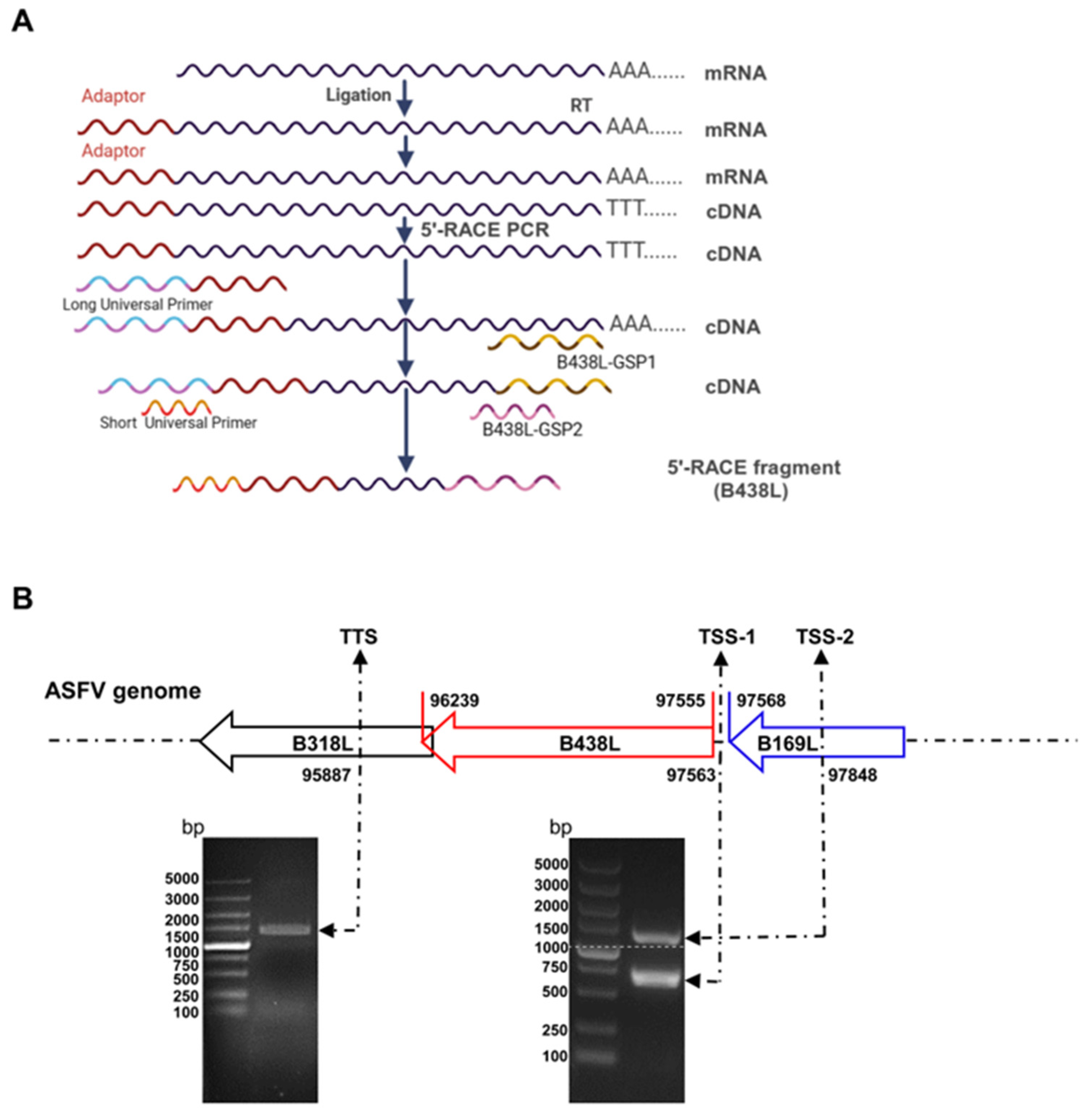
2.2. 5′ and 3′ Rapid Amplification of cDNA Ends (RACE) Assay

The total RNA was extracted from the ASFV-infected PAMs (the target cells of ASFV) at a multiplicity of infection (MOI) of 2 using an RNA purification kit (catalog no. BSC52M1; BioFlux, Hangzhou, China) according to the manufacturer’s instructions. The 5′RACE was performed using the 5′-Full RACE kit (catalog no. 638858; TaKaRa, Mountain View, CA, USA) and the B438L-specific primers (B438L-GSP1 and -GSP2 primers). The gene products of the 5′RACE polymerase chain reaction (PCR) were cloned into the pMD19-T vector (catalog no. 639648; TaKaRa, Mountain View, CA, USA) and sequenced using the M13 primers. All the primers in this study are shown in Table 1.

2.3. Construction of Plasmids

The genomic DNA of ASFV was extracted using a MagaBio plus virus DNA purification kit (catalog no. 9109; BioFlux, Hangzhou, China) according to the manufacturer’s instructions. Six primer pairs were designed for the amplification of the B438L-1 to -6 genes using the genomic DNA as a template. To generate the gene constructs of B169L-2 and B169L-mATG-2 (the mutation of the second ATG in B169L), a series of reverse primers and intermediate primers were generated and subjected to PCR using the B438L-1 gene as a template. The mutation and deletion of the GC and TATA boxes in B438L-7 and -5 were obtained by sequence overlapping extension (SOE) PCR, and the primers used in this study are shown in Table 1. All the PCR products were purified by using a gel extraction kit (catalog no. 740609.10; TaKaRa, Mountain View, CA, USA) and subsequently digested with the endonucleases KpnI (catalog no. 1618; TaKaRa, Mountain View, CA, USA) and XhoI (catalog no. 1635; TaKaRa, Mountain View, CA, USA), followed by a second gel purification for subsequent assays. The sequences of the B438L promoters were cloned into the pGL3-Basic luciferase reporter plasmid (catalog no. E1751; Promega, Madison, WI, USA). The recombinant plasmids were subjected to DNA sequencing.

2.4. Luciferase Reporter Assay

HEK293T cells cultured in 24-well plates were transfected with 0.5 μg of a series of reporter plasmids harboring different B438L promoters, a negative control (pGL3-Basic), and a positive control (pGL3-Control), respectively, together with 0.05 μg of an internal control plasmid pRL-TK expressing the Renilla luciferase.

2.5. Western Blotting Analysis

The HEK293T cells transfected with the plasmids were lysed with RIPA buffer and subjected to SDS-PAGE analysis, and then, the protein bands were electro-transferred onto nylon membranes. Subsequently, the membranes were blocked in TBS containing 5% skimmed milk for 2 h at room temperature and incubated with a mouse anti-Flag monoclonal antibody (MAb) (catalog no. F7425, Abcam, Cambridge, UK) or mouse anti-β-actin polyclonal antibodies (PAb) (catalog no. ZMS1156; Sigma-Aldrich, Darmstadt, Germany) for 2 h. The membranes were rinsed three times using TBS containing 0.05% Tween 20 (TBST) and incubated with IRDye 800CW goat anti-mouse IgG secondary antibody (catalog no. 926-32210; LI-COR, Lincoln, Dearborn, MI, USA) for 1 h at room temperature. Next, the membranes were scanned using an Odyssey infrared imaging system (LiCor BioSciences).

2.6. RNA Extraction and RT-qPCR

Total RNA was extracted from the PAMs treated with specific inhibitors or virus infection by a Simply P total RNA extraction kit (catalog no. BSC52M1; BioFlux, Hangzhou, China). The isolated RNA was reverse-transcribed to cDNA using a FastKing gDNA Dispelling RT SuperMix (catalog no. KR118-02; Tiangen, Shanghai, China) according to the manufacturer’s protocols. The cDNAs were used to analyze the mRNA transcription of B169L-2 by RT-qPCR using a QuantStudio system (Applied Biosystems), as described previously [11]. The relative mRNA levels of the target genes were normalized to that of the internal reference glyceraldehyde-3-phosphate dehydrogenase (GAPDH), and all primers are listed in Table 1. The relative fold changes in gene expression were determined by the threshold cycle (2−ΔΔCT) method [12].

2.7. Statistical Analysis

Statistical analysis was performed using GraphPad Prism 8.0 (San Diego, CA, USA). Statistical differences between groups were assessed by Student’s t test. All the experiments were performed independently in triplicates. Error bars represent standard deviations (SDs) or standard errors of the mean (SEM) in each group, as indicated in the figure legends (ns, not significant, p > 0.05; *, p < 0.05; **, p < 0.01; ***, p < 0.001). A p-value of < 0.05 was considered significant.

3. Results

3.1. Two Distinct Initiation Sites for B438L Transcription

To analyze the TSS located upstream of the B438L ATG, 5′RACE assay was conducted using the total RNA of the PAMs infected with the ASFV HLJ/2018 strain at 24 h postinfection (hpi). The first round of amplification was run by using the B438L-GSP1 primer pairs. The products of the first round of PCR (5 μL) were used as a template to perform the second round of amplification using the B438L-GSP2 primer pairs (Figure 1A). As shown in Figure 1B, PCR products were generated, indicating that the ASFV transcripts were successfully obtained. Approximately 20–25 bacterial clones from each 5′RACE product were sequenced and confirmed to harbor identical 5′-ends, indicating that they were derived from the same B438L mRNA transcript. The two positive bands with 700 and 1100 bp, respectively, were mainly amplified by PCR (Figure 1B). Upon sequence alignment, we identified two distinct TSSs located at nt 97563 (TSS-1) and 97848 (TSS-2) of the genome of the ASFV HLJ/2018 strain (Figure 1B). In addition, 3′RACE assay was performed to ascertain the transcription termination site of B438L, which was determined to be located at nt 95887 (Figure 1B). Collectively, these findings suggest the presence of two TSSs for the B438L gene.

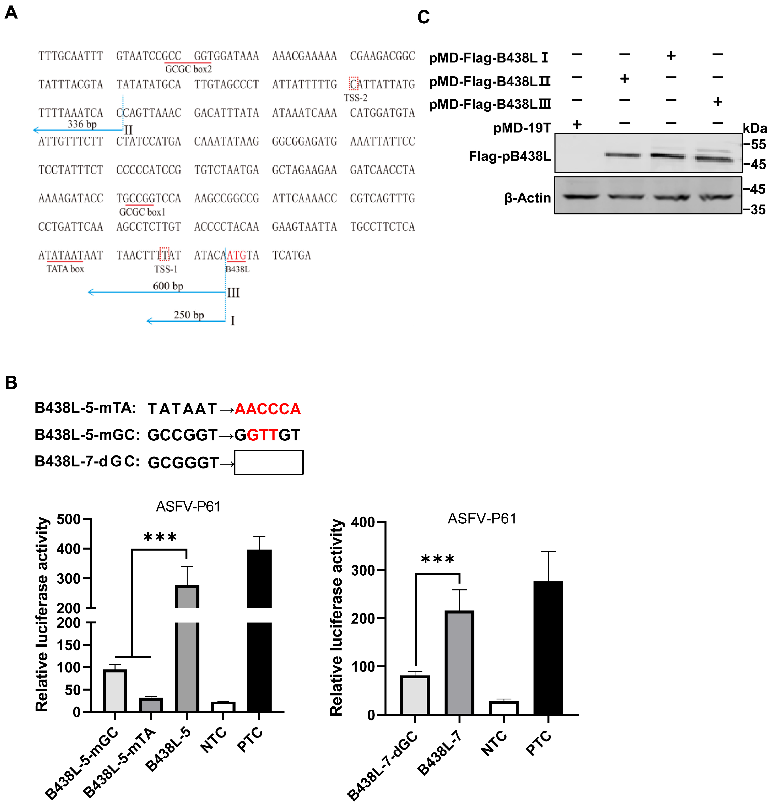
3.2. Mapping of the B438L Promoter Region Located in the B169L Gene

To delineate the promoter regions of ASFV B438L, a series of primers were designed to amplify seven DNA fragments of the B438L gene upstream, using the ASFV genomic DNA as the template (Table 1). Each promoter region was delineated with respect to an individual TSS position, as illustrated in Figure 2A. Notably, these selected promoter regions were designed to lack the ATG codon in the 3′ flanking sequence downstream of the TSS, thereby preventing any potential interference with the translation of the luciferase gene. To evaluate the potential promoter activity of these regions, the DNA fragments were cloned into pGL3-Basic to give rise to the luciferase reporter plasmids.
Subsequently, HEK293T cells were transfected with each of the reporter plasmids of different promoter regions and the internal control plasmid pRL-TK for 24 h. Afterward, the cells were lysed with the passive buffer, and the cell lysates were subjected to the examination of the luciferase activities using the dual-luciferase reporter assay system according to the manufacturer’s instructions. As shown in Figure 2B, none of the reporter plasmids displayed any promoter activities. In light of this observation, we presumed that the transcription of the B438L gene is likely to depend on ASFV-specific RNA polymerases and other essential transcription factors. To verify the hypothesis, HEK293T cells were transfected with the B438L gene reporter plasmids for 12 h. Subsequently, the cells were infected with an HEK293T cell-adapted ASFV, the ASFV-P61 strain [13], at an MOI of 0.1 for 24 h. Next, the luciferase activities were examined as described above. The results showed that all the reporter plasmids displayed promoter activities, while the B438L-1, B438L-3, and B438L-5 exhibited higher activities in comparison with B438L-2, B438L-4, and B438L-6 (Figure 2C). More specifically, the promoter activities of B438L-2 and B438L-4 were shown to be lower than those of B438L-3 and B438L-5, respectively (Figure 2D and E). To further determine whether a promoter exists between B438L-3 and B438L-5, we constructed a luciferase reporter plasmid in the genome region, and the data showed that B438L-7 also exhibited higher activity compared with the negative control (Figure 2F). Taken together, these findings suggest that B438L-5 and B438L-7 are responsible for initiating B438L transcription at TSS-1 and TSS-2, respectively.

3.3. Identification of the TATA and GC Boxes in the B438L Promoter

We identified two upstream fragments of the B438L TSS as promoters. Utilizing a comprehensive analysis of the locations of TATA or GC box within the promoter regions, along with computational analysis [14,15,16,17], we predicted that the putative TATA and GC boxes are positioned between 10 and 100 nt, respectively, upstream of TSS (Figure 3A). The TSSs of ASFV-B438L were determined at nt 97563 and 97848 in the ASFV HLJ/2018 strain.
We predicted the GC and TATA boxes in the two promoters of the B438L gene. The primers for amplifying the B438L promoter regions are shown in Table 1. The B438L-GSP1 and -GSP2 primers were designed based on the genome of the ASFV HLJ/2018 strain, while the 5′RACE Long and Short Universal Primers were provided by the 5′RACE kit. The TATA box or GC box was predicted as described above.
To identify the GC box or TATA box in the regions of B438L-7 and B438L-5, we introduced mutations or deletions (Figure 3B, upper panel) in these cis-acting elements using the mutant primers (Table 1). As shown in Figure 3B, the mutation (B438L-5-mGC) or deletion (B438L-7-dGC) of the GC box and the mutation of the TATA box (B438L-5-mTA) resulted in a reduction in the promoter activity. The data indicate that the B438L promoters contain functional TATA and GC boxes, which are indispensable for the promoter activity.
To further validate the identified promoter regions, we generated a series of recombinant plasmids expressing B438L using the clonal vector pMD-19T, each of which carries one or two of the two B438L promoter regions. Subsequently, HEK293T cells were transfected with the plasmids harboring B438L under the control of various regions of the promoters, pMD-Flag-B438L-I (the B438L-5 region), -II (the B438L-7 region), and -III (the B438L-3 region), or the empty pMD-19T vector, respectively. At 36 h post-transfection (hpt), the cell lysates were analyzed by Western blotting to verify the expression of pB438L. Notably, pB438L was efficiently expressed in the cells transfected with different plasmids (Figure 3C), indicating that both the distal and proximal promoters of B438L can initiate the expression of pB438L.

3.4. Identification of the B438L Distal Promoter That Initiates Transcription of B169L-2

Since the B438L distal promoter is located in the B169L gene, we investigated whether the promoter affects the B169L expression. To this end, we reviewed the relevant data and found that there is B169L-mRNA2 encoding a truncated pB169L (tpB169L) (aa 92–169). The sequence of B169L-2 is located in the downstream of the B438L distal promoter. Thus, we verified whether the B438L distal promoter is capable of initiating the transcription of B169L-2. Firstly, we constructed the recombinant plasmid pMD-Flag-tB169L, which carries the gene sequence of B169L-2 as well as the distal promoter sequence of B438L. Secondly, HEK293T cells were transfected with pMD-Flag-tB169L or pMD-Flag-B438L-II and infected with ASFV-P61. At 24 hpi, the cell lysates were analyzed by Western blotting to verify the expression of tpB169. Notably, tpB169L was expressed in the pMD-Flag-tB169L-transfected HEK293T cells (Figure 4A), indicating that the distal promoter of B438L can initiate the expression of tpB169L.
We further analyzed the sequences of B169L-2 and revealed that the gene contains two ATGs (ATG-2 and ATG-3), located in the region between TSS-2 and the termination codon TAA-1 (Figure 4A). Further investigations demonstrated that there is a termination codon (TAA-2) located in the 15 nt downstream of ATG-2, and ATG-3 is required for the translation of B169L-2. We speculated that the transcription efficiency of B169L-2 was impaired due to the presence of a mini transcript (located in the region of ATG-2 and TAA-2). Thus, we mutated the ATG-2 of B169L and constructed the Flag-tagged mutant plasmid pMD-B169L-mATG-2. Next, HEK293T cells were transfected with pMD-tB169L and pMD-B169L-mATG-2 and infected with ASFV-P61. At 24 hpi, cell lysates were collected to examine the protein expression of pMD-Flag-tB169L and pMD-tB169L-mATG2 by Western blotting, and total RNA was extracted for reverse transcription–quantitative PCR (RT-qPCR) to determine the mRNA transcription. The results showed that the transcription and translation efficiency of pMD-tB169L-mATG2 were significantly higher than those of pMD-Flag-tB169L (Figure 4B,C), indicating that the B438L distal promoter is capable of initiating the transcription of B438L as well as B169L-2, and that the mutation of the ATG-2 located between the TSS-2 of B438L and the ATG of B169L-2 (ATG-3) can efficiently enhance the expression of B169L-2.

4. Discussion

Several studies have focused on the regulation of gene transcription in eukaryotic cells. However, the modulation of viral gene transcription is still insufficient, especially in ASFV, a large DNA virus. The regulatory mechanisms of the genome transcription of ASFV remain largely unknown. The transcription of the ASFV genome exhibits different time courses, which are regulated by early or late promoters [18]. The ASFV p30 (encoded by the CP204L gene) and p72 (encoded by the B646L gene), two important structural proteins of ASFV, are expressed in the early and late stages, respectively, of the virus life cycle [19,20]. However, the promoters and the transcription time courses of other viral genes remain to be elucidated.
In this study, we revealed that two promoter regions are responsible for the ASFV B438L transcription located upstream of the B438L ORF. In addition, we demonstrated that the B169L gene (the upstream gene of B438L) contains a distal promoter for B438L. Interestingly, this distal promoter not only initiates the transcription of the B438L gene but also governs the transcription of B169L-2, suggesting the precise regulatory association of the adjacent viral genes. To further identify the key elements of the promoters, the TATA and GC box regions of the B438L promoters were predicted and verified by using site-directed mutagenesis and serial deletions of the boxes. The promoter activity significantly decreased upon mutation of the TATA box. Similarly, mutation or deletion of the GC box also led to a significant reduction in promoter activity. Collectively, we established a novel approach to elucidate the transcriptional association between B438L and B169L, wherein viral gene promoters were identified through the transfection of a promoter reporter in conjunction with the infection with an HEK293T cell-adapted ASFV mutant.
Furthermore, we have also observed an initiation codon (ATG-2) located upstream of B169L-2 (Figure 4A). Unexpectedly, the substitution of the ATG with GCT resulted in an enhanced transcription and translation efficiency for B169L-2 (Figure 4B). The presence of the ATG-2 is likely to be involved in modulating the expression of the downstream B169L-2, while the mutation of ATG-2 is speculated to abolish the binding between transcription factors and the promoter, thereby releasing the transcription factors and favoring the downstream gene transcription. The transcription time course of B169L-2 is also different from that of B169L, since the late promoter initiates the B169L transcription, while the early promoter initiates the transcription of B169L-2. Here, we identified the transcriptional regulatory element of B169L-2, indicating that ASFV employs multiple strategies to regulate gene expression. Future studies are required to investigate the functional role of B169L-2 during ASFV replication and the design of the B169L gene-deleted ASFV. The identification of early and late promoters of ASFV will greatly facilitate the investigation into the regulatory mechanisms governing the expression of viral proteins during viral infection.
In addition, in the absence of viral infection, all of the luciferase reporter plasmids were devoid of promoter activity. In contrast, in the presence of viral infection, all of the luciferase reporter plasmids exhibited promoter activities, suggesting that the regulation of the two promoters requires the participation of the ASFV-encoded polymerases and their associated factors (Figure 2B,C). ASFV is a large nucleoplasmic DNA virus that replicates predominantly in the cytoplasm and is involved in the nuclear translocation of the viral genome [21]. However, there is no convincing evidence to support genome replication in the nucleus, which appears to be similar to the members of the Poxviridae family that replicate exclusively in the cytoplasm [22]. It is likely that the transcription of several viral genes occurs in the nucleus in the early stage of ASFV replication, and then, the mRNAs are subjected to nuclear export and subsequent translation [23]. The soluble extracts of ASFV particles were found to possess the ability of initiating the transcription system in vitro [24,25], indicating that the ASFV genome encodes multiple viral RNA polymerases and transcription factors.
Viruses have evolved multiple strategies to regulate viral gene expression to complete the virus life cycle, some of which contain multiple promoters to facilitate the selective activation of gene transcription in response to diverse conditions, thereby enhancing viral adaptation in host cells. It has been shown that the X gene of hepatitis B virus (HBV), the nef gene of HIV-1, and the UL34 gene (encoding ICP0) of herpes simplex virus 1 (HSV-1) contain multiple promoters, which utilize multiple promoters to regulate gene expression patterns for the adaptation to host diversity, maintaining stable functionality at multiple stages of the virus life cycle [26,27,28,29,30,31]. Moreover, the presence of multiple promoters contributes to the genetic diversity of viral genes, thereby facilitating immune evasion of host cells. The IE1 gene of human cytomegalovirus (CMV) and the LMP1 gene of Epstein–Barr virus (EBV) also have multiple promoters, with these promoters playing a pivotal role in viral infection. Different promoters are activated during various stages of the viral life cycle, favoring virus replication and transmission [32,33,34,35]. In this study, we demonstrated that the distal promoter of the B438L gene initiates the transcription of both the B438L mRNA and B169L mRNA2, enhancing the understanding of viral gene transcription. Interestingly, in comparison with the promoters of different ASFV genes, we observed a significant enrichment in A and T in the promoters (Figure 5), indicating that ASFV has evolved unique regulatory mechanisms for gene transcription.

5. Conclusions

In conclusion, we demonstrated for the first time that the ORF of the ASFV B438L gene was governed by two different promoters, and the distal promoter could also initiate the transcription of both the B438L and B169L-2 genes (Figure 6). A comprehensive understanding of the multiple promoters of viral genes can facilitate the development of more effective antiviral strategies.

Author Contributions

Conceptualization, H.C., H.D. and Y.W.; methodology, H.C., H.D. and Y.W.; software, H.C., H.D. and D.L.; validation, H.C., H.D. and Y.W.; formal analysis, H.C., M.L. and L.L.; investigation, H.C.; resources, H.Q.; data curation, S.L.; writing—original draft preparation, H.C.; writing—review and editing, H.Q. and S.L; visualization, D.P., J.D. and J.L.; supervision, S.L.; project administration, H.Q.; funding acquisition, H.Q. and S.L. All authors have read and agreed to the published version of the manuscript.

Funding

This work was supported by the Natural Science Foundation of China (grants 32072866 and U20A2060), and the Heilongjiang Provincial Natural Science Foundation of China (grant YQ2022C043).

Institutional Review Board Statement

This study was conducted in compliance with the Animal Welfare Act and Guide for the Care and Use of Laboratory Animals, approved by the Laboratory Animal Welfare Committee of Harbin Veterinary Research Institute (approval number 220602-01-GR).

Informed Consent Statement

Not applicable.

Data Availability Statement

Data supporting the reported results are available in this article.

Conflicts of Interest

The authors declare no conflicts of interest in this work.

References

  1. Iyer, L.M.; Aravind, L.; Koonin, E.V. Common origin of four diverse families of large eukaryotic DNA viruses. J. Virol. 2001, 75, 11720–11734. [Google Scholar] [CrossRef] [PubMed]
  2. Alejo, A.; Andrés, G.; Salas, M.L. African swine fever virus proteinase is essential for core maturation and infectivity. J. Virol. 2003, 77, 5571–5577. [Google Scholar] [CrossRef] [PubMed]
  3. Carrascosa, A.L.; Bustos, M.J.; de Leon, P. Methods for growing and titrating African swine fever virus: Field and laboratory samples. Curr. Protoc. Cell Biol. 2011, 26, 21–26. [Google Scholar] [CrossRef] [PubMed]
  4. Chen, W.; Zhao, D.; He, X.; Liu, R.; Wang, Z.; Zhang, X.; Li, F.; Shan, D.; Chen, H.; Zhang, J.; et al. A seven-gene-deleted African swine fever virus is safe and effective as a live attenuated vaccine in pigs. Sci. China Life Sci. 2020, 63, 623–634. [Google Scholar] [CrossRef] [PubMed]
  5. Dixon, L.K.; Chapman, D.A.; Netherton, C.L.; Upton, C. African swine fever virus replication and genomics. Virus Res. 2013, 173, 3–14. [Google Scholar] [CrossRef]
  6. Cackett, G.; Matelska, D.; Sýkora, M.; Portugal, R.; Malecki, M.; Bähler, J.; Dixon, L.; Werner, F. The African swine fever virus transcriptome. J. Virol. 2020, 94, e00119-20. [Google Scholar] [CrossRef] [PubMed]
  7. Epifano, C.; Krijnse-Locker, J.; Salas, M.L.; Salas, J.; Rodríguez, J.M. Generation of filamentous instead of icosahedral particles by repression of African swine fever virus structural protein pB438L. J. Virol. 2006, 80, 11456–11466. [Google Scholar] [CrossRef] [PubMed]
  8. Zhan, Y.; Zhang, L.H.; Lin, Y.; Cai, Y.F.; Zou, Y.W.; Hao, Z.Y.; Luo, Z.H.; Wang, N.D.; Deng, Z.B.; Yang, Y.; et al. Development and preliminary testing of a probe-based duplex real-time PCR assay for the detection of African swine fever virus. Mol. Cell. Probes 2021, 59, 101764. [Google Scholar] [CrossRef]
  9. Cackett, G.; Sýkora, M.; Werner, F. Transcriptome view of a killer: African swine fever virus. Biochem. Soc. Trans. 2020, 48, 1569–1581. [Google Scholar] [CrossRef]
  10. Zhao, D.; Liu, R.; Zhang, X.; Li, F.; Wang, J.; Zhang, J.; Liu, X.; Wang, L.; Zhang, J.; Wu, X.; et al. Replication and virulence in pigs of the first African swine fever virus isolated in China. Emerg. Microbes Infect. 2019, 8, 438–447. [Google Scholar] [CrossRef] [PubMed]
  11. Zhou, P.; Li, L.F.; Zhang, K.; Wang, B.; Tang, L.; Li, M.; Wang, T.; Sun, Y.; Li, S.; Qiu, H.J. Deletion of the H240R gene of African swine fever virus decreases infectious progeny virus production due to aberrant virion morphogenesis and enhances inflammatory cytokine expression in porcine macrophages. J. Virol. 2022, 96, e0166721. [Google Scholar] [CrossRef] [PubMed]
  12. Livak, K.J.; Schmittgen, T.D. Analysis of relative gene expression data using real-time quantitative PCR and the 2−ΔΔCT method. Methods 2001, 25, 402–408. [Google Scholar] [CrossRef] [PubMed]
  13. Wang, T.; Wang, L.; Han, Y.; Pan, L.; Yang, J.; Sun, M.; Zhou, P.; Sun, Y.; Bi, Y.; Qiu, H.J. Adaptation of African swine fever virus to HEK293T cells. Transbound. Emerg. Dis. 2021, 68, 2853–2866. [Google Scholar] [CrossRef] [PubMed]
  14. Lee, T.Y.; Chang, W.C.; Hsu, J.B.; Chang, T.H.; Shien, D.M. GPMiner: An integrated system for mining combinatorial cis-regulatory elements in mammalian gene group. BMC. Genomics 2012, 13 (Suppl. S1), S3. [Google Scholar] [CrossRef] [PubMed]
  15. Shrivastava, S.; Bhanja, C.J.; Steele, R.; Ray, R.; Ray, R.B. Hepatitis C virus upregulates beclin1 for induction of autophagy and activates mTOR signaling. J. Virol. 2012, 86, 8705–8712. [Google Scholar] [CrossRef]
  16. Smale, S.T.; Kadonaga, J.T. The RNA polymerase II core promoter. Annu. Rev. Biochem. 2003, 72, 449–479. [Google Scholar] [CrossRef]
  17. Wasserman, W.W.; Sandelin, A. Applied bioinformatics for the identification of regulatory elements. Nat. Rev. Genet. 2004, 5, 276–287. [Google Scholar] [CrossRef]
  18. Rodríguez, J.M.; Salas, M.L. African swine fever virus transcription. Virus Res. 2013, 173, 15–28. [Google Scholar] [CrossRef] [PubMed]
  19. Clark, M.B.; Amaral, P.P.; Schlesinger, F.J.; Dinger, M.E.; Taft, R.J.; Rinn, J.L.; Ponting, C.P.; Stadler, P.F.; Morris, K.V.; Morillon, A.; et al. The reality of pervasive transcription. PLoS Biol. 2011, 9, e1000625. [Google Scholar] [CrossRef]
  20. García-Escudero, R.; Viñuela, E. Structure of African swine fever virus late promoters: Requirement of a TATA sequence at the initiation region. J. Virol. 2000, 74, 8176–8182. [Google Scholar] [CrossRef]
  21. Yang, Z.; Martens, C.A.; Bruno, D.P.; Porcella, S.F.; Moss, B. Pervasive initiation and 3′-end formation of poxvirus postreplicative RNAs. J. Biol. Chem. 2012, 287, 31050–31060. [Google Scholar] [CrossRef] [PubMed]
  22. Koonin, E.V.; Yutin, N. Origin and evolution of eukaryotic large nucleo-cytoplasmic DNA viruses. Intervirology 2010, 53, 284–292. [Google Scholar] [CrossRef] [PubMed]
  23. Reteno, D.G.; Benamar, S.; Khalil, J.B.; Andreani, J.; Armstrong, N.; Klose, T.; Rossmann, M.; Colson, P.; Raoult, D.; Lascola, B. Faustovirus, an Asfarvirus-related new lineage of giant viruses infecting amoebae. J. Virol. 2015, 89, 6585–6594. [Google Scholar] [CrossRef] [PubMed]
  24. Alejo, A.; Matamoros, T.; Guerra, M.; Andrés, G. A proteomic atlas of the African swine fever virus particle. J. Virol. 2018, 92, e01293-18. [Google Scholar] [CrossRef] [PubMed]
  25. Salas, M.L.; Kuznar, J.; Viñuela, E. Polyadenylation, methylation, and capping of the RNA synthesized in vitro by African swine fever virus. Virology 1981, 113, 484–491. [Google Scholar] [CrossRef]
  26. Furtado, M.R.; Balachandran, R.; Gupta, P.; Wolinsky, S.M. Analysis of alternatively spliced human immunodeficiency virus type-1 mRNA species, one of which encodes a novel Tat-Env fusion protein. Virology 1991, 185, 258–270. [Google Scholar] [CrossRef] [PubMed]
  27. Telwatte, S.; Lee, S.; Somsouk, M.; Hatano, H.; Baker, C.; Kaiser, P.; Kim, P.; Chen, T.H.; Milush, J.; Hunt, P.W.; et al. Gut and blood differ in constitutive blocks to HIV transcription, suggesting tissue-specific differences in the mechanisms that govern HIV latency. PLoS Pathog. 2018, 14, e1007357. [Google Scholar] [CrossRef]
  28. Shimoda, A.; Sugata, F.; Chen, H.S.; Miller, R.H.; Purcell, R.H. Evidence for a bidirectional promoter complex within the X gene of woodchuck hepatitis virus. Virus Res. 1998, 56, 25–39. [Google Scholar] [CrossRef] [PubMed]
  29. Slagle, B.L.; Bouchard, M.J. Hepatitis B virus X and regulation of viral gene expression. Cold Spring Harb. Perspect. Med. 2016, 6, a021402. [Google Scholar] [CrossRef]
  30. Kawaguchi, Y.; Tanaka, M.; Yokoymama, A.; Matsuda, G.; Kato, K.; Kagawa, H.; Hirai, K.; Roizman, B. Herpes simplex virus 1 alpha regulatory protein ICP0 functionally interacts with cellular transcription factor BMAL1. Proc. Natl. Acad. Sci. USA 2001, 98, 1877–1882. [Google Scholar] [CrossRef]
  31. Wijesekera, N.; Hazell, N.; Jones, C. Independent cis-regulatory modules within the herpes simplex virus 1 infected cell protein 0 (ICP0) promoter are transactivated by Krüppel-like factor 15 and glucocorticoid receptor. Viruses 2022, 14, 1284. [Google Scholar] [CrossRef] [PubMed]
  32. Jeang, K.T.; Rawlins, D.R.; Rosenfeld, P.J.; Shero, J.H.; Kelly, T.J.; Hayward, G.S. Multiple tandemly repeated binding sites for cellular nuclear factor 1 that surround the major immediate-early promoters of simian and human cytomegalovirus. J. Virol. 1987, 61, 1559–1570. [Google Scholar] [CrossRef] [PubMed]
  33. Stinski, M.F.; Isomura, H. Role of the cytomegalovirus major immediate early enhancer in acute infection and reactivation from latency. Med. Microbiol. Immunol. 2008, 197, 223–231. [Google Scholar] [CrossRef] [PubMed]
  34. Pai, S.; Khanna, R. Role of LMP1 in immune control of EBV infection. Semin. Cancer Biol. 2001, 11, 455–460. [Google Scholar] [CrossRef] [PubMed]
  35. Ryan, J.L.; Morgan, D.R.; Dominguez, R.L.; Thorne, L.B.; Elmore, S.H.; Mino-Kenudson, M.; Lauwers, G.Y.; Booker, J.K.; Gulley, M.L. High levels of Epstein-Barr virus DNA in latently infected gastric adenocarcinoma. Lab. Investig. 2009, 89, 80–90. [Google Scholar] [CrossRef]
Figure 1. Two distinct initiation sites for the transcription of the B438L gene of African swine fever virus. (A) Preparation of cDNAs. Total RNA was obtained from the ASFV-infected PAMs at 24 h postinfection, and cDNAs were synthesized using the SMARTer RACE 5′/3′ kit. After removal of the 5′ phosphate groups and the 5′ cap structure using calf intestine alkaline phosphatase and tumor abnormal protein, respectively, the adaptor was fused to the 5′-terminus of the mRNA using T4 RNA ligase. RT, reverse transcription. (B) Identification of the B438L transcript. At the upper panel, the numbers represent the respective loci of the initiation codon ATG and the stop codon TAG of the B438L gene in the genome of the ASFV HLJ/2018 strain. The bottom panel shows that the PCR products were run in a 1.2% agarose gel. The numbers on either side of the image indicate the corresponding sites of TSS-1 (lower band), TSS-2 (upper band), and the TTS in the ASFV genome. TSS, transcription start site; TTS, transcription termination site.
Viruses 16 01058 g001
Figure 2. Mapping of the B438L promoter in the B169L gene. (A) Schematic diagram of the B438L promoters. Each DNA fragment was obtained by PCR using the primers listed in Table 1. (B) None of the promoters of B438L exhibited promoter activities. HEK293T cells in 6-well plates were transfected with the reporter plasmids (2 μg/each) of B438L-1 to -6 for 36 h, respectively. Subsequently, the cells were lysed, and the activities of the B438L promoters were analyzed by luciferase reporter assay. (C) B438L-1, -3, and -5 exhibited robust promoter activities upon ASFV-P61 infection. HEK293T cells were transfected with the reporter plasmids as described above. Then, the cells were infected with an ASFV-P61 strain at a multiplicity of infection of 0.1 for 24 h. The cells were lysed and subjected to the examination of firefly luciferase and Renilla luciferase activities. The data represent the firefly luciferase activity, normalized to that of the Renilla luciferase. (D and E) The comparison of promoter activities among B438L-2, -3, and -4 (D) or B438L-4, -5, and -6 (E). (F) The identification of the promoter activity of B438L-7. The schematic diagram of the promoters of B438L-3, -5, and -7 is shown in the left panel. The promoter activities of B438L-3, -5, and -7 are shown in the right panel. All the experiments were performed in triplicates. NTC, negative control, pGL3-Basic; PTC, positive control, pGL3-Control. All the data were analyzed using the Student’s t test. *, p < 0.05; ** p < 0.01; *** p < 0.001; **** p < 0.0001, ns, not significant.
Viruses 16 01058 g002
Figure 3. Identification of the TATA and GC boxes in the B438L promoter. (A) The predication of TATA and GC boxes in the B438L promoter. The TATA and GC boxes were predicted and analyzed by GPMiner (https://bio.tools/GPMiner (accessed on 13 June 2023)), JASPAR (https://jaspar.genereg.net (accessed on 14 June 2023)), and Softberry (https://www.softberry.com (accessed on 16 June 2023)). The sequences shown in Figure 3A represent the reverse-complemented DNA sequences of the promoter regions. The TSSs were examined by 5′RACE and indicated as TSS-1, -2, respectively. (B) The identification of the functional TATA and GC boxes in the B438L promoter. HEK293T cells in 24-well plates were transfected with the reporter plasmids (2 μg/each) with the mutation or deletion of TATA and the GC box of the B438L promoter, and the cells were infected with the ASFV-P61 strain at an MOI of 0.1 for 24 h. The activities of the B438L promoters were determined by luciferase reporter assay. The data represent the firefly luciferase activity, normalized to the Renilla luciferase activity. The experiments were carried out in triplicates. (C) Both promoter regions (B438L-5, -7) initiate the expression of B438L. The plasmids harboring B438L under the control of various promoter sequences (2 μg/each), i.e., pMD-Flag-B438L-I (B438L-5, the region of nt 97555–97804), -II (B438L-7, the region of nt 97819–98154), or -III (B438L-3, the region of nt 97555–98154) or the empty vector pMD-19T, were transfected into HEK293T cells for 24 h. Then, the cells were infected with the ASFV-P61 strain as described above. Subsequently, the immunoblotting analyses were performed with an anti-Flag monoclonal antibody. NTC, negative control, pGL3-Basic; PTC, positive control, pGL3-Control. All the data were analyzed using the Student’s t test. *** p < 0.001.
Viruses 16 01058 g003
Figure 4. Identification of the B438L promoter that initiates the transcription of B169L-2. (A) The B438L distal promoter is capable of initiating the transcription of B169L-2. HEK293T cells in 6-well plates were transfected with pMD-Flag-tB169L or pMD-Flag-B438L-II (2 μg/each), followed by infection with the ASFV-P61 strain as described above. Subsequently, the immunoblotting analysis was performed with an anti-Flag monoclonal antibody (MAb). The sequences shown in Figure 4A (left panel) represent the reverse-complementary DNA sequences of the promoter regions. (B,C) The B169L-2 transcription efficiency is affected by the second initiation codon (ATG-2). HEK293T cells were transfected with pMD-Flag-tB169L or pMD-Flag-B169L-mATG-2 (2 μg/each) and infected with the ASFV-P61 strain as described above. Subsequently, the transcription level of B169L-2 was measured by relative reverse transcription–quantitative PCR (B), and the protein expression of ptB169L (encoding by B169L-mRNA2) was analyzed by immunoblotting analysis using the anti-Flag MAb (C). All the data were analyzed using the Student’s t test. *, p < 0.05.
Viruses 16 01058 g004
Figure 5. Alignment of 5′-flanking sequences of the ASFV genes. The selected 50-bp sequences from the upstream of transcription start site were aligned to compare the nucleotide sequence identities.
Viruses 16 01058 g005
Figure 6. A working model for the transcription of the B438L and B169L-2 genes of ASFV. The B438L gene is governed by two different promoters, and the distal promoter also initiates the transcription of both the B438L and B169L-2 genes.
Viruses 16 01058 g006
Table 1. The sequences of the primers used for PCR.
PrimersSequences (5′–3′)Description
Long Universal PrimerCTAATACGACTCACTATAGGGCAAGCAGTGGTATCAACGCAGAGTB438L
Short Universal PrimerCTAATACGACTCACTATAGGGCB438L
GSP1GATTACGCCAAGCTTAGCATCGGCACGTCCGTGTAGGTACB438L
GSP2GATTACGCCAAGCTTGTGTGCTCGCTGAACCTCGTAGAAGB438L
B438L-1.FCCGCTCGAGTGTATATAAAAGTTAATTATTATATTG
B438L-1.RGGGGTACCTGCGACTACATTAGCAATCTGGGCACCB438L-1
B438L-2.FGGGGTACCCCATTAGATAACTATCCCGTGCCACB438L-2
B438L-3.FGGGGTACCAGTCGGGCCTTTTTGAAGAATCTTCB438L-3
B438L-4.FGGGGTACCTAGATTTTATTGCGGGTATCAATAAB438L-4
B438L-5.FGGGGTACCGCCCTATTATTTTTGCATTATTATGB438L-5
B438L-6.FGGGGTACCATAAGGGCGGAGATGAAATTATTCCB438L-6
B438L-7.FGGGGTACCTGCGACTACATTAGCAATCTGGGCACCB438L-7
B438L-7.RCCGCTCGAGTACAATGCATATATATACGTAAATAGC
B438L-5-mTA.FCCTTCTCAAAACCAAAATAATTAACB438L-5-mTA
B438L-5-mTA.RGTTAATTATTTTGGTTTTGAGAAGG
B438L-5-mGC.FATTATGCCTTCTCAAAATTAACTTTB438L-5-mGC
B438L-5-mGC.RAAAGTTAATTTTGAGAAGGCATAAT
B438L-7-dGC.FAATGTAGATTTTATTATCAATAATTTAGGAB438L-7-dGC.
B438L-7-d.GC.RTCCTAAATTATTGATAATAAAATCTACATT
pMD-Flag-B438L-I.FCGGATCCTATAATAAATCAAACATGGATGTApMD-Flag-B438L-I
pMD-Flag-B438L-I.RTAAGAACTACTTATCGTCGTCATCCTTGTAATCCAATGATGGAGATATAGATG
pMD-Flag-B438L-II.1FCGGATCCGTCGGGCCTTTTTGAAGAATCTTCApMD-Flag-B438L-II
pMD-Flag-B438L-II.1RCATAATCATGATACATTGATTTAAAACATAA
pMD-Flag-B438L-II.2FTTATGTTTTAAATCAATGTATCATGATTATG
pMD-Flag-B438L-II.2RTAAGAACTACTTATCGTCGTCATCCTTGTAATCCAATGATGGAGATATAGATG
pMD-Flag-B438L-III.FCGGATCCGTCGGGCCTTTTTGAAGAATCTTCApMD-Flag-B438L-III
pMD-Flag-B438L-III.RTAAGAACTACTTATCGTCGTCATCCTTGTAATCCAATGATGGAGATATAGATG
pMD-Flag-tB169L.FCGGATCCTCCTTTCAAAATCCTTTCATTGTGGCpMD-Flag-tB169L
pMD-Flag-tB169L.RGGAATTCTTACTTGTCGTCATCGTCTTTGTAGTCATTATTATATTGAGAAGGC
pMD-Flag-B169-mATG-2.FTATTATTTTTGCATTATTGCTTTTTAAATCACCAGTTAApMD-Flag-B169-mATG-2
pMD-Flag-tB169-mATG-2.RTTAACTGGTGATTTAAAACATAATAATGCAAAAATAATA
qB169L-2-FTTTGCAATTTGTAATCCGCCGGTGRT-qPCR for B169L-mRNA2
qB169L-2-RACTTCTTGTAGGGGTACAAGAGG
Disclaimer/Publisher’s Note: The statements, opinions and data contained in all publications are solely those of the individual author(s) and contributor(s) and not of MDPI and/or the editor(s). MDPI and/or the editor(s) disclaim responsibility for any injury to people or property resulting from any ideas, methods, instructions or products referred to in the content.

