Article

A Standardised Method to Quantify the Infectious Titre of Rabbit Haemorrhagic Disease Virus

Elizabeth Macarthur Agricultural Institute, NSW Department of Primary Industries and Regional Development, Menangle, NSW 2568, Australia
*
Author to whom correspondence should be addressed.
Viruses 2025, 17(5), 609; https://doi.org/10.3390/v17050609
Submission received: 1 March 2025 / Revised: 4 April 2025 / Accepted: 18 April 2025 / Published: 24 April 2025
(This article belongs to the Special Issue Monitoring New Viral Diseases in Wild Rabbit and Hares (Lagomorphs))

Abstract

:
Quantifying the infectious titre of preparations containing rabbit haemorrhagic disease virus (RHDV) is an essential virological technique during RHDV research. The infectious titre of an RHDV preparation is determined using a bioassay to identify the endpoint dilution at which 50% of rabbits become infected (RID50). Previous publications have briefly described the method for estimating the infectious titre of RHDV preparations by challenging rabbits with 10-fold serial dilutions. However, these descriptions lack the critical considerations for a standardised method to estimate RID50. These details are presented here, along with a comparison between the Reed–Muench, Dragstedt–Behrens, Spearman–Kärber, and probit regression methods for calculating the RID50. All the statistical approaches demonstrated a high level of agreement in calculating the RID50. To help assess the precision of the estimated infectious titre, the improved Spearman–Kärber and probit regression methods provide the 95% confidence intervals. The method outlined improves the accuracy of results when undertaking studies of pathogenicity, host resistance, and the production of vaccines against RHDV.

1. Introduction

Globally, rabbit haemorrhagic disease virus (RHDV; genus Lagovirus, family Caliciviridae) is an important pathogen of lagomorph populations. The emergence of genotype GI.2 (RHDV2) has resulted in the increase in the host range of RHDV from European rabbits (Oryctolagus cuniculus) to wild populations of Antelope rabbits (Lepus alleni), Desert cottontails (Sylvilagus audubonii), Mountain cottontails (Sylvilagus nuttallii), and Eastern cottontails (Sylvilagus floridanus) [1,2]. Unlike RHDV GI.1 (RHDV1), RHDV2 also causes disease in hares (Lepus capensis var. mediterraneus, Lepus corsicanus, Lepus europaeus, and Lepus timidus) [3,4,5,6]. Research has been undertaken to compare the virulence of RHDV strains [7,8,9], assess host resistance [2,10], and evaluate vaccines [6].
The multiplication of RHDV cannot be achieved in vitro using cell cultures; each of these investigations is heavily dependent on both the in vivo replication of the virus in rabbits and subsequent quantification using an in vivo bioassay based on the inoculation of rabbits. RHDV preparations used in experimental studies often consist of liver homogenates with different levels of purification and of varying concentrations [7]. Differences in the quality of a preparation, susceptibility of rabbits used in a bioassay, and methods used to quantify the titre of infectious virus can all impact on experimental outcomes, with significant variations observed even within single studies [7]. Furthermore, comparisons between studies are difficult when the methods used to quantify the amount of virus used have not been calculated on the basis of infectivity [7].
Methods that estimate RNA or antigen concentrations are unlikely to provide an indication of the amount of infectious virus in an inoculum with sufficient precision to give a reliable outcome. These assays cannot distinguish between viable virus and non-infectious viral components [11,12]. While RHDV frequently causes death, the lethal dose may not provide a valid estimate of infectivity as the occurrence of death is influenced by the virulence and pathogenicity of a strain of virus and host factors that influence pathogenicity [13,14]. Additionally, incomplete or defective virus particles that lack infectivity can lead to differences between RNA and antigen concentrations and infectivity [12].
For RHDV, the infectious unit is expressed as the median rabbit infectious dose (RID50), which represents the endpoint dilution at which 50% of rabbits become infected [8,15]. The titre of an RHDV preparation is expressed as the number of infectious units in a specified volume, usually as RID50 per mL.
Quantifying the infectious titre of an RHDV preparation involves the inoculation of groups of rabbits with serial dilutions of the preparation in question. Only two peer-reviewed papers describe the titration of RHDV in rabbits [8,15]. Both publications provide a single-sentence description of the experimental method: “For titration, groups of six adult rabbits were inoculated intramuscularly with 10-fold serial dilutions of the concentrated virus stock, and the Reed-Muench method was used to determine the 50% endpoint”. However, factors that could impact on RHDV replication in rabbits are missing from these descriptions and could affect the repeatability and reproducibility for estimates of the RID50 from the bioassay. The virus titre (expressed as RID50/mL) can be calculated using interpolation or curve-fitting methods, determined from the number of infected and non-infected rabbits in the bioassay [16,17]. The Reed–Muench (RM) method has been used frequently to quantify RHDV preparations [8,15,18] by interpolation between two doses that have endpoints based on counts above and below 50% [16]. Alternative approaches using interpolation include the Dragstedt–Behrens (DB) [19] and Spearman–Kärber (SK) methods [20,21]. Mathematically, the improved SK method may be the most reliable [17]. In addition, the estimates for the 50% endpoint across all three interpolation methods are consistent and can be calculated using spreadsheets or even manually [22,23]. In contrast, curve-fitting methods, like probit regression, have seen limited use in virology, partly due to the challenges in implementing this statistical approach [24,25,26,27]. To address accessibility, we have developed an online calculator for the probit regression method to enable researchers to input data and obtain RID50 estimates without specialised statistical software (https://virus-shiny-data.shinyapps.io/RID50/, accessed on 28 February 2025). Similar to the improved SK method, probit regression also provides confidence limits for the estimate which facilitates the evaluation of the precision of the calculated RID50.
In order to ensure that estimates of RID50 are accurate and repeatable there are a number of essential considerations, including rabbit selection and the absence of immunity to RHDV, inoculum preparation, infection control measures, the duration of the bioassay, criteria used to define infection status, and the statistical method (s) used for calculations. The objectives of this study were to (1) describe a standardised method for estimating RID50 and (2) compare the statistical methods used to calculate RID50 including approaches that can provide a measure of precision.

2. Materials and Methods

2.1. Rabbit Selection and Housing

New Zealand white unvaccinated rabbits of either sex, older than 12 weeks, and free of antibodies to all strains of RHDV are selected for the bioassay. Blood samples are collected before selection and tested for RHDV antibodies using the three ELISAs previously described for RHDV1, RHDV2, and RHDV GI.4 (RCV-A1) [28]. A minimum of 28 rabbits should be selected and assigned at random to one of 4 groups to provide at least 6 rabbits for each dose of diluted RHDV inoculum and one uninfected control to be co-housed with each group.
For the duration of the bioassay, the rabbits are housed in insect-proof biological containment, following the guidelines stipulated for the care and management of rabbits in scientific institutions [29] and the protocol approved by the institute’s Animal Ethics Committee. The rabbits are transferred to housing that segregates each rabbit in a manner that prevents direct contact and short distance aerosol transmission between individual rabbits and between groups. Practically, this is achieved by placing seven appropriately sized cages in a separate room for groups of rabbits inoculated with each dilution and the unchallenged control. They are acclimated for at least 24 h before inoculation.
An RHDV inoculum usually consists of homogenates of infected rabbit livers, which can be prepared at varying levels of concentration or purification [30,31]. An estimation of the range of dilutions to be prepared is based on past experience and may be guided by hemagglutination or qRT-PCR. At least four ten-fold dilutions should be prepared using sterile phosphate-buffered saline (pH 7.2) as the diluent. The dilution with the highest concentration of virus should be 1 log10 higher than the predicted 50% endpoint with 3 additional dilutions prepared to give lower virus concentrations. The accuracy of the dilution series is confirmed using a reverse-transcription, real-time PCR (RT-qPCR) specific to RHDV1 [32] or RHDV2 [33] depending on the virus used as the inoculum.
The rabbits are given an intramuscular injection with 1 mL of the diluted RHDV inoculum and observed 2–3 times daily for seven days, after which the bioassay is terminated by sedation prior to euthanasia to ensure that the infection status of each rabbit can be determined.
Following the bioassay’s conclusion, all consumables are autoclaved and discarded to prevent the potential carryover of infectious RHDV between experiments. The equipment is cleaned and soaked in a suitable disinfectant (e.g., 1% F10SC Veterinary Disinfectant, Health and Hygiene, South Africa) for at least 18 h. The animal holding facility is also cleaned and disinfected in a similar manner by removing all organic matter, cleaning and applying a disinfectant spray, and then leaving for the relevant contact time. Finally, after all the equipment and the facility are dry, the room and remaining equipment are fumigated (e.g., paraformaldehyde prilled, Merck Life Science, Melbourne, Australia) to ensure thorough decontamination.

2.2. Criteria Used to Determine Infection Status

Seven days after inoculation, liver samples are collected from each rabbit. The liver is exposed by making a ventral midline incision with a sterile scalpel. A second sterile scalpel is used to incise the surface of the liver across multiple lobes. A sterile dry swab is passed vigorously across the freshly cut liver surfaces until saturated and placed into 3 mL of sterile phosphate-buffered saline (pH 7.2) supplemented with 0.5% gelatin (PBGS). The PBGS from each rabbit is tested by RT-qPCR to detect RHDV RNA [32,33].
A rabbit is classified as RHDV-infected if there is evidence of viral replication. It is considered that virus replication has occurred when the cycle-threshold (Ct) value from the liver swab is less than the Ct value of the inoculum administered to the rabbit. A rabbit was classified as not-infected if virus replication is not detected.

2.3. Calculating the Median Rabbit Infectious Dose and Infectious Titre of the RHDV Inoculum

For each dose of diluted RHDV inoculum, the count of RHDV-infected and non-infected rabbits is used to construct the dose–response relationship and calculate the infectious virus load from the RID50. An example of the statistical analyses used to calculate RID50, is provided in Appendix A to illustrate the RM [18], DB [19], SK [20,21], and probit regression [24,25] methods. The analyses were performed in R (v4.4.0; R Core Team, 2024 [34]) using the skrmdb (version 4.5.0) and the MASS (version 7.3-60.2) packages [35,36]. To remove the need for statistical software to complete the probit regression, an online calculator including the profile likelihood (PL) function for the 95% confidence interval was developed using Shiny [37]. This calculator is available at https://virus-shiny-data.shinyapps.io/RID50/. After taking into account the dilution factor at the chosen endpoint, the infectious titre of the RHDV preparation is calculated and is expressed as RID50 per mL.

2.4. Assessing the Agreement Between the Calculation Approaches for RID50

The agreement between the analyses used to calculate RID50 was evaluated using data from 26 separate bioassays undertaken to quantify the infectious titre of different preparations of RHDV. The calculated RID50 from the RM, DB, SK, and probit regression methods were visualised using the Bland–Altman plot [38] and assessed using the intra-class correlation coefficient (ICC) calculated using the irr (version 0.84.1) package in R [39]. A two-way random-effects model was selected with an absolute agreement definition. The ICC results were interpreted as follows: values below 0.50 indicate poor agreement, 0.50–0.75 indicate moderate agreement, 0.75–0.90 indicate good agreement, and values above 0.90 indicate excellent agreement [40].

3. Results

From the 26 bioassays, a total of 542 rabbits were challenged with doses in a ten-fold dilution series from 10−2 to 10−8 (all <10,000 RID50/mL, Figure 1). An average of six rabbits were inoculated per dose (min. 4, max. 9) across at least four dilutions, typically from 10−3 to 10−6.
Liver swabs from RHDV-infected rabbits had a range of 6.74 to 25.1 (Q1 11.4, median 12.7 and Q3 14.3, Figure 1). The Ct difference between the inoculum and the liver swab from RHDV-infected rabbits ranged from 3.7 to 22.6 (Q1 9.1, median 12.1 and Q3 15.6).
RHDV RNA was detected in non-infected rabbits on 13 occasions (5.7%, out of 230 non-infected rabbits). For these 13 non-infected rabbits, the liver swabs ranged from 30.5 to 39.3 (Q1 33.6, median 35.84 and Q3 38.4). The Ct values for liver swabs from non-infected rabbits were higher than those of the inoculum by a range of 0.64 to 6.9 cycles (Q1 2.9, median 3.0 and Q3 3.3).
The results from the 26 bioassays were used to compare the statistical approaches used to calculate the infectious titre of RHDV (Table 1; Supplementary Figure S1). The three interpolative methods approached complete agreement: RM and DB (ICC = 0.999), RM and SK (ICC = 0.998), and DB and SK (ICC = 0.998) (Figure 2a–c).
The ICC between probit and RM (ICC = 0.861), probit and DB (ICC = 0.853), and probit and SK (ICC = 0.864) methods fall within the good agreement range (Figure 2d–f). The Bland–Altman plots support this, showing mean differences close to zero with most data points within the 95% limits of agreement.

4. Discussion

Accurately quantifying the infectious titre of an RHDV preparation is crucial to ensure that a consistent viral dose is used in experimental studies undertaken to compare virulence of strains, assess host resistance or to demonstrate vaccine efficacy. A bioassay that provides a repeatable and reproducible measure of the RID50 is essential.
Selection of rabbits
Rabbits need to be over 12 weeks of age as younger rabbits are not susceptible to RHDV1 infection [41,42,43]. To ensure susceptibility, all the rabbits should be tested for RHDV antibodies and demonstrated to be seronegative for all genogroups. Pre-existing immunity from vaccination, natural exposure or maternal antibodies will impact infection outcomes [28,44,45]. Further, the breed and strain of rabbits is important as genetic resistance to infection with RHDV has been reported in some rabbit populations [9,46,47]. Typically, laboratory-bred New Zealand white rabbits are used, although other albino breeds may be suitable [7,8,28]. Albino rabbits are preferred because melatonin could provide some protective effects against RHDV infection [48]. Rabbits from a source farm with potential selection for resistance to RHDV infection should be avoided.
Inoculum preparation
The infectious titre must be quantified from an aliquot stored in the same manner as the material intended for experimental investigation. Repeated freezing and thawing can significantly reduce the infectious titre of RHDV [49]. The range of dilutions tested must to extend across the entire dose–response curve from 100% of rabbits being infected at the lowest dilution to no evidence of infection at the highest dilution [16]. The accuracy of the 10-fold dilution series is confirmed prior to inoculation by quantifying RHDV RNA by RT-qPCR [50], whereby each log10 dilution should result in an increase in Ct value of approximately 3.3 [51]. A dilution series is considered suitable if the Ct values increase by 3.3 ± 0.5.
Bioassay conditions
Infection control measures in the animal housing facility are important to ensure that there is no extraneous source of RHDV, minimising the risk of rabbits being exposed to a challenge that is different from the intended dose of inoculum, or cross-contamination during sample collection. Additionally, rabbits are protected from sources of RHDV infection from fomites and vectors external to the study. Natural transmission of RHDV generally occurs by direct contact with an infected animal, indirectly on fomites such as feed, bedding, cages and equipment, or by mechanical transmission by flying insects [52,53]. Rabbits are housed individually in insect-proof rooms with equipment that has been autoclaved, fumigated, or disinfected before re-use. This ensures that the only source of infection is the measured dose of inoculum. A control rabbit in the room used for each dilution is used to assess if inadvertent transmission has occurred. A seven-day period allows RHDV to replicate in the liver if the rabbit is infected [7,53,54,55]. Antibodies against RHDV develop between 5 and 7 days post-inoculation [56] However, seven days post-inoculation, RHDV RNA is still readily detected in the liver of surviving infected rabbits [11,56,57].
Definition of infection
RHDV infection, or the lack thereof, is established by molecular tests of liver samples from challenged rabbits. Infected rabbits have a high concentration of RHDV; therefore, sample collection needs to be conducted in a manner that minimises the risk of cross-contamination. If viral replication has occurred, this is identified by increased amounts of viral RNA, which is indicated by a lower Ct value in the RT-qPCR assay compared to the inoculum. Non-infected rabbits had higher Cts when compared to the inoculum. This prevents the misclassification of non-infected rabbits with high Ct values, which may result from residual viral RNA from the inoculum.
Data analysis
There were high levels of agreement between the RM, DB, and SK methods, along with probit regression. However, it must be acknowledged that the three interpolation analyses share a common bias [16]. These interpolative methods bias RID50 towards the middle of the dose range [16,17,22]. This explains the differences observed between the estimates of the interpolative methods and probit regression, such as in E1, E4, E18, and E22 (Table 1). Nonetheless, there was a good level of agreement between the calculation approaches (Figure 2), which is consistent with previous comparisons and supports the use of an alternative analysis to calculate RID50 [16,22,23,58].
A calculation approach that includes an uncertainty estimate is beneficial for assessing the precision of the measurement. Although the RM method is frequently used to calculate RID50, this approach cannot provide an uncertainty estimate [17]. In contrast, the improved SK and probit regression methods provide a statistical measure of precision [16]. These estimates assume the homogeneity of the rabbits challenged in the bioassay, a condition that is met by rabbit selection and random assignment to treatment groups [16]. Similar to TCID50 calculations in cell cultures, the 95% confidence intervals for RID50 are wider with the probit method than with the improved SK method (Table 1) [58]. For instance, the probit PL method gave infinite 95% CI (Table 1). A regression that cannot resolve the confidence limits indicates that the experiment has not included the relevant dilution range to calculate the 50% end point. Similarly, if the choice of dilutions remains entirely above or below the 50% infectious dose, such as in E6 and E26, the RM and DB methods cannot interpolate the endpoint (Table 1). When interpolation is not possible, repeat titrations are necessary to obtain an accurate RID50. The improved SK or probit regression methods can inform the choice of dilution factors for the subsequent bioassay based on the confidence limit. For example, the online calculator for probit regression (https://virus-shiny-data.shinyapps.io/RID50/) will identify the dilutions predicted to find an endpoint and refine the sample size and dose range required for the desired level of precision before challenging animals.

5. Conclusions

This study outlines a standardised method for the bioassay of RHDV preparations and the key details for obtaining an accurate measure of the concentration of infectious virus expressed as RID50. The standardisation of bioassay conditions with consideration of the important aspects of RHDV infection of rabbits provides a reliable measure of RID50 that facilitates comparison of virulence, pathogenicity, and vaccine efficacy both within and between scientific institutions.

Supplementary Materials

The following supporting information can be downloaded at: https://www.mdpi.com/article/10.3390/v17050609/s1, Figure S1: Estimates of the median rabbit infectious dose (RID50).

Author Contributions

Conceptualisation, T.W.O., D.C., A.J.R. and P.M.H.; methodology, T.W.O., D.C., A.J.R. and P.M.H.; software, T.W.O. and D.C.; validation, T.W.O. and D.C.; formal analysis, T.W.O. and D.C.; investigation, T.W.O., D.C., A.J.R. and P.M.H.; resources, A.J.R., P.M.H. and P.D.K.; data curation, T.W.O., D.C. and A.J.R.; writing—original draft preparation, T.W.O., D.C., A.J.R. and P.M.H.; writing—review and editing, T.W.O., D.C., A.J.R., P.M.H. and P.D.K.; visualisation, T.W.O. and D.C.; supervision, A.J.R., P.M.H. and P.D.K.; project administration, A.J.R., P.M.H. and P.D.K.; and funding acquisition, A.J.R., P.M.H. and P.D.K. All authors have read and agreed to the published version of the manuscript.

Funding

This research received no external funding.

Institutional Review Board Statement

The bioassays described in this manuscript were approved by the AEC for the Elizabeth Macarthur Agriculture Institute, Menangle, NSW Australia.

Data Availability Statement

The original contributions presented in this study are included in the article/Supplementary Material. Further inquiries can be directed to the corresponding author.

Acknowledgments

We would like to thank Nataly Boskoski, Bernadette Furnari, Adrian Culasing, and Kim Koeford for their invaluable assistance in the care of the rabbits during this study and the staff of the EMAI Virology Laboratory for the completion of the testing required to determine the suitability and infection status of rabbits in experiments from which these data were derived.

Conflicts of Interest

The authors declare no conflicts of interest.

Abbreviations

The following abbreviations are used in this manuscript:
CtCycle-threshold value
DBDragstedt–Behrens
GGenogroup
ICCIntra-class correlation coefficient
RHDVRabbit haemorrhagic disease virus
RID50Median rabbit infectious dose
RMReed–Muench
RT-qPCRReverse transcription real-time PCR
SKSpearman–Kärber
TCID50Median tissue culture infectious dose

Appendix A

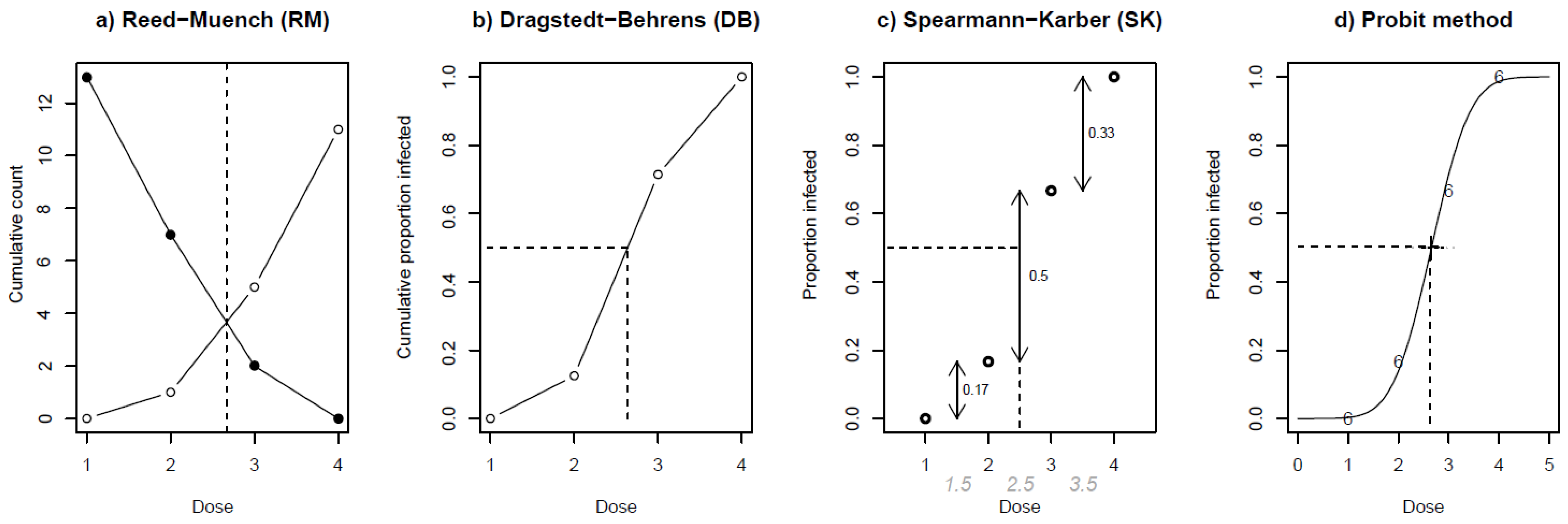
This example has been provided to demonstrate the four approaches to calculate RID50. An inoculum of RHDV was prepared in a ten-fold dilution series with no infected rabbits (0/6) at the first dose, one infected rabbit at the second dose (1/6), four infected rabbits at the third dose (4/6), and finally all six infected rabbits at the fourth dose (6/6). The RM [18], DB [19], and SK methods [20,21] are interpolative methods based on the formation of cumulative sums or proportions (Table A1). These methods follow the assumption that an infected animal will also be infected at higher doses and that an animal that is not infected will not be infected at lower doses (higher dilutions). The RM method uses the cumulative counts (Figure A1a). The DB method uses cumulative proportions (Figure A1b). Finally, the SK method calculates each dose’s weighted mean infection proportion (Figure A1c).
Table A1. An example of the values obtained from the bioassay to determine the median rabbit infectious dose of a hypothetical inoculum of rabbit haemorrhagic disease virus. The counts of infected and non-infected rabbits and cumulative proportions are presented as the preliminary steps for applying the Reed–Muench and Dragstedt–Behrens methods to estimate the 50% endpoint.
Doselog10 Dilution FactorCountsCumulative
InfectedNon-
Infected
TotalProportion
Infected
(Infected/Total)
InfectedNon-
Infected
TotalProportion Infected
(Infected/Total)
1−40660013130
2−31560.1671780.125
3−24260.6675270.714
4−16061110111
Figure A1. Depiction of the four approaches used to calculate the median infectious dose of an inoculum of rabbit haemorrhagic disease virus, based on a hypothetical bioassay using four doses with none (0/6), one (1/6), four (4/6), and six (6/6) rabbits infected from a total of six rabbits per dilution. The dashed lines on each plot visualise each method’s estimated 50% endpoint. Plots (a,b) demonstrate the calculation of the 50% endpoint via cumulative counts and proportions, as used in the Reed–Muench (RM) and Dragstedt–Behrens (DB) methods, respectively. To interpolate the 50% endpoint from the RM, two curves are plotted using the cumulative counts of infected (white) and non-infected (black) rabbits. Plot (c) shows the Spearman–Kärber (SK) method, which calculates the weighted mean of the doses based on observed proportions. Plot (d) depicts the Probit method, which fits a sigmoid curve to the dose–response data to estimate the 50% endpoint.
Viruses 17 00609 g0a1
As a curve-fitting method, probit regression does not rely on an assumption of successive infection probability and accounts for the tolerance distribution for individual rabbits. To estimate RID50, a normally distributed “tolerance distribution” is assumed. That is, each rabbit can ‘tolerate’ a specific dose of RHDV before becoming infected (Figure A2) [17,59]. When these tolerances are normally distributed across a hypothetical population of rabbits, the resulting dose–response relationship can be fitted in a sigmoid regression curve (Figure A1d).
Figure A2. Depiction of the tolerance distribution based on a hypothetical bioassay using four log10 doses with 0, 1, 4, and 6 rabbits infected (as plotted in the red–lined area under the curve) from a total of 6 replicates challenged per dilution. The dose–response relationship from this tolerance distribution can be fitted to a sigmoid curve to estimate the 50% endpoint for the probit method.
Viruses 17 00609 g0a2
The probit regression curve can be fitted using maximum likelihood, and this can be analysed as a generalised linear model (GLM) with a binomial distribution and a probit link function. The probit model estimates the relationship between the dilution ( d i ) of the inoculum and the probability of infection ( p i ) and can be summarised as follows:
p i   = ϕ ( α + β d i )
where Φ is the cumulative normal density function and α and β are the two parameters to be estimated. Once estimates α and β are obtained, the infective titre of an inoculum at which 50% of rabbits become infected (RID50) can be estimated as −α/β, and the standard error can be extracted from the model.
The 95% confidence interval can be determined using a profile likelihood function (PL). To estimate the PL, the probit model (A1) can also be rewritten as a function of the RID50 (A2).
p i   = ϕ ( β d i R I D 50 )
Using Equation (2), a 95% profile likelihood confidence interval can be defined as the set of all RID50 values satisfying the following:
l β ^ ( R I D 50 ) , R I D 50   l ( β , ^ R I D 50 ^ ) 0.5 × χ 1 ;   0.95 2
where ℓ is the log-likelihood function, β ^ and R I D 50 ^ are the maximum likelihood estimates, β ^ ( R I D 50 ) is the maximum likelihood estimate of β   given RID50, and χ 1 ;   0.95 2 is the 95% point of a χ 2 distribution with 1 degree of freedom. The 95% confidence interval will be defined by the minimum and maximum values where Equation (A3) is satisfied.

References

  1. Capucci, L.; Cavadini, P.; Lavazza, A. Viral haemorrhagic disease: RHDV type 2 ten years later. World Rabbit. Sci. 2022, 30, 1–11. [Google Scholar] [CrossRef]
  2. Lavazza, A.; Cavadini, P.; Barbieri, I.; Tizzani, P.; Pinheiro, A.; Abrantes, J.; Esteves, P.J.; Grilli, G.; Gioia, E.; Zanoni, M.; et al. Field and experimental data indicate that the eastern cottontail (Sylvilagus floridanus) is susceptible to infection with European brown hare syndrome (EBHS) virus and not with rabbit haemorrhagic disease (RHD) virus. Vet. Res. 2015, 46, 13. [Google Scholar] [CrossRef]
  3. Puggioni, G.; Cavadini, P.; Maestrale, C.; Scivoli, R.; Botti, G.; Ligios, C.; Le Gall-Reculé, G.; Lavazza, A.; Capucci, L. The new French 2010 Rabbit Hemorrhagic Disease Virus causes an RHD-like disease in the Sardinian Cape hare (Lepus capensis mediterraneus). Vet. Res. 2013, 44, 96. [Google Scholar] [CrossRef] [PubMed]
  4. Camarda, A.; Pugliese, N.; Cavadini, P.; Circella, E.; Capucci, L.; Caroli, A.; Legretto, M.; Mallia, E.; Lavazza, A. Detection of the new emerging rabbit haemorrhagic disease type 2 virus (RHDV2) in Sicily from rabbit (Oryctolagus cuniculus) and Italian hare (Lepus corsicanus). Res. Vet. Sci. 2014, 97, 642–645. [Google Scholar] [CrossRef] [PubMed]
  5. Velarde, R.; Cavadini, P.; Neimanis, A.; Cabezón, O.; Chiari, M.; Gaffuri, A.; Lavín, S.; Grilli, G.; Gavier-Widén, D.; Lavazza, A.; et al. Spillover Events of Infection of Brown Hares (Lepus europaeus) with Rabbit Haemorrhagic Disease Type 2 Virus (RHDV2) Caused Sporadic Cases of an European Brown Hare Syndrome-Like Disease in Italy and Spain. Transbound. Emerg. Dis. 2017, 64, 1750–1761. [Google Scholar] [CrossRef] [PubMed] [PubMed Central]
  6. Neimanis, A.S.; Ahola, H.; Larsson Pettersson, U.; Lopes, A.M.; Abrantes, J.; Zohari, S.; Esteves, P.J.; Gavier-Widén, D. Overcoming species barriers: An outbreak of Lagovirus europaeus GI.2/RHDV2 in an isolated population of mountain hares (Lepus timidus). BMC Vet. Res. 2018, 14, 367. [Google Scholar] [CrossRef]
  7. Hall, R.N.; King, T.; O’Connor, T.; Read, A.J.; Arrow, J.; Trought, K.; Duckworth, J.; Piper, M.; Strive, T. Age and Infectious Dose Significantly Affect Disease Progression after RHDV2 Infection in Naïve Domestic Rabbits. Viruses 2021, 13, 1184. [Google Scholar] [CrossRef] [PubMed] [PubMed Central]
  8. Tu, T.; Zhou, Y.; Jiang, D.; Pang, M.; Wu, X.; Yao, X.; Luo, Y.; Yang, Z.; Ren, M.; Wang, Y. The pathogenicity comparison of Lagovirus europaeus GI.1 and GI.2 strains in China by using relative quantitative assay. Sci. Rep. 2022, 12, 20518. [Google Scholar] [CrossRef]
  9. Elsworth, P.; Cooke, B.D.; Kovaliski, J.; Sinclair, R.; Holmes, E.C.; Strive, T. Increased virulence of rabbit haemorrhagic disease virus associated with genetic resistance in wild Australian rabbits (Oryctolagus cuniculus). Virology 2014, 464, 415–423. [Google Scholar] [CrossRef]
  10. Mohamed, F.; Gidlewski, T.; Berninger, M.L.; Petrowski, H.M.; Bracht, A.J.; de Rueda, C.B.; Barrette, R.W.; Grady, M.; O’Hearn, E.S.; Lewis, C.E.; et al. Comparative susceptibility of eastern cottontails and New Zealand white rabbits to classical rabbit haemorrhagic disease virus (RHDV) and RHDV2. Transbound. Emerg. Dis. 2022, 69, e968–e978. [Google Scholar] [CrossRef]
  11. Droillard, C.; Lemaitre, E.; Amelot, M.; Blanchard, Y.; Keita, A.; Eterradossi, N.; Le Gall-Reculé, G. Rabbit haemorrhagic disease virus Lagovirus europaeus/GI.1d strain: Genome sequencing, in vivo virus replication kinetics, and viral dose effect. BMC Vet. Res. 2021, 17, 257. [Google Scholar] [CrossRef] [PubMed] [PubMed Central]
  12. Le Gall-Reculé, G.; Lavazza, A.; Marchandeau, S.; Bertagnoli, S.; Zwingelstein, F.; Cavadini, P.; Martinelli, N.; Lombardi, G.; Guérin, J.-L.; Lemaitre, E.; et al. Emergence of a new lagovirus related to Rabbit Haemorrhagic Disease Virus. Vet. Res. 2013, 44, 81. [Google Scholar] [CrossRef] [PubMed]
  13. Kieran, T.J.; Sun, X.; Maines, T.R.; Beauchemin, C.A.A.; Belser, J.A. Exploring associations between viral titer measurements and disease outcomes in ferrets inoculated with 125 contemporary influenza A viruses. J. Virol. 2024, 98, e0166123. [Google Scholar] [CrossRef] [PubMed] [PubMed Central]
  14. Saganuwan, S.A. Application of median lethal concentration (LC(50)) of pathogenic microorganisms and their antigens in vaccine development. BMC Res. Notes 2020, 13, 289. [Google Scholar] [CrossRef] [PubMed] [PubMed Central]
  15. Read, A.J.; Kirkland, P.D. Efficacy of a commercial vaccine against different strains of rabbit haemorrhagic disease virus. Aust. Vet. J. 2017, 95, 223–226. [Google Scholar] [CrossRef]
  16. Armitage, P.; Allen, I. Methods of estimating the LD 50 in quantal response data. Epidemiol. Infect. 1950, 48, 298–322. [Google Scholar] [CrossRef] [PubMed] [PubMed Central]
  17. Finney, D. Statistical Method in Biological Assay, 2nd ed; Griffin: London, UK, 1964. [Google Scholar]
  18. Reed, L.J.; Muench, H. A simple method of estimating fifty percent endpoints. Am. J. Epidemiol. 1938, 27, 493–497. [Google Scholar] [CrossRef]
  19. Behrens, B. Zur Auswertung der Digitalisblätter im Froschversuch. Naunyn-Schmiedebergs Arch. Für Exp. Pathol. Und Pharmakologie 1929, 140, 237–256. [Google Scholar] [CrossRef]
  20. Kärber, G. Beitrag zur kollektiven Behandlung pharmakologischer Reihenversuche. Naunyn-Schmiedeberg’s Arch. Pharmacol. 1931, 162, 480–483. [Google Scholar] [CrossRef]
  21. Spearman, C. The method of “right and wrong cases” (constant stimuli) without Gauss’s formula. Br. J. Psychol. 1908, 2, 227–242. [Google Scholar] [CrossRef]
  22. Miller, R.G. Nonparametric Estimators of the Mean Tolerance in Bioassay. Biometrika 1973, 60, 535–542. [Google Scholar] [CrossRef]
  23. Ramakrishnan, M.A. Determination of 50% endpoint titer using a simple formula. World J. Virol. 2016, 5, 85–86. [Google Scholar] [CrossRef] [PubMed] [PubMed Central]
  24. Labarre, D.D.; Lowy, R.J. Improvements in methods for calculating virus titer estimates from TCID50 and plaque assays. J. Virol. Methods 2001, 96, 107–126. [Google Scholar] [CrossRef]
  25. Bryan, W.R. Interpretation of host response in quantitative studies on animal viruses. Ann. N. Y. Acad. Sci. 1957, 69, 698–728. [Google Scholar] [CrossRef]
  26. Cresta, D.; Warren, D.C.; Quirouette, C.; Smith, A.P.; Lane, L.C.; Smith, A.M.; Beauchemin, C.A.A. Time to revisit the endpoint dilution assay and to replace the TCID50 as a measure of a virus sample’s infection concentration. PLoS Comput. Biol. 2021, 17, e1009480. [Google Scholar] [CrossRef]
  27. Ramakrishnan, M.; Dhanavelu, M. Influence of Reed-Muench Median Dose Calculation Method in Virology in the Millennium. Antivir. Res. 2018, 28, 16–18. [Google Scholar]
  28. O’Connor, T.W.; Read, A.J.; Hall, R.N.; Strive, T.; Kirkland, P.D. Immunological Cross-Protection between Different Rabbit Hemorrhagic Disease Viruses—Implications for Rabbit Biocontrol and Vaccine Development. Vaccines 2022, 10, 666. [Google Scholar] [CrossRef] [PubMed] [PubMed Central]
  29. Animal Research Review Panel. Guidelines for the Care and Management of Rabbits in Scientific Institutions in: Department of Primary Industries; New South Wales Government: Orange, Australia, 2023. [Google Scholar]
  30. Teixeira, L.; Marques, R.M.; Águas, A.P.; Ferreira, P.G. A simple and rapid method for isolation of caliciviruses from liver of infected rabbits. Res. Vet. Sci. 2011, 91, 164–166. [Google Scholar] [CrossRef] [PubMed]
  31. World Organisation for Animal Health (WOAH). Terrestrial Manual, Chapter 3.7.2 Rabbit Haemorrahgic Disease 2023. Available online: https://www.woah.org/fileadmin/Home/eng/Health_standards/tahm/3.07.02_RHD.pdf (accessed on 28 February 2025).
  32. Gall, A.; Hoffmann, B.; Teifke, J.P.; Lange, B.; Schirrmeier, H. Persistence of viral RNA in rabbits which overcome an experimental RHDV infection detected by a highly sensitive multiplex real-time RT-PCR. Vet. Microbiol. 2007, 120, 17–32. [Google Scholar] [CrossRef]
  33. Hall, R.N.; Mahar, J.E.; Read, A.J.; Mourant, R.; Piper, M.; Huang, N.; Strive, T. A strain-specific multiplex RT-PCR for Australian rabbit haemorrhagic disease viruses uncovers a new recombinant virus variant in rabbits and hares. Transbound. Emerg. Dis. 2018, 65, e444–e456. [Google Scholar] [CrossRef]
  34. R Core Team. R: A Language and Environment for Statistical Computing; R Foundation for Statistical Computing: Vienna, Austria, 2024. [Google Scholar]
  35. Siev, D.; Vendettuoli, M.; Kent, T. skrmdb: Package to estimate ED50. 4.5.0. 2024. Available online: https://github.com/ABS-dev/skrmdb (accessed on 18 October 2024).
  36. Venables, W.N.; Ripley, B.D. Modern Applied Statistics with S; Springer: New York, NY, USA, 2002. [Google Scholar]
  37. Chang, W.; Cheng, J.; Allaire, J.J.; Sievert, C.; Schloerke, B.; Xie, Y.; Allen, J.; McPherson, J.; Dipert, A.; Borges, B. Shiny: Web Application Framework for R. 1.10.0. 2025. Available online: https://github.com/rstudio/shiny (accessed on 18 October 2024).
  38. Bland, J.M.; Altman, D.G. Measuring agreement in method comparison studies. Stat. Methods Med. Res. 1999, 8, 135–160. [Google Scholar] [CrossRef] [PubMed]
  39. Gamer, M.; Lemon, J.; Fellows, I.; Singh, P. irr: Various Coefficients of Interrater Reliability and Agreement. 0.84.1. 2019. Available online: https://CRAN.R-project.org/package=irr (accessed on 18 October 2024).
  40. Bartko, J. The intraclass correlation coefficient as a measure of reliability. Psychol. Rep. 1966, 19, 3–11. [Google Scholar] [CrossRef] [PubMed]
  41. Šmíd, B.; Valíček, L.; Rodak, L.; Štěpánek, J.; Jurak, E.J.V.M. Rabbit haemorrhagic disease: An investigation of some properties of the virus and evaluation of an inactivated vaccine. Veter-Microbiol. 1991, 26, 77–85. [Google Scholar] [CrossRef] [PubMed]
  42. Marcato, P.; Benazzi, C.; Vecchi, G.; Galeotti, M.; Della Salda, L.; Sarli, G.; Lucidi, P. Clinical and pathological features of viral haemorrhagic disease of rabbits and the European brown hare syndrome. Rev. Sci. Tech. l’OIE 1991, 10, 371–392. [Google Scholar] [CrossRef]
  43. Dalton, K.P.; Nicieza, I.; Balseiro, A.; Muguerza, M.A.; Rosell, J.M.; Casais, R.; Álvarez, Á.L.; Parra, F. Variant rabbit hemorrhagic disease virus in young rabbits, Spain. Emerg. Infect. Dis. 2012, 18, 2009–2012. [Google Scholar] [CrossRef] [PubMed] [PubMed Central]
  44. Cooke, B.D.; McPhee, S.; Robinson, A.J.; Capucci, L. Rabbit haemorrhagic disease: Does a pre-existing RHDV-like virus reduce the effectiveness of RHD as a biological control in Australia? Wildl. Res. 2002, 29, 673–682. [Google Scholar] [CrossRef]
  45. Baratelli, M.; Molist-Badiola, J.; Puigredon-Fontanet, A.; Pascual, M.; Boix, O.; Mora-Igual, F.X.; Woodward, M.; Lavazza, A.; Capucci, L. Characterization of the Maternally Derived Antibody Immunity against Rhdv-2 after Administration in Breeding Does of an Inactivated Vaccine. Vaccines 2020, 8, 484. [Google Scholar] [CrossRef] [PubMed] [PubMed Central]
  46. Elsworth, P.G.; Kovaliski, J.; Cooke, B.D. Rabbit haemorrhagic disease: Are Australian rabbits (Oryctolagus cuniculus) evolving resistance to infection with Czech CAPM 351 RHDV? Epidemiol. Infect. 2012, 140, 1972–1981. [Google Scholar] [CrossRef]
  47. Schwensow, N.; Pederson, S.; Peacock, D.; Cooke, B.; Cassey, P. Adaptive changes in the genomes of wild rabbits after 16 years of viral epidemics. Mol. Ecol. 2020, 29, 3777–3794. [Google Scholar] [CrossRef]
  48. Crespo, I.; Miguel, B.S.; Laliena, A.; Alvarez, M.; Culebras, J.M.; González-Gallego, J.; Tuñón, M.J. Melatonin prevents the decreased activity of antioxidant enzymes and activates nuclear erythroid 2-related factor 2 signaling in an animal model of fulminant hepatic failure of viral origin. J. Pineal Res. 2010, 49, 193–200. [Google Scholar] [CrossRef] [PubMed]
  49. Liu, S.; Xue, H.; Pu, B.; Qian, N. A new viral disease in rabbits. Anim. Husb. Vet. Med. 1984, 16, 253–255. [Google Scholar]
  50. Bustin, S.A. Absolute quantification of mRNA using real-time reverse transcription polymerase chain reaction assays. J. Mol. Endocrinol. 2000, 25, 169–193. [Google Scholar] [CrossRef] [PubMed]
  51. Bustin, S. Quantification of mRNA using real-time reverse transcription PCR (RT-PCR): Trends and problems. J. Mol. Endocrinol. 2002, 29, 23–39. [Google Scholar] [CrossRef]
  52. Cooke, B. Rabbit haemorrhagic disease: Field epidemiology and the management of wild rabbit populations. Rev. Sci. et Tech. de l’OIE 2002, 21, 347–358. [Google Scholar] [CrossRef]
  53. Abrantes, J.; van Der Loo, W.; Le Pendu, J.; Esteves, P.J. Rabbit haemorrhagic disease (RHD) and rabbit haemorrhagic disease virus (RHDV): A review. Vet. Res. 2012, 43, 1–19. [Google Scholar] [CrossRef]
  54. Matthaei, M.; Kerr, P.J.; Read, A.J.; Hick, P.; Haboury, S.; Wright, J.D.; Strive, T. Comparative quantitative monitoring of rabbit haemorrhagic disease viruses in rabbit kittens. Virol. J. 2014, 11, 109. [Google Scholar] [CrossRef]
  55. Lavazza, A.; Capucci, L. How Many Caliciviruses Are There in Rabbits? A Review on RHDV and Correlated Viruses, Lagomorph Biology: Evolution, Ecology, and Conservation; Springer: Berlin, Germany, 2008; pp. 263–278. [Google Scholar]
  56. Shien, J.H.; Shieh, H.K.; Lee, L.H. Experimental infections of rabbits with rabbit haemorrhagic disease virus monitored by polymerase chain reaction. Res. Vet. Sci. 2000, 68, 255–259. [Google Scholar] [CrossRef]
  57. Guittré, C.; Ruvoen-Clouet, N.; Barraud, L.; Cherel, Y.; Baginski, I.; Prave, M.; Ganiere, J.P.; Trépo, C.; Cova, L. Early Stages of Rabbit Haemorrhagic Disease Virus Infection Monitored by Polymerase Chain Reaction. J. Vet. Med. 1996, 43, 109–118. [Google Scholar] [CrossRef]
  58. Lei, C.; Yang, J.; Hu, J.; Sun, X. On the Calculation of TCID(50) for Quantitation of Virus Infectivity. Virol. Sin. 2021, 36, 141–144. [Google Scholar] [CrossRef] [PubMed] [PubMed Central]
  59. Finney, D. Probit Analysis; Cambridge University Press: London, UK, 1971. [Google Scholar]
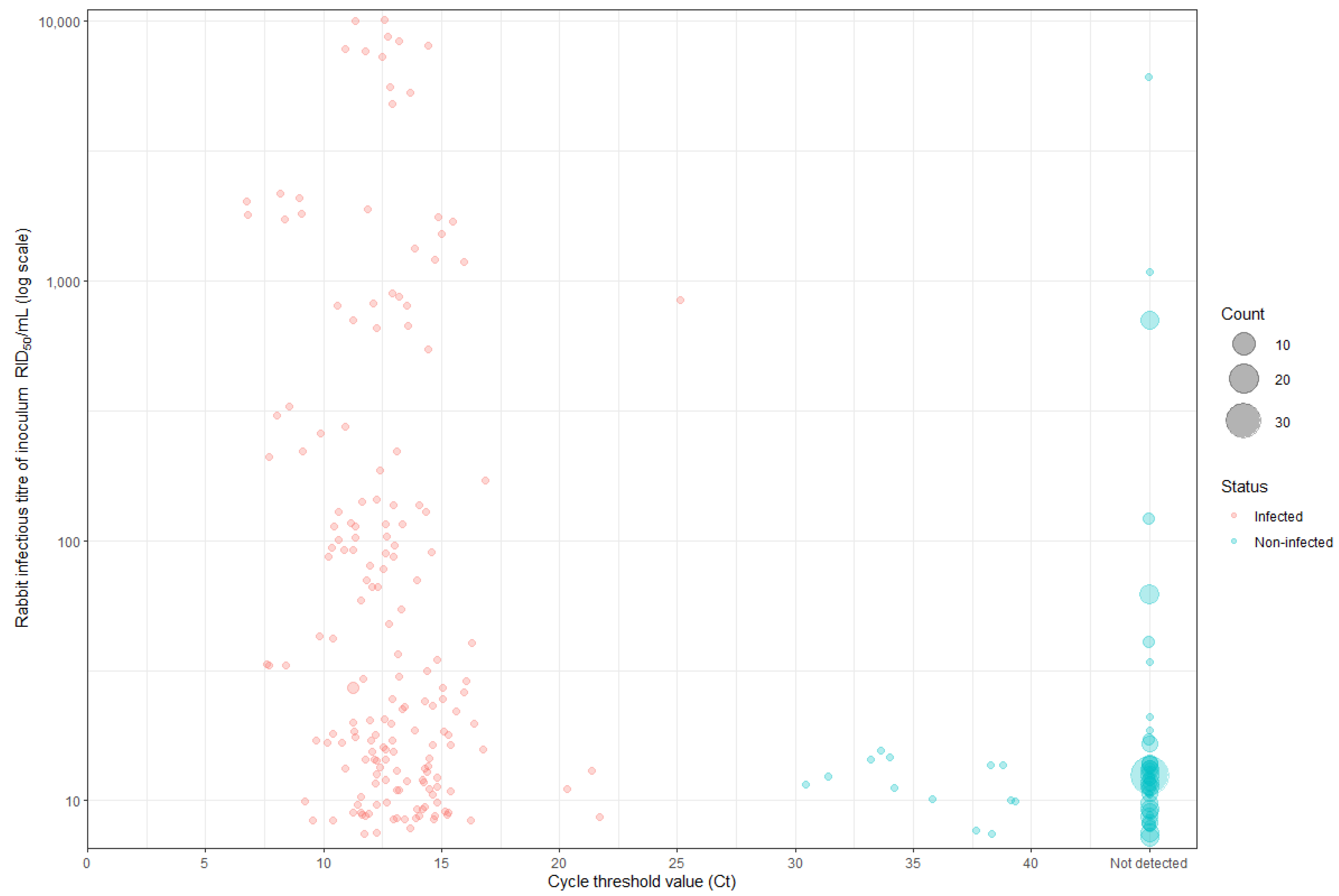
Figure 1. Distribution of cycle threshold (Ct) values from liver swabs collected from all the rabbits in the bioassays to determine the infectious titre (RID50/mL) of rabbit haemorrhagic disease virus (RHDV) preparations. The RID50/mL values, depicted on a log10 scale, were calculated using the Reed–Muench method. Individual data points represent single rabbits, with RHDV-infected rabbits in red and non-infected rabbits in blue. The point size also corresponds to the number of rabbits that received the same dose and had the same RT-qPCR result.
Viruses 17 00609 g001
Figure 2. Bland–Altman plots comparing the agreement between different methods to calculate the median rabbit infectious dose (RID50). Each plot (af) displays the difference between the two methods against their mean, with the blue dashed line representing the mean difference and the red dashed lines indicating the 95% limits of agreement. The intraclass correlation coefficient (ICC) for each pair is displayed in each plot.
Viruses 17 00609 g002
Table 1. The log10 titre determined for 26 different preparations of RHDV expressed as median rabbit infectious dose (RID50) per mL using four calculation methods: Reed–Muench, Dragstedt–Behrens, improved Spearman–Kärber, and probit analysis. The 95% confidence intervals (CI) for the improved Spearman–Kärber and probit estimates are provided. Experiments where a 50% endpoint could not be determined are noted as “Not available; NA”. Where the upper or lower bounds of the CI are infinite, this has been indicated as “∞”.
ExperimentReed–MuenchDragstedt–Behrens Spearman–Kärber 95% CIProbit95% CI
E13.603.623.672.84–4.504.01−∞ to +∞
E24.974.935.004.60–5.405.194.65–5.47
E34.204.214.373.79–4.944.231.85–5.06
E44.634.584.513.81–5.226.35−∞ to +∞
E54.004.004.003.55–4.454.003.60–5.43
E6NANA4.173.75–4.594.073.76 to +∞
E74.384.384.333.65–5.024.463.72–6.51
E86.096.206.085.25–6.925.354.90–7.01
E93.003.003.002.55–3.453.002.60–3.40
E105.835.835.835.07–6.595.964.84–8.84
E113.503.503.502.90–4.103.502.94–4.06
E126.506.506.505.90–7.106.825.93 to +∞
E133.503.503.503.03–3.973.502.90–3.96
E143.503.503.502.90–4.103.451.99–4.06
E153.333.363.332.80–3.873.312.05–3.84
E166.476.486.475.95–6.996.475.92–7.11
E176.436.456.505.80–7.206.535.65–7.38
E183.573.593.672.92–4.412.69−∞ to +∞
E193.713.693.673.05–4.283.612.16–4.31
E204.714.684.704.07–5.334.764.12–7.93
E213.133.173.102.61–3.593.042.12–3.51
E226.506.456.335.56–7.119.43−∞ to +∞
E234.004.004.003.55–4.454.003.60–4.40
E244.004.004.003.55–4.454.003.60–4.40
E253.703.703.833.20–4.473.78−∞ to +∞
E26NANA7.337.00–7.677.176.95 to +∞
Disclaimer/Publisher’s Note: The statements, opinions and data contained in all publications are solely those of the individual author(s) and contributor(s) and not of MDPI and/or the editor(s). MDPI and/or the editor(s) disclaim responsibility for any injury to people or property resulting from any ideas, methods, instructions or products referred to in the content.