Article

Respiratory Syncytial Virus Persistence in Murine Macrophages Impairs IFN-β Response but Not Synthesis

by
Evelyn Rivera-Toledo
,
Laura Torres-González
and
Beatriz Gómez
*
Department of Microbiology and Parasitology, Faculty of Medicine, National Autonomous University of Mexico (UNAM), Ciudad Universitaria, D.F. Mexico C.P. 04510, Mexico
*
Author to whom correspondence should be addressed.
Viruses 2015, 7(10), 5361-5374; https://doi.org/10.3390/v7102879
Submission received: 23 April 2015 / Revised: 11 September 2015 / Accepted: 12 October 2015 / Published: 16 October 2015
(This article belongs to the Section Animal Viruses)

Abstract

:
Type-I interferon (IFN-I) production is an early response to viral infection and pathogenic viruses have evolved multiple strategies to evade this cellular defense. Some viruses can establish and maintain persistent infections by altering the IFN-I signaling pathway. Here, we studied IFN-I synthesis and response in an in vitro model of persistent infection by respiratory syncytial virus (RSV) in a murine macrophage-like cell line. In this model, interferon regulatory factor 3 was constitutively active and located at nuclei of persistently infected cells, inducing expression of IFN-beta mRNA and protein. However, persistently infected macrophages did not respond in an autocrine manner to the secreted-IFN-beta or to recombinant-IFN-beta, since phosphorylated-STAT1 was not detected by western blot and transcription of the interferon-stimulated genes (ISGs) Mx1 and ISG56 was not induced. Treatment of non-infected macrophages with supernatants from persistently infected cells induced STAT1 phosphorylation and ISGs expression, mediated by the IFN-I present in the supernatants, because blocking the IFN-I receptor inhibited STAT1 phosphorylation. Results suggest that the lack of autocrine response to IFN-I by the host cell may be one mechanism for maintenance of RSV persistence. Furthermore, STAT1 phosphorylation and ISGs expression induced in non-infected cells by supernatants from persistently infected macrophages suggest that RSV persistence may trigger a proinflammatory phenotype in non-infected cells as part of the pathogenesis of RSV infection.

1. Introduction

Respiratory syncytial virus (RSV), a negative-sense, single-stranded RNA virus of the family Paraxmyxoviridae (genus Pneumovirus), is considered the main cause of bronchiolitis and pneumonia in children under two years of age [1,2,3]. Most infants experience RSV infection during the first year of life [4,5] and after severe RSV disease, 30%–40% of this affected population will undergo recurrent wheezing or asthma-like symptoms in later childhood [6,7,8]. The mechanisms underlying long-term airway hyperactivity are incompletely understood, but may be explained in part by a low-level, persistent viral infection, which may cause a chronically stimulated immune response [7,9]. RSV persistence in humans, although not confirmed, has been suggested by studies that reported the isolation of RSV from the nasopharynx of apparently healthy children [10], the presence of genomic RSV-RNA in respiratory-tract secretions from adult patients with stable chronic obstructive pulmonary disease [11], and the detection of viral mRNA in post-mortem lung tissue from infants who died without apparent clinical disease [12]. Moreover, the RSV genome has been detected in primary human bone-marrow stromal cells from adult and child donors [13], suggesting that RSV may establish persistent infections in respiratory and non-respiratory tissues. To study the mechanisms and consequences of persistent viral infections, cell cultures and animal models are useful tools; RSV persistence has been established in both types of models [14,15,16,17,18].
Interferon regulatory factor 3 (IRF3) is a transcription factor that mediates type-I interferon (IFN-I) expression, such as IFN-α and IFN-β, after viral infection [19,20]. As IFN-I activity is an efficient mechanism to control viral replication, many pathogenic viruses have developed strategies to overcome IFN-I expression in order to guarantee their replication and spread. IRF3 inactivation, achieved through several viral strategies, has been reported to be associated with persistent infections [21,22,23].
Triggering of the IFN-I signaling pathway during viral replication implies recognition of viral 5′-triphosphate-RNA and double-stranded RNA intermediates by the cytosolic, retinoic acid-inducible gene I (RIG-I)-like receptors (RLRs) of the innate immune system, such as RIG-I and MDA5 [24]. Upon ligand binding, RLRs form homo-dimers that migrate to the outer membrane of mitochondria, where a macromolecular signaling complex is assembled [25,26]. Consequently, IRF3 undergoes phosphorylation, dimerization, and nuclear translocation where along with the transcription factors NF-kB and AP-1, it binds the IFN-β promoter to induce gene expression [27,28]. Secreted IFN-β acts in an autocrine/paracrine manner via the IFNα/β receptor (IFNAR), eliciting phosphorylation of STAT2 and STAT1 through the Jak and Tyk2 kinases. Phosphorylated-STAT1 (p-STAT1), with STAT2 and IRF9, forms the interferon-stimulated gene factor-3 complex, which is translocated to the nucleus, thereby inducing the expression of a variety of interferon-stimulated genes (ISGs), including myxovirus-resistance gene 1 (Mx1) and interferon-induced protein with tetratricopeptide repeats 1 (IFIT1/ISG56), to create an antiviral state [29,30].
Acute RSV infection of epithelial cell lines and of primary macrophages induces a relatively low production of IFN-α and -β. Synthesis of IFN-α and -β is significantly augmented, when infection is achieved with mutant RSV that lack the non-structural viral genes 1 and 2 (NS1 and NS2) [31]. RSV NS1 and NS2 proteins are responsible for disrupting cellular IFN-I signaling at the levels of expression and response by acting on multiple targets, such as IRF-3 and STAT2, respectively [32,33,34,35,36].
Whereas most studies that were focused on determining the mechanisms by which RSV evades the IFN-I response have been performed during acute infections, we studied the state of activation of the IFN-I signaling pathway at the level of synthesis and response during persistent RSV infection by using a previously established in vitro model of persistently RSV-infected murine macrophages (MΦP) [14]. Here, we report that MΦP constitutively expressed the active form of IRF3 and consequently IFN-β, but these macrophages were not competent to respond to this cytokine in an autocrine manner, as phosphorylation of STAT1 was not detected, neither expression of the ISGs Mx1 nor ISG56. However, IFN-β secreted by MΦP was able to activate STAT1 in non-infected macrophages along with Mx1 and ISG56 mRNA expression, thereby suggesting that persistent RSV infection may not only affect the host cell, but also indirectly affect surrounding cells by altering their state of activation, contributing with the pathogenesis of RSV infection.

2. Materials and Methods

2.1. Cell Lines and Virus

The MΦP culture was obtained in our laboratory as previously reported [14]. Briefly, the murine macrophage-like cell line P388D1, obtained from the American Type culture Collection (ATCC, TIB 63; Rockville, MD, USA), was infected at a multiplicity of infection (m.o.i.) of 1 by using the RSV wild-type strain Long (ATCC, VR-26) (wtRSV). Surviving cells were subcultured for ten passages and then were superinfected (m.o.i. of 1), resulting in 90%–95% of cells expressing viral antigens. After ten passages of that superinfected culture, neither cellular destruction nor syncytia formation were observed, although more than 90% of cells expressed viral antigens and the supernatants had low levels of infectious viruses (1–3 × 10² 50% tissue culture infective dose (TCID50)/mL) [14]. In parallel to MΦP, the original P388D1 cell line was propagated as a non-infected control cell line (MΦN) in supplemented-RPMI (RPMI 1640 cell-culture medium (GIBCO/BRL, Grand Island, NY, USA), containing 5% fetal bovine serum (Biowest, Veracruz, Mexico), 1% penicillin-streptomycin (Invitro S.A., Mexico City), and 1 μM 2-mercaptoethanol) (37 °C; 5% CO2).
The human epithelial cell line Hep-2, was cultured in Dulbecco’s Modified Eagle Medium (GIBCO/BRL, Grand Island, NY, USA), supplemented with 5% fetal bovine serum, 10 mM HEPES (SIGMA, St. Louis, MO, USA), and 1% penicillin-streptomycin. Hep-2 cells were used to propagate the wtRSV and to determine viral infectivity, expressed in TCID50/mL [37]. The wtRSV obtained in this manner was used to infect cultures of P388D1 macrophages (m.o.i. of 2) for 6, 24, and 48 h, in order to evaluate the expression of p-IRF3 in these acutely RSV-infected macrophages (MΦA). Under such conditions, we observed by direct immunofluorescence 65%–75% and 85%–90% of infected cells at 24 and 48 h post-infection (p.i.), respectively. The percentage of acutely infected cells at 48 h p.i., was similar to the percentage of persistently infected macrophages in the MΦP cultures.

2.2. Confirmation of Persistent RSV Infection in MΦP

In this study, MΦP from passages 85–105 were used to perform the experiments. Throughout the passages, RSV persistence was monitored (every two or three weeks) by immunofluorescence to determine the expression of viral antigens and by conventional RT-PCR to detect mRNA of the nucleocapsid (N) gene. Direct immunofluorescence was performed with a FITC-labeled, polyclonal antibody anti-RSV directed against the N and F viral proteins (Oxoid, Hampshire, UK). Briefly, 0.5 × 10⁶ MΦP or MΦN were fixed with 4% p-formaldehyde (15 min) and permeabilized with 0.3% saponine (15 min). After non-specific binding sites were blocked by incubating cells (25 min; on ice), with blocking solution (PBS containing 5% neonate bovine serum (Bioexport, Mexico)), a dilution of polyclonal anti-RSV (1:10 in blocking solution) was added to the cells and the mixture was incubated (35 min; on ice). The percentage of FITC-positive cells, as determined by flow cytometry (FACScan; Becton Dickinson, Mountain View, CA, USA) was reported. The expression of N gene mRNA was analyzed by using a primer pair to amplify a 1187-bp segment; as a control, mRNA of the GAPDH gene was determined with a primer pair to amplify a 260-bp segment, as previously described [38].

2.3. Western Blot

Whole-cell extracts were prepared from MΦN and MΦP, and where indicated, from MΦA. Ice-cold lysing buffer (50 mM Tris-HCl, pH 8.0, 150 mM NaCl, 1% Triton X-100, 1% sodium deoxycholate, and 0.1% SDS), containing EDTA-free complete protease inhibitor cocktail (Roche, Indianapolis, IN, USA), was added to 3–5 × 10⁶ cells. After incubation (15 min; on ice), lysates were centrifuged (13,000 rpm) and supernatants collected. The concentration of total protein in each sample was determined by using Bradford reagent (Bio-Rad, Hercules, CA, USA). Proteins (30 μg) were separated by SDS-polyacrylamide gel electrophoresis on a NuPAGE bis-Tris gradient gel with 4%–12% acrylamide (Invitrogen, Carlsbad, CA, USA) and transferred to polyvinylidene difluoride membranes (Amersham, Piscataway, NJ, USA). Specific proteins were analyzed with the primary monoclonal antibodies, anti-p-IRF3 or anti-p-STAT1 (Cell Signaling, Beverly, MA, USA), diluted 1:1000 in TBS-T buffer with 5% BSA. Monoclonal anti-STAT1 was diluted 1:500, while polyclonal anti-IRF3 and anti-β-tubulin (Santa Cruz Biotechnology, CA, USA) were diluted at 1:1000 in TBS-T buffer containing 5% non-fat milk. All membranes were incubated with primary antibody (overnight; 4 °C), washed three times with TBS-T and incubated with the appropriate peroxidase-conjugated secondary antibody, diluted 1:3000 in TBS-T containing 5% non-fat milk (2 h; room temperature).
Western blots were analyzed by chemiluminescence with the SuperSignal kit (Pierce Biotechnology, Rockford, IL, USA). Densitometry analysis of the resulting bands was performed with images obtained with the ChemiDoc™ XRS (Bio-Rad) and analyzed by using QuantityOne software (Bio-Rad).

2.4. Subcellular Fractionation

Cell pellets with 5 × 10⁶ MΦN or MΦP were resuspended in a cold cytoplasmic-lysing buffer (10 mM HEPES pH 7.9, 1.5 mM MgCl2, 10 mM KCl, 0.5 mM DTT, 0.5% Igepal, and EDTA-complete protease inhibitor cocktail) and incubated (15 min; on ice). After centrifugation (3500 rpm; 4 °C; 15 min), each supernatant, which contained predominantly cytoplasmic proteins, was collected. Each remaining pellet was washed two times and resuspended in nuclei-lysing buffer (5 mM HEPES pH 7.9, 1.5 mM MgCl2, 0.2 mM EDTA, 0.5 mM DTT, 25% glycerol and EDTA-free complete protease inhibitor cocktail) and incubated (4 °C; 25 min) with rocking agitation. Each supernatant, which contained predominantly total nuclear proteins, was collected after centrifugation (13,000 rpm; 4 °C; 5 min). Analysis of protein (20 μg) from cytoplasmic or nuclear cellular fractions was performed by western blot, as described above, using the antibodies anti-p-IRF3, anti-β-tubulin, and the polyclonal anti-Histone-3 (Cell Signaling), each at 1:1000 dilution.

2.5. Supernatant Collection and mRNA Extraction

MΦN or MΦP were cultured in 35-mm-diameter plastic Petri dishes (NUNC). Once 90% confluence was reached, each culture medium was replaced with RPMI containing 2% fetal bovine serum, to a final concentration of 1 × 10⁶ cells/mL. After the cultures were incubated (37 °C; 5% CO2) for the specified time (6, 24, or 48 h), supernatants were collected. Samples were frozen at −75 °C until use. After removing supernatants, total RNA from cells was extracted with the AxyPrep Multisource Total RNA Miniprep kit (Axygen Scientific, Union City, CA, USA), according to the manufacturer’s instructions.

2.6. Quantitation of IFN-β by ELISA

The concentration of IFN-β protein in MΦN and MΦP supernatants from 6, 24 and 48 h was determined by using a commercially available VeriKine™ mouse IFN-β sandwich ELISA kit (PBL interferon source, Piscataway, NJ, USA), according to the manufacturer’s instructions. From the IFN-β standard curve, samples with unknown concentration were interpolated. The assay range was 15.6–1000 pg/mL.

2.7. Quantitative RT-PCR

In a two-step RT-PCR assay, RNA (2 μg) was reverse transcribed with RNA transcriptase Superscript II (Invitrogen). TaqMan real-time RT-PCR was performed with primers and probes (Assay on Demand 20× mix) for murine IFN-β, Mx1, ISG56 and GAPDH (endogenous control), using TaqMan assay reagent master mix (Applied Biosystems, Foster City, CA, USA). After determining the dynamic range, a cDNA dilution (1:25) was analyzed for expression of IFN-β, Mx1, ISG56 and GAPDH genes. Cycling parameters were established according to the manufacturer’s protocol. Triplicate Ct values were analyzed by using the comparative Ct (ΔCt) method, as described by the manufacturer (Applied Biosystems). The relative amount of mRNA of each gene was obtained by normalization to the amount of mRNA of cellular GAPDH, as endogenous reference sample.

2.8. Macrophage Treatment with Recombinant IFN-β or Supernatants

Cultures of MΦN and MΦP, at 90% confluence, were each treated with 100 U/mL (26 ng/mL) of recombinant mouse-IFN-β (r-IFN-β) (Millipore Corporation, Temecula, CA, USA) (45 min; 37 °C; 5% CO2), as reported for RAW macrophages [39]. In addition, other MΦN were treated with supernatants collected from 24-h cultures of MΦP, previously exposed to U.V. light to inactivate the infectious RSV produced by MΦP and to eliminate the possibility of STAT1 activation by virus infection. Before U.V. light exposure, supernatants had a viral titer of 3.71 × 10² ± 104 TCID50/mL; after treatment infectivity was null. Total RNA and whole cell-extracts were obtained as mentioned above and were analyzed by quantitative RT-PCR regarding Mx1 and ISG56 gene expression or by western blot with anti-STAT1, anti-p-STAT1 and anti-β-tubulin.
In order to analyze the activity of IFN-β from MΦP supernatants on STAT1 phosphorylation, IFN-α/β receptors expressed on MΦN were blocked with polyclonal mouse anti-IFNAR (R&D systems, Minneapolis, MN, USA) at a concentration of 10 μg/mL (30 min; 37 °C); thus treated, the MΦN were then incubated with the MΦP supernatants (45 min; 37 °C). Expression of p-STAT1 was evaluated by western blot.

2.9. Statistics

Two-sided, unpaired, Student’s t-test was applied to determine differences between groups, using GraphPad Prism 5.0 software (GraphPad, San Diego, CA, USA). A p-value of <0.05 was considered to indicate a statistically significant difference.

3. Results

3.1. Persistent RSV Infection is Maintained throughout Passages of MΦP

Persistent RSV-infection in the murine macrophage-like cell line P388D1 was monitored constantly by evaluating the expression of the viral structural proteins N and F and the expression of mRNA for the viral N gene. Flow cytometry showed that at least 96% of macrophages from passage 95 expressed RSV antigens (Supplementary Figure S1A) and the N gene mRNA was also expressed in passages 95 and 97 (Supplementary Figure S1B). These results were representative of the cellular passages used for this study, indicating that effectively, RSV is infecting persistently the MΦP culture.

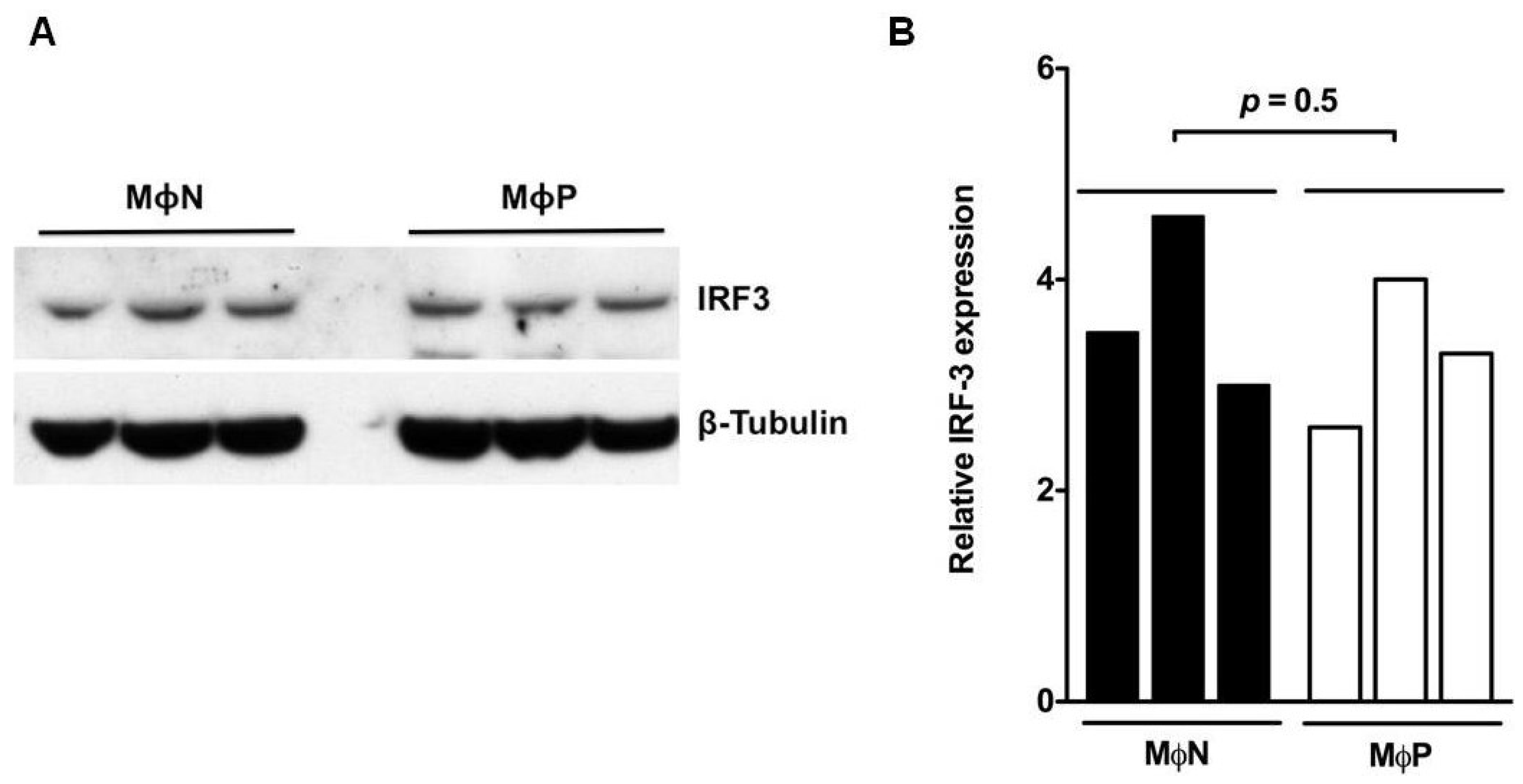
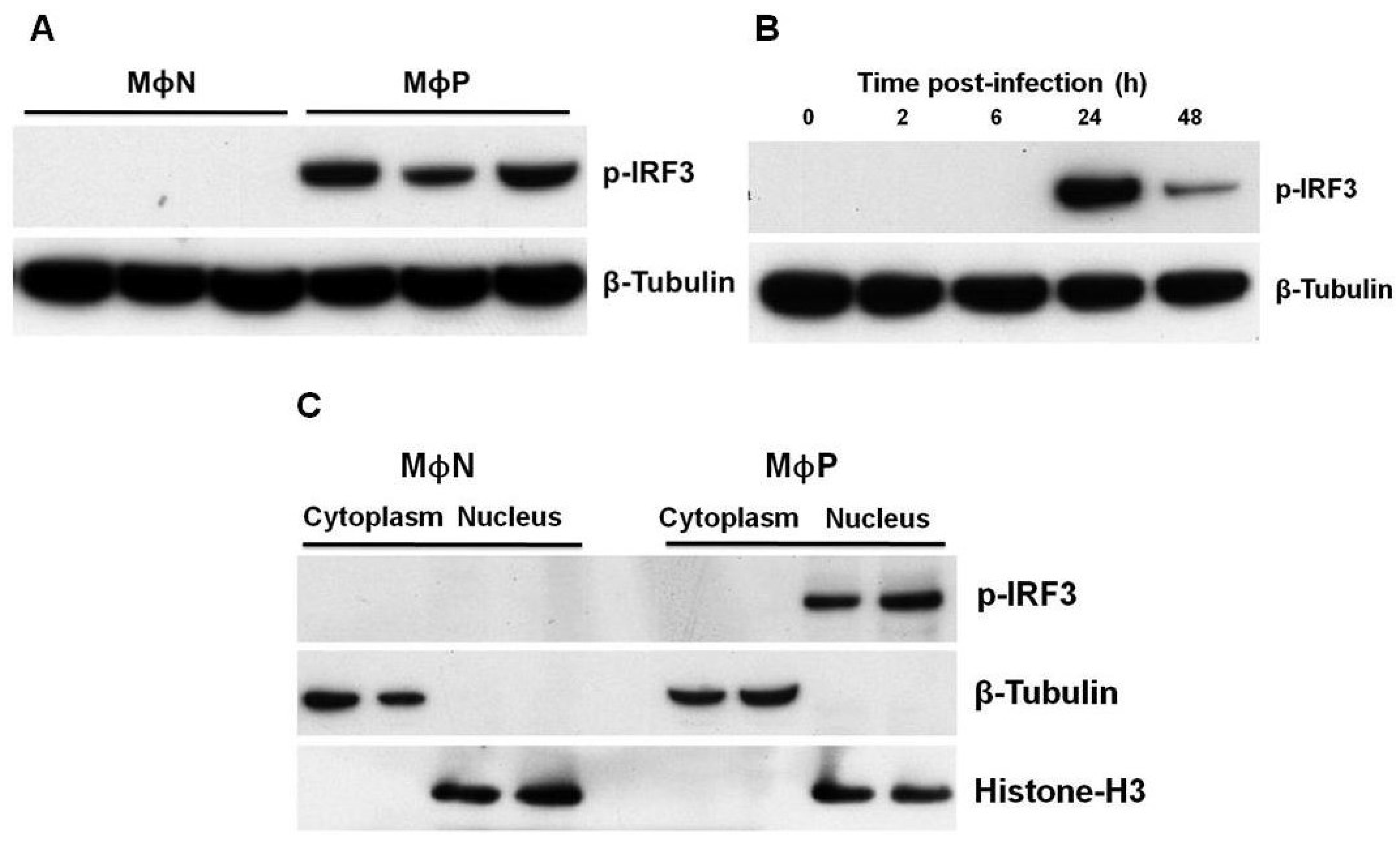
3.2. IRF3 was Constitutively Expressed in Its Phosphorylated Form in MΦP

The state of activation of the IFN-I signaling pathway during persistent RSV-infection was evaluated at the level of synthesis, by initial analysis of expression of the transcription factor IRF3. Western blot analysis (Figure 1A) showed that the levels of expression of total IRF3 in MΦP and MΦN were similar. Densitometry analysis (Figure 1B) showed no statistical difference between the average of three different passages from MΦN and MΦP (p = 0.5), indicating that constitutive expression of total IRF3 is not altered by persistent RSV-infection.
Figure 1. Total interferon regulatory factor 3 (IRF3) expression in persistently RSV-infected macrophages. (A) Total cell lysates from passages 86, 90 and 93 of non-infected macrophages (MΦN) and from passages 85, 86 and 88 of persistently RSV-infected macrophages (MΦP) were analyzed by western blot to detect total IRF3. The expression of β-tubulin protein was evaluated as loading control; (B) Semi-quantitative analysis was performed through densitometry with the software QuantityOne. Student-t test was performed to compare the average of the density values of the three passages of MΦN or MΦP. Results were reproduced in seven cellular passages analyzed (biological replicates).
Viruses 07 02879 g001
During acute viral infections, including those by RSV, IRF3 is activated through serine/threonine phosphorylation, leading to its homo-dimerization and nuclear translocation [19]. Whether persistent RSV also changes the state of activation of IRF3 in MΦP, was analyzed through western blot. Figure 2A shows that MΦP express constitutively p-IRF3 while in MΦN this active form of IRF3 was not detected. As a control to verify that RSV infection is sufficient to induce IRF3 activation, MΦN were infected in acute manner (MΦA). It was observed in MΦA phosphorylation of IRF3 at 24 h p.i., (Figure 2B) with a significant reduction by 48 h p.i., confirming that RSV infection promotes activation of IRF3.
Figure 2. Phosphorylation and location of interferon regulatory factor 3 (IRF3) in persistently RSV-infected macrophages. (A) Phospho-IRF3 (p-IRF3) expression was analyzed by western blot in total cell lysates from passages 86, 90 and 93 of non-infected macrophages (MΦN) and from passages 85, 86 and 88 of persistently RSV-infected macrophages (MΦP); (B) As a positive control, induction of IRF3 phosphorylation was analyzed in acutely RSV-infected macrophages (m.o.i. of 2) at 2, 6, 24, and 48 h p.i. As loading control, the expression of β-tubulin was evaluated; (C) Cytoplasmic and nuclear subcellular fractions from passages 94 and 95 of MΦN and from passages 90 and 91 of MΦP were evaluated for localization of p-IRF3. To demonstrate the purity of the fractions, histone-H3 and β-tubulin (nuclear and cytoplasmic markers, respectively) were determined. Results were reproduced in six cellular passages analyzed (biological replicates).
Viruses 07 02879 g002
To determine the subcellular localization of p-IRF3, cells were fractionated into nuclear and cytoplasmic extracts. For MΦP, p-IRF3 was located exclusively in nuclei, whereas for MΦN, it was not detected in either cellular fraction, thus indicating that persistence of RSV induces constitutive phosphorylation of IRF3 and its translocation to the nuclei (Figure 2C). Then, p-IRF3 may be able to induce gene expression in a constitutive manner.

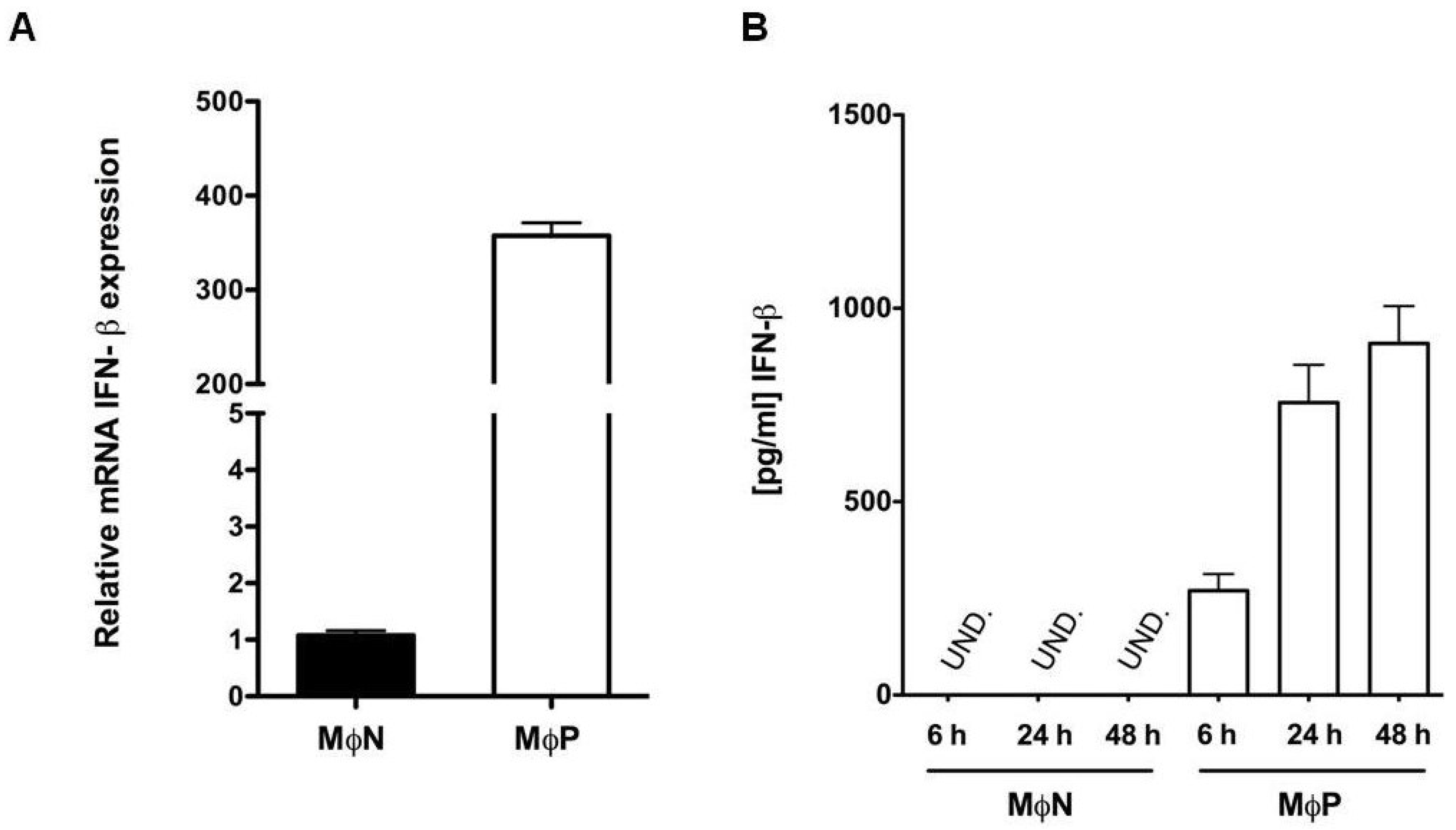
3.3. Persistent RSV Infection Induced Constitutive Expression of IFN-β

To evaluate whether nuclear-p-IRF3 is active as transcription factor, it was determined the expression level of mRNA for IFN-β, as this gene is under control of IRF-3. MΦN and MΦP at 24 h of culture were analyzed by quantitative RT-PCR (qRT-PCR) by the ΔCt method. Results showed that expression level of IFN-β mRNA in MΦP cultured for 24 h was more than 300-fold greater than that in MΦN (p < 0.0001) cultured in similar conditions (Figure 3A). No significant difference in IFN-β mRNA expression was observed between MΦP cultured for 6 and 24 h (351.6 ± 24.73- and 339.7 ± 22.07-fold increase, respectively in regard to MΦN). In order to determine whether IFN-β mRNA levels correlated with protein expression, supernatants from MΦP cultured for 6, 24, and 48 h were collected and analyzed by ELISA. Figure 3B shows a gradual increase in IFN-β expression over time, in the MΦP supernatants, possibly due to accumulation (270.1 ± 42.6, 756.20 ± 97.4, and 909.17 ± 96.6 pg/mL, respectively). No IFN-β was detected in the MΦN supernatants, despite their being cultured for 48 h. This result indicates that, although RSV persistence induces a constitutive production of IFN-β, MΦP are not able to eliminate the virus.
Figure 3. Expression of interferon-β (IFN-β) mRNA and protein in persistently RSV-infected macrophages. (A) Quantitative RT-PCR was performed with three different passages of non-infected macrophages (MΦN) and of persistently RSV-infected macrophages (MΦP), all cultured for 24 h, to determine the mRNA expression level of IFN-β by the ΔCt method. The values from the three different passages were averaged and plotted as relative expression of mRNA IFN-β; (B) Quantitative analysis by ELISA of the expression of IFN-β protein in supernatants of MΦN and MΦP cultured for 6, 24, and 48 h. UND: undetected. IFN-β mRNA and protein expression were evaluated in passages 93, 95 and 96 of MΦN and 92, 93 and 94 of MΦP. Results were reproduced in six cellular passages analyzed (biological replicates).
Viruses 07 02879 g003

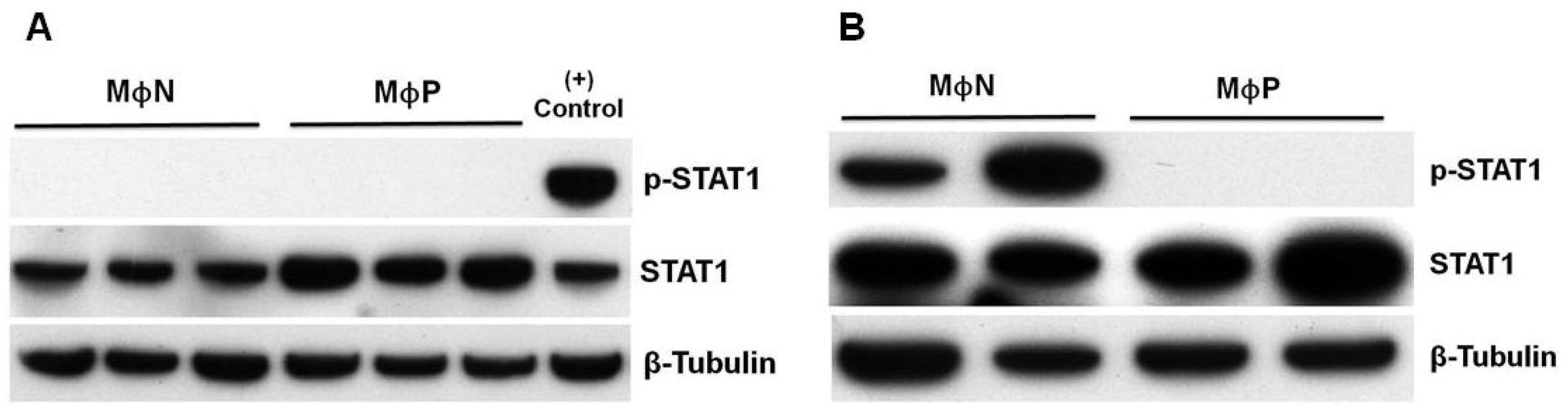
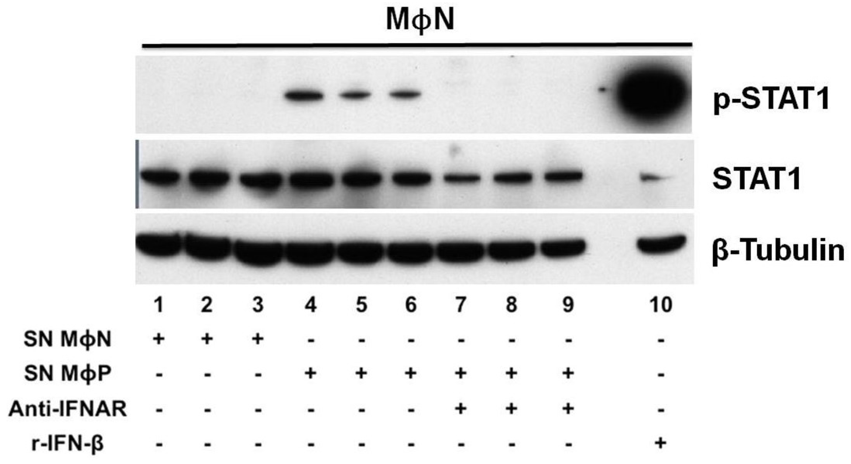
3.4. Persistent RSV Infection Impaired STAT1 Activation and ISGs Expression in Response to IFN-I

The activation of the IFN-I signaling pathway at the level of response was first evaluated in MΦP through the expression and activation of STAT1, a key player of the Jak/STAT signaling pathway, which is activated down-stream binding of IFNAR [40]. Total STAT1 was expressed constitutively in both MΦN and MΦP cultured for 48 h, but was slightly increased in MΦP (Figure 4A). However, p-STAT1 was not observed in any of the samples, although it had been expected that MΦP would have been positive for p-STAT1, because MΦP were constantly exposed to the IFN-β that they produced and secreted. A possible deficiency to signal in response to IFN-I by the macrophage-like cell line P388D1 was ruled out, because added commercial r-IFN-β induced phosphorylation of STAT1 in MΦN ((+) control, Figure 4A). In contrast, treatment of MΦP with a high concentration of r-IFN-β did not induce activation of STAT-1 (Figure 4B), confirming that activation of STAT1 during RSV persistence was impaired.
Figure 4. State of activation of transcription factor STAT1 in response to interferon-β (IFN-β) in persistently RSV-infected macrophages. (A) Total cell lysates of passages 95, 96 and 97 of non-infected macrophages (MΦN) and of passages 94, 95 and 96 of persistently RSV-infected macrophages (MΦP) were analyzed by western blot to detect total STAT1 and p-STAT1. As a positive control (+), MΦN were treated with 100 U/mL of recombinant-IFN-β for 45 min; (B) As MΦP do not respond to the IFN-β secreted by them, recombinant IFN-β (100 U/mL) was added to two passages of MΦP for 45 min. Two passages of MΦN were used as positive control. Results were reproduced in six cellular passages analyzed (biological replicates).
Viruses 07 02879 g004
A late consequence of the cellular response to IFN-I is the expression of antiviral genes [29]; thus the level of mRNA expression of the Mx1 and ISG56 genes in MΦP in respect to mock- or r-IFN-β-treated MΦN was then evaluated. Such genes have been previously reported as part of the early antiviral response during acute RSV infection [41,42]. As expected, relative expression of Mx1 and ISG56 mRNA in MΦP was similar to that in MΦN, notwithstanding constant exposure of MΦP to autocrine IFN-β (Figure 5A,B). Such result correlates with the lack of STAT1 phosphorylation in MΦP.
Figure 5. Mx1 and ISG56 mRNA expression level in persistently RSV-infected macrophages. Quantitative RT-PCR was performed with passages 107, 108 and 109 of mock-treated, non-infected macrophages (MΦN) and with passages 102, 103 and 104 of persistently RSV-infected macrophages (MΦP), to determine mRNA expression level of Mx1 (A) and ISG56 (B) by the ΔCt method. Also, MΦN were treated with 100 U/mL of recombinant-IFN-β (r-IFN-β) or supernatants from passages 87, 90 and 96 of MΦP (SN MΦP) for 45 min. Subsequently, Mx1 and ISG56 gene expression was evaluated. The values from the three different passages were averaged and plotted as relative expression of each gene. Treat: treatment. Results were reproduced in six cellular passages analyzed (biological replicates).
Viruses 07 02879 g005
To test whether the lack of autocrine response to IFN-β in MΦP was due to a deficient production of this cytokine or to impaired biological activity, three different passages of MΦN were treated with supernatants from MΦP cultures. In this case, STAT1 phosphorylation was induced in MΦN (Figure 6), along with the transcriptional activation of Mx1 and ISG56 genes (Figure 5A,B). Even more, when the IFN-I-receptor (IFNAR) in MΦN was blocked with a high concentration of a specific polyclonal antibody, subsequent exposition to supernatants from MΦP induced null levels of p-STAT1 (Figure 6). These findings indicate that the IFN-β produced by MΦP was of sufficient quantity and was capable of activating its specific signaling pathway in MΦN, but persistent RSV induced a refractory state to the IFN-I response in the host cell.
Figure 6. Activation of transcription factor STAT1 in non-infected macrophages in response to the interferon-β (IFN-β) produced by persistently RSV-infected macrophages. Three different passages of non-infected macrophages (MΦN) were incubated (45 min) with supernatants from three different passages of MΦN (lanes 1, 2, and 3) or with supernatants from passages 87, 90 and 96 of MΦP (lanes 4, 5, and 6), the latter supernatants containing MΦP-produced interferon-β (IFN-β). In addition, three passages of MΦN were pre-treated (30 min) with a polyclonal antibody against IFN α/β receptor (anti-IFNAR), at a concentration of 10 μg/mL, and then were incubated (45 min) with MΦP supernatants (lanes 7, 8, and 9). Total cell lysates were analyzed by western blot to detect total STAT1, p-STAT1, and β-tubulin as loading control. Infectious viruses present in the MΦP supernatants were inactivated by exposure to U.V. light to avoid viral infection as inducer of STAT1 activation. Treatment of a passage of MΦN with r-IFN-β (100 U/mL) was used as positive control (lane 10). Results were reproduced in six cellular passages analyzed (biological replicates).
Viruses 07 02879 g006

4. Discussion

Many viruses can establish and maintain persistent infections through interfering with, and controlling, antiviral cellular processes such as apoptosis and production of IFN-I [43]. Here, we report that RSV interfered with the IFN-I response during persistent infection in an in vitro model established in our laboratory, which consists of a murine macrophage cell line persistently infected with RSV (MΦP). The MΦP culture has been maintained over several passages, as confirmed through determining expression of viral N gene mRNA and the viral structural proteins N and F. Using this model, we observed that persistent RSV infection did not prevent either IRF3 activation or IFN-β expression in the host cell. However, the results indicate that persistent RSV infection inhibits the autocrine response to IFN-β down-stream binding the IFNAR, because neither STAT1 was phosphorylated nor Mx1 and ISG56 gene transcription were active in MΦP, even though we observed that this cytokine was constitutively produced and secreted into supernatants in sufficient quantities to activate MΦN. Moreover, MΦP were refractory to r-IFN-β stimuli, which induced STAT1 phosphorylation as well as antiviral genes expression in MΦN. Such results suggest that the lack of response to IFN-β by the persistently RSV-infected host cell may be one of several mechanisms for maintenance of RSV persistence.
Most studies conducted to analyze the consequences of RSV infection on IFN-I production and response have been performed by using models of acute infection. In comparison to other RNA viruses such as influenza and hMPV, RSV is considered as a low inducer of IFN-I due to the biological activity of the viral NS1 and NS2 proteins [44,45]. Epithelial cell lines and primary macrophages infected with mutant RSV lacking the NS1/NS2 genes produce higher levels of IFN-I than when infected with wtRSV [31,34,46]. Particularly, it has been reported that wtRSV infection of A546 cells induces activation and nuclear localization of IRF3; however, detection of this transcription factor is dramatically diminished with the onset of NS1/NS2 expression [34], due mainly to protein degradation [47]. Comparison between MΦP and MΦN showed no difference in total IRF3 expression, but the phosphorylated form was detected only in RSV-infected cultures. For MΦA, a high level of p-IRF3 was detected at 24 h p.i., with an important reduction by 48 h p.i., possibly associated with the expression of NS1/NS2. In contrast, that p-IRF3 was expressed in MΦP at similar levels after 24 or 48 h (data not shown), suggests equilibrium between production of viral and cellular proteins in the host cell during persistent RSV infection.
Nuclear localization of p-IRF3 correlated with the constitutive expression of mRNA of IFN-β gene and the secreted protein, indicating that its activation and role as a transcription factor is not impaired in macrophages by RSV persistence. To our knowledge, there is only one study directed to evaluate the state of activation of the IFN-I signaling pathway during RSV persistence and it was performed in in Hep-2 cells [48]. In agreement with our observations in kinetic assays to quantify the IFN-β produced at 6, 24 and 48 h by MΦP (shown in Figure 3B), it was reported that persistent RSV infection in Hep-2 cells also induces IFN-β expression, although it was quantified after 8 h of culture [48]. Expression of other genes directly controlled by IRF3 was not analyzed in our in vitro model, however we have observed high production of the chemokine RANTES [49], which is product of a gene that requires cooperation between IRF3 and NF-kB [50]. Such observations suggest that a subset of genes under the control of IRF3 may be constitutively expressed in MΦP.
We found the continuous production of IFN-β in MΦP is paradoxical, because these cells do not eliminate the persistent RSV. This finding may be explained by the lack of autocrine response to the constitutively produced IFN-β. This deficiency in response to IFN-I is in agreement with previous reports in which acute RSV infection in primary dendritic cells and RAW macrophages was shown to induce expression of total STAT1 and STAT2, whereas their phosphorylated forms were not found—Even after r-IFN-β had been added [39,51]. In contrast to reports of epithelial cells in which STAT2 is degraded by the expression of NS2 or during RSV infection [35,47,52], it appears that inhibition of response to IFN-I in myeloid cells is mediated by increased phosphatase activity, preventing Tyk2 phosphorylation with a subsequent deficiency in STAT2 and STAT1 activation, rather than degradation [39]. Otherwise, overexpression of suppressor of cytokine signaling 1 (SOCS1) during persistent RSV infection in Hep-2 cells is also a possible mechanism responsible for inhibition of STAT1/STAT2 phosphorylation [48]. In MΦP, we observed a slightly higher expression of total STAT1, than that in MΦN, but p-STAT1 was not detected after addition of r-IFN-β at a concentration that had been sufficient to activate STAT1 in MΦN. These results suggest that persistent RSV more efficiently controls the response to IFN-I than its production, by inhibition of STAT1 phosphorylation with the concomitant lack of ISGs transcription, allowing viral persistence. The biological activity of the IFN-β produced and secreted by MΦP was evidenced, because induced STAT1 phosphorylation and ISGs expression in MΦN and their activation was inhibited blocking the IFNAR with a specific polyclonal antibody. In future investigations, we will study the mechanism involved in the impairment of the IFN-I response in MΦP. Importantly, the paracrine effect of supernatants from MΦP on MΦN may lead to the acquisition of a proinflammatory phenotype, since antiviral response was activated in non-infected macrophages. In fact, supernatants from MΦP were more efficient than r-IFN-β to induce expression of the ISG56 gene (shown in Figure 5B).
Studying the infectious process by RSV in macrophages is relevant since during severe acute lower-respiratory tract disease in children, analysis of bronchioalveolar lavages has shown that alveolar macrophages are target cells for this virus, because they express RSV antigens and proinflammatory cytokines [53]. Also, it has been reported that isolated human alveolar macrophages can support a productive RSV infection in vitro at least for 25 days without a notable reduction in viability [54], suggesting that macrophages may be an important reservoir for RSV during acute infection in vivo and possibly during persistent infection, although this type of infection has not been confirmed in humans.

5. Conclusions

Overall, our work extends the information about the state of activation of the IFN-I signaling pathway during persistent RSV infection in macrophages, through three meaningful findings: 1) a constitutive state of activation of IRF3 resulting in a continual IFN-β production and secretion; 2) the lack of autocrine response to the constitutively produced IFN-β by inhibition of STAT1 phosphorylation, preventing transcription of antiviral genes and consequently allowing maintenance of persistent RSV infection; 3) the paracrine effect of the constitutively produced IFN-β, on non-infected macrophages inducing STAT1 activation and transcription of antiviral genes. We hypothesized that the paracrine effect of the IFN-I expressed by persistently infected macrophages, on non-infected cells may have relevant implications for the development of a chronic proinflammatory environment contributing to the pathogenesis of RSV infection.

Supplementary Files

Supplementary File 1

Acknowledgments

The authors thank Carlos Santiago Olivares and Jorge Gaona Bernal for constructive comments during the course of this work, and Veronica Yakoleff for helpful comments and English editing. The research was supported by grants from the Consejo Nacional de Ciencia y Tecnología, México (Grant 179838), the Dirección General de Asuntos del Personal Académico, Universidad Nacional Autónoma de México (Grant PAPIIT IN215913), and the Facultad de Medicina, UNAM.

Author Contributions

ERT and BG designed the experiments; ERT and LTG performed the experiments; ERT and BG prepared the manuscript.

Conflicts of Interest

The authors declare no conflict of interest.

References

  1. Nair, H.; Nokes, D.J.; Gessner, B.D.; Dherani, M.; Madhi, S.A.; Singleton, R.J.; O’Brien, K.L.; Roca, A.; Wright, P.F.; Bruce, N.; et al. Global burden of acute lower respiratory infections due to respiratory syncytial virus in young children: A systematic review and meta-analysis. Lancet 2010, 375, 1545–1555. [Google Scholar] [CrossRef]
  2. Hall, C.B.; Simőes, E.A.; Anderson, L.J. Clinical and epidemiologic features of respiratory syncytial virus. Curr. Top. Microbiol. Immunol. 2013, 372, 39–57. [Google Scholar] [PubMed]
  3. Bardach, A.; Rey-Ares, L.; Cafferata, M.L.; Cormick, G.; Romano, M.; Ruvinsky, S.; Savy, V. Systematic review and meta-analysis of respiratory syncytial virus infection epidemiology in Latin America. Rev. Med. Virol. 2014, 24, 76–89. [Google Scholar] [CrossRef] [PubMed]
  4. Staat, M.A. Respiratory syncytial virus infections in children. Semin. Respir. Infect. 2002, 17, 15–20. [Google Scholar] [CrossRef] [PubMed]
  5. Parrott, R.H.; Kim, H.W.; Brandt, C.D.; Chanock, R.M. Respiratory syncytial virus in infants and children. Prev. Med. 1974, 3, 473–480. [Google Scholar] [CrossRef]
  6. Sigurs, N.; Bjarnason, R.; Sigurbergsson, F.; Kjellman, B. Respiratory syncytial virus bronchiolitis in infancy is an important risk factor for asthma and allergy at age 7. Am. J. Respir. Crit. Care Med. 2000, 16, 1501–1507. [Google Scholar] [CrossRef] [PubMed]
  7. Piedimonte, G. Respiratory syncytial virus and asthma: Speed-dating or long-term relationship? Curr. Opin. Pediatr. 2013, 25, 344–349. [Google Scholar] [CrossRef] [PubMed]
  8. Zomer-Kooijker, K.; van der Ent, C.K.; Ermers, M.J.; Uiterwaal, C.S.; Rovers, M.M.; Bont, L.J.; RSV Corticosteroid Study Group. Increased risk of wheeze and decreased lung function after respiratory syncytial virus infection. PLoS ONE 2014, 9, e87162. [Google Scholar] [PubMed]
  9. Mejías, A.; Chávez-Bueno, S.; Ramilo, O. Respiratory syncytial virus pneumonia: Mechanisms of inflammation and prolonged airway hyperresponsiveness. Curr. Opin. Infect. Dis. 2005, 18, 199–204. [Google Scholar] [CrossRef] [PubMed]
  10. Isaia, G.; Teodosiu, O.; Popescu, G.; Athanasiu, P.; Sternberg, I.; Dumitriu, Z. Persistence of viruses in the nasopharynx of apparently healthy children aged 0–5 years. Results of investigations performed in 1982–1983. Virologie 1985, 36, 175–179. [Google Scholar] [PubMed]
  11. Borg, I.; Rohde, G.; Löseke, S.; Bittscheidt, J.; Schultze-Werninghaus, G.; Stephan, V.; Bufe, A. Evaluation of a quantitative real-time PCR for the detection of respiratory syncytial virus in pulmonary diseases. Eur. Respir. J. 2003, 21, 944–951. [Google Scholar] [CrossRef] [PubMed]
  12. Cubie, H.A.; Duncan, L.A.; Marshall, L.A.; Smith, N.M. Detection of respiratory syncytial virus nucleic acid in archival postmortem tissue from infants. Pediatr. Pathol. Lab. Med. 1997, 17, 927–938. [Google Scholar] [CrossRef] [PubMed]
  13. Rezaee, F.; Gibson, L.F.; Piktel, D.; Othumpangat, S.; Piedimonte, G. Respiratory syncytial virus infection in human bone marrow stromal cells. Am. J. Respir. Cell Mol. Biol. 2011, 45, 277–286. [Google Scholar] [PubMed]
  14. Sarmiento, R.E.; Tirado, R.; Gómez, B. Characteristics of a respiratory syncytial virus persistently infected macrophage-like culture. Virus Res. 2002, 84, 45–58. [Google Scholar] [CrossRef]
  15. Martínez, I.; Lombardía, L.; Herranz, C.; García-Barreno, B.; Domínguez, O.; Melero, J.A. Cultures of HEp-2 cells persistently infected by human respiratory syncytial virus differ in chemokine expression and resistance to apoptosis as compared to lytic infections of the same cell type. Virology 2009, 388, 31–41. [Google Scholar] [CrossRef] [PubMed]
  16. Schwarze, J.; O’Donnell, D.R.; Rohwedder, A.; Openshaw, P.J. Latency and persistence of respiratory syncytial virus despite T cell immunity. Am. J. Respir. Crit. Care Med. 2004, 169, 801–805. [Google Scholar] [CrossRef] [PubMed]
  17. Estripeaut, D.; Torres, J.P.; Somers, C.S.; Tagliabue, C.; Khokhar, S.; Bhoj, V.G.; Grube, S.M.; Wozniakowski, A.; Gomez, A.M.; Ramilo, O.; et al. Respiratory syncytial virus persistence in the lungs correlates with airway hyperreactivity in the mouse model. J. Infect. Dis. 2008, 198, 1435–1443. [Google Scholar] [CrossRef] [PubMed]
  18. Hegele, R.G.; Hayashi, S.; Bramley, A.M.; Hogg, J.C. Persistence of respiratory syncytial virus genome and protein after acute bronchiolitis in guinea pigs. Chest 1994, 105, 1848–1854. [Google Scholar] [CrossRef] [PubMed]
  19. Yoneyama, M.; Suhara, W.; Fukuhara, Y.; Fukuda, M.; Nishida, E.; Fujita, T. Direct triggering of the type I interferon system by virus infection: Activation of a transcription factor complex containing IRF-3 and CBP/p300. EMBO J. 1998, 17, 1087–1095. [Google Scholar] [CrossRef] [PubMed]
  20. Wathelet, M.G.; Lin, C.H.; Parekh, B.S.; Ronco, L.V.; Howley, P.M.; Maniatis, T. Virus infection induces the assembly of coordinately activated transcription factors on the IFN-beta enhancer in vivo. Mol. Cell 1998, 1, 507–518. [Google Scholar] [CrossRef]
  21. Borrow, P.; Martínez-Sobrido, L.; de la Torre, J.C. Inhibition of the type I interferon antiviral response during arenavirus infection. Viruses 2010, 2, 2443–2480. [Google Scholar] [CrossRef] [PubMed]
  22. Foy, E.; Li, K.; Wang, C.; Sumpter, R., Jr.; Ikeda, M.; Lemon, S.M.; Gale, M., Jr. Regulation of interferon regulatory factor-3 by the hepatitis C virus serine protease. Science 2003, 300, 1145–1148. [Google Scholar] [CrossRef] [PubMed]
  23. Ye, J.; Maniatis, T. Negative regulation of interferon-β gene expression during acute and persistent virus infections. PLoS ONE 2011, 6, e20681. [Google Scholar] [CrossRef] [PubMed]
  24. Schlee, M. Master sensors of pathogenic RNA - RIG-I like receptors. Immunobiology 2013, 218, 1322–1335. [Google Scholar] [CrossRef] [PubMed]
  25. Fitzgerald, K.A.; McWhirter, S.M.; Faia, K.L.; Rowe, D.C.; Latz, E.; Golenbock, D.T.; Coyle, A.J.; Liao, S.M.; Maniatis, T. IKKepsilon and TBK1 are essential components of the IRF3 signaling pathway. Nat. Immunol. 2003, 4, 491–496. [Google Scholar] [CrossRef] [PubMed]
  26. Seth, R.B.; Sun, L.; Ea, C.K.; Chen, Z.J. Identification and characterization of MAVS, a mitochondrial antiviral signaling protein that activates NF-kappaB and IRF3. Cell 2005, 122, 669–682. [Google Scholar] [CrossRef] [PubMed]
  27. Conzelmann, K.K. Transcriptional activation of alpha/beta interferon genes: Interference by nonsegmented negative-strand RNA viruses. J. Virol. 2005, 79, 5241–5248. [Google Scholar] [CrossRef] [PubMed]
  28. Yang, H.; Ma, G.; Lin, C.H.; Orr, M.; Wathelet, M.G. Mechanism for transcriptional synergy between interferon regulatory factor (IRF)-3 and IRF-7 in activation of the interferon-beta gene promoter. Eur. J. Biochem. 2004, 271, 3693–3703. [Google Scholar] [CrossRef] [PubMed]
  29. Kraus, T.A.; Lau, J.F.; Parisien, J.P.; Horvath, C.M. A hybrid IRF9-STAT2 protein recapitulates interferon-stimulated gene expression and antiviral response. J. Biol. Chem. 2003, 278, 13033–13038. [Google Scholar] [CrossRef] [PubMed]
  30. Schindler, C.; Levy, D.E.; Decker, T. JAK-STAT signaling: From interferons to cytokines. J. Biol. Chem. 2007, 282, 20059–20063. [Google Scholar] [CrossRef] [PubMed]
  31. Spann, K.M.; Tran, K.C.; Chi, B.; Rabin, R.L.; Collins, P.L. Suppression of the induction of alpha, beta, and lambda interferons by the NS1 and NS2 proteins of human respiratory syncytial virus in human epithelial cells and macrophages. J. Virol. 2004, 78, 4363–4369. [Google Scholar] [CrossRef] [PubMed]
  32. Barik, S. Respiratory syncytial virus mechanisms to interfere with type 1 interferons. Curr. Top. Microbiol. Immunol. 2013, 372, 173–191. [Google Scholar] [PubMed]
  33. Lo, M.S.; Brazas, R.M.; Holtzman, M.J. Respiratory syncytial virus nonstructural proteins NS1 and NS2 mediate inhibition of Stat2 expression and alpha/beta interferon responsiveness. J. Virol. 2005, 79, 9315–9319. [Google Scholar] [CrossRef] [PubMed]
  34. Spann, K.M.; Tran, K.C.; Collins, P.L. Effects of nonstructural proteins NS1 and NS2 of human respiratory syncytial virus on interferon regulatory factor 3, NF-kappaB, and proinflammatory cytokines. J. Virol. 2005, 79, 5353–5362. [Google Scholar] [CrossRef] [PubMed]
  35. Swedan, S.; Musiyenko, A.; Barik, S. Respiratory syncytial virus nonstructural proteins decrease levels of multiple members of the cellular interferon pathways. J. Virol. 2009, 83, 9682–9693. [Google Scholar] [CrossRef] [PubMed]
  36. Ren, J.; Liu, T.; Pang, L.; Li, K.; Garofalo, R.P.; Casola, A.; Bao, X. A novel mechanism for the inhibition of interferon regulatory factor-3-dependent gene expression by human respiratory syncytial virus NS1 protein. J. Gen. Virol. 2011, 92, 2153–2159. [Google Scholar] [CrossRef] [PubMed]
  37. Payment, P.; Trudel, M. Methods and Techniques in Virology; Marcel Dekker: New York, NY, USA, 1993; Volume 36–37, pp. 73–74. [Google Scholar]
  38. Gaona, J.; Santiago-Olivares, C.; Ortega, E.; Gómez, B. Respiratory syncytial virus persistence in macrophages upregulates Fcgamma receptors expression. Viruses 2014, 6, 624–639. [Google Scholar] [CrossRef] [PubMed]
  39. Senft, A.P.; Taylor, R.H.; Lei, W.; Campbell, S.A.; Tipper, J.L.; Martinez, M.J.; Witt, T.L.; Clay, C.C.; Harrod, K.S. Respiratory syncytial virus impairs macrophage IFN-alpha/beta- and IFN-gamma-stimulated transcription by distinct mechanisms. Am. J. Respir. Cell Mol. Biol. 2010, 42, 404–414. [Google Scholar] [CrossRef] [PubMed]
  40. Ivashkiv, L.B.; Donlin, L.T. Regulation of type I interferon responses. Nat. Rev. Immunol. 2014, 14, 36–49. [Google Scholar] [CrossRef] [PubMed]
  41. Pletneva, L.M.; Haller, O.; Porter, D.D.; Prince, G.A.; Blanco, J.C. Induction of type I interferons and interferon-inducible Mx genes during respiratory syncytial virus infection and reinfection in cotton rats. J. Gen. Virol. 2008, 89, 261–270. [Google Scholar] [CrossRef] [PubMed]
  42. Tian, B.; Zhao, Y.; Kalita, M.; Edeh, C.B.; Paessler, S.; Casola, A.; Teng, M.N.; Garofalo, R.P.; Brasier, A.R. CDK9-dependent transcriptional elongation in the innate interferon-stimulated gene response to respiratory syncytial virus infection in airway epithelial cells. J. Virol. 2013, 87, 7075–7092. [Google Scholar] [CrossRef] [PubMed]
  43. Kane, M.; Golovkina, T. Common threads in persistent viral infections. J. Virol. 2010, 84, 4116–4123. [Google Scholar] [CrossRef] [PubMed]
  44. Chonmaitree, T.; Roberts, N.J., Jr.; Douglas, R.G., Jr.; Hall, C.B.; Simons, R.L. Interferon production by human mononuclear leukocytes: Differences between respiratory syncytial virus and influenza viruses. Infect. Immun. 1981, 32, 300–303. [Google Scholar] [PubMed]
  45. Guerrero-Plata, A.; Casola, A.; Suarez, G.; Yu, X.; Spetch, L.; Peeples, M.E.; Garofalo, R.P. Differential response of dendritic cells to human metapneumovirus and respiratory syncytial virus. Am. J. Respir. Cell Mol. Biol. 2006, 34, 320–329. [Google Scholar] [CrossRef] [PubMed]
  46. Ling, Z.; Tran, K.C.; Teng, M.N. Human respiratory syncytial virus nonstructural protein NS2 antagonizes the activation of beta interferon transcription by interacting with RIG-I. J. Virol. 2009, 83, 3734–3742. [Google Scholar] [CrossRef] [PubMed]
  47. Goswami, R.; Majumdar, T.; Dhar, J.; Chattopadhyay, S.; Bandyopadhyay, S.K.; Verbovetskaya, V.; Sen, G.C.; Barik, S. Viral degradasome hijacks mitochondria to suppress innate immunity. Cell Res. 2013, 23, 1025–1042. [Google Scholar] [CrossRef] [PubMed]
  48. Zheng, J.; Yang, P.; Tang, Y.; Zhao, D. A respiratory syncytial virus persistent-infected cell line system reveals the involvement of SOCS1 in the innate antiviral response. Virol. Sin. 2015, 30, 190–199. [Google Scholar] [CrossRef] [PubMed]
  49. Rivera-Toledo, E.; Gómez, B. National Autonomous University of Mexico (UNAM); Ciudad Universitaria: D.F., Mexico, 2014. [Google Scholar]
  50. Génin, P.; Algarté, M.; Roof, P.; Lin, R.; Hiscott, J. Regulation of RANTES chemokine gene expression requires cooperativity between NF-kappa B and IFN-regulatory factor transcription factors. J. Immunol. 2000, 164, 5352–5361. [Google Scholar] [CrossRef] [PubMed]
  51. Jie, Z.; Dinwiddie, D.L.; Senft, A.P.; Harrod, K.S. Regulation of STAT signaling in mouse bone marrow derived dendritic cells by respiratory syncytial virus. Virus Res. 2011, 156, 127–133. [Google Scholar] [CrossRef] [PubMed]
  52. Ramaswamy, M.; Shi, L.; Varga, S.M.; Barik, S.; Behlke, M.A.; Look, D.C. Respiratory syncytial virus nonstructural protein 2 specifically inhibits type I interferon signal transduction. Virology 2006, 344, 328–339. [Google Scholar] [CrossRef] [PubMed]
  53. Midulla, F.; Villani, A.; Panuska, J.R.; Dab, I.; Kolls, J.K.; Merolla, R.; Ronchetti, R. Respiratory syncytial virus lung infection in infants: Immunoregulatory role of infected alveolar macrophages. J. Infect. Dis. 1993, 168, 1515–1519. [Google Scholar] [CrossRef] [PubMed]
  54. Panuska, J.R.; Cirino, N.M.; Midulla, F.; Despot, J.E.; McFadden, E.R., Jr.; Huang, Y.T. Productive infection of isolated human alveolar macrophages by respiratory syncytial virus. J. Clin. Investig. 1990, 83, 113–119. [Google Scholar] [CrossRef] [PubMed]

