Review

The Roles of Platelet-Activating Factor and Magnesium in Pathophysiology of Hypertension, Atherogenesis, Cardiovascular Disease, Stroke and Aging

Touro College of Osteopathic Medicine, 60 Prospect Ave, Middletown, NY 10940, USA
*
Author to whom correspondence should be addressed.
Cardiogenetics 2022, 12(1), 49-62; https://doi.org/10.3390/cardiogenetics12010005
Submission received: 13 October 2021 / Revised: 16 January 2022 / Accepted: 26 January 2022 / Published: 2 February 2022
(This article belongs to the Special Issue Cardiogenetics: Feature Papers 2021)

Abstract

:
Hypertension and atherosclerosis are debilitating diseases that affect millions each year. Long-term consequences include but are not limited to stroke, myocardial infarction, and kidney failure. Platelet-activating factor (PAF) is a proinflammatory mediator synthesized from a subclass of phosphatidylcholines that increases platelet activation, leukocyte adhesion, infiltration of macrophages, and intracellular lipid accumulation, thereby contributing to atherosclerosis. Magnesium, a key micronutrient and free radical scavenger, is a water-soluble mineral that regulates peripheral vasodilation and calcium, phosphate, and hydroxyapatite homeostasis. Magnesium’s antihypertensive ability stems from its role as a natural calcium antagonist and promoter of vasodilatory mediators, such as nitric oxide. Platelet-activating factor and magnesium share an inverse relationship, and elevated magnesium levels have been shown to have protective effects against plaque formation as well as antihypertensive and antiarrhythmic effects, all of which allow for healthier aging. The purpose of this literature review is to investigate the role of platelet-activating factor and magnesium in the pathophysiology of hypertension, atherosclerosis, cardiovascular disease, stroke, and aging. Since the pathophysiology of the platelet-activating factor biomolecule is underexplored, further research studies are warranted in order to navigate the putative signaling pathways involved in the cardioprotective effects of dietary magnesium as a natural anti-PAF agent.

1. Introduction

The homeostatic function of peripheral, coronary, and cerebral vasculature is dependent on the molecular functions of numerous chemical mediators. Magnesium, being one of these mediators, is a micronutrient that has been shown to help reduce the risk of developing cardiovascular complications resulting from hypertension [1]. This mineral has also been implicated in decreasing levels of platelet activating factor (PAF), a proinflammatory mediator involved in the development of thrombogenic plaques [2,3].
In this review, we will discuss hypertension, followed by the role of PAF in atherogenesis as it relates to causing hypertension. Finally, magnesium will be introduced along with its beneficial role in reducing hypertension and antagonism of PAF. Ultimately, this study will explore the roles of PAF and magnesium in cardiovascular disease pertaining to atherosclerosis, hypertension, and other relevant cardiac pathology.

2. Hypertension: Risk Factors and Link to Atherosclerosis

Hypertension is a public health menace with an estimated prevalence rate of 45.4% in adults in the United States [4]. Defined as an increase in pressure exerted by blood in the arteries, a common metric used to assess hypertension is blood pressure [5]. The current guidelines by the American College of Cardiology categorize the levels of blood pressures into four different stages. These criteria are organized in Table 1. However, in order to stage a diagnosis of hypertension, an average of two or more readings must be performed [6].
The exact cause of hypertension in approximately 95% of cases is unknown. This is termed as ‘essential hypertension’ [7]. Both genetic and environmental factors underlie essential hypertension [7]. On the other hand, if the underlying cause of hypertension is known, such as reduction in blood flow, kidney dysfunctions, or endocrine pathologies, it is termed as “secondary hypertension” [8]. A plethora of lifestyle factors such as smoking, psychological stressors, low vegetable/fruit consumption, excess alcohol, and obesity increase the chances of hypertension; with obesity and excess alcohol being at the forefront [9]. Furthermore, several genetic and epigenetic variations affecting the Renin-Angiotensin-Aldosterone System (RAAS) and endothelial elasticity can directly affect renal plasma flow, fluid/electrolyte balance, and systemic/peripheral vascular resistance, thereby predisposing individuals to hypertension [3,4,5,6,7,8].
Hypertensive patients are at higher risk for atherothrombotic events such as ischemic and hemorrhagic stroke, retinopathies, and myocardial infarctions [10]. Hypertension is typically preceded by prothrombotic events such as vascular dysfunction, imbalance of procoagulants, and fibrinolytic activity, which ultimately affects platelet function [11]. Platelets play a critical role in maintaining vascular homeostasis and as such their maladaptive function underlies atherosclerosis [12]. Even though the sequence of events leading to atherogenic episodes is understood, the precise mechanism that activates platelets remains largely unknown [13,14,15]. Platelets when activated by Platelet-Activating Factor (PAF) or environmental stressors are prone to producing platelet derived microparticles (PMPs). These PMPs have been associated with numerous cardiovascular risk factors, one of them being hypertension [14]. PAF, a phospholipid mediator, may have a key role in linking maladaptive platelet function to atherosclerosis and subsequent hypertension.

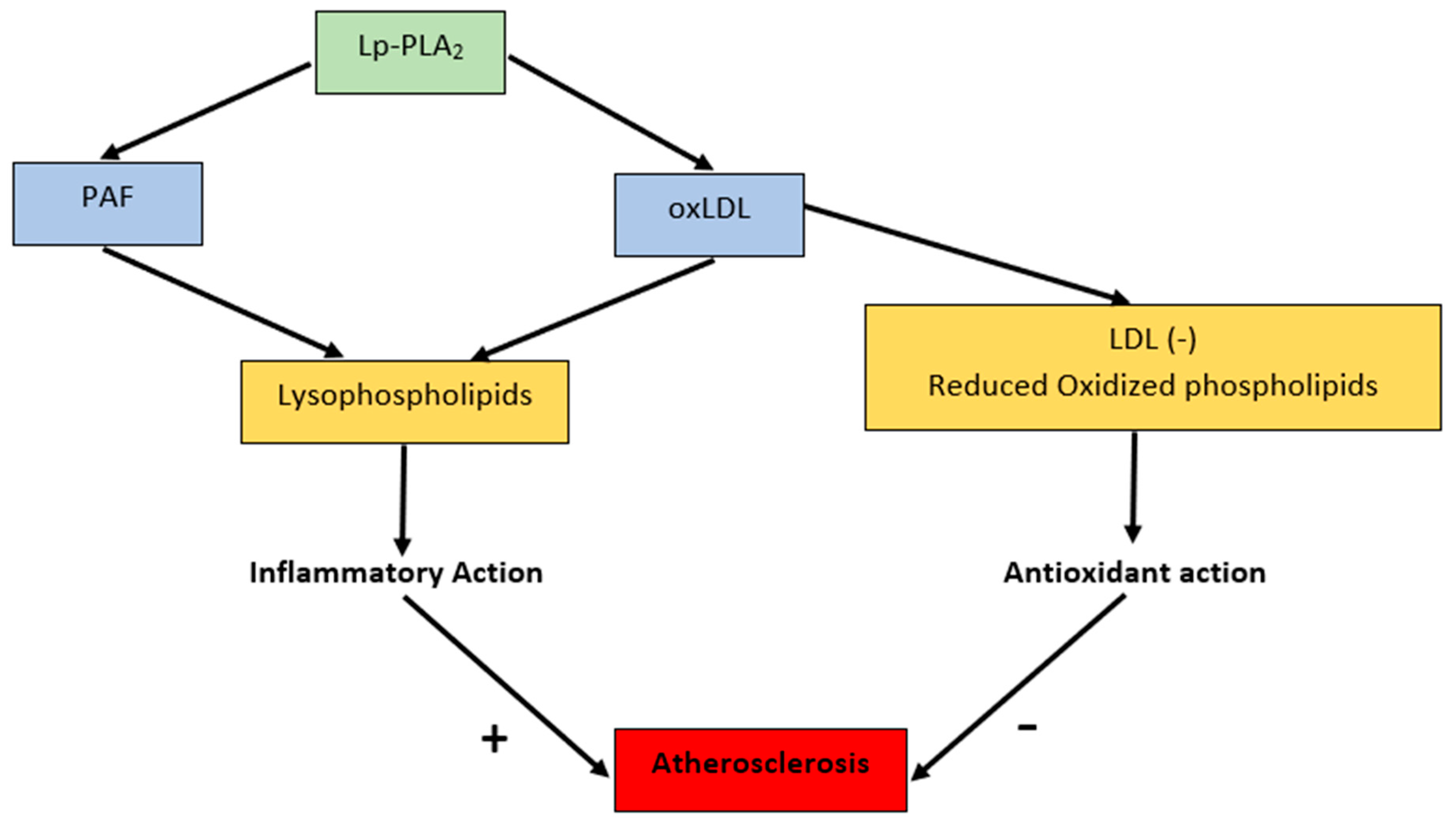
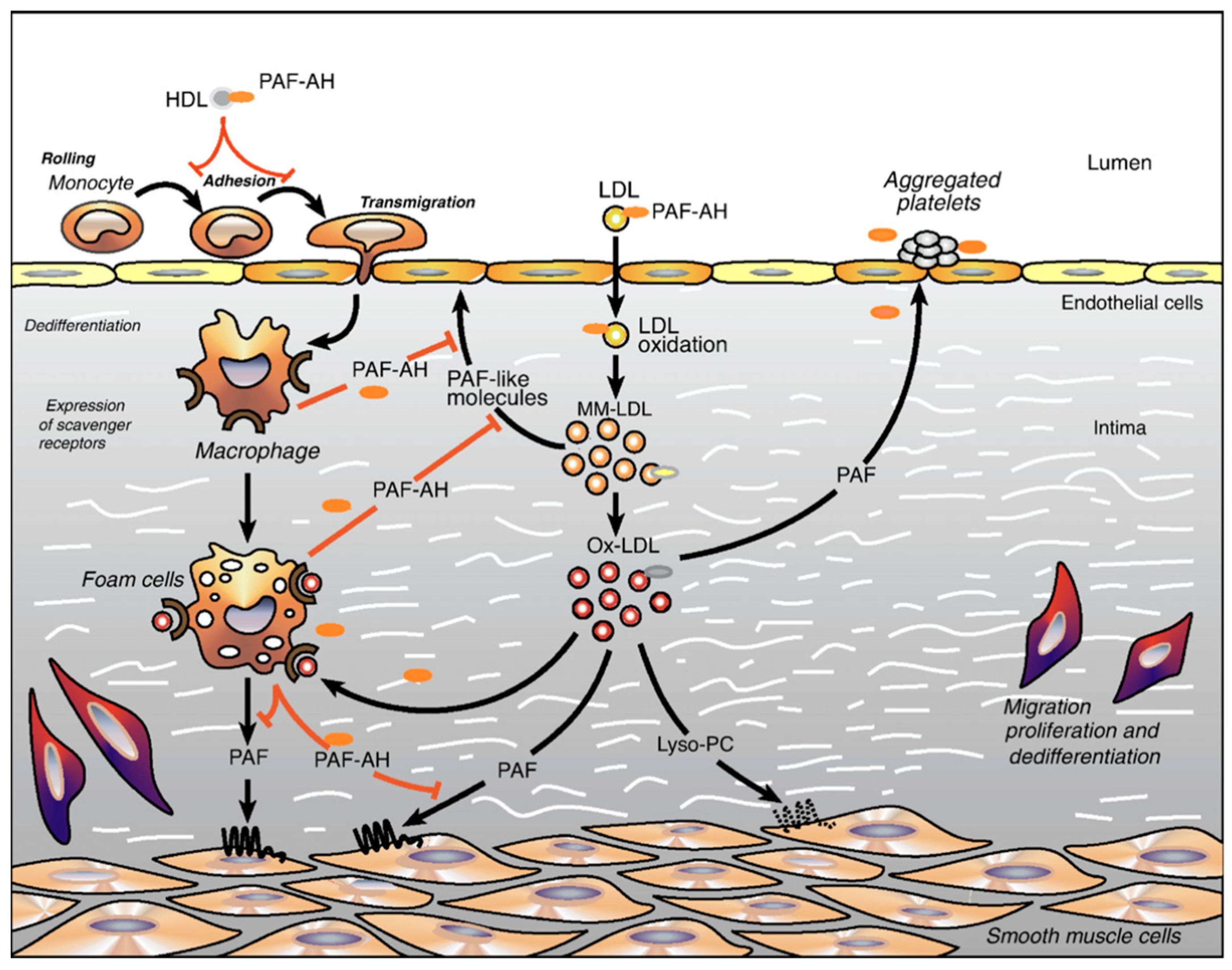
3. PAF in the Pathophysiology of Atherosclerosis

One of the common pathways that has been linked to atherogenesis is the Nuclear Factor Kappa-B (NF-kB) pathway (Figure 1) [15]. NF-kB is an important nuclear transcription factor involved in cytokine activation through regulation of inflammation [16]. Cytokines released by cells or oxidative stress can activate NF-kB, which upregulates genes such as VCAM1, a cell adhesion molecule, and tumor necrosis factor (TNF-α) in the blood vessels [17,18]. Constitutive expression of the NF-kB signal in blood vessels and smooth muscle cells leads to a chronic increase in oxidative stress, which further promotes cytokine activation. This causes a systemic inflammatory response, ultimately resulting in atherogenesis [15]. The evidence for the role of NF-kB pathway in facilitating atherogenesis has been found by the detection of NF-kB associated proteins in atherosclerotic vessels; it is not present in healthy blood vessels that do not exhibit evidence of atherogenesis [18,19].
PAF is considered to play an important role in atherosclerotic plaque formation by supplementing the inflammatory processes that take place within the NF-kB pathway [21]. Clinical studies have shown an observable correlation between patients with severe atherosclerosis and elevated PAF levels in coronary artery circulation [22]. PAF is released by endothelial cells in response to thrombin, vasoactive mediators, and pro-inflammatory cytokines [9]. It is a known vasoactive mediator that causes increased leukocyte adhesion and infiltration of macrophages, resulting in cytokine release, inflammation, and intracellular lipid accumulation [3]. Macrophages are specifically activated by PAF, leading to an increase in intracellular calcium levels. This calcium triggers further downstream effects by first leading to macrophage adhesion to LDL [23], which results in the oxidation of LDL by macrophages. This is an important step in the atherogenic mechanism [24]. The oxidized LDL are taken up by macrophages and generate foam cells along blood vessel walls, which are seen in atherosclerotic plaques, indicated by Figure 2. In addition, macrophages can also release PAF, which further facilitates plaque formation [25]. PAF also increases oxidative stress in the blood vessels through indirect generation of reactive oxygen species and increasing the vascular permeability of arteries [26]. Furthermore, PAF directly activates platelets, causing them to aggregate and adhere to the injured endothelium, thereby initiating the plaque-forming cascade in blood vessels (Figure 3) [16].
PAF’s role in hypertension and arrhythmias is of concern. A study performed in rats has shown that low endogenous levels of PAF correlated with peripheral vasodilation, thereby highlighting a potential protective effect on peripheral vascular resistance and hypertension [27]. Furthermore, PAF levels were elevated in ischemic myocardium, particularly in conjunction with arrhythmias [28]. Hence, evidence suggests a potential role of PAF in mediating fatal arrhythmias such as ventricular fibrillation that are a known complication of ischemic myocardial injury [28].
Figure 2. Role of PAF in atherosclerosis [29].
Cardiogenetics 12 00005 g002
Figure 3. Relationship between PAF and atherosclerosis [30].
Cardiogenetics 12 00005 g003

4. PAF Structure and Molecular Mechanism

PAF’s (1-O-alkyl-2-acetyl-sn-glycero-3-phosphocholine) unique structure offers further insights into its role in atherosclerosis. PAF is synthesized from a subclass of phosphatidylcholines, which contain an ether bond instead of an ester bond with the backbone of glycerol [20]. It is a minor component of low-density lipoprotein (LDL), which, along with platelets, is another key pro-atherogenic mediator [27]. Moreover, the precursors of PAF can also undergo oxidative attack since they contain polyunsaturated fatty acids. Such oxidative damage could result in further numerous PAF-like molecules [31].
The PAF receptor is present on various cell types including immune, endothelium, smooth muscle, platelets, and cardiomyocytes [19,32]. It belongs to a G-protein linked receptor superfamily, which transduces a variety of functions via multiple heterotrimeric G proteins [19,32]. Protein kinase C activation, tyrosine phosphorylation, and proto-oncogene expression are among the diverse effects observed upon PAF receptor activation [33]. The inflammatory cascade also involves the stimulation of phospholipase A2 and subsequent production of arachidonic acid, which is a key precursor for several inflammatory mediators [33].

5. PAF in Cardiac Pathophysiology and Stroke

PAF release has been noted to increase in the heart post-ischemia, indicating that myocardial tissue can produce and release PAF in the absence of perfusion. In addition, reperfusion of cardiac tissue post-ischemia has been followed by an increase in PAF release, which contributes to inflammation [34]. Sources of the rise in PAF levels include platelets, polymorphonuclear leukocytes, monocytes, mast cells, macrophages, and even cardiac myocytes [34]. PAF reciprocally promotes recruitment of these polymorphonuclear leukocytes, monocytes, and eosinophils to release pro-inflammatory cytokines, causing endothelial damage and inflammation [35]. Reactive oxygen species then oxidize low density lipoprotein, which contributes to atherosclerotic plaque formation. Subsequent recruitment of Th-1 cells leads to further inflammation, and disruption of the atherosclerotic plaque, causing acute cardiovascular disease [35].
Generally, PAF has a depressive effect on the cardiovascular system’s function. It causes a decrease in venous return by inducing systemic venous vasodilation and increasing vascular permeability [34]. In addition, PAF is strongly associated with coronary vasoconstriction, believed to be mediated by serotonin, thromboxane, and leukotriene, which reduces coronary artery perfusion and oxygen supply [34]. PAF is also believed to have a minor impact on cardiac conduction, causing cardiac arrhythmias [34]. Studies conducted with both isolated hearts and cultured myocyte samples have shown that, when exposed to PAF, there is a decrease in contractile force, beat amplitude, and velocities of contraction and relaxation [34].
As in the myocardium, PAF levels have been noticed to increase in cerebrovascular tissues post-ischemia. After an ischemic event, cerebral tissue undergoes stroke due to hypoxia, and PAF is believed to be responsible for the vasoconstriction of vessels that are supplying the ischemic regions of the brain [36]. Some studies have shown that the administration of a PAF receptor antagonist, such as indomethacin, decreases the PAF-mediated ischemia and hypoxia seen in stroke-affected cerebral tissues [36]. One study, conducted by K. Satoh et. al. in 1992, looked at PAF blood levels in stroke patients by performing a radioimmunoassay. When compared to the controls, post-stroke patients were measured to have an increase in PAF levels in the blood [37].
Hypersensitivity reactions by the immune system can target the cardiovascular system, and PAF is the major factor mediating these reactions. PAF, along with cyclooxygenase and leukotrienes, is responsible for the coronary vasoconstriction, arrhythmias, and decreased cardiac contractility seen in hypersensitivity reactions [34]. In addition, administration of PAF receptor-specific antagonists has been shown to decrease the cardiovascular effects of these hypersensitivity reactions.

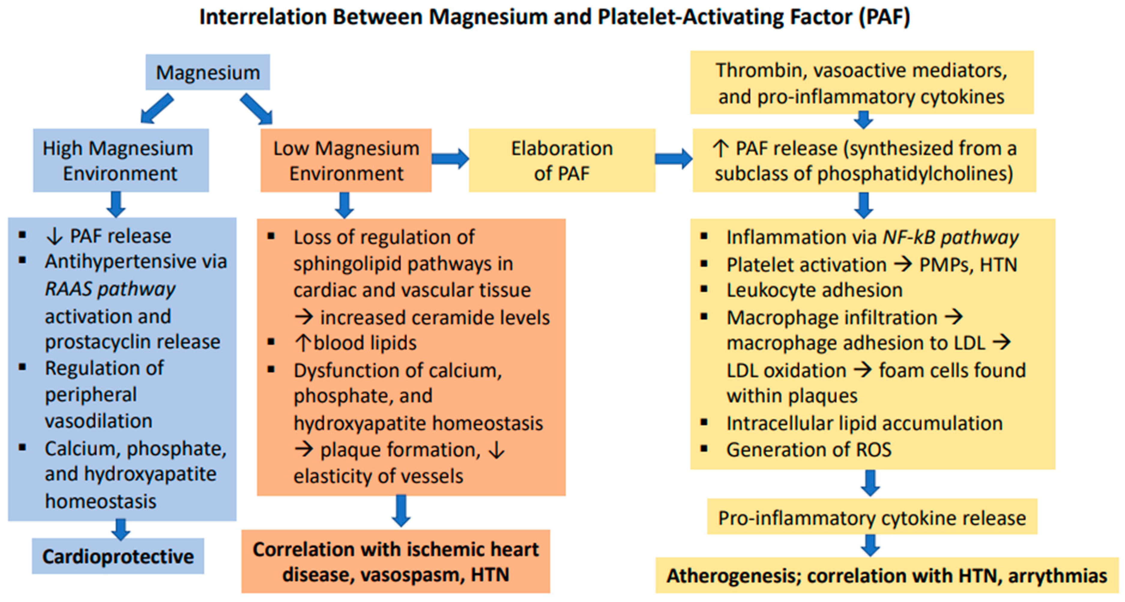
6. Magnesium in the Pathophysiology of Atherosclerosis and Hypertension

PAF has been found to be elevated in blood and tissues of animals deficient in magnesium [38]. A study using proton nuclear magnetic resonance spectroscopy on single vascular smooth muscle cells, excised canine and rat aortic, coronary, and cerebral arterial vessels illustrated that low levels of magnesium led to rapid PAF synthesis [39].
Magnesium is also independently implicated in causing maladaptive changes that lead to atherosclerosis. Deficiency of magnesium leads to a loss of its regulatory effects on sphingolipid pathways in cardiac and vascular smooth muscle cells, resulting in elevated ceramide levels from sphingolipid metabolism [40]. Elevated ceramide concentrations may lead to erosion of existing atherosclerotic plaques, thereby inducing thrombosis and plaque remodeling [39]. Deficiency of magnesium worsens lipid metabolism, resulting in a build-up of cholesterol, triglycerides, VLDL, and LDL [41,42]. These lipids pose a great danger as they can aggregate within the vasculature and cause atherosclerosis. They are also accompanied by enhanced oxidation of very-low-density lipoproteins and low-density lipoproteins (VLDL)/(LDL) and lipid peroxidation in cardiac myocytes. This can increase reactive oxygen species to levels that are known to perpetuate the inflammation preceding plaque formation [32,38]. Magnesium also helps maintain the elasticity of vessels by antagonizing calcium deposition [41]. Magnesium binds phosphate in the gastrointestinal tract, effectively inhibiting plaque formation as both calcium and phosphate are needed [43]. It also inhibits the maturation of hydroxyapatite, which is the most abundant crystal present in atherosclerotic plaques [43].
The antihypertensive effect of magnesium is supported by studies showing that decreased magnesium levels caused activation of the RAAS pathway, increased angiotensin II induced plasma aldosterone concentration, production of thromboxane, and vasoconstrictor prostaglandins [44]. Magnesium also increases prostacyclin release in cultured cells as well as healthy individuals [45]. Normally, the endothelium regulates its vasomotor tone by synthesizing prostacyclin [45]. Magnesium promotes vasodilatory effects of blood vessels through increasing prostacyclin release, thus possessing potential antihypertensive effects [45].
Long term and significant magnesium deficiency were associated with overactive RAAS, hypertension, and oxidative stress that induced damage to the endothelium [46]. Figure 4 demonstrates the far-reaching, deleterious effects of magnesium deficiency.

7. Magnesium’s Role in Cardiac Pathophysiology

Figure 5 demonstrates the multitude of effects that magnesium has on cellular transporters and channels. The regulation of cardiac conduction and contractility is highly dependent on maintaining sufficient magnesium ion levels. Magnesium can regulate K+ and Na+ ion transport within cardiac myocytes by acting on the ATPase to hydrolyze ATP in order to promote Na+/K+ ATPase pump activity [42]. Deficiency of magnesium has been implicated in, but not limited to, coronary artery disease, cardiac arrhythmia, and heart failure. Magnesium deficiency can lead to cardiac thickening and calcifications, specifically causing left ventricular hypertrophy and coronary artery calcification [42]. Magnesium deficiency, also known as hypomagnesemia, typically occurs with hypokalemia, resulting in significant cardiac arrhythmias. One meta-analysis found that patients with acute myocardial infarctions, after treatment with intravenous magnesium, experienced a 49% reduction in ventricular tachycardia and ventricular fibrillation [42].
Magnesium also plays an important role in the prevention of heart failure, by ensuring proper cardiac function and blood pressure. Magnesium promotes ATP synthesis and regulates intracellular Ca2+ levels to promote cardiac contraction, while decreasing aldosterone levels to decrease blood volume and blood pressure [42]. Patients with chronic hypomagnesemia are seen to suffer from heart failure due to the loss of these protective effects.

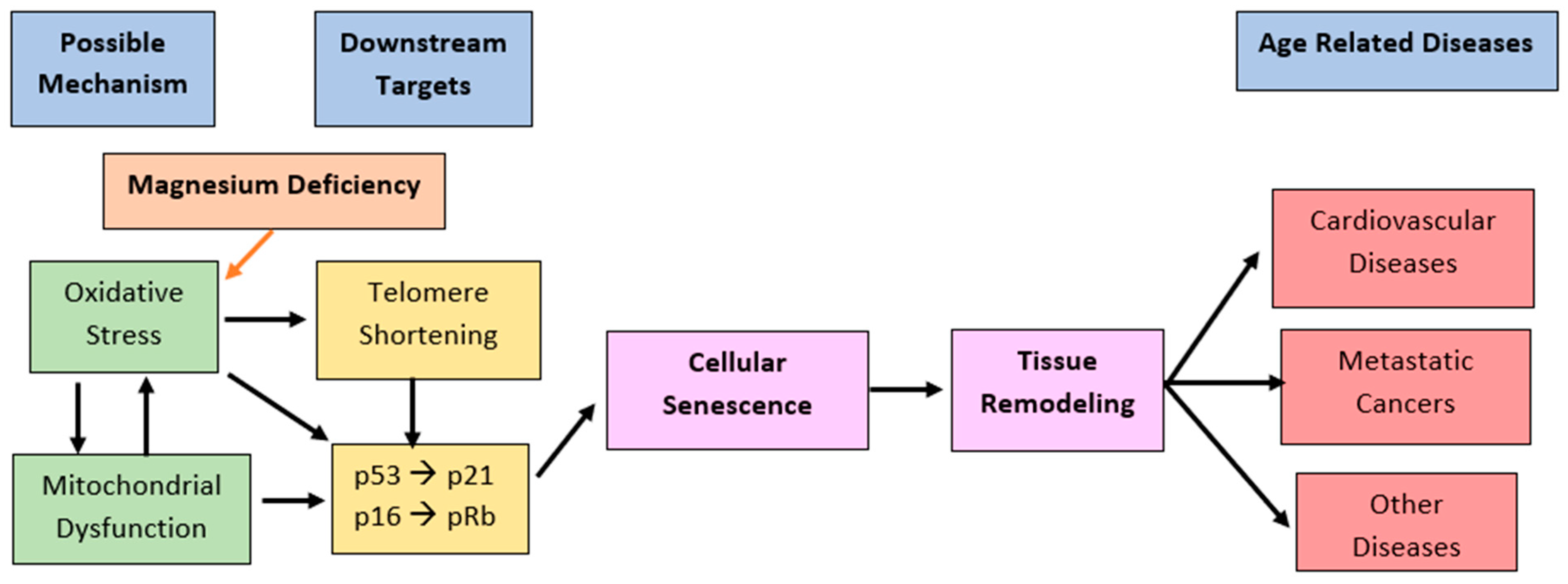
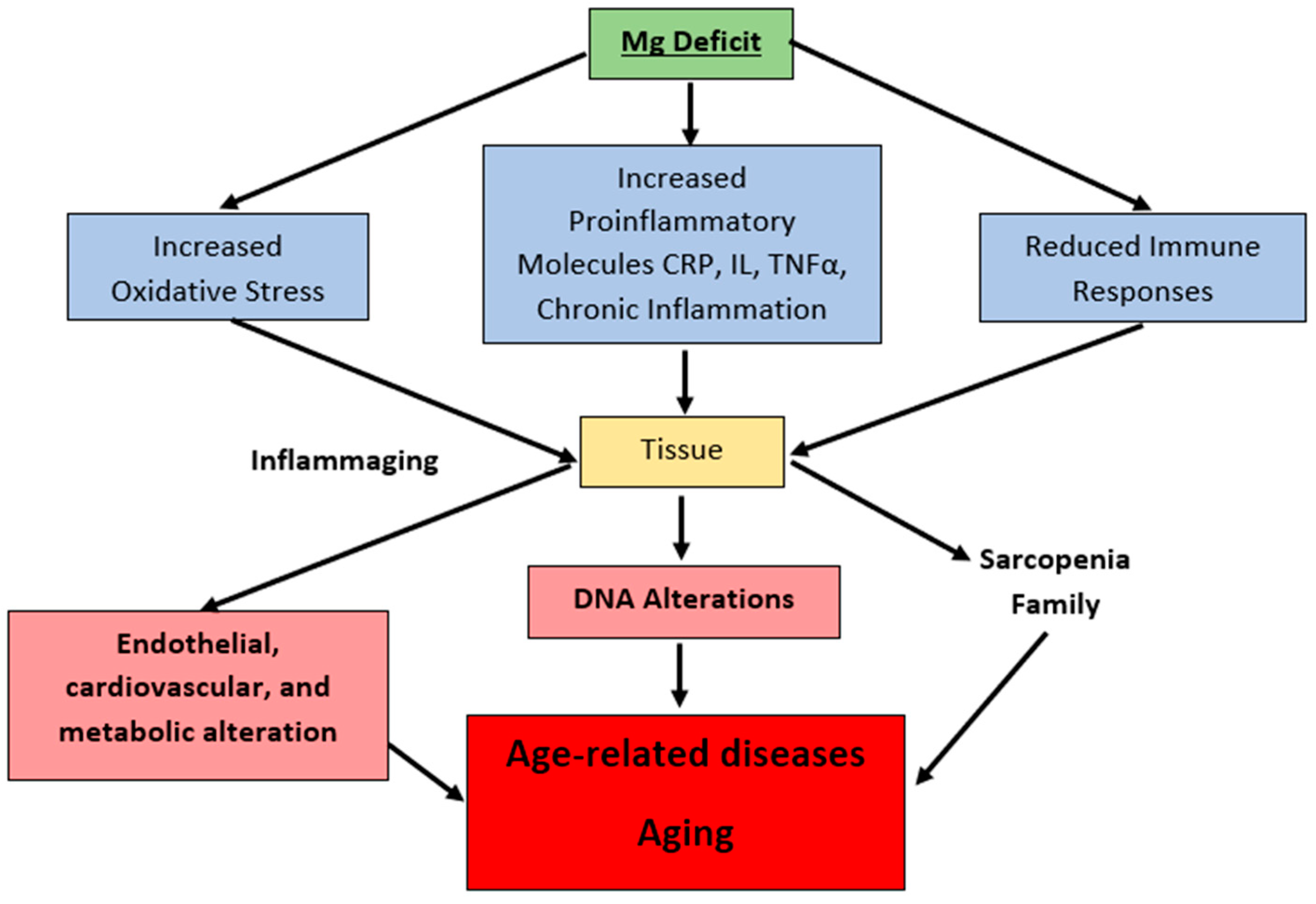
8. Magnesium in the Pathophysiology of Aging

As the world’s population continues to age, many people have shown signs of aging including metabolic decline, atherosclerosis, high blood pressure, cardiovascular diseases, and type 2 diabetes. These symptoms of aging have been associated both experimentally and clinically with the presence of Mg-deficient states [47].
Magnesium deficiency has been found to accelerate the cellular aging process. Studies performed by Shah et al. suggested that short term magnesium deficiency resulted in an upregulation of p53 and neutral sphingomyelinase (N-SMAse) in heart cells and smooth muscle cells of the aorta [47]. N-SMAse upregulation leads to the synthesis and release of ceramide and possibly other sphingolipid products [47]. Ceramide synthesis has been shown to downregulate telomerase activity, which is required for maintaining telomere length, but further studies are warranted [47]. All together, these have pivotal roles in atherogenesis, hypertension, and heart failure, all of which are involved in the aging process (Figure 6) [47]. These results uphold that magnesium deficiency, unless corrected early, will contribute significantly to aging [47].
Aging has also been associated with an increase in the levels of proinflammatory cytokines in tissues and cells [47]. Interestingly, recent findings in Mg-deficient animals, tissues, and different cell types have shown elevated levels of many of the proinflammatory cytokines such as IL-1, IL-6, TNF-alpha, among others [47]. TNF-alpha is known to be associated negatively on telomerase activity in some cell types [47]. Furthermore, magnesium acts as an antioxidant against free radical damage of the mitochondria. Chronic inflammation and oxidative stress are pathogenic factors in aging and age-related diseases (Figure 7) [49].
It is crucial to supplement magnesium especially in the elderly population. Studies have shown that simple increases in daily intake of magnesium can allow for healthier aging [47].

9. Magnesium: An Essential Nutrient

The recommended allowance of magnesium in both males and females is 400–420 mg/day and 310–320 mg/day, respectively [50]. An essential feature of heart failure associated with complex ventricular arrhythmias is hypomagnesemia, most likely related to increased urine magnesium excretion. By supplementing with magnesium, these arrhythmias can be alleviated or abolished [51]. Magnesium supplementation above 15 mmol per day is required to normalize high blood pressure in unmedicated hypertensive patients as well as lower high blood pressure in patients treated with anti-hypertensive medications [1]. Unfortunately, the average dietary intake of magnesium has decreased among both men and women living in North America [52]. Several studies have shown a relative decreased intake in magnesium content in people following Western diets [53]. In addition, areas with soft water have low magnesium content in drinking water [54]. Residents of areas with soft-water and magnesium poor-soil have a higher tendency to suffer from ischemic heart disease (IHD), coronary vasospasm, hypertension, and sudden cardiac death (SCD) [55]. Low levels of magnesium are also associated with prehypertension and hypertension in children [56].
In the general population, a meta-analysis of one million patients revealed reduced risk of heart failure, stroke, diabetes, and all-cause mortality in those receiving magnesium supplementation [57]. Intravenous magnesium can be therapeutically administered to reduce the risk of potentially fatal arrhythmias after a myocardial infarction [58,59]. Studies have also shown that complex ventricular arrhythmias in patients with heart failure can be abolished by magnesium supplementation [52]. Overall, these findings highlight the importance of magnesium in preventing and treating cardiovascular disease.

10. Conclusions and Discussion

Hypertension leads to changes in blood vessels that contribute to atherosclerosis and platelet activation. This in turn contributes to cardiovascular and cerebrovascular disease through oxidative stress. Oxidative stress via low magnesium and increased PAF has also been found to reduce telomerase activity and shorten telomeres, which directly contributes to aging and is correlated to increased myocardial infarction risk [48]. In this review we have discussed the role of PAF and magnesium in the pathophysiology of these conditions. Hypertension was defined within the context of clinical medicine and its pathophysiology was explained. Secondly, PAF’s structure and function were discussed. Thirdly, magnesium was introduced as a micronutrient that plays a key role as an antihypertensive. Through calcium deposition antagonism and release of vasodilatory prostacyclin, magnesium is theorized to reduce high blood pressure. Experimental evidence has shown that supplementing magnesium deficiency with dietary magnesium has significantly improved cardiovascular health [60]. In a study performed by Altura et al. on rabbits who were fed a cholesterol diet, oral supplementation with Mg salt magnesium aspartate hydrochloride lowered levels of cholesterol and triglycerides in normal (25–35%) and atherosclerotic (20–40%) animals and inhibited the atherosclerotic pathway [60].
There is clinical evidence to support that hypomagnesemia contributes to vasospasm and ischemic injury through several mechanisms, including induction of mitochondrial dysfunction, activation of apoptosis, and facilitation of ceramide synthesis and release. Furthermore, platelet-activating factor (PAF) has been implicated in atherogenesis and inflammation, especially in relation to cardiovascular and cerebrovascular injury. The promotion of platelet aggregation, oxidative stress, and vascular permeability were some of the highlighted mechanisms that PAF augments in atherogenesis. Figure 8 below summarizes the interrelationship between magnesium and PAF in the context of atherogenesis and vascular injury.
As discussed, PAF has been implicated in inflammatory processes in relation to the NF-kB pathway. In 2016, Altura et al. provided evidence for a novel hypothesis interrelating PAF with activation of the NF-kB pathway, proto-oncogenes c-fos and c-jun, and ceramide synthesis, in a low-Mg2+ environment, which in turn contributes to the elaboration of PAF [39]. However, the precise interrelation between free Mg2+ concentration and PAF in the context of vascular disease is not yet clear. Ultimately, the present review (summarized below in Figure 9) encourages further investigation into platelet-activating factor and the cardioprotective role of dietary magnesium supplements, particularly in patients with a history of coronary artery disease and arrhythmias. Further investigation into the crosstalk between magnesium and PAF in the prevention of atherosclerosis and hypertension is also warranted.

Author Contributions

Conceptualization, N.S., R.S. and S.S.; investigation, N.S., R.S., S.S. and K.J.; writing—original draft preparation, N.S.; writing—review and editing, N.S., R.S., S.S., K.J., J.D., Y.C. and C.S.; supervision, H.W. All authors have read and agreed to the published version of the manuscript.

Funding

This research did not receive any specific grant from funding agencies in the public, commercial, or not-for-profit sectors.

Institutional Review Board Statement

Not applicable.

Informed Consent Statement

Not applicable.

Data Availability Statement

Not applicable.

Acknowledgments

We are very grateful to Zumit Shah, Laxit Shah, Sasha Taylor, Ved Bhavin Patel, Radha Sanjay Shah, and Kena Piyush Patel for their technical assistance and generous help in writing this manuscript.

Conflicts of Interest

The authors declare no conflict of interest.

References

  1. Dominguez, L.; Veronese, N.; Barbagallo, M. Magnesium and Hypertension in Old Age. Nutrients 2020, 13, 139. [Google Scholar] [CrossRef] [PubMed]
  2. Wegner, M.; Araszkiewicz, A.; Zozulińska-Ziółkiewicz, D.; Wierusz-Wysocka, B.; Pioruńska-Mikołajczak, A.; Pioruńska-Stolzmann, M. The relationship between concentrations of magnesium and oxidized low density lipoprotein and the activity of platelet activating factor acetylhydrolase in the serum of patients with type 1 diabetes. Magnes. Res. 2010, 23, 97–104. [Google Scholar] [CrossRef] [PubMed]
  3. Ross, R. The pathogenesis of atherosclerosis: A perspective for the 1990s. Nature 1993, 362, 801–809. [Google Scholar] [CrossRef] [PubMed]
  4. Ostchega, Y.; Fryar, C.D.; Nwankwo, T.; Nguyen, D.T. Hypertension Prevalence among Adults Aged 18 and Over: United States, 2017–2018; NCHS Data Brief, No. 364; National Center for Health Statistics: Hyattsville, MD, USA, 2020.
  5. High Blood Pressure: Also Known as Hypertension. U.S. Department of Health & Human Services: National Heart, Lung and Blood. Available online: https://www.nhlbi.nih.gov/health-topics/high-blood-pressure (accessed on 2 June 2021).
  6. Whelton, P.K.; Carey, R.M.; Aronow, W.S.; Casey, D.E.; Collins, K.J.; Dennison Himmelfarb, C.; DePalma, S.M.; Gidding, S.; Jamerson, K.A.; Jones, D.W.; et al. 2017 ACC/AHA/AAPA/ABC/ACPM/AGS/APhA/ASH/ASPC/NMA/PCNA Guideline for the Prevention, Detection, Evaluation, and Management of High Blood Pressure in Adults. J. Am. Coll. Cardiol. 2018, 71, e127–e248. [Google Scholar] [CrossRef]
  7. Poulter, N.R.; Prabhakaran, D.; Caulfield, M. Hypertension. Lancet Lond Engl. 2015, 386, 801–812. [Google Scholar] [CrossRef]
  8. Charles, L.; Triscott, J.; Dobbs, B. Secondary Hypertension: Discovering the Underlying Cause. Am. Fam. Physician 2017, 96, 453–461. [Google Scholar]
  9. Nguyen, B.; Bauman, A.; Ding, D. Association between lifestyle risk factors and incident hypertension among middle-aged and older Australians. Prev. Med. 2019, 118, 73–80. [Google Scholar] [CrossRef]
  10. Jamrozik, K. Age-specific relevance of usual blood pressure to vascular mortality: A meta-analysis of individual data for one million adults in 61 prospective studies. Lancet 2002, 360, 1903–1913. [Google Scholar] [CrossRef]
  11. Konukoglu, D.; Uzun, H. Endothelial Dysfunction and Hypertension. Adv. Exp. Med. Biol. 2017, 956, 511–540. [Google Scholar] [CrossRef]
  12. Gimbrone, M.A.; García-Cardeña, G. Endothelial Cell Dysfunction and the Pathobiology of Atherosclerosis. Circ Res. 2016, 118, 620–636. [Google Scholar] [CrossRef] [Green Version]
  13. Marketou, M.; Kontaraki, J.; Papadakis, J.; Kochiadakis, G.; Vrentzos, G.; Maragkoudakis, S.; Fragkiadakis, K.; Katsouli, E.; Plataki, M.; Patrianakos, A.; et al. Platelet microRNAs in hypertensive patients with and without cardiovascular disease. J. Hum. Hypertens 2019, 33, 149–156. [Google Scholar] [CrossRef] [PubMed]
  14. Barale, C.; Russo, I. Influence of Cardiometabolic Risk Factors on Platelet Function. Int. J. Mol. Sci. 2020, 21, 623. [Google Scholar] [CrossRef] [PubMed] [Green Version]
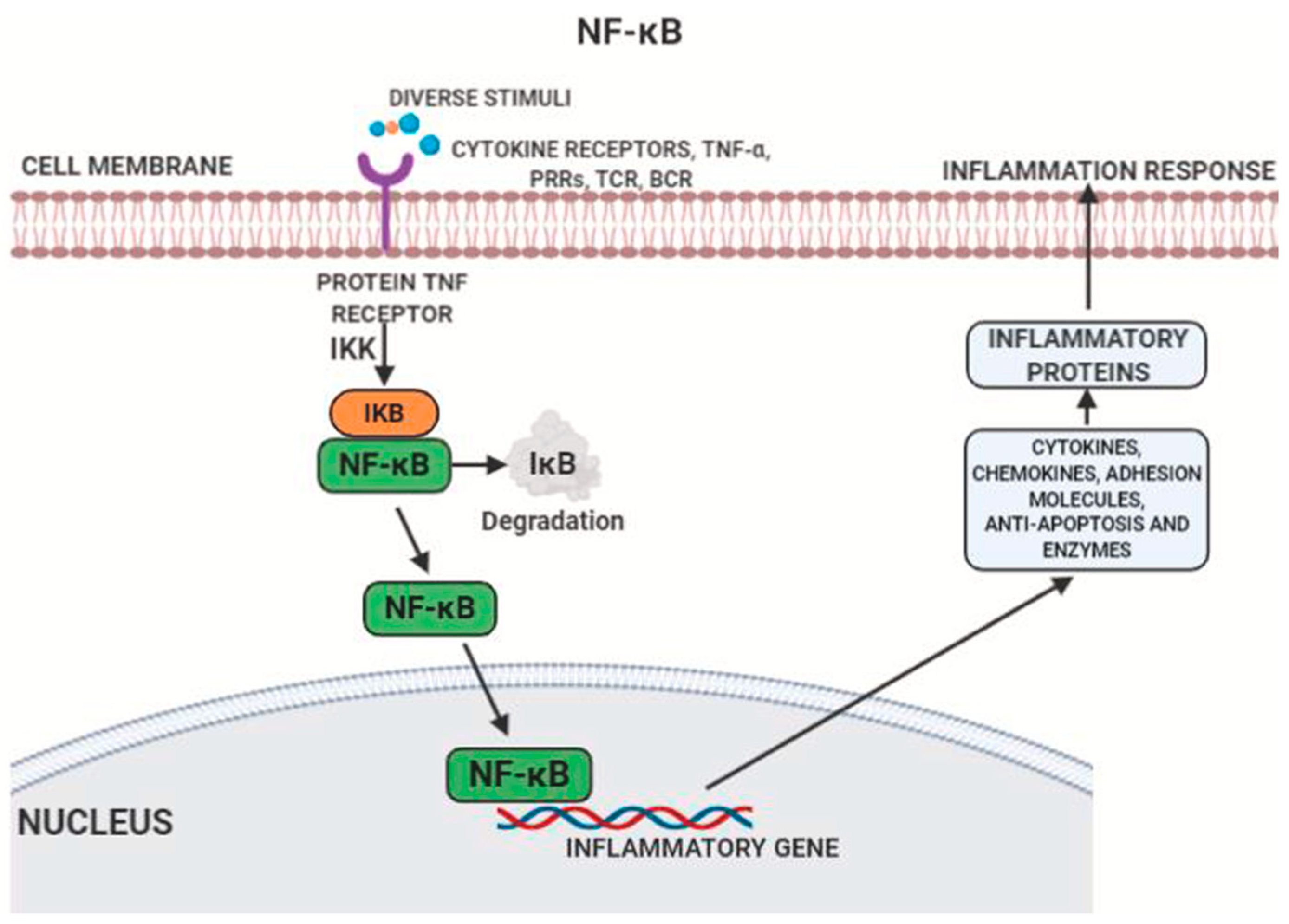
  15. Mussbacher, M.; Salzmann, M.; Brostjan, C.; Hoesel, B.; Schoergenhofer, C.; Datler, H.; Hohensinner, P.; Basílio, J.; Petzelbauer, P.; Assinger, A.; et al. Cell Type-Specific Roles of NF-κB Linking Inflammation and Thrombosis. Front. Immunol. 2019, 10, 85. [Google Scholar] [CrossRef] [PubMed] [Green Version]
  16. Liu, T.; Zhang, L.; Joo, D.; Sun, S.-C. NF-κB signaling in inflammation. Signal Transduct. Target. Ther. 2017, 21, 7023. [Google Scholar] [CrossRef] [Green Version]
  17. Monaco, C.; Andreakos, E.; Kiriakidis, S.; Mauri, C.; Bicknell, C.; Foxwell, B.; Cheshire, N.; Paleolog, E.; Feldmann, M. Canonical pathway of nuclear factor B activation selectively regulates proinflammatory and prothrombotic responses in human atherosclerosis. Proc. Natl. Acad. Sci. USA 2004, 101, 5634–5639. [Google Scholar] [CrossRef] [Green Version]
  18. Pandey, A.K.; Singhi, E.K.; Arroyo, J.P.; Ikizler, T.A.; Gould, E.R.; Brown, J.; Beckman, J.A.; Harrison, D.G.; Moslehi, J. Mechanisms of VEGF (Vascular Endothelial Growth Factor) Inhibitor–Associated Hypertension and Vascular Disease. Hypertension 2018, 71, e1–e8. [Google Scholar] [CrossRef]
  19. Honda, Z.I.; Ishii, S.; Shimizu, T. Platelet-Activating Factor Receptor. J. Biochem. 2002, 131, 773–779. [Google Scholar] [CrossRef]
  20. Kumar, A.; Sawhney, G.; Kumar Nagar, R.; Chauhan, N.; Gupta, N.; Kaul, A.; Ahmed, Z.; Sangwan, P.L.; Satheesh Kumar, P.; Yadav, G. Evaluation of the immunomodulatory and anti-inflammatory activity of Bakuchiol using RAW 264.7 macrophage cell lines and in animal models stimulated by lipopolysaccharide (LPS). Int. Immunopharmacol. 2021, 91, 107264. [Google Scholar] [CrossRef]
  21. Tsoupras, A.; Lordan, R.; Zabetakis, I. Inflammation, not Cholesterol, Is a Cause of Chronic Disease. Nutrients 2018, 10, 604. [Google Scholar] [CrossRef] [Green Version]
  22. Mueller, H.W.; Haught, C.A.; McNatt, J.M.; Cui, K.; Gaskell, S.J.; Johnston, D.A.; Willerson, J.T. Measurement of platelet-activating factor in a canine model of coronary thrombosis and in endarterectomy samples from patients with advanced coronary artery disease. Circ. Res. 1995, 77, 54–63. [Google Scholar] [CrossRef]
  23. Garrido, D.; Chanteloup, N.K.; Trotereau, A.; Lion, A.; Bailleul, G.; Esnault, E.; Trapp, S.; Quéré, P.; Schouler, C.; Guabiraba, R. Characterization of the Phospholipid Platelet-Activating Factor As a Mediator of Inflammation in Chickens. Front. Vet. Sci. 2017, 4, 226. [Google Scholar] [CrossRef] [PubMed] [Green Version]
  24. Rouis, M.; Nigon, F.; John Chapman, M. Platelet activating factor is a potent stimulant of the production of active oxygen species by human monocyte-derived macrophages. Biochem. Biophys. Res. Commun. 1988, 156, 1293–1301. [Google Scholar] [CrossRef]
  25. Dentan, C.; Lesnik, P.; Chapman, M.J.; Ninio, E. Phagocytic Activation Induces Formation of Platelet-Activating Factor in Human Monocyte-Derived Macrophages and in Macrophage-Derived foam Cells. Relevance to the Inflammatory Reaction in Atherogenesis. Eur. J. Biochem. 1996, 236, 48–55. [Google Scholar] [CrossRef] [PubMed]
  26. Mittal, M.; Siddiqui, M.R.; Tran, K.; Reddy, S.P.; Malik, A.B. Reactive Oxygen Species in Inflammation and Tissue Injury. Antioxid. Redox Signal. 2014, 20, 1126–1167. [Google Scholar] [CrossRef] [Green Version]
  27. Kamata, K.; Mori, T.; Shigenobu, K.; Kasuya, Y. Endothelium-dependent vasodilator effects of platelet activating factor on rat resistance vessels. Br. J. Pharmacol. 1989, 98, 1360–1364. [Google Scholar] [CrossRef] [Green Version]
  28. Tao, Y.K.; Zhao, S.P.; Yu, P.L.; Shi, J.; Gu, C.D.; Sun, H.T.; Zhang, G.Q. Elevated platelet activating factor level in ischemia-related arrhythmia and its electrophysiological effect on myocardium. Biomed. Environ. Sci. 2013, 26, 365–370. [Google Scholar] [CrossRef]
  29. Silva, I.T.; Mello, A.P.; Damasceno, N.R. Antioxidant and inflammatory aspects of lipoprotein-associated phospholipase A2(Lp-PLA2): A review. Lipids Health Dis. 2011, 10, 170. [Google Scholar] [CrossRef] [Green Version]
  30. Karabina, S.-A.; Ninio, E. Plasma PAF-acetylhydrolase: An unfulfilled promise? Biochim. Biophys. Acta (BBA)-Mol. Cell Biol. Lipids 2006, 1761, 1351–1358. [Google Scholar] [CrossRef]
  31. Fruhwirth, G.O.; Loidl, A.; Hermetter, A. Oxidized phospholipids: From molecular properties to disease. Biochim. Biophys. Acta BBA-Mol. Basis Dis. 2007, 1772, 718–736. [Google Scholar] [CrossRef] [Green Version]
  32. Ishii, S.; Nagase, T.; Shimizu, T. Platelet-activating factor receptor. Prostaglandins Other Lipid Mediat. 2002, 68–69, 599–609. [Google Scholar] [CrossRef]
  33. Chao, W.; Olson, M.S. Platelet-activating factor: Receptors and signal transduction. Biochem. J. 1993, 292, 617–629. [Google Scholar] [CrossRef] [PubMed] [Green Version]
  34. Feuerstein, G.; Rabinovici, R.; Leor, J.; Winkler, J.D.; Vonhof, S. Platelet-activating factor and cardiac diseases: Therapeutic potential for PAF inhibitors. J. Lipid Mediat. Cell Signal. 1997, 15, 255–284. [Google Scholar] [CrossRef]
  35. Palur Ramakrishnan, A.V.; Varghese, T.P.; Vanapalli, S.; Nair, N.K.; Mingate, M.D. Platelet activating factor: A potential biomarker in acute coronary syndrome? Cardiovasc. Ther. 2017, 35, 64–70. [Google Scholar] [CrossRef] [PubMed]
  36. Lindsberg, P.J.; Hallenbeck, J.M.; Feuerstein, G. Platelet-activating factor in stroke and brain injury. Ann. Neurol. 1991, 30, 117–129. [Google Scholar] [CrossRef] [PubMed]
  37. Satoh, K.; Imaizumi, T.; Yoshida, H.; Hiramoto, M.; Takamatsu, S. Increased levels of blood platelet-activating factor (PAF) and PAF-like lipids in patients with ischemic stroke. Acta Neurol. Scand. 1992, 85, 122–127. [Google Scholar] [CrossRef]
  38. Morrill, G.A.; Gupta, R.K.; Kostellow, A.B.; Ma, G.Y.; Zhang, A.; Altura, B.T.; Altura, B.M. Mg2+ modulates membrane lipids in vascular smooth muscle: A link to atherogenesis. FEBS Lett. 1997, 408, 191–194. [Google Scholar] [CrossRef] [Green Version]
  39. Altura, B.M.; Li, W.; Zhang, A.; Zheng, T.; Shah, N.C.; Shah, G.J.; Altura, B.T. The Expression of Platelet-Activating Factor is Induced by Low Extracellular Mg2+ in Aortic, Cerebral and Neonatal Coronary Vascular Smooth Muscle; Cross Talk with Ceramide Production, NF–kB and Proto-Oncogenes: Possible Links to Atherogenesis and Sudden Cardiac Death in Children and Infants, and Aging; Hypothesis, Review and Viewpoint. Int. J. Cardiol. Res. 2016, 3, 47–67. [Google Scholar] [CrossRef]
  40. Rayssiguier, Y.; Gueux, E.; Bussière, L.; Durlach, J.; Mazur, A. Dietary magnesium affects susceptibility of lipoproteins and tissues to peroxidation in rats. J. Am. Coll. Nutr. 1993, 12, 133–137. [Google Scholar] [CrossRef]
  41. Kostov, K.; Halacheva, L. Role of Magnesium Deficiency in Promoting Atherosclerosis, Endothelial Dysfunction, and Arterial Stiffening as Risk Factors for Hypertension. Int. J. Mol. Sci. 2018, 19, 1724. [Google Scholar] [CrossRef] [Green Version]
  42. Tangvoraphonkchai, K.; Davenport, A. Magnesium and Cardiovascular Disease. Adv. Chronic Kidney Dis. 2018, 25, 251–260. [Google Scholar] [CrossRef]
  43. ter Braake, A.D.; Shanahan, C.M.; de Baaij, J.H.F. Magnesium Counteracts Vascular Calcification: Passive Interference or Active Modulation? Arter. Thromb Vasc Biol. 2017, 37, 1431–1445. [Google Scholar] [CrossRef] [PubMed] [Green Version]
  44. Swaminathan, R. Magnesium metabolism and its disorders. Clin. Biochem. Rev. 2003, 24, 47–66. [Google Scholar] [PubMed]
  45. Yolcu, M.; Ipek, E.; Turkmen, S.; Ozen, Y.; Yıldırım, E.; Sertcelik, A.; Ulusoy, F.; Mustafa, Y.; Emrah, I.; Erkan, Y.; et al. The relationship between elevated magnesium levels and coronary artery ectasia. Cardiovasc. J. Afr. 2016, 27, 294–298. [Google Scholar] [CrossRef] [PubMed] [Green Version]
  46. Cunha, A.R.; Umbelino, B.; Correia, M.L.; Neves, M.F. Magnesium and vascular changes in hypertension. Int. J. Hypertens. 2012, 2012, 754250. [Google Scholar] [CrossRef] [Green Version]
  47. Shah, N.C.; Shah, G.J.; Li, Z.; Jiang, X.C.; Altura, B.T.; Altura, B.M. Short-term magnesium deficiency downregulates telomerase, upregulates neutral sphingomyelinase and induces oxidative DNA damage in cardiovascular tissues: Relevance to atherogenesis, cardiovascular diseases and aging. Int. J. Clin. Exp. Med. 2014, 74, 97–514. [Google Scholar]
  48. Killilea, D.W.; Maier, J.A.M. A connection between magnesium deficiency and aging: New insights from cellular studies. Magnes. Res. 2008, 21, 77–82. [Google Scholar] [CrossRef]
  49. Barbagallo, M.; Dominguez, L.J. Magnesium and aging. Curr. Pharm. Des. 2010, 16, 832–839. [Google Scholar] [CrossRef] [PubMed]
  50. Dietary Reference Intakes for Calcium, Phosphorus, Magnesium, Vitamin D, and Fluoride; National Academies Press: Washington, DC, USA, 1997. [CrossRef]
  51. Ceremuzyński, L.; Gebalska, J.; Wolk, R.; Makowska, E. Hypomagnesemia in heart failure with ventricular arrhythmias. Beneficial effects of magnesium supplementation. J. Intern. Med. 2000, 247, 78–86. [Google Scholar] [CrossRef]
  52. Bailey, R.L.; Fulgoni, V.L.; Keast, D.R.; Dwyer, J.T. Dietary supplement use is associated with higher intakes of minerals from food sources. Am. J. Clin. Nutr. 2011, 94, 1376–1381. [Google Scholar] [CrossRef] [Green Version]
  53. DiNicolantonio, J.J.; O’Keefe, J.H.; Wilson, W. Subclinical magnesium deficiency: A principal driver of cardiovascular disease and a public health crisis. Open Heart 2018, 5, e000668. [Google Scholar] [CrossRef]
  54. Burton, A. Cardiovascular Health: Hard Data for Hard Water. Environ. Health Perspect. 2008, 116, A114. [Google Scholar] [CrossRef] [PubMed] [Green Version]
  55. Eisenberg, M.J. Magnesium deficiency and sudden death. Am. Heart J. 1992, 124, 544–549. [Google Scholar] [CrossRef]
  56. Han, H.; Fang, X.; Wei, X.; Liu, Y.; Jin, Z.; Chen, Q.; Fan, Z.; Aaseth, J.; Hiyoshi, A.; He, J.; et al. Dose-response relationship between dietary magnesium intake, serum magnesium concentration and risk of hypertension: A systematic review and meta-analysis of prospective cohort studies. Nutr. J. 2017, 16, 26. [Google Scholar] [CrossRef] [PubMed]
  57. Fang, X.; Wang, K.; Han, D.; He, X.; Wei, J.; Zhao, L.; Imam, M.U.; Ping, Z.; Li, Y.; Xu, Y.; et al. Dietary magnesium intake and the risk of cardiovascular disease, type 2 diabetes, and all-cause mortality: A dose–response meta-analysis of prospective cohort studies. BMC Med. 2016, 14, 210. [Google Scholar] [CrossRef] [Green Version]
  58. Rasmussen, H.S.; Suenson, M.; Mcnair, P.; Nørregård, P.; Balslev, S. Magnesium infusion reduces the incidence of arrhythmias in acute myocardial infarction. A double-blind placebo-controlled study. Clin. Cardiol. 1987, 10, 351–356. [Google Scholar] [CrossRef] [PubMed]
  59. Abraham, A.S. Magnesium in the Prevention of Lethal Arrhythmias in Acute Myocardial Infarction. Arch. Intern. Med. 1987, 147, 753. [Google Scholar] [CrossRef]
  60. Altura, B.T.; Brust, M.; Bloom, S.; Barbour, R.L.; Stempak, J.G.; Altura, B.M. Magnesium dietary intake modulates blood lipid levels and atherogenesis. Proc. Natl. Acad. Sci. USA 1990, 87, 1840–1844. [Google Scholar] [CrossRef] [Green Version]
Figure 1. NF-kB signaling [20].
Cardiogenetics 12 00005 g001
Figure 4. Cardiovascular implications of magnesium deficiency.
Cardiogenetics 12 00005 g004
Figure 5. Effect of magnesium (Mg2+) on potassium (K+) channels, sodium (Na+) channels, and sodium/potassium (Na+/K+) ATPases transporting ions through cardiac myocytes.
Cardiogenetics 12 00005 g005
Figure 6. Impact of magnesium deficient states on telomerase activity [48].
Cardiogenetics 12 00005 g006
Figure 7. Effect of magnesium deficient states on age related diseases [49].
Cardiogenetics 12 00005 g007
Figure 8. Interrelation between magnesium and platelet-activating factor (PAF).
Cardiogenetics 12 00005 g008
Figure 9. Effects of low magnesium and increased PAF on hypertension, atherogenesis, cardiovascular disease, stroke, and aging.
Cardiogenetics 12 00005 g009
Table 1. Current blood pressure guidelines by American College of Cardiology (ACC).
Blood PressureSystolic (mmHg)Diastolic (mmHg)
Normal<120<80
Elevated129–120<80
Stage 1 Hypertension139–13089–80
Stage 2 Hypertension≥140≥90
Publisher’s Note: MDPI stays neutral with regard to jurisdictional claims in published maps and institutional affiliations.

