Review

Gene Therapy for Inherited Arrhythmia Syndromes

1
Department of Medicine, University of British Columbia, Vancouver, BC V6T 2Z3, Canada
2
School of Biomedical Engineering, University of British Columbia, Vancouver, BC V6T 2B9, Canada
3
Centre for Heart Lung Innovation, UBC and St. Paul’s Hospital, Vancouver, BC V6Z 1Y6, Canada
*
Author to whom correspondence should be addressed.
Cardiogenetics 2024, 14(3), 132-148; https://doi.org/10.3390/cardiogenetics14030011
Submission received: 17 March 2024 / Revised: 7 June 2024 / Accepted: 19 July 2024 / Published: 2 August 2024

Abstract

:
The emergence of gene therapy offers opportunities for treating a myriad of genetic disorders and complex diseases that previously had limited or no treatment options. The key basic strategies for gene therapy involve either the addition, inhibition, or introduction of a new gene, with a crucial component being the use of a delivery vector to effectively target cells. Particularly promising is the application of gene therapy for the treatment of inherited arrhythmia syndromes, conditions associated with significant mortality and morbidity that have limited treatment options, and a paucity of disease modifying therapy. This review aims to summarize the utility of gene therapy for the treatment of inherited arrhythmia syndromes by exploring the current state of knowledge, limitations, and future directions.

1. Introduction

Inherited arrhythmia syndromes are a group of genetic disorders affecting cardiac ion channels and interrupt the normal heart rhythm. The main inherited arrhythmia syndromes include long QT syndrome (LQTS), Brugada syndrome (BrS), arrhythmogenic right ventricular cardiomyopathy (ARVC), and catecholaminergic polymorphic ventricular tachycardia (CPVT). Symptoms vary depending on the particular syndrome, but typically include syncope and predisposition to sudden cardiac death. Diagnosis is usually established using a combination of ECG features, clinical and family history, exercise stress testing, and genetic testing [1]. Treatment options for inherited arrhythmia syndromes are limited. The mainstay of treatment is usually pharmacological treatment with beta-blockers and/or sodium and transient outward current blockers. However, individuals at higher risk may receive treatment with implantable cardioverter defibrillators (ICDs) and catheter ablation of arrhythmogenic substrates (when the substrate is focal). Currently, there are no disease-modifying therapeutics available for inherited arrhythmia syndromes, thus making gene therapy a promising area of research.
Gene therapy is the genetic manipulation and modification of cells to produce a desired therapeutic effect [2,3]. These therapies are administered through various vectors including viruses, genetically engineered microorganisms, lipid nanoparticles, liposomes, or nucleic acids alone [4]. Gene therapies can be administered outside the body with cells removed from the patient and then re-introduced (ex vivo gene therapy) or administered in vivo where genetic material is delivered directly into the patient. Broadly speaking, there are three different gene therapy strategies: gene augmentation/replacement, gene inhibition, and gene editing (Table 1).

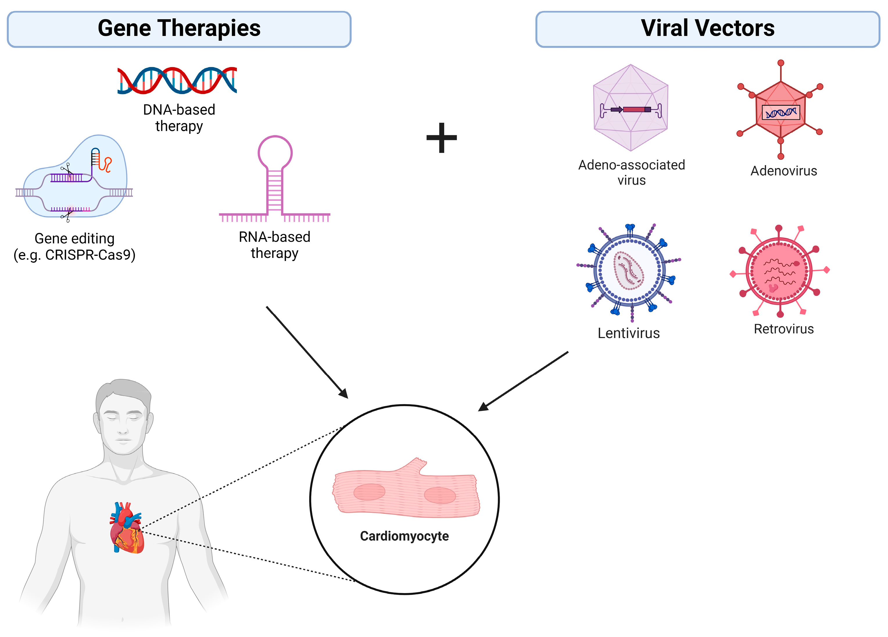
1.1. Gene Augmentation/Replacement

Gene augmentation is the most straightforward approach. It involves replacement of a faulty gene with a functional version and is most appropriate for the treatment of monogenic recessive disorders [5]. For more complex multigenic disorders or monogenic dominant disorders, other approaches, such as gene inhibition or editing, are more appropriate. Several gene-augmentation-based therapies have passed clinical trials and obtained FDA approval. Examples include voretigene nepavovec (brand name Luxturna) for leber congenital amourosis (LCA), valoctocogene roxaparvovec (brand name Roctavian) for the treatment of hemophilia A, fidanacogene elaparvovec-dzkt (brand name Beqvez) for the treatment of hemophilia B, and onasemnogene abeparvovec (brand name Zolgensma) for the treatment of spinal muscular atrophy 1 (SMA1) [6,7,8,9].

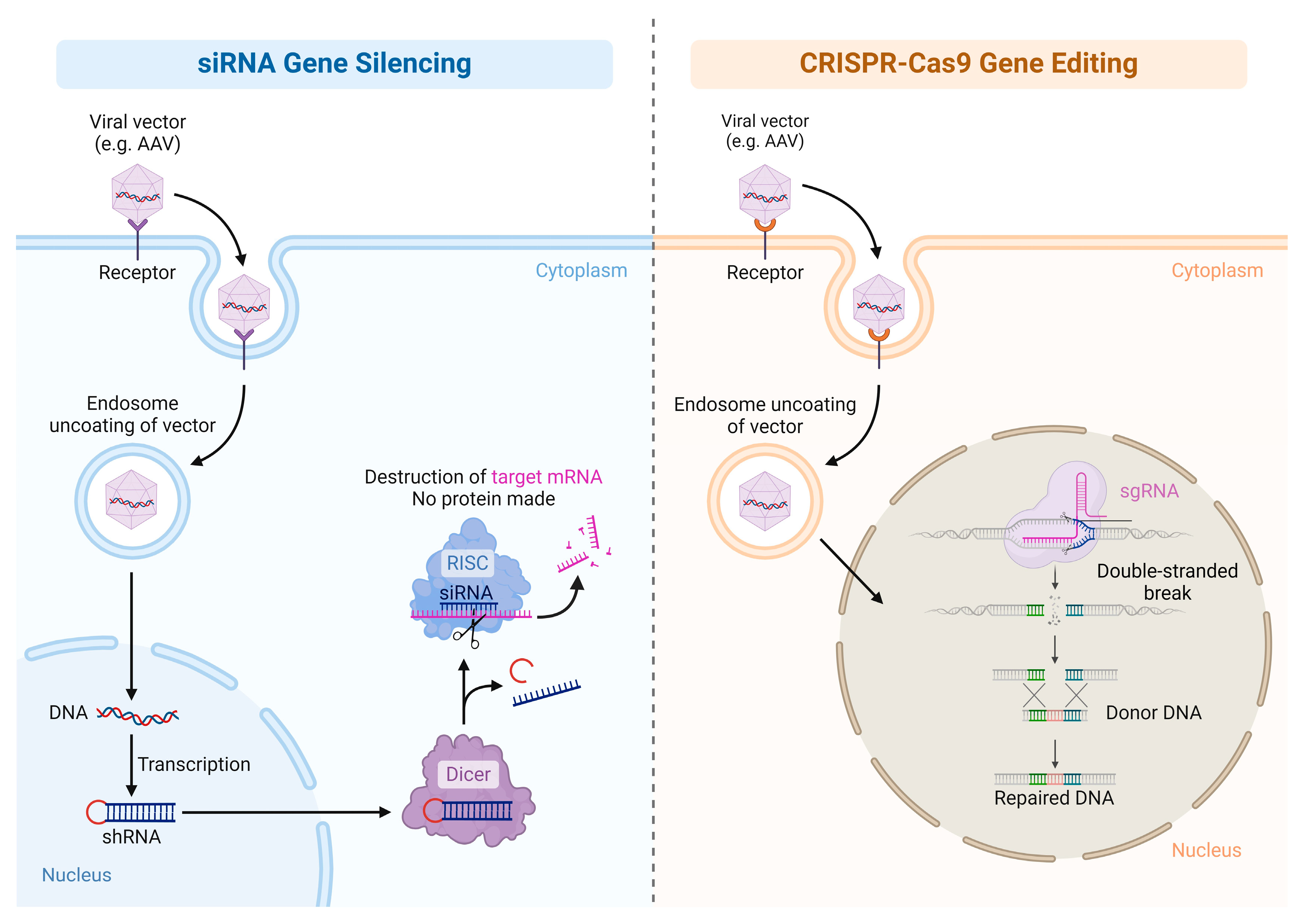
1.2. Gene Inhibition Therapy

Gene inhibition therapy involves downregulating aberrant gene expression, typically using either RNA interference (RNAi) against the mRNA of interest or using an antibody to knock down protein expression. RNAi typically involves the use of either a small interfering RNA (siRNA) or microRNA (miRNA) (Figure 1). miRNAs are endogenous, non-coding, ssRNA molecules which regulate post-transcriptional gene expression [10]. Biologically, miRNAs are important in the development of multiple organ systems, including musculoskeletal, integumental, reproductive, endocrine, and nervous systems, amongst others. In contrast, siRNAs are short, exogenous dsRNA molecules that can induce gene silencing without triggering an endogenous interferon response [11]. When compared to miRNAs, siRNAs trigger more efficient and specific gene silencing and involve targeted mRNA degradation [11]. A number of gene inhibition therapies have also received FDA approval. For instance, patisiran (brand name Onpattro) is an siRNA-based therapeutic delivered in an LNP used to treat polyneuropathy due to hereditary transthyretin-mediated amyloidosis, which received FDA approval in 2018 [12]. Similarly, Givlaari (brand name Givosiran), used for the treatment of acute intermittent porphyria, is also an siRNA-based and delivered by an LNP vector that received FDA approval in 2019 [13].

1.3. Gene-Editing Therapy

Gene-editing therapy is the modification of DNA of living cells in order to produce a therapeutic effect. This section will discuss a brief history of the development of gene-editing technologies and its current position in the medical field.
Early genetic engineering advances in the 1970s, such as recombinant DNA technology, set the groundwork for modern gene-editing technologies. One notable development in recombinant DNA technology was Cre-Lox recombination. Cre-Lox recombination is a site-specific recombination technology derived from P1 bacteriophages that allows for the inversion, translocation, or deletion of specific DNA sequences [14]. The Cre-Lox system is composed of a lox recombination site and the Cre protein. The lox site is made of two palindromic sequences separated by an 8 bp spacer region, which determines the directionality of recombination. The lox sites flank the DNA region of interest. lox sites oriented parallel to each other result in the inversion of the DNA segment between them, whereas sites oriented antiparallel to each other result in the excision of the DNA segment. The Cre protein facilitates the recombination reaction.
The 1990s saw major advances in the field of genetic engineering with the development of the first gene-editing tool, zinc finger nucleases (ZFNs). ZFNs are comprised of a DNA-binding domain and an endonuclease domain, which can be engineered to bind and cleave specific DNA sequences [15]. However, ZFNs have many limitations such as a complex and labor-intensive manufacturing process, limited target site selection, and concerns about off-target effects [16]. In the early 2010s, transcription-activator-like effector nucleases (TALENs) were developed as an alternative to ZFNs. TALENs are derived from transcription-activator-like effectors (TALEs), a class of proteins found in certain plant pathogenic bacteria [16]. TALENs have advantages over ZFNs such as allowing for the recognition of larger binding sequences and fewer constraints on site selection [17]. However, TALENs share the same issues of off-target effects, and although they are simpler to design than ZFNs, they still require considerable labor and expertise to manufacture [16].
Around the same time, the field of genetic engineering had a major breakthrough through the groundbreaking discovery of a novel gene-editing technique, which overcame many of its predecessors’ shortcomings. Clustered regularly interspaced short palindromic repeats (CRISPR) and CRISPR-associated proteins (Cas) are essential components of the immune system of prokaryotic bacteria and archaea, but have proven to be very effective gene-editing tools, allowing for precise genome editing with a simple implementation process [18]. The gene-editing technology CRISPR-Cas9 utilizes CRISPR-associated protein 9 (Cas9), an endonuclease, paired with a guide RNA (sgRNA), which subsequently determines the double-stranded breaks [15]. CRISPR-Cas9 allows the deletion of base pairs, the insertion of new DNA base pairs including modified ribonucleotides, and the deletion of DNA pairs [15]. Its delivery to cells is facilitated by a vector, such as an adeno-associated virus vector (Figure 1).
CRISPR-Cas9 has been the most effective gene-editing tool for gene therapy. Currently, several CRISPR-Cas9-based FDA-approved therapeutics exist. Exagamglogene autotemcel (brand name Casgevy) and lovotibeglogene autotemcel (brand name Lyfgenia) are gene therapies used to treat sickle cell disease. Exagamglogene autotemcel is a cell-based therapy involving infusion with autologous hematopoietic stem cell progenitors that have been genetically edited to express fetal hemoglobin using CRISPR-Cas9 [19]. Similarly, lovotibeglogene autotemcel is also a cell-based therapy where the patient’s peripheral blood-mobilized stem cells are modified using CRISPR-Cas9 to express a functional version of hemoglobin A [20]. Gene editing has also found utility within the realm of cancer immunotherapy. Chimeric antigen receptor (CAR) T cells are genetically modified T cells that can recognize and attack specific cell surface antigens expressed on tumor cells. In 2017, the first CAR T cell therapeutic, tisagenlecleucel (Kymriah) was approved by the FDA for the treatment of acute lymphoblastic leukemia [21]. Despite the success of these novel therapeutics, existing CRISPR-Cas9-based gene therapies are mostly limited to ex vivo approaches. The direct in vivo modification of cells, which may be necessary for inherited arrhythmia syndromes, requires further research before being applied in a clinical setting.

2. Gene Therapy Vectors—The Evolution, Where We Are, and Where We Are Going

2.1. Viral Vectors

The modality of delivery of the gene therapy is equally important to the actual nucleic acid therapy itself, with respect to both safety and efficacy in a clinical setting. Viral vectors were the first delivery modality capable of gene transfer in human cells and are by far the most researched with respect to the number of clinical trials (Figure 2) [22]. Viruses are commonly used for gene therapy as they are capable of taking in genetic material and efficiently introducing and maintaining it in human cells. Adenoviruses, adeno-associated viruses, lentiviruses, and retroviruses have been employed most commonly. Recombinant viral vectors are specifically designed to capitalize on a virus’s ability to integrate its genome within the host genome while simultaneously minimizing the adverse effects and toxicities associated with wild-type viruses [22]. Specifically, toxicity may occur due to viral integration with the host genome and insertional mutagenesis at the site of DNA integration. Modern viral delivery vectors overcome this source of genotoxicity within post-mitotic cells (i.e., neurons and myocytes) by delivering non-integrating vectors that can be maintained with stability in an extrachromosomal form.
Recombinant AAVs (rAAVs) are the most widely used and researched viral vectors for cardiac gene therapy as a result of their safety and ability to maintain expression for many years [23]. Moreover, many serotypes of rAAVs also demonstrate tropism for cardiac myocytes with serotypes 6, 8, and 9, showing significant tropism for mice cardiomyocytes [23].
However, there are several concerns regarding the safety and efficacy of viral vectors for gene therapy. Adenovirus, lentivirus, and retroviral vectors pose a risk of insertional mutagenesis, which could subsequently lead to aberrant genetic expression and tumor formation [24]. With respect to AAVs, there is a risk of cross-reactivity and adaptive immunity due to many patients already having antibodies to the vector, thereby inactivating the gene therapy. Additionally, AAVs are relatively small, with a cargo capacity that is limited to ~4.5 kb, limiting the size of the gene used in therapy [23]. Despite these limitations, viral vectors have been shown to be a promising delivery method for gene therapy.

2.2. Non-Viral Vectors

Non-viral vectors include delivery systems such as lipid-based nanoparticles and plasmid DNA. Unlike viral vectors, non-viral vectors may pose a lower risk of inducing host immune responses, which allows for repeated administration and reduced side effects. Liposomes are spherical vesicles consisting of cholesterol and phospholipids that can encapsulate DNA or RNA and fuse with cell membranes. Liposomal nanoparticles are conjugated with ligands to help facilitate and direct uptake in specific cells, are readily able to merge with the lipid bilayer of cell membranes, protect mRNA molecules from nucleases present in the extracellular matrix, and can aid in escape from endosomes that facilitate degradation of foreign materials [25,26]. Evers et al. (2022) investigated the potential of lipid nanoparticles to deliver functionally modified mRNA to the myocardium after a myocardial infarction in mouse models [27]. However, the study noted that higher levels of lipid nanoparticles accumulated in organs such as the liver and spleen, which suggests that while lipid nanoparticles can reach the heart, the level of gene expression might not be sufficient for therapeutic effects. A recent study developed heart-targeted nanoparticles using three cardiac-targeting peptides (CTP) and demonstrated increased binding of nanoparticles to human and mouse cardiomyocytes in vitro when conjugated with CTPs [28]. Additionally, the group demonstrated increased accumulation in the heart, rather than other organs, when tracking nanoparticles in mice. While the study did not observe immunogenicity, initial safety assessments showed no induced cardiomyocyte apoptosis. Labonia et al. (2023) also demonstrated the use of mRNA–lipid nanoparticle delivery in cardiac cells; however, there appeared to be higher levels of mRNA accumulation in the liver [29]. Currently, the exploration of lipid nanoparticle gene therapy for cardiac cells is limited and more investigation is required to analyze safety profiles, demonstrate a sufficient therapeutic effect, and identify specific ligands/receptors to enhance specificity.
On the other hand, plasmid-mediated gene transfer introduces plasmid DNA into target cells through transfection or transformation. Electroporation, which involves applying an electrical stimulus to the heart tissue, temporarily opens the cell membrane and allows plasmids to enter. While the creation of plasmids is fairly simple and allows for cheaper, large-scale production, gene transduction into host cells usually occurs at clinically insignificant levels [30]. This is due to the degradation of plasmids by extracellular nucleases and elimination by the liver, the inadequate crossing of cell membranes, and the non-specific targeting of cardiac cells [31]. Decreased specificity to cardiac cells and insufficient gene transfer are major limitations of non-viral gene therapies. However, nanomedicine may hold potential for delivering genetic material without the use of viral carriers or physical methods, such as electroporation and microinjection.

3. Basic Science and Animal Studies Pertaining to Gene Therapies and Inherited Arrhythmia Syndromes

The proper management of inherited arrhythmia syndromes is of the utmost importance to prevent severe cardiac-related events and death. However, most inherited arrhythmia syndromes have limited effective treatment options. This section will explore the genetic mechanisms and basic science research on gene therapy for the treatment of several inherited arrhythmia syndromes: congenital long QT syndrome (LQTS), catecholaminergic polymorphic ventricular tachycardia (CPVT), Brugada syndrome (BrS), and ARVC (Table 2).

3.1. Long QT Syndrome

Congenital LQTS is an inherited cardiac arrhythmia disorder characterized by a prolongation of the QTc interval and T-wave abnormalities on an ECG [42]. Many individuals are asymptomatic. However, LQTS predisposes individuals to torsades de pointes during periods of sympathetic excitation, which may lead to syncope, seizures, cardiac arrest, and/or sudden cardiac death. ECG features alone are insufficient to diagnose LQTS. The diagnosis of LQTS is based on the Schwartz score and made using a combination of factors such as personal and family history, genetic testing, and exercise stress testing, in addition to the ECG [43].
Many mutations are associated with LQTS, but mutations in three major genes are implicated in ~90% of patients [42]. All three of these genes encode for various subunits of cardiac sodium or potassium channels, which regulate the cardiac action potential. Normal cardiac action potential is dependent on an intricate balance between depolarization, mediated by voltage-gated sodium and calcium channels, and repolarization, mediated by voltage-gated potassium channels. During depolarization, an outward current is generated through the influx of Na+ into cardiomyocytes through voltage-gated sodium and calcium channels. During repolarization, an inward current is evoked through the opening of voltage-gated potassium channels. These include the slow delayed rectifier (IKs), the rapid delayed rectifier (IKr), and the inwardly rectifying current (IK1).
Types 1 and 2 LQTS are associated with loss-of-function mutations in voltage-gated potassium channels, whereas Type 3 LQTS (LQT3) is associated with gain-of-function mutations in voltage-gated sodium channels. Type 1 LQTS (LQT1) makes up the majority of LQTS cases (~35%), and is caused by a loss-of-function mutation in the KCNQ1 gene, which encodes for the α-subunit of the Kv7.1 voltage-gated potassium channel [42]. This potassium channel plays a crucial role in the repolarization of the cardiac action potential by generating IKs. Mutations in KCNQ1 lead to a disease that follows an autosomal dominant pattern of inheritance. On the other hand, Type 2 LQTS (LQT2) is the second most common form of LQTS, making up around 30% of cases [44]. It is associated with loss-of-function mutations in the KCNH2 gene encoding the α-subunit of the Kv11.1 voltage-gated potassium channel, which conducts the IKr. Finally, LQT3, which comprises ≤10% of cases, is associated with a gain-of-function mutation in the SCN5a gene, encoding for voltage-gated Nav1.5 sodium channel [44]. LQT3 increases the amplitude of the fast inward cardiac sodium current INa, prolonging the QTc interval.

3.1.1. Jervell and Lange-Niellson Syndrome, Anderson Tawil Syndrome, and Timothy Syndrome

Other rarer types of LQTS include Jervell and Lange-Niellson syndrome (JLNS), Anderson Tawil syndrome (ATS), and Timothy syndrome (TS). JLNS is an autosomal recessive version of LQTS, which is characterized by bilateral sensorineural hearing loss in addition to QT prolongation. It is associated with deletion mutations in KCNQ1 or KCNE1, which disrupt the function of potassium channels found in the heart and in the cochlea [45]. The prognosis of JLNS is more severe than most other forms of LQTS. Even with the use of beta-blockers, JLNS is associated with high rates of symptoms and SCD [46].
ATS (also known as LQTS Type 7) is another subtype of LQTS, characterized by episodic flaccid muscle weakness, ventricular arrhythmias, prolonged QT interval, and dysmorphic features (e.g., low-set ears, widely spaced eyes, syndactyly, amongst others) [47]. Similar to JLNS, ATS follows an autosomal dominant pattern of inheritance and is associated with a mutation in KCNJ2.
Lastly, TS, another rare autosomal dominant disorder, is characterized by QT prolongation, structural heart defects, syndactyly, and autism spectrum disorders [48]. In contrast to JLNS and ATS, TS is associated with mutations in CACNA1C, which encodes for CaV1.2, a critical unit of cardiac L-type voltage-gated calcium channels [48].

3.1.2. Management and Gene Therapy for LQTS

LQTS is routinely managed with beta-blockers. However, syncope, aborted cardiac arrest, and LQTS-related death still remain a considerable risk [49]. A recent systematic review showed that beta-blockers are very effective at reducing syncope and sudden cardiac death in LQT1, and effective, though perhaps to a lesser degree, in Type 2 LQT2 and LQT3 [50]. Beta-blockers reduced the risk of first cardiac events in LQT1 by 79% but only 63% in LQT2 [51]. High-risk patients may require implantable cardioverter defibrillators (ICDs), which carry their own risks and side effects, including surgical risks, acute and long-term risk of infection, deep vein thrombosis, inappropriate shocks, and device malfunction or recalls over time [52].
There has been much promise in the use of gene therapy for the treatment of both LQT1 and LQT2. Dotzler et al. (2021) provided a proof-of-principle gene therapy for LQT1 using a dual-component suppression-and-replacement (SupRep) KCNQ1 gene therapy [32]. The authors successfully transduced induced pluripotent stem cell cardiomyocytes (iPSC-CMs) with a lentiviral vector carrying KCNQ1-SupRep. Confocal microscopy showed good effect of using a short-hairpin RNA to knock down KCNQ1 expression and of replacing endogenous gene expression with an shRNA-immune (shIMM) KCNQ1 cDNA. Measuring the optical action potentials of LQT1 iPSC-CMs using FluoVolt voltage dye showed that treatment with the gene therapy shortened the cardiac action potential duration (APD). Shortening of the cardiac APD was also confirmed in a 3D organoid culture of LQT1 iPSC-CMs. Bains et al. (2022) utilized a similar SupRep technique in iPSC-CMs for the treatment of LQT2 [33]. Likewise, the concurrent treatment of cells with KCNH2-shIMM and KCNH2 cDNA showed selective knock-down of endogenous gene expression and effective replacement as measured by qRT-PCR and Western blot. In iPSC-CMs isolated from two LQTS2 patients, the gene therapy normalized the prolonged cardiac APD as measured by FluoVolt voltage dye. The success of these two studies suggests that the suppression and replacement of LQTS-associated genes may have potential as a therapeutic strategy for treating LQT1 and LQT2.
It is important to note that although the transduction of iPSC-CMs with lentiviral vectors is appropriate at the bench side, lentiviral vectors are inappropriate for use in human subjects due to the risk of integration into the host genome and insertional mutagenesis. AAV9 vectors are preferred for treatments involving human subjects due to their superior safety profile related in part to their high degree of cardiac transduction specificity [33].

3.2. Catecholaminergic Polymorphic Ventricular Tachycardia

CPVT commonly presents in children and adolescents and can cause SCD at a young age [53]. The disease is characterized by adrenergic-induced bidirectional and polymorphic VT. Clinically, the disease presents with a normal resting ECG, ventricular ectopy or VT reproducibly induced during exercise test, isoproterfenol infusion, or emotional stress [53]. When an associated gene can be identified, the disease is most commonly caused by an autosomal-dominant variant in the cardiac ryanodine receptor type 2 (RYR2) gene or by autosomal-recessive variants in the cardiac calsequestrin 2 (CASQ2) gene. Both RYR2 and CASQ2 are involved in regulating levels of intracellular calcium by either acting as an ion channel, in the case of RYR2, or sequestration of Ca2+ in the sarcoplasmic reticulum, in the case of CASQ2 [54]. When untreated, CPVT has an extremely poor prognosis of ~40% mortality within 10 years of diagnosis [55]. Although non-selective beta-blockers, and in some patients flecainide either in combination or as monotherapy, are clinically effective at reducing syncope, there is still a risk of life-threatening arrhythmic episodes [53,56]. Thus, there is a need to explore novel treatments for CPVT.
Four broad approaches to gene therapy have been explored, targeting both RYR2 and CASQ2: the replacement of the wild-type gene to provide a sufficient dosage to restore loss of function, the silencing of the mutant allele and the exertion of a dominant negative effect on the wild-type protein, CRISPR-Cas9-mediated gene editing, and the suppression of downstream molecules within relevant pathways [57].
With respect to therapies utilizing the replacement of wild-type genes, Denegri et al. were successful in using an AAV vector to deliver CASQ2 cDNA to CASQ2 knock-out (R33Q) mice [34]. The viral gene transfer to the mice restored ultrastructural abnormalities and normalized CASQ2 protein levels, from birth all to adulthood (1 year). A similar follow-up study was conducted where CASQ2 knock-out mice showed that the expression of ≥33% of normal CASQ2 levels was sufficient to protect against non-sustained VT and stress-induced premature ventricular contractions [35].
Another approach to gene therapy for CPVT is to silence the mutant allele via siRNA. Researchers screened 13 siRNAs and picked the best performing siRNA at knocking down the mutant RYR2 [36]. They then treated RyR2R4496C/+ mice with an AAV vector carrying the selected siRNA, resulting in a protective effect in the inducibility of polymorphic VT when animals were challenged with caffeine and epinephrine. These results suggest that the siRNA-induced silencing of RYR2 is a promising candidate for further investigation as a gene therapy for CPVT.
In contrast, a CRISPR-Cas9 gene-editing approach has shown promise in murine models of RYR2-mediated CPVT. Researchers transfected a CPVT murine model with a CRISPR-Cas9 system delivered in an AAV9 vector [37]. The treatment allowed for the successful and highly specific gene editing of the disease-causing R176Q allele with no detectable off-target mutagenesis. The R176Q/+ mice were resistant to arrhythmia induction after electrical stimulation and simulated adrenergic stimulation (isoproterenol and caffeine injection). Furthermore, confocal microscopy demonstrated the normalization of sarcoplasmic reticulum Ca2+ release in the mouse model’s cardiomyocytes. In combination, these results highlight the potential of CRISPR-Cas9 gene-editing therapy for the treatment of CPVT.
In CPVT, most cases of VT are triggered by the phosphorylation of RyR2 by Ca2+/calmodulin-dependent protein kinase II (CaMKII). Therefore, the inhibition of CaMKII is another potential therapeutic target for gene therapies. Researchers transfected mice and iPSCs from patients with CPVT with an AAV9 vector containing a CaMKII inhibitory peptide, autocamtide-2-related inhibitory peptide (AIP) [38]. The expression of AIP was specific to the heart, while extracardiac tissues did not express AIP. The gene therapy successfully suppressed ventricular arrhythmias induced by β-adrenergic stimulation or programmed ventricular pacing in a mouse model of CPVT. It also was effective in suppressing the arrhythmic phenotype in iPSCs from two CPVT patients with different disease-causing variants. In conclusion, multiple research studies have underscored the promise of gene replacement and gene editing for the treatment of CPVT.

3.3. Brugada Syndrome

BrS is a rare heritable disease that predisposes patients to potentially fatal cardiac arrhythmias. It has an incidence of 3 to 5 per 10,000 people [58]. Etiologically, variants in many different genes have been associated with BrS, but it is most associated with variants in the NaV1.5 cardiac voltage-gated sodium channel gene SCN5A [59]. This type of mutation follows an autosomal dominant pattern of inheritance. With regards to treatment, patients are counselled on lifestyle modifications, including the prompt treatment of fevers with antipyretics, avoiding pro-arrhythmic medication, and the avoidance of alcohol intake [58]. Ablation therapy has proven to be effective in a limited number of patients. In high-risk patients, quinidine medical therapy and or implantable cardioverter defibrillators (ICDs) are recommended to prevent SCD [58]. Unfortunately, ICDs do not prevent arrhythmias, are associated with risks and side effects, and are not curative of BrS. Therefore, novel treatments are needed.
Preliminary research on the use of gene therapy for the treatment of BrS is limited, but the initial data look promising. Yu et al. (2022) explored this in a heterozygous mouse model of BrS with a knocked-in SCN5A mutation that was homologous with the human p.G1746R mutation (seen in multiple families of BrS) [39]. Instead of using the SCN5 protein as the gene therapy, which was too large to fit in the small AAV9 vector, researchers used MOG1, a small 20 kDa chaperone protein that promotes the trafficking of NaV1.5 to the plasma membrane. The AAV9-MOG1 gene therapy normalized ECG readings by rescuing reported sinus mode dysfunction, cardiac conduction disease, and cardiac contractile dysfunction in the knock-in mice. Next, researchers tested whether or not the gene therapy could work in human-induced-pluripotent-stem-cell-derived cardiomyocytes (hiCMs). They found that treatment with the AAV9-MOG1 gene therapy restored cardiac sodium current densities in hiCMs with p.G1743R and p.1275N SCN5A mutations [39]. This preliminary data highlight the therapeutic potential of gene therapy for BrS.

3.4. Arrhythmogenic Right Ventricular Cardiomyopathy

Arrhythmogenic right ventricular cardiomyopathy (ARVC) is an inherited heart muscle disorder that is characterized by the progressive loss of the right ventricular myocardium and replacement by fibrofatty tissue [60]. Clinically, many patients will be asymptomatic despite significant myocardial structural abnormalities. SCD may be the first presentation of disease [61]. When symptoms do emerge, they typically present within patients ages 20–40 [61]. The disease most commonly presents with ventricular arrhythmias and related symptoms, including palpitations, effort-induced syncope, and cardiac arrest [61]. Genetically, the disease follows an autosomal dominant pattern of inheritance. Five desmosome-related genes have been implicated in ARVC, but only 30–50% of patients will show defects in one of these genes [62]. The most implicated gene is plakophilin 2 (PKP2), which can be affected by a variety of different types of mutations such as insertions, deletions, and nonsense and missense mutations, all leading to a loss of function and haploinsufficiency [63,64,65].
The goals of the management of ARVC consist of reducing the risk of SCD and improving the quality of life by symptomatic relief. Pharmacologically, treatment options include beta-blockers and antiarrhythmic drugs (AADs); however, there are currently no disease-modifying agents available [66]. Other treatment options include catheter ablation for atrial and ventricular rhythm disorders and ICD therapy.
The use of gene therapy for the treatment of ARVC is a novel and growing area of research, with few studies available. The first peer-reviewed preclinical study exploring the potential use of gene therapy for ARVC was published recently in December 2023. Bradford et al. (2023) generated a mouse model of ARVC with a PKP2 mutation that impaired RNA splicing, leading to a phenotype that captured key features of human ARVC (SCD, ventricular arrhythmia, desmosomal ultrastructural deficits, and fibrofatty replacement of myocardium) [40]. The researchers showed that treating the mice with a gene therapy consisting of an AAV vector carrying a replacement PKP2 gene could prevent disease progression and related mortality. Western blot and histological analysis showed rescued desmosome protein loss, desmosome cell junction defects, and normalized cardiac morphology in both early- and late-stage disease. Additionally, cardiac MRI and ECG showed that the gene therapy corrected the ejection fraction and the widened QRS complex in the disease model.
Similarly, Opbergen et al. (2023) utilized a cardiac-specific, tamoxifen (TAM)-activated PKP2 knock-out mouse model, which had 100% mortality within 50 days after injection [41]. The gene therapy consisted of an AAV vector encoding the human PKP2a variant. Treatment with the gene therapy allowed for 100% survival for more than 5 months. It also prevented right ventricle dilation, arrested left ventricle functional decline, and mitigated arrhythmia burden as measured on an echocardiogram. These two studies show promise for the use of gene therapy for the treatment of ARVC.

4. Discussion

This review has highlighted the current state of knowledge regarding gene therapies for inherited arrhythmia syndromes. Gene therapy is an emerging field with still nascent basic science research and emerging clinical trial data evaluating its efficacy and safety for the treatment of inherited arrhythmia syndromes. Here we discuss how gene therapy may be integrated with clinical medicine to improve patient outcomes, highlight key challenges, and explore future directions in the field of gene therapy.

4.1. Integration of Gene Therapy with Clinical Medicine

Integrating gene therapy with current standard-of-care treatments may improve patient outcomes. For instance, gene therapy could be offered for inherited arrhythmia syndromes with focal substrates, such as BrS and ARVC, in addition to catheter ablation and ICD implantation. Gene therapy may provide even greater benefits in inherited arrhythmia syndromes with no focal substrate in which catheter ablation is not appropriate (e.g., LQTS). Although ICDs may still be used to prevent SCD in such patients, there are currently no disease-modifying treatments available. Additionally, ICDs and catheter ablation carry the risk of adverse events such as device-related complications in the case of ICDs, and recurrence of arrhythmogenic substrates in the case of catheter ablation. Gene therapy may be of benefit for arrhythmia syndromes with focal and non-focal substrates by acting as a targeted disease-modifying treatment, directly correcting the underlying genetic abnormality. Additionally, while catheter ablation and ICDs may manage arrhythmic events, they do not prevent disease progression or reduce the need for lifelong pharmacological treatment. The development of effective gene therapy treatments may improve the quality of life and reduce complications related to catheter ablation and ICDs.

4.2. Limitations of Gene Therapy

Although gene therapies for inherited arrhythmia syndromes have shown promise in several studies on animal models, there are still challenges faced in translating gene therapies from bench to bedside. A major shortcoming is the lack of any clinical research studies. Pre-clinical studies on animal or cell models may not always accurately reflect human biology or potential adverse effects. There is a need for more comprehensive pre-clinical studies to assess the safety and efficacy of gene therapies for inherited arrhythmia syndromes to pave the way for human clinical studies. Additionally, due to the insufficient number of pre-clinical studies, there have been no clinical trials examining the efficacy of gene therapy in humans for the treatment of inherited arrhythmias.
Secondly, a major obstacle to gene therapy is that most inherited arrhythmia syndromes are not strictly monogenic—a wide variability of expression exists. Multigenic and multifactorial models better account for this wide variability of expression. Because of the complexity and variability of these conditions, gene therapy strategies targeting only a single gene may not be sufficient to achieve a therapeutic response.
Another challenge is in the area of the human immune system, which is a current topic of active investigation [38]. In large animals that have received large AAV gene therapy doses, the activation of the innate immune response has been reported, which has led to lethal systemic inflammation. The risk of unforeseen long-term toxicities and immune responses has made it challenging to conduct clinical trials investigating the therapeutic efficacy of gene therapy on various cardiac conditions in human patients [4].
Additionally, precise control over how much and when a therapeutic gene is expressed is critical in minimizing toxic effects and producing sufficient therapeutic effects. Given that cardiomyocytes exhibit limited turnover, the effectiveness of viral vectors that integrate into the host genome, such as lentiviruses, is compromised. These vectors depend on cell division to effectively incorporate their genetic material. Hence, the utilization of such vectors can make gene therapy less efficient and unpredictable. Vectors that often remain episomal, such as with AAVs, may be lost during rare instances of cardiomyocyte cell division, and potentially diminish long-term therapeutic effects.
When choosing promoters for cardiac gene therapy delivery, it is crucial to prioritize strong expression in the heart while minimizing expression in other tissues. As such, the CMV promoter has been employed in numerous cardiac gene therapy trials due to its robust expression in the human heart. However, the CMV promoter exhibits broad activity and relies solely on AAV isotype selection for cardiac targeting, which proves inadequate in restricting expression to the heart [38].
A persistent challenge is the need to enhance specificity and minimize off-target effects to ensure robust gene expression in the heart and reduce extracardiac expression. Sasaki et al. summarized multiple solutions to producing cardiac-cell-specific AAV capsids, including the isolation of naturally occurring AAVs from target organs, the selection of cardiotropic AAVs by directed evolution, and the barcode-seq technique to enhance the transduction of cardiac cells [67]. Breton et al. (2021) recently observed a decrease in unintended AAV activity in mouse models by decreasing nuclease expression, which was achieved through truncating its promotor sequence and self-targeting the protein for degradation [68]. Furthermore, a higher-affinity variant of the AAV9 vector, called AAVMYO, was combined with CRISPR base editors to repair cardiomyocyte gene defects in dilated cardiomyopathy-induced mouse models. In contrast to previous attempts, a higher number of cardiomyocytes were repaired using a lower amount of viral material, hence decreasing toxicity and associated immune responses [69]. Further research may explore the potential of extending the use of AAVMYO in gene therapy directed towards targeting inherited arrhythmia syndromes. Overall, it is evident that an integrated approach, which harnesses the advantages of various gene-editing techniques, may mitigate the shortcomings of existing methods.
Finally, the high cost of developing and producing gene therapies raises many concerns. For instance, the cost to treat a single eye of a patient with LCA is $425,000 USD [70]. Similarly, the cost of onasemnogene abeparvovec-xioi for the treatment of SMA1 is $2.1 million USD per patient. The high costs of gene therapies are often justified by pharmaceutical companies by their time-consuming nature, potential clinical benefits, and uncertainties in development. For pharmaceutical companies, there is a high risk associated with drug development: only 13.8% of drugs that enter phase 1 clinical testing end up receiving final approval from the FDA [71]. Furthermore, there is the issue of whether insurance companies will be willing to cover novel gene therapies. In the US, many health plans do not cover the existing approved gene therapies or implement policies that limit the number of patients eligible to receive a given patient each year [72]. It is unclear how much novel gene therapies will cost, and even less clear whether insurance companies will cover them.

4.3. Future Directions

Ongoing research into the genetic basis of inherited cardiac genetic disorders is shedding light on possible novel treatment methods. For example, Abramochkin et al. (2024) have recently identified a new gain-of-function gene variant in the KCNH2 gene of an individual diagnosed with BrS and demonstrated that a missense mutation may lead to mild QTc shortening [73]. Cruz et al. (2024) also examined the improper protein function of potassium channels implicated in ATS type 1 and demonstrated a mutation that led to disulfide bond instability impacting conduction of cardiomyocytes [74]. Understanding the exact subset of mutations that lead to certain cardiac arrhythmias may help guide the appropriate genetic therapy.
Additionally, genome-wide association studies (GWASs) and deep learning models have identified critical loci associated with hereditary arrhythmias and other cardiac disorders such as congenital heart disease, dilated cardiomyopathies, and genetic determinants of right heart phenotypes [75]. For example, Lahrouchi et al. (2020) conducted a transethnic GWAS that identified three loci associated with LQTS. Interestingly, the study also demonstrated that only about 15% of the variance in one’s susceptibility to LQTS could be explained by common genetic variants, such as single nucleotide polymorphisms. This helps clarify the genetic basis of LQTS while highlighting the limitations of genetic influence and the consideration of other non-genetic factors. Nonetheless, as fast and reliable sequencing technology becomes more readily accessible, researchers and healthcare professionals will be better able to identify the appropriate biomarkers to inform diagnosis and treatment [76].
Emerging research into nanocarriers has enabled targeted drug delivery and the reduction in immunogenicity due to their non-viral characteristics. Therefore, combining both nanotechnology and existing gene therapies may potentially increase treatment efficacy and reduce side effects through its well-controlled delivery paths. Although nanoparticles have demonstrated effectiveness in delivering CRISPR-Cas9 plasmids with high transfection and gene disruption efficiency in in vitro and mouse models, further studies are required to establish the suitability of nanoparticles and CRISPR-Cas9 in treating cardiac-related disorders [77]. Moreover, advancements in exosome-mediated gene therapy have suggested that these natural nanoparticles are superior drug delivery vehicles that are better able to cross biological barriers and confer lower immunogenicity than standard nanocarriers. In fact, the targeted delivery capabilities of exosomes have demonstrated their potential in combating certain cancers, suggesting a promising avenue for its application in the treatment of cardiac disorders [78].
The study of inherited cardiac disorders and guidance of novel gene therapies often hinges on the availability of reliable disease models to understand genetic alterations and pathophysiology. Accurate disease models are conducive to reliable animal-to-clinical translation. Hence, the inherent limitations of current in vivo models may be deterred using stem cell models, which may offer a more accurate and ethical means to investigate inherited cardiac disorders [79]. Induced-pluripotent-stem-cell-derived cardiomyocytes (iPSC-CMs) are noted to best emulate cardiac arrhythmia disorders and have been reported to reveal gene mutations underlying disease phenotypes. For example, Okata et al. (2016) reported a mutation in the SCN5A gene causing a BrS/LQTS disease state using an iPSC-CM model [80]. Chai et al. (2023) also corrected gene variants in hypertrophic cardiomyopathy using base-editing technology while using iPSC-CM models to emulate human tissue [81]. Additionally, emerging new stem cell therapy has also made waves in replacing damaged cells in the heart and providing regeneration capabilities [82]. However, stem cell replacement therapy has shown potential risks of causing arrhythmias [83]. Further evaluations of the electrophysiological attributes of transplanted cells and an investigation into the ability to regulate the expression of gap junctions and ion channels are required. Gene therapy may emerge as a promising avenue to address these challenges by precisely manipulating the genetic make-up of stem cells to selectively differentiate into a customized cardiac cell that elicits favorable electrical and functional properties. Hence, both gene and cell therapy could synergistically enhance regenerative capabilities and safety profiles. Understanding trends in the regulatory landscape for new and existing gene therapy investigations is essential for developers to ensure compliance, adapt to evolving standards, and navigate the approval process. This includes planning the path to market, optimizing gene therapies to ensure safety and efficacy features, and assessing translational viability from the laboratory to the clinic. Historically, the expansion of clinical gene therapy trials was hindered by the challenges of scaling the production of gene therapy medicinal products and the absence of comprehensive regulatory guidelines. However, recent advancements in large-scale industrial manufacturing techniques and purification of adenoviral vector-based products have enhanced the propensity of gene therapy development by biotech companies [84] In addition, regulatory agencies play a crucial role in ensuring reliable and accurate clinical investigations and good manufacturing practices of these therapies and must update, create new guidelines, and approval processes that are specific to gene therapies for cardiac disorders. While the FDA has not released specific guidelines catered to cardiac disorders, recent publications released in 2022 provide guidance in studying gene therapy products in early-phase clinical trials and products incorporating human genome editing [85] The rapid advancements in the field of cardiac-specific gene therapy warrants the establishment of dedicated guidelines to ensure safe and effective investigational practices. Furthermore, clinical trials are severely limited due to the advanced disease burden, low recruitment of participants, and lack of long-term follow-up [84].
Spartalis et al. (2023) raise interesting ethical questions involving the rapidly changing landscape of gene therapy use in the treatment and management of heart diseases [86]. The financial burden of gene-editing treatment may exacerbate existing disparities in healthcare access, making this innovative technology available to only a privileged few. In addition, Spartalis et al. (2023) suggested a risk stratification and effective patient selection tool to identify those who are most likely to see improvement from the therapy and minimize unnecessary treatments to reduce costs [86]. Papadopoulou et al. (2023) also presented the possible likelihood of eliminating monogenic cardiovascular disease through germline genome editing but questioned the current technological and ethical limitations [76]. Also, due to the uncertain prognosis of patients with cardiac disease, it may not be ethical to utilize placebo controls to conduct double-blind randomized studies.
Overall, advancements in next-generation sequencing, CRISPR-Cas9, and both viral and non-viral delivery systems have led to comprehensive genetic analysis, understanding, and enhancements of therapeutic interventions for inherited cardiac arrhythmias.

Author Contributions

Conceptualization Z.L. and C.J.L.; literature search, all authors contributed; writing—original draft preparation, all authors contributed; writing—review and editing, C.J.L., Z.L. and S.S.; table and figures, C.J.L.; supervision, Z.L. All authors have read and agreed to the published version of the manuscript.

Funding

This research received no external funding.

Institutional Review Board Statement

Not applicable.

Informed Consent Statement

Not applicable.

Data Availability Statement

Not applicable.

Acknowledgments

The authors would like to acknowledge that Figure 1 and Figure 2 were created with permission, using software from Biorender.com.

Conflicts of Interest

The authors declare no conflicts of interest.

References

  1. Schwartz, P.J.; Ackerman, M.J.; Antzelevitch, C.; Bezzina, C.R.; Borggrefe, M.; Cuneo, B.F.; Wilde, A.A.M. Inherited Cardiac Arrhythmias. Nat. Rev. Dis. Primer 2020, 6, 58. [Google Scholar] [CrossRef] [PubMed]
  2. Belete, T.M. The Current Status of Gene Therapy for the Treatment of Cancer. Biol. Targets Ther. 2021, 15, 67–77. [Google Scholar] [CrossRef] [PubMed]
  3. Kaji, E.H.; Leiden, J.M. Gene and Stem Cell Therapies. JAMA 2001, 285, 545–550. [Google Scholar] [CrossRef] [PubMed]
  4. Argirò, A.; Ding, J.; Adler, E. Gene Therapy for Heart Failure and Cardiomyopathies. Rev. Esp. Cardiol. Engl. Ed. 2023, 76, 1042–1054. [Google Scholar] [CrossRef] [PubMed]
  5. Nóbrega, C.; Mendonça, L.; Matos, C.A. Gene Therapy Strategies: Gene Augmentation. In A Handbook of Gene and Cell Therapy; Nóbrega, C., Mendonça, L., Matos, C.A., Eds.; Springer International Publishing: Cham, Switzerland, 2020; pp. 117–126. ISBN 978-3-030-41333-0. [Google Scholar]
  6. Gerhardt, M.J.; Priglinger, C.S.; Rudolph, G.; Hufendiek, K.; Framme, C.; Jägle, H.; Salchow, D.J.; Anschütz, A.; Michalakis, S.; Priglinger, S.G. Gene Therapy with Voretigene Neparvovec Improves Vision and Partially Restores Electrophysiological Function in Pre-School Children with Leber Congenital Amaurosis. Biomedicines 2022, 11, 103. [Google Scholar] [CrossRef] [PubMed]
  7. Ozelo, M.C.; Mahlangu, J.; Pasi, K.J.; Giermasz, A.; Leavitt, A.D.; Laffan, M.; Symington, E.; Quon, D.V.; Wang, J.-D.; Peerlinck, K.; et al. Valoctocogene Roxaparvovec Gene Therapy for Hemophilia A. N. Engl. J. Med. 2022, 386, 1013–1025. [Google Scholar] [CrossRef] [PubMed]
  8. Pipe, S.W.; Leebeek, F.W.; Recht, M.; Key, N.S.; Castaman, G.; Miesbach, W.; Lattimore, S.; Peerlinck, K.; Van der Valk, P.; Coppens, M.; et al. Gene Therapy with Etranacogene Dezaparvovec for Hemophilia B. N. Engl. J. Med. 2023, 388, 706–718. [Google Scholar] [CrossRef]
  9. Mendell, J.R.; Al-Zaidy, S.; Shell, R.; Arnold, W.D.; Rodino-Klapac, L.R.; Prior, T.W.; Lowes, L.; Alfano, L.; Berry, K.; Church, K.; et al. Single-Dose Gene-Replacement Therapy for Spinal Muscular Atrophy. N. Engl. J. Med. 2017, 377, 1713–1722. [Google Scholar] [CrossRef]
  10. Bartel, D.P. Metazoan MicroRNAs. Cell 2018, 173, 20–51. [Google Scholar] [CrossRef]
  11. Hu, B.; Zhong, L.; Weng, Y.; Peng, L.; Huang, Y.; Zhao, Y.; Liang, X.-J. Therapeutic siRNA: State of the Art. Signal Transduct. Target. Ther. 2020, 5, 1–25. [Google Scholar] [CrossRef]
  12. Akinc, A.; Maier, M.A.; Manoharan, M.; Fitzgerald, K.; Jayaraman, M.; Barros, S.; Ansell, S.; Du, X.; Hope, M.J.; Madden, T.D.; et al. The Onpattro Story and the Clinical Translation of Nanomedicines Containing Nucleic Acid-Based Drugs. Nat. Nanotechnol. 2019, 14, 1084–1087. [Google Scholar] [CrossRef]
  13. Phase 3 Trial of RNAi Therapeutic Givosiran for Acute Intermittent Porphyria | New England Journal of Medicine. Available online: https://www.nejm.org/doi/full/10.1056/NEJMoa1913147 (accessed on 12 May 2024).
  14. Hoess, R.H.; Abremski, K. The Cre-Lox Recombination System. In Nucleic Acids and Molecular Biology 4; Eckstein, F., Lilley, D.M.J., Eds.; Springer: Berlin/Heidelberg, Germany, 1990; pp. 99–109. ISBN 978-3-642-84150-7. [Google Scholar]
  15. Li, H.; Yang, Y.; Hong, W.; Huang, M.; Wu, M.; Zhao, X. Applications of Genome Editing Technology in the Targeted Therapy of Human Diseases: Mechanisms, Advances and Prospects. Signal Transduct. Target. Ther. 2020, 5, 1. [Google Scholar] [CrossRef]
  16. Gupta, R.M.; Musunuru, K. Expanding the Genetic Editing Tool Kit: ZFNs, TALENs, and CRISPR-Cas9. J. Clin. Investig. 2014, 124, 4154–4161. [Google Scholar] [CrossRef]
  17. Guilinger, J.P.; Pattanayak, V.; Reyon, D.; Tsai, S.Q.; Sander, J.D.; Joung, J.K.; Liu, D.R. Broad Specificity Profiling of TALENs Results in Engineered Nucleases With Improved DNA Cleavage Specificity. Nat. Methods 2014, 11, 429–435. [Google Scholar] [CrossRef]
  18. Barrangou, R. The Roles of CRISPR–Cas Systems in Adaptive Immunity and Beyond. Curr. Opin. Immunol. 2015, 32, 36–41. [Google Scholar] [CrossRef]
  19. Frangoul, H.; Locatelli, F.; Sharma, A.; Bhatia, M.; Mapara, M.; Molinari, L.; Wall, D.; Liem, R.I.; Telfer, P.; Shah, A.J.; et al. Exagamglogene Autotemcel for Severe Sickle Cell Disease. N. Engl. J. Med. 2024, 390, 1649–1662. [Google Scholar] [CrossRef]
  20. FDA Commissioner. FDA Approves First Gene Therapies to Treat Patients with Sickle Cell Disease. Available online: https://www.fda.gov/news-events/press-announcements/fda-approves-first-gene-therapies-treat-patients-sickle-cell-disease (accessed on 12 May 2024).
  21. Maude, S.L.; Laetsch, T.W.; Buechner, J.; Rives, S.; Boyer, M.; Bittencourt, H.; Bader, P.; Verneris, M.R.; Stefanski, H.E.; Myers, G.D.; et al. Tisagenlecleucel in Children and Young Adults with B-Cell Lymphoblastic Leukemia. N. Engl. J. Med. 2018, 378, 439–448. [Google Scholar] [CrossRef]
  22. Kotterman, M.A.; Chalberg, T.W.; Schaffer, D.V. Viral Vectors for Gene Therapy: Translational and Clinical Outlook. Annu. Rev. Biomed. Eng. 2015, 17, 63–89. [Google Scholar] [CrossRef]
  23. Bezzerides, V.J.; Prondzynski, M.; Carrier, L.; Pu, W.T. Gene Therapy for Inherited Arrhythmias. Cardiovasc. Res. 2020, 116, 1635–1650. [Google Scholar] [CrossRef]
  24. Romano, G. Development of Safer Gene Delivery Systems to Minimize the Risk of Insertional Mutagenesis-Related Malignancies: A Critical Issue for the Field of Gene Therapy. ISRN Oncol. 2012, 2012, 616310. [Google Scholar] [CrossRef]
  25. Zylberberg, C.; Gaskill, K.; Pasley, S.; Matosevic, S. Engineering Liposomal Nanoparticles for Targeted Gene Therapy. Gene Ther. 2017, 24, 441–452. [Google Scholar] [CrossRef]
  26. Nsairat, H.; Alshaer, W.; Odeh, F.; Esawi, E.; Khater, D.; Bawab, A.A.; El-Tanani, M.; Awidi, A.; Mubarak, M.S. Recent Advances in Using Liposomes for Delivery of Nucleic Acid-Based Therapeutics. OpenNano 2023, 11, 100132. [Google Scholar] [CrossRef]
  27. Evers, M.J.W.; Du, W.; Yang, Q.; Kooijmans, S.A.A.; Vink, A.; van Steenbergen, M.; Vader, P.; de Jager, S.C.A.; Fuchs, S.A.; Mastrobattista, E.; et al. Delivery of Modified mRNA to Damaged Myocardium by Systemic Administration of Lipid Nanoparticles. J. Control. Release 2022, 343, 207–216. [Google Scholar] [CrossRef] [PubMed]
  28. Li, D.; Taylor, A.; Shi, H.; Zhou, F.; Li, P.; Joshi, J.; Zhu, W.; Wang, S. Peptide-Guided Nanoparticle Drug Delivery for Cardiomyocytes. Biology 2024, 13, 47. [Google Scholar] [CrossRef]
  29. Labonia, M.C.I.; Estape Senti, M.; Van Der Kraak, P.H.; Brans, M.A.D.; Deshantri, A.K.; Dokter, I.; Schiffelers, R.M.; Sluijter, J.P.G.; Vader, P. Effective Cardiac mRNA Delivery Using Lipid Nanoparticles. Eur. Heart J. 2023, 44, ehad655.3301. [Google Scholar] [CrossRef]
  30. Khawajakhail, R.; Khan, R.U.; Gondal, M.U.R.; Toru, H.K.; Malik, M.; Iqbal, A.; Malik, J.; Faraz, M.; Awais, M. Advancements in Gene Therapy Approaches for Atrial Fibrillation: Targeted Delivery, Mechanistic Insights and Future Prospects. Curr. Probl. Cardiol. 2024, 49, 102431. [Google Scholar] [CrossRef]
  31. Williams, P.D.; Kingston, P.A. Plasmid-Mediated Gene Therapy for Cardiovascular Disease. Cardiovasc. Res. 2011, 91, 565–576. [Google Scholar] [CrossRef] [PubMed]
  32. Dotzler, S.M.; Kim, C.S.J.; Gendron, W.A.C.; Zhou, W.; Ye, D.; Bos, J.M.; Tester, D.J.; Barry, M.A.; Ackerman, M.J. Suppression-Replacement KCNQ1 Gene Therapy for Type 1 Long QT Syndrome. Circulation 2021, 143, 1411–1425. [Google Scholar] [CrossRef]
  33. Bains, S.; Zhou, W.; Dotzler, S.M.; Martinez, K.; Kim, C.J.; Tester, D.J.; Ye, D.; Ackerman, M.J. Suppression and Replacement Gene Therapy for KCNH2-Mediated Arrhythmias. Circ. Genomic Precis. Med. 2022, 15, e003719. [Google Scholar] [CrossRef]
  34. Denegri, M.; Bongianino, R.; Lodola, F.; Boncompagni, S.; De Giusti, V.C.; Avelino-Cruz, J.E.; Liu, N.; Persampieri, S.; Curcio, A.; Esposito, F.; et al. Single Delivery of an Adeno-Associated Viral Construct to Transfer the CASQ2 Gene to Knock-In Mice Affected by Catecholaminergic Polymorphic Ventricular Tachycardia Is Able to Cure the Disease From Birth to Advanced Age. Circulation 2014, 129, 2673–2681. [Google Scholar] [CrossRef]
  35. Kurtzwald-Josefson, E.; Yadin, D.; Harun-Khun, S.; Waldman, M.; Aravot, D.; Shainberg, A.; Eldar, M.; Hochhauser, E.; Arad, M. Viral Delivered Gene Therapy to Treat Catecholaminergic Polymorphic Ventricular Tachycardia (CPVT2) in Mouse Models. Heart Rhythm 2017, 14, 1053–1060. [Google Scholar] [CrossRef]
  36. Bongianino, R.; Denegri, M.; Mazzanti, A.; Lodola, F.; Vollero, A.; Boncompagni, S.; Fasciano, S.; Rizzo, G.; Mangione, D.; Barbaro, S.; et al. Allele-Specific Silencing of Mutant mRNA Rescues Ultrastructural and Arrhythmic Phenotype in Mice Carriers of the R4496C Mutation in the Ryanodine Receptor Gene (RYR2). Circ. Res. 2017, 121, 525–536. [Google Scholar] [CrossRef]
  37. Pan, X.; Philippen, L.; Lahiri, S.K.; Lee, C.; Park, S.H.; Word, T.A.; Li, N.; Jarrett, K.E.; Gupta, R.; Reynolds, J.O.; et al. In Vivo Ryr2 Editing Corrects Catecholaminergic Polymorphic Ventricular Tachycardia. Circ. Res. 2018, 123, 953–963. [Google Scholar] [CrossRef]
  38. Bezzerides, V.J.; Caballero, A.; Wang, S.; Ai, Y.; Hylind, R.J.; Lu, F.; Heims-Waldron, D.A.; Chambers, K.D.; Zhang, D.; Abrams, D.J.; et al. Gene Therapy for Catecholaminergic Polymorphic Ventricular Tachycardia by Inhibition of Ca2+/Calmodulin-Dependent Kinase II. Circulation 2019, 140, 405–419. [Google Scholar] [CrossRef]
  39. Yu, G.; Chakrabarti, S.; Tischenko, M.; Chen, A.-L.; Wang, Z.; Cho, H.; French, B.A.; Naga Prasad, S.V.; Chen, Q.; Wang, Q.K. Gene Therapy Targeting Protein Trafficking Regulator MOG1 in Mouse Models of Brugada Syndrome, Arrhythmias, and Mild Cardiomyopathy. Sci. Transl. Med. 2022, 14, eabf3136. [Google Scholar] [CrossRef]
  40. Bradford, W.H.; Zhang, J.; Gutierrez-Lara, E.J.; Liang, Y.; Do, A.; Wang, T.-M.; Nguyen, L.; Mataraarachchi, N.; Wang, J.; Gu, Y.; et al. Plakophilin 2 Gene Therapy Prevents and Rescues Arrhythmogenic Right Ventricular Cardiomyopathy in a Mouse Model Harboring Patient Genetics. Nat. Cardiovasc. Res. 2023, 2, 1246–1261. [Google Scholar] [CrossRef]
  41. van Opbergen, C.J.; Narayanan, B.; Sacramento, C.B.; Stiles, K.M.; Mishra, V.; Frenk, E.; Ricks, D.; Chen, G.; Zhang, M.; Yarabe, P.; et al. AAV-Mediated Delivery of Plakophilin-2a Arrests Progression of Arrhythmogenic Right Ventricular Cardiomyopathy in Murine Hearts: Preclinical Evidence Supporting Gene Therapy in Humans. Circ. Genom. Precis. Med. 2024, 17, e004305. [Google Scholar] [CrossRef]
  42. Schwartz, P.J.; Crotti, L.; Insolia, R. Long-QT Syndrome. Circ. Arrhythm. Electrophysiol. 2012, 5, 868–877. [Google Scholar] [CrossRef]
  43. Schwartz, P.J.; Crotti, L. QTc Behavior During Exercise and Genetic Testing for the Long-QT Syndrome. Circulation 2011, 124, 2181–2184. [Google Scholar] [CrossRef]
  44. Wilde, A.A.M.; Amin, A.S.; Postema, P.G. Diagnosis, Management and Therapeutic Strategies for Congenital Long QT Syndrome. Heart 2022, 108, 332–338. [Google Scholar] [CrossRef]
  45. Tranebjærg, L.; Samson, R.A.; Green, G.E. Jervell and Lange-Nielsen Syndrome. In GeneReviews®; Adam, M.P., Feldman, J., Mirzaa, G.M., Pagon, R.A., Wallace, S.E., Bean, L.J., Gripp, K.W., Amemiya, A., Eds.; University of Washington, Seattle: Seattle, WA, USA, 1993. [Google Scholar]
  46. Schwartz, P.J.; Spazzolini, C.; Crotti, L.; Bathen, J.; Amlie, J.P.; Timothy, K.; Shkolnikova, M.; Berul, C.I.; Bitner-Glindzicz, M.; Toivonen, L.; et al. The Jervell and Lange-Nielsen Syndrome: Natural History, Molecular Basis, and Clinical Outcome. Circulation 2006, 113, 783–790. [Google Scholar] [CrossRef]
  47. Veerapandiyan, A.; Statland, J.M.; Tawil, R. Andersen-Tawil Syndrome. In GeneReviews®; Adam, M.P., Feldman, J., Mirzaa, G.M., Pagon, R.A., Wallace, S.E., Bean, L.J., Gripp, K.W., Amemiya, A., Eds.; University of Washington, Seattle: Seattle, WA, USA, 1993. [Google Scholar]
  48. Jiang, C.; Zhang, Y. Current Updates on Arrhythmia within Timothy Syndrome: Genetics, Mechanisms and Therapeutics. Expert Rev. Mol. Med. 2023, 25, e17. [Google Scholar] [CrossRef]
  49. Moss, A.J.; Zareba, W.; Hall, W.J.; Schwartz, P.J.; Crampton, R.S.; Benhorin, J.; Vincent, G.M.; Locati, E.H.; Priori, S.G.; Napolitano, C.; et al. Effectiveness and Limitations of β-Blocker Therapy in Congenital Long-QT Syndrome. Circulation 2000, 101, 616–623. [Google Scholar] [CrossRef]
  50. Went, T.R.; Sultan, W.; Sapkota, A.; Khurshid, H.; Qureshi, I.A.; Jahan, N.; Tara, A.; Win, M.; Wiltshire, D.A.; Kannan, A.; et al. A Systematic Review on the Role of Βeta-Blockers in Reducing Cardiac Arrhythmias in Long QT Syndrome Subtypes 1-3. Cureus 2021, 13, e17632. [Google Scholar] [CrossRef]
  51. Saadeh, K.; Shivkumar, K.; Jeevaratnam, K. Targeting the β-Adrenergic Receptor in the Clinical Management of Congenital Long QT Syndrome. Ann. N. Y. Acad. Sci. 2020, 1474, 27–46. [Google Scholar] [CrossRef]
  52. Ghzally, Y.; Mahajan, K. Implantable Defibrillator. In StatPearls; StatPearls Publishing: Treasure Island, FL, USA, 2023. [Google Scholar]
  53. Leenhardt, A.; Denjoy, I.; Guicheney, P. Catecholaminergic Polymorphic Ventricular Tachycardia. Circ. Arrhythm. Electrophysiol. 2012, 5, 1044–1052. [Google Scholar] [CrossRef]
  54. Györke, S. Molecular Basis of Catecholaminergic Polymorphic Ventricular Tachycardia. Heart Rhythm 2009, 6, 123–129. [Google Scholar] [CrossRef]
  55. Sumitomo, N.; Harada, K.; Nagashima, M.; Yasuda, T.; Nakamura, Y.; Aragaki, Y.; Saito, A.; Kurosaki, K.; Jouo, K.; Koujiro, M.; et al. Catecholaminergic Polymorphic Ventricular Tachycardia: Electrocardiographic Characteristics and Optimal Therapeutic Strategies to Prevent Sudden Death. Heart 2003, 89, 66–70. [Google Scholar] [CrossRef]
  56. Peltenburg, P.J.; Kallas, D.; Bos, J.M.; Lieve, K.V.V.; Franciosi, S.; Roston, T.M.; Denjoy, I.; Sorensen, K.B.; Ohno, S.; Roses-Noguer, F.; et al. An International Multicenter Cohort Study on β-Blockers for the Treatment of Symptomatic Children with Catecholaminergic Polymorphic Ventricular Tachycardia. Circulation 2022, 145, 333–344. [Google Scholar] [CrossRef]
  57. Pérez, P.R.; Hylind, R.J.; Roston, T.M.; Bezzerides, V.J.; Abrams, D.J. Gene Therapy for Catecholaminergic Polymorphic Ventricular Tachycardia. Heart Lung Circ. 2023, 32, 790–797. [Google Scholar] [CrossRef]
  58. El Sayed, M.; Goyal, A.; Callahan, A.L. Brugada Syndrome. In StatPearls; StatPearls Publishing: Treasure Island, FL, USA, 2023. [Google Scholar]
  59. Antzelevitch, C. Genetic Basis of Brugada Syndrome. Heart Rhythm Off. J. Heart Rhythm Soc. 2007, 4, 756–757. [Google Scholar] [CrossRef]
  60. Thiene, G.; Nava, A.; Corrado, D.; Rossi, L.; Pennelli, N. Right Ventricular Cardiomyopathy and Sudden Death in Young People. N. Engl. J. Med. 1988, 318, 129–133. [Google Scholar] [CrossRef]
  61. Corrado, D.; Link, M.S.; Calkins, H. Arrhythmogenic Right Ventricular Cardiomyopathy. N. Engl. J. Med. 2017, 376, 61–72. [Google Scholar] [CrossRef]
  62. Marcus, F.I.; Edson, S.; Towbin, J.A. Genetics of Arrhythmogenic Right Ventricular Cardiomyopathy. J. Am. Coll. Cardiol. 2013, 61, 1945–1948. [Google Scholar] [CrossRef]
  63. Gerull, B.; Heuser, A.; Wichter, T.; Paul, M.; Basson, C.T.; McDermott, D.A.; Lerman, B.B.; Markowitz, S.M.; Ellinor, P.T.; MacRae, C.A.; et al. Mutations in the Desmosomal Protein Plakophilin-2 Are Common in Arrhythmogenic Right Ventricular Cardiomyopathy. Nat. Genet. 2004, 36, 1162–1164. [Google Scholar] [CrossRef]
  64. Kirchner, F.; Schuetz, A.; Boldt, L.-H.; Martens, K.; Dittmar, G.; Haverkamp, W.; Thierfelder, L.; Heinemann, U.; Gerull, B. Molecular Insights into Arrhythmogenic Right Ventricular Cardiomyopathy Caused by Plakophilin-2 Missense Mutations. Circ. Cardiovasc. Genet. 2012, 5, 400–411. [Google Scholar] [CrossRef]
  65. Rasmussen, T.B.; Nissen, P.H.; Palmfeldt, J.; Gehmlich, K.; Dalager, S.; Jensen, U.B.; Kim, W.Y.; Heickendorff, L.; Mølgaard, H.; Jensen, H.K.; et al. Truncating Plakophilin-2 Mutations in Arrhythmogenic Cardiomyopathy Are Associated with Protein Haploinsufficiency in Both Myocardium and Epidermis. Circ. Cardiovasc. Genet. 2014, 7, 230–240. [Google Scholar] [CrossRef]
  66. Corrado, D.; Wichter, T.; Link, M.S.; Hauer, R.; Marchlinski, F.; Anastasakis, A.; Bauce, B.; Basso, C.; Brunckhorst, C.; Tsatsopoulou, A.; et al. Treatment of Arrhythmogenic Right Ventricular Cardiomyopathy/Dysplasia: An International Task Force Consensus Statement. Eur. Heart J. 2015, 36, 3227–3237. [Google Scholar] [CrossRef]
  67. Sasaki, N.; Kok, C.Y.; Westhaus, A.; Alexander, I.E.; Lisowski, L.; Kizana, E. In Search of Adeno-Associated Virus Vectors with Enhanced Cardiac Tropism for Gene Therapy. Heart Lung Circ. 2023, 32, 816–824. [Google Scholar] [CrossRef]
  68. Breton, C.; Furmanak, T.; Avitto, A.N.; Smith, M.K.; Latshaw, C.; Yan, H.; Greig, J.A.; Wilson, J.M. Increasing the Specificity of AAV-Based Gene Editing through Self-Targeting and Short-Promoter Strategies. Mol. Ther. 2021, 29, 1047–1056. [Google Scholar] [CrossRef]
  69. Grosch, M.; Schraft, L.; Chan, A.; Küchenhoff, L.; Rapti, K.; Ferreira, A.-M.; Kornienko, J.; Li, S.; Radke, M.H.; Krämer, C.; et al. Striated Muscle-Specific Base Editing Enables Correction of Mutations Causing Dilated Cardiomyopathy. Nat. Commun. 2023, 14, 3714. [Google Scholar] [CrossRef] [PubMed]
  70. Wong, C.H.; Li, D.; Wang, N.; Gruber, J.; Lo, A.W.; Conti, R.M. The Estimated Annual Financial Impact of Gene Therapy in the United States. Gene Ther. 2023, 30, 761–773. [Google Scholar] [CrossRef] [PubMed]
  71. Wong, C.H.; Siah, K.W.; Lo, A.W. Estimation of Clinical Trial Success Rates and Related Parameters. Biostatistics 2019, 20, 273–286. [Google Scholar] [CrossRef] [PubMed]
  72. Chambers, J.D.; Panzer, A.D.; Kim, D.D.; Margaretos, N.M.; Neumann, P.J. Variation in US Private Health Plans’ Coverage of Orphan Drugs. Am. J. Manag. Care 2019, 25, 508–512. [Google Scholar] [PubMed]
  73. Abramochkin, D.; Li, B.; Zhang, H.; Kravchuk, E.; Nesterova, T.; Glukhov, G.; Shestak, A.; Zaklyazminskaya, E.; Sokolova, O.S. Novel Gain-of-Function Mutation in the Kv11.1 Channel Found in the Patient with Brugada Syndrome and Mild QTc Shortening. Biochem. Mosc. 2024, 89, 543–552. [Google Scholar] [CrossRef] [PubMed]
  74. Cruz, F.M.; Macías, Á.; Moreno-Manuel, A.I.; Gutiérrez, L.K.; Vera-Pedrosa, M.L.; Martínez-Carrascoso, I.; Pérez, P.S.; Robles, J.M.; Bermúdez-Jiménez, F.J.; Díaz-Agustín, A.; et al. Extracellular Cysteine Disulfide Bond Break at Cys122 Disrupts PIP2-Dependent Kir2.1 Channel Function and Leads to Arrhythmias in Andersen-Tawil Syndrome. bioRxiv 2023. [Google Scholar] [CrossRef]
  75. Pirruccello, J.P.; Di Achille, P.; Nauffal, V.; Nekoui, M.; Friedman, S.F.; Klarqvist, M.D.R.; Chaffin, M.D.; Weng, L.-C.; Cunningham, J.W.; Khurshid, S.; et al. Genetic Analysis of Right Heart Structure and Function in 40,000 People. Nat. Genet. 2022, 54, 792–803. [Google Scholar] [CrossRef] [PubMed]
  76. Papadopoulou, E.; Bouzarelou, D.; Tsaousis, G.; Papathanasiou, A.; Vogiatzi, G.; Vlachopoulos, C.; Miliou, A.; Papachristou, P.; Prappa, E.; Servos, G.; et al. Application of next Generation Sequencing in Cardiology: Current and Future Precision Medicine Implications. Front. Cardiovasc. Med. 2023, 10, 1202381. [Google Scholar] [CrossRef]
  77. Deng, H.; Huang, W.; Zhang, Z. Nanotechnology Based CRISPR/Cas9 System Delivery for Genome Editing: Progress and Prospect. Nano Res. 2019, 12, 2437–2450. [Google Scholar] [CrossRef]
  78. Zhang, Y.; Liu, Q.; Zhang, X.; Huang, H.; Tang, S.; Chai, Y.; Xu, Z.; Li, M.; Chen, X.; Liu, J.; et al. Recent Advances in Exosome-Mediated Nucleic Acid Delivery for Cancer Therapy. J. Nanobiotechnol. 2022, 20, 279. [Google Scholar] [CrossRef]
  79. Nijak, A.; Saenen, J.; Labro, A.J.; Schepers, D.; Loeys, B.L.; Alaerts, M. iPSC-Cardiomyocyte Models of Brugada Syndrome—Achievements, Challenges and Future Perspectives. Int. J. Mol. Sci. 2021, 22, 2825. [Google Scholar] [CrossRef] [PubMed]
  80. Okata, S.; Yuasa, S.; Suzuki, T.; Ito, S.; Makita, N.; Yoshida, T.; Li, M.; Kurokawa, J.; Seki, T.; Egashira, T.; et al. Embryonic Type Na+ Channel β-Subunit, SCN3B Masks the Disease Phenotype of Brugada Syndrome. Sci. Rep. 2016, 6, 34198. [Google Scholar] [CrossRef] [PubMed]
  81. Chai, A.C.; Cui, M.; Chemello, F.; Li, H.; Chen, K.; Tan, W.; Atmanli, A.; McAnally, J.R.; Zhang, Y.; Xu, L.; et al. Base Editing Correction of Hypertrophic Cardiomyopathy in Human Cardiomyocytes and Humanized Mice. Nat. Med. 2023, 29, 401–411. [Google Scholar] [CrossRef] [PubMed]
  82. Abubakar, M.; Masood, M.F.; Javed, I.; Adil, H.; Faraz, M.A.; Bhat, R.R.; Fatima, M.; Abdelkhalek, A.M.; Buccilli, B.; Raza, S.; et al. Unlocking the Mysteries, Bridging the Gap, and Unveiling the Multifaceted Potential of Stem Cell Therapy for Cardiac Tissue Regeneration: A Narrative Review of Current Literature, Ethical Challenges, and Future Perspectives. Cureus 2023, 15, e41533. [Google Scholar] [CrossRef] [PubMed]
  83. Chen, K.; Huang, Y.; Singh, R.; Wang, Z.Z. Arrhythmogenic Risks of Stem Cell Replacement Therapy for Cardiovascular Diseases. J. Cell. Physiol. 2020, 235, 6257–6267. [Google Scholar] [CrossRef] [PubMed]
  84. Schwartze, J.T.; Havenga, M.; Bakker, W.A.M.; Bradshaw, A.C.; Nicklin, S.A. Adenoviral Vectors for Cardiovascular Gene Therapy Applications: A Clinical and Industry Perspective. J. Mol. Med. Berl. Ger. 2022, 100, 875–901. [Google Scholar] [CrossRef]
  85. Center for Biologics Evaluation and Research. Cellular & Gene Therapy Guidances; FDA: Rockville, MD, USA, 2023.
  86. Spartalis, M.; Spartalis, E.; Siasos, G. Inherited Arrhythmias and Gene Therapy: Are There Any Ethical Considerations to Take into Account? World J. Cardiol. 2023, 15, 623–626. [Google Scholar] [CrossRef]
Figure 1. Cellular basis of siRNA gene silencing and CRISPR-Cas9 gene editing. Adapted with permission from a template by BioRender.com (2020). Retrieved from https://app.biorender.com/biorender-templates (accessed on 28 February 2024).
Cardiogenetics 14 00011 g001
Figure 2. Overview of cardiac gene therapy. Different combinations of gene therapies and vector options may be used to target and treat cardiomyocytes.
Cardiogenetics 14 00011 g002
Table 1. Gene therapy strategies.
Gene Therapy StrategyDescriptionTechnologiesClinical Applications
Gene augmentation/replacementReplacement of a defective gene with a functional versionViral vectors (e.g., adenovirus, AAV, lentivirus), non-viral vectors (e.g., LNPs)Spinal muscular atrophy 1, hemophilia A, hemophilia B, leber congenital amourosis
Gene inhibitionDownregulation of disease associated gene expressionsiRNA, miRNA. shRNAhATTR polyneuropathy, hepatic porphyria
Gene editingPrecise genome modification in living cellsZFNs, TALENs, CRISPR-Cas9Sickle cell disease, acute lymphoblastic leukemia
AAV = adeno-associated virus; LNP = lipid nanoparticle; siRNA = small-interfering RNA; shRNA = short-hairpin RNA; miRNA = microRNA; ZFN = zinc finger nuclease; TALEN = transcription-activator-like effector nuclease; CRISPR = clustered regularly interspaced short palindromic repeats; hATTR = hereditary transthyretin-mediated amyloidosis.
Table 2. Gene therapies for inherited arrhythmia syndromes.
DiseaseStudyVectorGene Therapy TypeDisease Model
LQT1Dotzler et al. (2021) [32] LentivirusSupRep—short hairpin RNA + cDNAiPSC-CM
LQT2Bains et al. (2022) [33]AAV9SupRep—short hairpin RNA + cDNAiPSC-CM
CPVTDenegri et al. (2014) [34]AAV9cDNACASQ2 knock-out mice
Kurtzwald-Josefson et al. (2017) [35]AAV9cDNACASQ2 knock-out mice
Bongianino et al. (2017) [36]AAV9siRNARyR2R4496C/+ mice
Pan et al. (2018) [37]AAV9CRISPR-Cas9RyR2R176Q/+ mice
Bezzerides et al. (2019) [38]AAV9Inhibitory peptideRyR2R176Q/+ mice
BrSYu et al. (2022) [39]AAV9Gene replacementhiCM
ARVCBradford et al. (2023) [40]AAV9Gene replacementPKP2 RNA splice acceptor mutation (PKP2 IVS10-1G>C)
van Opbergen et al. (2023) [41]AAV9Gene replacementPKP2 knock-out mice
LQT1 = type 1 long QT syndrome; LQT2 = type 2 long QT syndrome; CPVT = catecholaminergic polymorphic ventricular tachycardia; BrS = Brugada syndrome; ARVC = arrythmogenic right ventricular cardiomyopathy; SupRep = suppression and replacement; iPSC-CM = induced pluripotent stem cell cardiomyocyte; hiCM = human induced pluripotent stem cell cardiomyocyte.
Disclaimer/Publisher’s Note: The statements, opinions and data contained in all publications are solely those of the individual author(s) and contributor(s) and not of MDPI and/or the editor(s). MDPI and/or the editor(s) disclaim responsibility for any injury to people or property resulting from any ideas, methods, instructions or products referred to in the content.

