Review

Exosome-Derived microRNAs in Hypertrophic Cardiomyopathy

by
Brian Xiangzhi Wang
Department of Cardiology, Jersey General Hospital, Gloucester Street, St. Helier, Jersey JE1 3QS, UK
Cardiogenetics 2024, 14(4), 228-253; https://doi.org/10.3390/cardiogenetics14040019
Submission received: 22 September 2024 / Revised: 4 November 2024 / Accepted: 27 November 2024 / Published: 9 December 2024
(This article belongs to the Section Biomarkers)

Abstract

:
Hypertrophic cardiomyopathy (HCM), characterized by myocardial hypertrophy and an increased risk of sudden cardiac death, poses a significant health burden worldwide. Recent studies have revealed the involvement of exosome-derived microRNAs (miRNAs) in the pathogenesis of HCM, shedding light on novel regulatory mechanisms in cardiac remodeling and dysfunction. This literature review synthesizes current evidence on the role of exosome-derived miRNAs in HCM. It discusses key miRNAs identified from diverse cellular origins, including cardiomyocytes, stem cells, and conduction cells, elucidating their contributions to hypertrophic signaling pathways, fibrosis, and changes in cellular metabolism. Notable miRNAs highly expressed in exosomes such as miR-1, miR-133, and miR-208 are highlighted for their implications in HCM pathophysiology. Moreover, this review explores the diagnostic and therapeutic potential of exosome-derived miRNAs as biomarkers and therapeutic targets in HCM management. The studies summarized in this review demonstrate that exosome-derived miRNAs play a crucial role in orchestrating the molecular events underlying HCM, offering new insights into disease mechanisms and potential therapeutic avenues. Understanding the intricate interplay between exosome-mediated miRNA communication and HCM pathophysiology holds promise for the development of personalized diagnostic tools and targeted therapies to improve patient outcomes in HCM.

1. Introduction

Hypertrophic cardiomyopathy (HCM) is a genetically complex and clinically heterogenous cardiovascular disorder characterized by abnormal thickening of the heart muscle that leads to structural and functional impairments in the myocardium [1]. It represents the most common inherited cardiac condition, with variable disease penetrance and presentation. HCM can manifest as a life-threatening condition, and it is a leading cause of sudden cardiac death (SCD), particularly in young athletes and individuals with familial predisposition. Despite advances in cardiovascular research, the underlying molecular mechanisms that drive the onset and progression of HCM remain poorly understood. Addressing these knowledge gaps is crucial for improving diagnosis and treatment strategies.
Recent advancements in cardiovascular research have improved our understanding of the genetic and molecular underpinnings of HCM, such as identifying mutations in sarcomeric proteins like MYH7 and MYBPC3, which play key roles in disease pathogenesis [2,3,4,5]. The mutation in MYH7, encoding the β-myosin heavy chain (β-MHC), is linked to the contractile dysfunction observed in HCM, while MYBPC3 mutations are associated with disruption in sarcomere assembly, as shown in studies of familial HCM cases [4,6]. Furthermore, research into myocardial fibrosis has revealed that fibrotic remodeling in HCM is driven by signaling pathways involving the transforming growth factor (TGF)-β, connective tissue growth factor, and various inflammatory cytokines, all of which contribute to adverse cardiac remodeling [7]. Studies using murine models have illustrated the role of the phosphoinositide 3-kinase/protein kinase B (PI3K/AKT) pathway in hypertrophic signaling, demonstrating how the dysregulation of these pathways promotes pathological hypertrophy and fibrosis in HCM [8].
A growing body of evidence suggests that intercellular communication via extracellular vesicles (EVs), specifically exosomes, play a significant role in the pathophysiology of HCM [9]. Exosomes are small EVs secreted by various cell types, facilitating the transfer of bioactive molecules, including microRNAs (miRNAs), between cells [10]. These exosome-derived miRNAs are increasingly recognized for their role in gene regulation, particularly in post-transcriptional modification, and have been implicated in various cardiovascular diseases, including HCM [11]. While miRNAs regulate key cellular processes such as proliferation, differentiation, apoptosis, and metabolism, their precise role in the context of HCM pathogenesis are not yet fully elucidated.

Research Gaps and Objective

The current literature on HCM highlights multiple genetic and molecular contributors to disease development, yet the specific role of exosome-derived miRNAs in modulating the hypertrophic and fibrotic processes in the heart is not well established. Given their potential as biomarkers and therapeutic targets, understanding the involvement of these miRNAs in HCM pathology is critical. However, much remains to be learned about how exosome-mediated miRNA signaling influences the complex cardiac remodeling processes underlying HCM.
This review aims to provide a comprehensive overview of the current understanding of the involvement of exosome-derived miRNAs in the pathogenesis and progression of HCM. By consolidating findings from both preclinical and clinical studies, this review will examine the contributions of exosomal miRNAs in HCM and explore their therapeutic applications, highlighting their potential as diagnostic biomarkers. Ultimately, this review will propose new directions for research, addressing key gaps in our understanding of how exosome-derived miRNAs contribute to HCM pathophysiology.

2. HCM

HCM is characterized by a complex array of structural, functional, and metabolic changes that collectively contribute to disease pathogenesis and progression. As Figure 1 illustrates, the pathogenesis of HCM unfolds as a timeline of escalating cardiac dysfunction, transitioning from early myocardial hypertrophy to advanced stages of systolic dysfunction and heart failure (Figure 1). Each stage is marked by specific alterations in the myocardium that exacerbate clinical symptoms and increase the risk of severe complications.

2.1. Hypertrophic Changes

One of the hallmark features of HCM is asymmetric septal hypertrophy, commonly present in early disease stages. This disproportionate thickening of the interventricular septum leads to left ventricular outflow tract obstruction (LVOT), which impacts blood flow and increases left ventricular pressures. As seen in Figure 1, myocardial hypertrophy is often an initial pathological adaptation, but as the disease progresses, hypertrophy leads to diastolic dysfunction, where the heart struggles to relax and fill efficiently during diastole. This early transition is associated with compensatory mechanisms that gradually diminish cardiac efficiency and contribute to symptom onset, including shortness of breath and chest pain [12].
Over time, persistent hypertrophy and high intramyocardial pressures promote the development of myocardial disarray and fibrosis. Myocyte disarray—disorganized alignment of cardiomyocytes—interferes with electrical conductivity, predisposing patients to arrhythmias [13]. This progression underlies the increased risk of SCD, particularly in younger patients or those with a family history of HCM. The incidence of SCD in childhood in patients with HCM over the course of 5 years is approximately 10%, making HCM the most common cause of SCD in young people [14,15]. As fibrosis accumulates, the myocardial tissue loses elasticity, leading to further diastolic dysfunction and ventricular stiffness, both of which are shown as intermediate stages in Figure 1’s timeline.
The hypertrophic process in HCM also leads to increases in cell size and protein content [16]. This cellular enlargement is often accompanied by alterations in gene expression and changes in the structure of sarcomeres, the fundamental units of muscle contraction [17]. Altered gene expression and protein synthesis in hypertrophied cardiomyopathies further contribute to the inefficiency, exacerbating the heart’s inability to contract and relax normally.

2.2. Fibrotic Changes

Fibrotic remodeling is a key element of HCM progression and is shown in Figure 1 as an evolving feature from early myocardial stiffness to end-stage systolic dysfunction and heart failure [18]. Fibrosis in HCM is observed in two main forms. One form of fibrosis is interstitial fibrosis, where there is excessive deposition of collagen and other extracellular matrix proteins between cardiomyocytes [19]. This leads to myocardial stiffness, impaired relaxation, and diastolic dysfunction as the heart muscle becomes less flexible and more resistant to filling with blood. Over time, this stiffness contributes to heart failure with preserved ejection fraction (HFpEF), a common outcome in patients with HCM [20].
Replacement fibrosis, where areas of fibrotic tissue replace damaged or ischemic myocardium, further impairs the heart’s contractile function and contributes to arrhythmogenic substrates. This process often occurs in response to chronic ischemia or infarction, which is more likely found in hypertrophied hearts with elevated oxygen demands [21]. The fibrotic areas disrupt normal electrical conduction and exacerbate the risk of fatal arrhythmias.
Fibrotic remodeling in HCM is driven by the activation of fibroblasts [18]. These cells become activated in response to mechanical stress and neurohormonal signaling, such as angiotensin II and TGF-β [22]. Once activated, fibroblasts proliferate and secrete collagen, contributing to the extensive fibrotic changes seen in the hearts of individuals with HCM [18]. This process further compromises myocardial function and contributes to disease progression.

2.3. Metabolic Changes

Cardiomyocytes in HCM also exhibit significant metabolic remodeling. There is a notable shift in substrate utilization and energy production pathways, with a preference for glycolysis over oxidative phosphorylation [23]. This shift leads to impaired efficiency in adenosine triphosphate (ATP) production, which contributes to contractile dysfunction. The reliance on less efficient energy pathways puts additional strain on the already hypertrophied myocardium, exacerbating heart failure symptoms.
Mitochondrial dysfunction is another critical aspect of metabolic changes in HCM. In hypertrophied cardiomyocytes, mitochondrial abnormalities include altered morphology, impaired respiratory chain function, and a shift from oxidative phosphorylation to glycolysis, all of which compromise ATP production and efficiency [24]. An abnormal mitochondrial structure is often associated with mutations in mitochondrial and sarcomeric genes, contributing to an energy deficit that exacerbates the high-energy demands of hypertrophied myocardial tissue. Rodent models with HCM-related mutations (e.g., MYH7 transgenic mice) have revealed altered mitochondrial morphology and respiratory chain function, contributing to energy deficits in oxidative stress in hypertrophied hearts [25]. This energy imbalance leads to the increased production of reactive oxygen species (ROS), resulting in oxidative stress and further cell damage. Elevated ROS levels have downstream effects, including the activation of pro-hypertrophic signaling pathways and apoptosis, both of which accelerate HCM progression [26]. Additionally, the impairment in mitochondrial calcium handling, particularly through dysfunctional calcium uniporters, disrupts the delicate calcium balance that is critical for synchronized cardiomyocyte contraction and relaxation, further increasing arrhythmogenic risks. Thus, mitochondrial dysfunction in HCM not only contributes to metabolic inefficiency but also amplifies structural and functional impairments within the myocardium.
Furthermore, the dysregulation of calcium handling is a significant metabolic change in HCM. Calcium plays a vital role in cardiac contraction and relaxation, and proteins involved in calcium homeostasis, such as ryanodine receptors and sarcoplasmic/endoplasmic reticulum Ca2+-ATPase 2a (SERCA2a), are often dysfunctional [27,28]. This dysregulation disrupts intracellular calcium levels, leading to inefficient contraction and relaxation as well as an increased risk of arrhythmogenesis. Abnormal calcium dynamics contribute to both mechanical dysfunction and the development of arrhythmias, further worsening the clinical outcomes of patients with HCM [29].

2.4. Progression to Atrial and Ventricular Dysfunction

In the later stages of pathogenesis in HCM, as shown in Figure 1, structural abnormalities extend to the atria, often leading to atrial dilatation and atrial fibrillation. AF is a common arrhythmia in HCM and worsens cardiac function by reducing effective ventricular filling. Patients with advanced HCM may experience systolic dysfunction as the heart becomes increasingly unable to contract adequately, culminating in heart failure [30]. This transition from diastolic dysfunction to systolic impairment highlights the cumulative burden of the fibrotic, hypertrophic, and metabolic changes occurring over the patient’s lifetime.

3. Exosomes: Mediators of Intercellular Communication

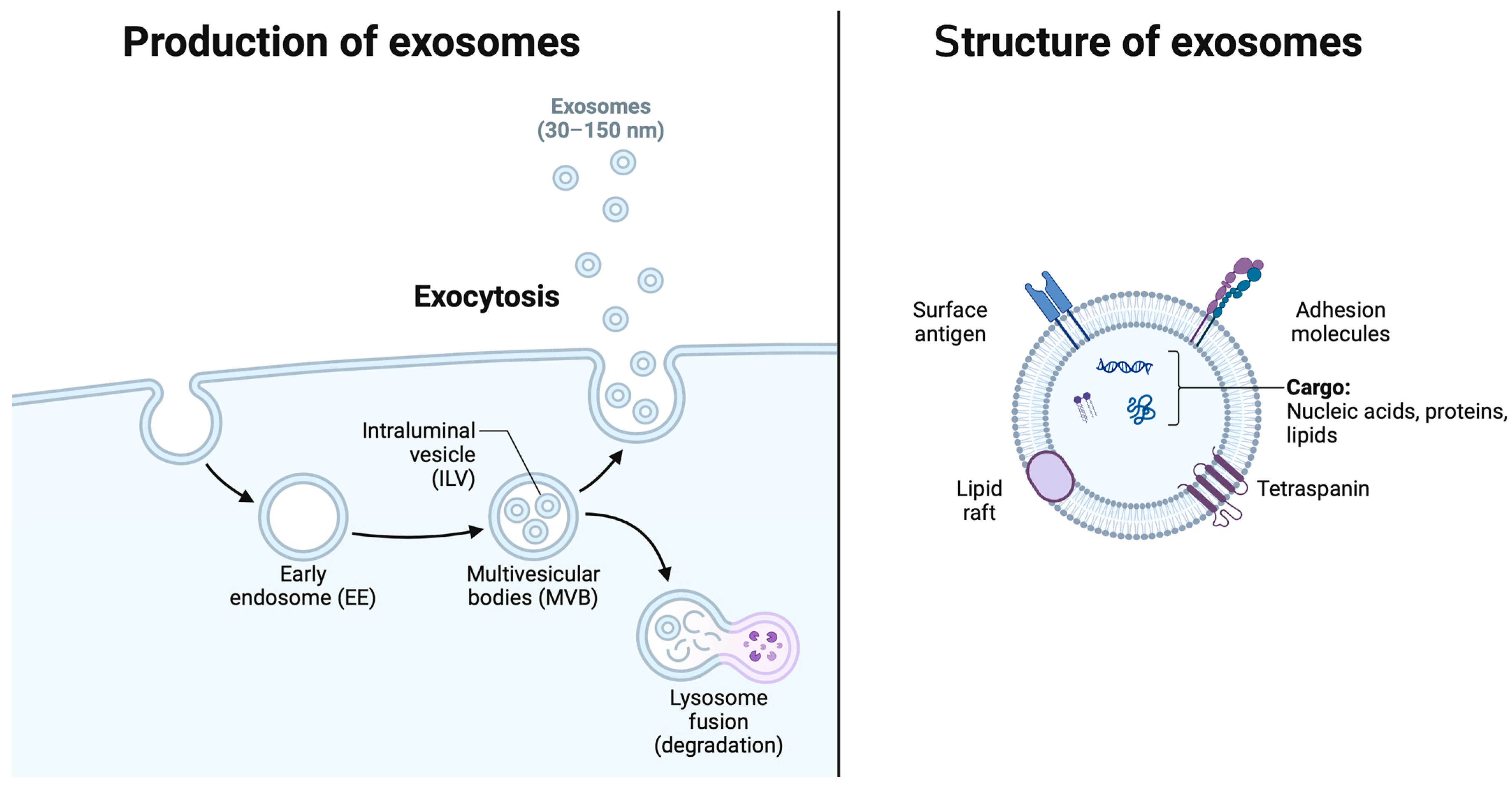
Exosomes are small EVs with a diameter ranging from 30 to 150 nanometers, originating from the endosomal system and secreted by various cell types into the extracellular milieu. These nanovesicles serve as vital mediators of intercellular communication, facilitating the exchange of bioactive molecules between cells and modulating diverse physiological and pathological processes. The distinctive characteristics of exosomes (Figure 2), including their lipid bilayer membrane and enriched cargo composition, underpin their role as key players in cell-to-cell communication networks.

3.1. Definition and Characteristics of Exosomes

Exosomes are distinguished by their unique biophysical properties and molecular composition. Their biogenesis begins within the endosomal compartment, where inward budding of the endosomal membrane forms intraluminal vesicles (ILVs) within multivesicular endosomes (MVEs) [31]. The subsequent fusion of MVEs with the plasma membrane results in the release of ILVs into the extracellular space as exosomes. These vesicles are characterized by a lipid bilayer membrane enriched in cholesterol, sphingolipids, and tetraspanin proteins (e.g., CD9, CD63, and CD81), which confer stability and facilitate their uptake by recipient cells [32]. Importantly, exosomes possess a diverse cargo of proteins, lipids, and nucleic acids, including miRNAs, messenger RNAs (mRNAs), and various classes of non-coding RNAs, reflecting their origin from parent cells and influencing their biological functions [9,33].

3.2. Biogenesis and Secretion Mechanisms

The biogenesis of exosomes is intricately regulated by a series of molecular machineries involved in endosomal sorting, membrane trafficking, and vesicle release. This process is extensively outlined elsewhere [34]. Central to this process is the endosomal sorting complex required for transport (ESCRT), which comprises multiple protein subunits (ESCRT-0, -I, -II, and -III) responsible for sorting ubiquitinated cargoes into ILVs and facilitating their budding into MVEs [35]. Additionally, ESCRT-independent mechanisms, such as the ceramide-dependent pathway and tetraspanin-enriched microdomains, also contribute to exosome biogenesis and cargo sorting [36]. Upon the fusion of MVEs with the plasma membrane, exosomes are released into the extracellular milieu through a process mediated by soluble N-ethylmaleimide-sensitive factor attachment protein receptors (SNAREs) and other vesicle trafficking proteins [37].

3.3. Cargo Composition: Proteins, Lipids, and Nucleic Acids

Exosomes harbor a diverse array of biomolecules derived from their parent cells, reflecting their cellular origin and physiological state. A proteomic analysis of exosome cargo has identified a plethora of proteins with diverse functions, including cell surface receptors, cytoskeletal proteins, heat shock proteins (HSPs), and enzymes involved in metabolism and signaling pathways [38]. Notably, exosomes are enriched in tetraspanins (e.g., CD9, CD63, and CD81), HSPs (e.g., HSP70 and HSP90), and cytoskeletal proteins (e.g., actin and tubulin), which are commonly used as exosomal markers [39,40]. Exosomes from human cardiomyocytes have demonstrated preferential uptake by human endothelial cells, and the composition of the exosomal membrane may contribute to the selective uptake of exosomes by target cells [10]. Furthermore, exosomes contain a repertoire of lipids, including cholesterol, sphingomyelin, phosphatidylserine, and various bioactive lipids, which contribute to their membrane stability and biophysical properties [41]. Importantly, exosomes encapsulate nucleic acids, including miRNAs, mRNAs, long non-coding RNAs, and circular RNAs, which can be transferred to recipient cells, modulating gene expression and cellular functions [42].

4. MiRNAs: Regulators of Gene Expression

MiRNAs are a class of small non-coding RNAs, typically 19–25 nucleotides in length, that play crucial roles in post-transcriptional gene regulation [43]. Through their interaction with the 3′ untranslated region of target mRNAs, miRNAs can mediate mRNA degradation or translational repression, thereby fine-tuning gene expression profiles and influencing various cellular processes.

4.1. Biogenesis and Mechanisms of Action

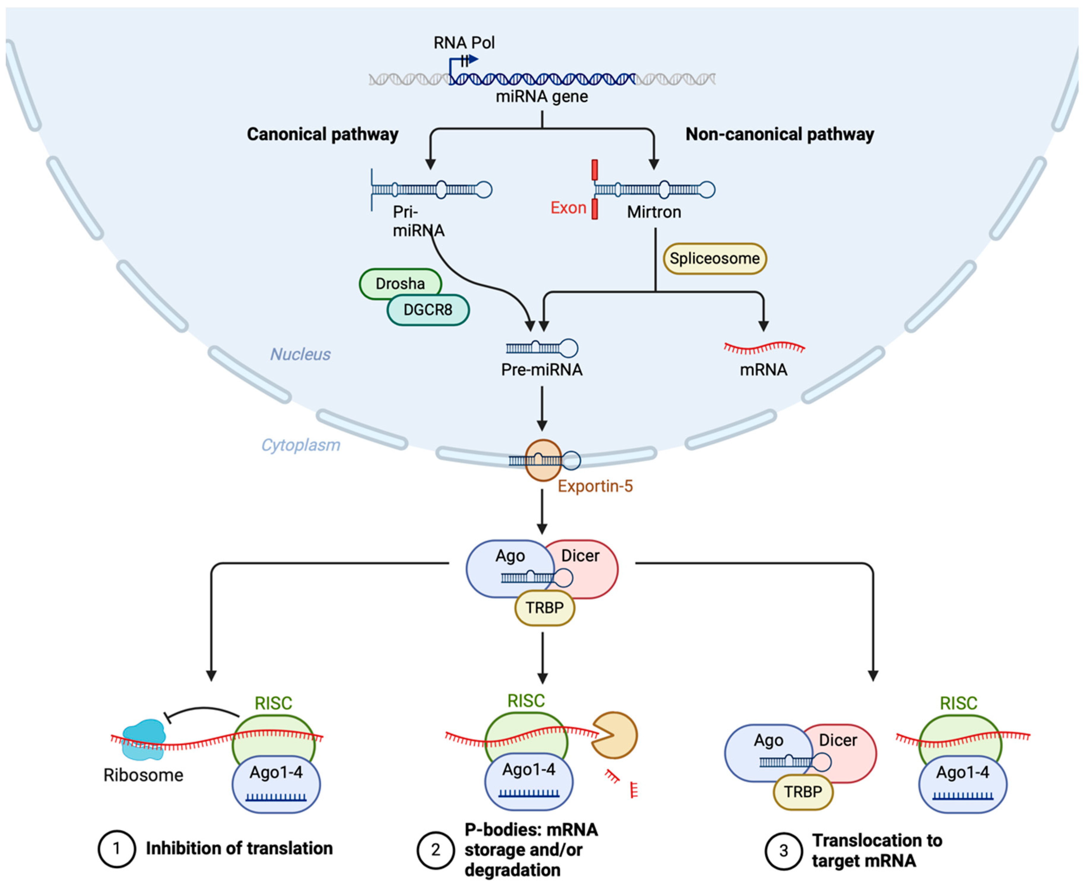
MiRNA biogenesis involves a series of coordinated steps that can be broadly categorized into canonical and non-canonical pathways (Figure 3). Both pathways convert primary miRNA transcripts (pri-miRNAs) into mature miRNAs, which are then incorporated into the RNA-induced silencing complex (RISC) to guide gene silencing.
In the canonical pathway, miRNA biogenesis begins in the nucleus with the transcription of pri-miRNAs by RNA polymerase II [44]. These pri-miRNAs contain a hairpin structure essential for further processing. The microprocessor complex, composed of the RNase III enzyme Drosha and its cofactor DGCR8 (DiGeorge Syndrome Critical Region 8), cleaves the pri-miRNA near the base of the hairpin to release a precursor miRNA (pre-miRNA) approximately 70 nucleotides long [45]. The pre-miRNA is then transported from the nucleus to the cytoplasm by Exportin-5 in a RanGTP-dependent manner. In the cytoplasm, the pre-miRNA is further processed by another RNase III enzyme, Dicer, in conjunction with the transactivation response RNA-binding protein (TRBP) [46]. This processing results in a double-stranded miRNA duplex approximately 22 nucleotides in length.
The miRNA duplex is then loaded onto an Argonaute protein (Ago1-4) to form the core of the RISC. One strand of the duplex, known as the guide strand, is retained while the passenger strand is usually degraded. The mature RISC, guided by the miRNA, binds to complementary sequences in target mRNAs, leading to translational repression or mRNA degradation [47]. The ribosome and spliceosome also play roles in the regulation of translation and splicing, which are influenced by miRNA activity.
Non-canonical miRNA biogenesis pathways deviate from the canonical Drosha-DGCR8-Dicer pathway and can be categorized into several types, outlined in detail elsewhere [48]. Mirtrons, for example, are miRNAs derived from the introns of host genes that bypass Drosha processing. Following splicing by the spliceosome, the intron lariat is debranched and refolded into a pre-miRNA-like hairpin structure, which is then exported to the cytoplasm and processed by Dicer. Similarly, simtrons are short introns processed independently of Drosha, utilizing spliceosome machinery but possibly requiring additional nucleases before Dicer processing [49].
Certain miRNAs can be loaded directly onto Argonaute proteins without the need for Dicer cleavage, constituting another non-canonical pathway described in detail elsewhere [50]. These pathways may involve the direct transcription of short hairpin RNAs that can be incorporated into RISC without further processing. Additionally, tRNA-derived fragments can act as miRNAs. These fragments are processed from tRNAs by specific nucleases and incorporated into RISC, bypassing the canonical miRNA processing machinery [50].

4.2. Functions of miRNAs in Cardiovascular Biology

MiRNAs play diverse and critical roles in cardiovascular biology, influencing processes such as cardiomyocyte proliferation, differentiation, apoptosis, angiogenesis, and contractility. These small regulatory molecules are involved in the maintenance of cardiac homeostasis and the response to various physiological and pathological stimuli. For instance, miR-1 and miR-133 are abundantly expressed in the heart and play key roles in cardiac development and function [51]. MiR-1 is involved in the regulation of cardiac hypertrophy and arrhythmogenesis, while miR-133 contributes to myocyte proliferation and differentiation [51,52,53]. Similarly, miR-208a, a cardiac-specific miRNA encoded within the intron of the α-myosin heavy chain (α-MHC) gene (Myh6), is essential for cardiac conduction and contractility.

5. Exosome-Derived miRNAs in HCM: Clinical Significance

Exosome-derived miRNAs are increasingly recognized for their role in the pathogenesis and progression of HCM, offering potential as biomarkers and therapeutic targets. Their role in modulating hypertrophic signaling, fibrotic remodeling, and cardiac metabolism underscores their relevance in HCM diagnosis and treatment.

5.1. Identification of Differential Expression Profiles

Numerous studies have highlighted distinct miRNA expression profiles within exosomes derived from HCM patients compared to those from healthy individuals or patients with other cardiac conditions. High-throughput sequencing and quantitative PCR have identified exosome-derived miRNAs as key regulators in cardiac hypertrophy and fibrosis [54,55,56]. The differential expressions of miRNas, including miR-1, miR-133, and miR-208, for example, highlight their potential as diagnostic biomarkers and therapeutic targets.

5.2. Potential Biomarkers for Diagnosis and Prognosis

Exosome-derived miRNAs hold significant promise as non-invasive biomarkers for diagnosing and monitoring HCM. Circulating exosomes can be isolated from biological fluids, including blood and urine, offering a minimally invasive approach to disease-specific molecular profiling. Specific miRNAs within exosomes can potentially differentiate between HCM and other cardiac conditions, assess disease severity, and predict outcomes, thus enabling more personalized management [57,58,59]. The stability of these miRNAs in circulation further supports their value for tracking disease progression [54,55,56].

5.3. Therapeutic Implications

The dysregulation of exosome-derived miRNAs in HCM presents novel opportunities for therapeutic intervention. By targeting specific miRNAs implicated in disease pathogenesis, researchers aim to modulate cardiac hypertrophy, fibrosis, and remodeling processes, ultimately ameliorating disease progression and improving clinical outcomes. Strategies for therapeutic intervention include the use of antagomirs, oligonucleotide-based inhibitors that specifically target and inhibit the function of dysregulated miRNAs, as well as the delivery of exogenous miRNAs or miRNA mimics to restore normal gene expression patterns [60,61,62]. Moreover, engineered exosomes loaded with therapeutic miRNAs or other bioactive molecules represent a promising approach for targeted drug delivery to the heart, circumventing the limitations of conventional pharmacological agents [63].

5.4. Comparison Between Physiological and Pathological Hypertrophy

Emerging evidence suggests distinct differences in the expression patterns of exosome-derived mRNAs in physiological (adaptive) versus pathological (maladaptive) cardiac hypertrophy. In physiological hypertrophy, as seen in athletes or during controlled cardiac growth, miRNAs like miR-1 and miR-133 are typically upregulated to support adaptive responses, aiding in healthy cellular proliferation and contractility without detrimental remodeling [64]. In contrast, pathological hypertrophy—characteristic of HCM and other cardiac disorders—often exhibits elevated levels of miRNAs such as miR-21 and miR-208, which are implicated in promoting fibrosis, arrhythmias, and hypertrophic signaling [65]. These maladaptive responses underscore the value of miRNAs as biomarkers for distinguishing between adaptive and pathological cardiac stages, with exosome-derived mRNAs providing insight into specific stages of disease progression in HCM. Further exploration into these distinct miRNA profiles may lead to more refined diagnostic and therapeutic strategies, targeting the early maladaptive changes associated with pathological hypertrophy.

6. Exosomal miRNAs That Attenuate HCM

6.1. MiR-1 from Cardiomyocytes

MiR-1 is one of the most abundant miRNAs found in cardiomyocytes and has been shown to be highly expressed in cardiomyocyte-secreted exosomes, but its expression is reduced in the serum of patients with HCM [66,67]. MiR-1 has been implicated as a significant protective factor against cardiac hypertrophy by targeting several pro-hypertrophic signaling pathways [68].
Dysregulation in calcium signaling is associated with HCM, wherein elevated intracellular Ca2+ levels trigger hypertrophic responses within the myocardium by enhancing cardiac output [69]. Calcineurin is a calcium-dependent enzyme shown to play a pivotal role in this process by activating the transcription factor, the nuclear factor of activated T cells (NFATC3) [70,71]. Studies involving human heart tissue samples and mouse cardiomyocytes have shown that the increased expression of miR-1 mitigates cardiac hypertrophy by downregulating NFATC3 expression. The downregulation of NFATC3 caused a reduction in the cardiomyocyte surface area and a decrease in the levels of β-MHCs, a hallmark of cardiac hypertrophy [72,73].
Further studies have identified that exosomal miRNA-1 from cardiomyocytes may exert its anti-hypertrophic effect through the modulation of Ca2+ uptake within mitochondria. Mitochondrial Ca2+ uniporter (MCU) is the core channel subunit of the mithochondrial Ca2+ uniporter complex (MCUC). MCUC plays a critical role in cardiomyocyte stress adaptation by modulating Ca2+ uptake within mitochondria [74]. Investigations into the way miRNA-1 can impose a protective role against cardiac hypertrophy have identified the modulation of the cyclin D kinase 6-retinoblastoma (CDK6-Rb) pathway and thyroid hormone levels as significant mechanisms [75,76]. Studies involving hypertrophic rat cardiomyocytes transfected with miR-1 mimics or cyclin D kinase 6 (CDK6) siRNA demonstrated the inhibition of CDK6-Rb pathway activation, a significant mechanism in regulating cell cycle progression contributing to cardiac hypertrophy [77]. Diniz and colleagues identified that the overexpression of miR-1 countered thyroid hormone-induced cardiac hypertrophy, evidenced by decreased levels of atrial natriuretic peptide (ANP) and α-MHC, both indicators of cardiac hypertrophy [76]. In addition, the study found that thyroid hormone induces hypertrophy by targeting histone deacetylase-4 (HDAC4), an effect mitigated by miR-1 overexpression [76]. In this way, the selective delivery of exosomal miR-1 to cardiomyocytes can prevent the progression of pathogenesis in HCM.
We now know that a key downstream target of miR-1 is the cytoskeletal regulatory protein twinfilin-1 (TWF1), which governs actin dynamics, thereby influencing various cellular processes [78,79]. The expression pattern of TWF1 has been shown to be inversely related to miR-1 levels [78]. Rat cardiomyocytes overexpressing miR-1 exhibit a reduced cell size and suppressed TWF1 protein expression, contrasting with hypertrophic rat hearts and phenylephrine-induced HCM, where downregulated miR-1 is associated with upregulated TWF1 and actin protein levels [78].
Furthermore, insulin-like growth factor-1 (IGF-1) has emerged as one of the cardiac targets influenced by miR-1 in modulating cardiac hypertrophy [80]. IGF-1 is recognized for its role in regulating cardiomyocyte size and contractile function [81]. Studies utilizing the transverse aortic constriction model and Akt-transgenic animal model revealed that the repression of miR-1 coincided with increased IGF-1 protein levels and its receptor, IGF-1R [80,82]. This finding was further supported by clinical evidence demonstrating reduced miR-1 levels in biopsies from patients with acromegaly, a condition characterized by elevated production of growth hormone and IGF-1 alongside an increased cardiomyocyte size [80].

6.2. MiR-133a from Cardiomyocytes

Similarly to miR-1, miR-133a is a miRNA found in muscle tissue, which was demonstrated to counteract hypertrophy by affecting targets within calcium signaling, cell growth, and cell development pathways [83,84,85,86,87,88]. MiR-133 is highly expressed in exosomes secreted from cardiomyocytes, and reduced expression in a normal adult cardiomyocyte is enough to induce cardiac hypertrophy [89,90,91]. Introducing miR-133a decreases cardiac hypertrophy by reducing the expression of calcineurin [83]. Additionally, miR-133a transfection has been found to impede cardiomyocyte hypertrophy induced by phenylephrine by downregulating the expression of NFATC4 [84]. Studies using transverse aortic constriction (TAC) in mice demonstrated that miR-133 overexpression mitigates cardiac hypertrophy by downregulating the calcineurin/NFAT pathway, highlighting its potential protective effects in HCM [92].
The anti-hypertrophic effects of the miR-133 family of miRNAs were observed in a rat model of triiodothyronine-induced hypertrophy, where reduced levels of miR-133 were linked, in part, to the angiotensin II receptor subtype 1 [87]. Angiotensin II receptor subtype 1 plays a dual role by both decreasing miR-133 levels and increasing the expression of calcineurin and SERCA2a, both known targets of miR-133 [87].
Recent studies have identified a potential interaction between miR-133a and α1-adrenergic receptor-mediated signaling, impacting calcium signaling dysregulation that drives cardiomyocyte hypertrophy. MiR-133a inhibits norepinephrine-induced hypertrophic changes in cardiomyocytes by downregulating protein kinase C (PKC) and Gq protein levels [85]. Norepinephrine binding to α1-AR triggers a stress response by activating PKC and phospholipase C signaling pathways. PKC activates transcription factors linked to cardiac hypertrophy through a mitogen-activated protein kinase (MAPK) cascade, while phospholipase C generates inositol 1,4,5-triphosphate and diacylglycerol, activating calcium signaling and PKC, respectively [93]. By targeting PKC and Gq, miR-133a can inhibit the rise in intracellular calcium levels and the subsequent activation of hypertrophic transcription factors such as c-Jun and c-Myc [93].
In addition, exosomal miR-133a has the capability to diminish cardiac hypertrophy by inhibiting the expression of serum response factor and cyclin D2 [87]. These factors contribute to abnormal growth of heart muscle cells and subsequent heart dysfunction. Furthermore, miR-133a targets other key players in cardiac function, such as myocyte enhancer factor 2 (MEF2), serum- and glucocorticoid-responsive kinase-1 (SGK1), and IGF-1R [88]. When cardiomyocytes undergo hypertrophy due to glucose stimulation, there is an increase in MEF2, SGK1, and IGF-1R levels alongside a decrease in miR-133a expression. Introducing miR-133a mimics counteracted the upregulation of these hypertrophy-related molecules, suggesting that MEF2, SGK1, and IGF-1R are influenced by miR-133a in the pathological remodeling that occurs in HCM [88].

6.3. MiR-30 from Endothelial Cells and Cardiomyocytes

MiR-30 family members, including miR-30a, miR-30b, miR-30c, and miR-30d, exert pleiotropic effects on cardiac fibrosis by targeting multiple fibrogenic pathways. These miRNAs modulate the expression of genes involved in transforming growth factor-beta (TGF-β) signaling, collagen synthesis, and myofibroblast differentiation. The dysregulation of miR-30 expression has been implicated in the pathogenesis of cardiac fibrosis, with the therapeutic restoration of miR-30 levels attenuating fibrotic remodeling in experimental models of HCM.
Endothelial cell-derived exosomes highly express miRNA-30a, and an investigation of the role that miR-30a plays in cardiac hypertrophy using male mice subjected to thoracic aortic constriction demonstrated that miR-30a expression in the myocardium is reduced in mouse models of hypertrophy [94]. The inhibition of miR-30a markedly increased the mRNA expression of cardiac hypertrophy markers such as atrial natriuretic factor and brain peptide and greatly increased cell size [94].
MiR-30b-5p is an mRNA found highly expressed in endothelial cells and is highly expressed within plasma exosomes [95,96]. Computational and expressional analyses have shown that it is downregulated in cardiac hypertrophy, and restoration inhibits the expression of hypertrophic signaling marker Ca2+/calmodulin-dependent protein kinase [97].
A study of the role of miR-30c in high-glucose-induced cardiomyocyte hypertrophy identified that it plays a key role in regulating the p53-p21 pathway that mediates cardiomyocyte hypertrophy. MiR-30c has been identified to target p53, and the expression of this miRNA was significantly decreased in high-glucose-treated cardiomyocytes, together with a concomitant increase in the myocardial expression of p53 and p21 genes. The overexpression of miR-30c decreased the expression of p53, p21, cardiomyocyte cell size, and apoptosis in high-glucose-treated cardiomyocytes [98].
Cardiomyocytes have been identified as the major cell type that expresses miR-30d and secretes them packaged in EVs [99]. A study using murine and neonatal rat cardiomyocyte models of hypertrophy identified that miR-30d prevented pathological cardiac hypertrophy via negatively regulating its target genes MAP4K4 and GRP78 and inhibiting pro-hypertrophic NFATC [100].

6.4. MiR-499 from Cardiomyocytes

MiR-499 has been recognized for its role in cardiac remodeling and stress response, particularly in the context of HCM. As a miRNA found predominantly in cardiac muscle, miR-499 modulates various pathways associated with cardiomyocyte apoptosis, mitochondrial function, and muscle differentiation. It has been demonstrated that elevated levels of miR-499 contribute to reduced apoptosis and improved survival of cardiomyocytes under stress conditions, potentially countering the adverse effects associated with HCM, such as cell death and maladaptive remodeling [101].
A study using murine models revealed that miR-499 overexpression inhibited apoptosis by targeting pro-apoptotic factors like calcineurin and activating anti-apoptotic pathways mediated by Bcl-2 [102]. This activity makes miR-499 a compelling candidate for therapeutic modulation in HCM as it can mitigate the hypertrophic response through enhanced cellular survival and maintenance of cardiac function. These findings suggest that the exosome-mediated delivery of miR-499 could serve as a cardioprotective mechanism for patients with HCM, particularly those at risk of progressive cardiac remodeling.

6.5. Other Exosomal MiRNAs

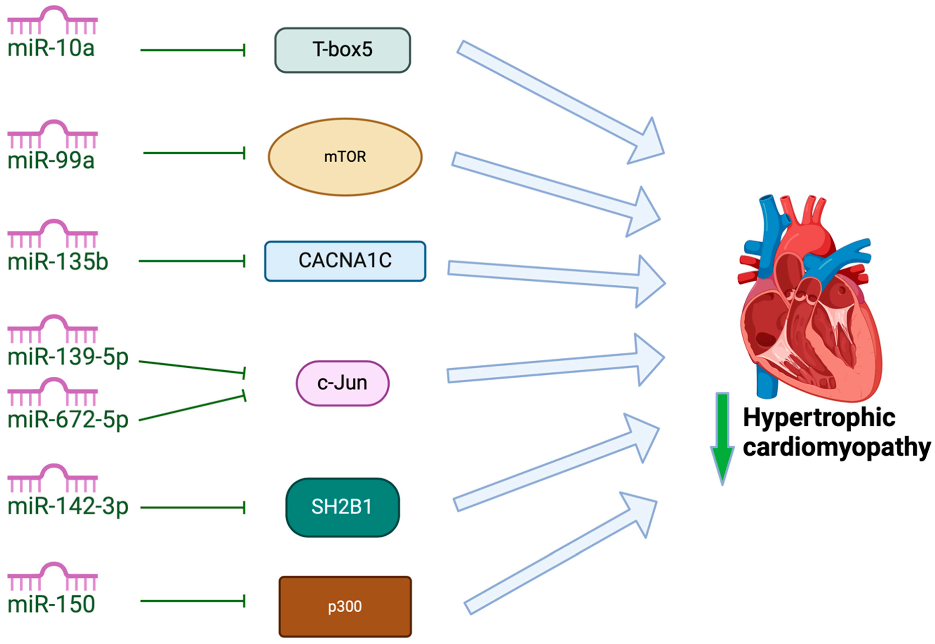
In addition to the miRNAs mentioned above, other less-studied miRNAs attenuate cardiac hypertrophy (Figure 4). Some of these miRNAs mediate their effects by regulating factors involved in cardiac development or factors involved in calcium signaling and energy metabolism. MiR-10a has been shown to target T-box5 in rat models, decreasing the expression of natriuretic peptide A and myosin heavy chain 7 cardiac muscle beta, attenuating cardiac hypertrophy [103]. MiR-135b overexpression has been shown to attenuate cardiac hypertrophy by targeting CACNA1C, which encodes L-type calcium channels in cardiomyocytes, attenuating hypertrophic signaling [104]. The overexpression of miR-142-3p has been shown to inhibit Src homology 2 B adaptor protein 1. In doing so, miR-142-3p ameliorates cardiac mitochondrial dysfunction and improves mitochondrial function in rat models of HCM [105]. Both miR-139-5p and miR-672 have recently been identified to reduce cardiac hypertrophy by inhibiting the expression of transcription factor JUN [106,107], and a study by Duan and colleagues identified that miR-150 regulates high-glucose-induced cardiac hypertrophy by targeting the transcriptional co-activator p300 [108]. The overexpression of miR-99a attenuated cardiac hypertrophy in mouse models of cardiac hypertrophy induced by TAC, likely through the downregulated expression of mammalian target of rapamycin (mTOR) [109]. Further studies are required to understand the role that these miRNAs can play in therapeutic strategies for HCM.

7. Exosomal miRNAs That Promote HCM

7.1. MiR-200 from Cardiomyocytes

The exosomal miR-200 family has been implicated in the cellular crosstalk between cardiomyocytes and endothelial cells in health and pathology [110]. One of the targets of miR-200c is dual-specific phosphatase-1 (DUSP-1). This target has been shown to prevent cardiac hypertrophy by deactivating MAPKs, including extracellular signal-regulated kinase, c-Jun N-terminal kinase, and p38 [111]. In high-glucose-treated cardiomyocytes, miR-200c downregulates DUSP-1 expression, leading to the activation of MAPK proteins, thereby inducing diabetes-associated cardiac hypertrophy [112].
Further studies have demonstrated that exosomal miR-200c promotes cardiac hypertrophy by directly targeting myosin light chain kinase (MLCK). This enzyme is pivotal in cardiovascular physiology and pathophysiology [113]. Elevated levels of miR-200c correlate with a reduction in MLCK expression [114]. In hypertrophic models, the overexpression of miR-200c causes an increase in the production of ROS and apoptosis, characterized by the heightened expression of pro-apoptotic markers including Bax and caspase-3 [114]. Hypertrophic growth in the myocardium then occurs in cardiac remodeling secondary to stress signaling.

7.2. MiR-155 from Immune Cells

Studies have indicated that exosomal miR-155 secreted from immune cells are involved in the promotion of HCM by targeting inflammation and calcium signaling pathways [115,116,117,118]. Macrophages expressing miR-155 activate the Janus kinase (JAK)/signal transducer and activator of transcription 3 (STAT3) pathway, thereby facilitating hypertrophic remodeling in the myocardium. MiR-155 is believed to inhibit the suppressor of cytokine signaling 1 (SOCS1), leading to STAT3 phosphorylation [119]. Phosphorylated STAT3 has been widely shown to initiate pro-hypertrophic signaling in cardiomyocytes. In this way, miR-155 secreted within macrophage exosomes can contribute to HCM, and the therapeutic inhibition of miR-155 presents itself as a potential avenue to protect the heart from pathological cardiac hypertrophy [115].

7.3. MiR-21 from Immune Cells and Cardiac Fibroblasts

Exosomal miR-21 has drawn considerable attention in recent years for the role it may play in HCM [120,121]. MiR-21 is abundantly expressed in immune cells, such as T cells and macrophages, as well as in cardiac fibroblasts. To date, several genes have been identified as miR-21 targets involved in cardiomyopathy. Programmed cell death 4 (PDCD4) and phosphatase and tensin homolog deleted from chromosome 10 (PTEN) are the two most recognized target genes involved in anti-apoptosis [122,123]. PTEN acts as a negative regulator of the PI3K/Akt/mTOR pathway, and it is pivotal in cardiac function [124]. By targeting these genes, as well as other negative regulators of hypertrophic signaling including sprout homolog 1 (SPRY1), immune cell- and fibroblast-derived exosomes enriched with miR-21 can promote cardiomyocyte hypertrophy [125,126].
While primarily associated with hypertrophic signaling, mir-21 also plays a crucial role in cardiac fibrosis in HCM. MiR-21 promotes fibroblast activation and extracellular matrix deposition [126] by targeting inhibitors of the TGF-β pathway, such as SMAD7 [127]. Exosomes enriched with miR-21 have been implicated in the transmission of pro-fibrotic signals to fibroblasts, exacerbating fibrotic remodeling in the hypertrophic heart and causing a decrease in miR-21 as antagomir-21 prevents fibrosis following myocardial injury [128].

7.4. MiR-22 from Smooth Muscle Cells

MiR-22 is highly expressed by smooth muscle and mesenchymal stromal cells, and the exosomal transfer of these miRNAs is hypothesized to play a pivotal role in crosstalk with cardiomyocytes [129]. MiR-22 has been shown to be upregulated during myocardial differentiation and cardiac hypertrophy [130]. Similarly to miR-155, miR-22 is implicated in promoting HCM by targeting the calcineurin pathway, as a loss of miR-22 represses calcineurin-induced cardiac hypertrophy [130]. The inhibition of miR-22 or the observation of cardiomyocytes from miR-22 knockout mice led to heightened levels of HDAC4 and sirtuin 1 (SIRT1) [130]. Both are known to play protective roles against cardiac hypertrophy [131,132].
Further studies have also demonstrated that miR-22-induced hypertrophy may involve the modulation of the PTEN pathway. The overexpression of miR-22 in HCM leads to reduced PTEN protein levels, accompanied by increased cell surface area and expression of cardiac hypertrophy markers [133]. This inverse relationship between miR-22 and PTEN in cardiac hypertrophy was further supported by another study, demonstrating that atorvastatin, a cholesterol-lowering medication known for its anti-hypertrophic effects, reversed the consequences of miR-22 overexpression by elevating PTEN levels [134]. Nonetheless, clinical evidence regarding the impact of atorvastatin on human patients with cardiac hypertrophy remains scarce, and further studies are required to delineate the mechanisms by which it attenuates cardiac hypertrophy.

7.5. MiR-21 from Cardiomyocytes

MiR-217 is highly expressed in cardiomyocytes, and cardiomyocyte-derived exosomes containing miR-217 has been shown to enhance the proliferation of fibroblasts in vitro [135]. MiR-217 appears to function similarly to miR-22 regarding its involvement in HCM, particularly through its interaction with PTEN. The overexpression of miR-217 leads to cardiac hypertrophy, which can be mitigated by restoring PTEN expression, as evidenced by decreased levels of hypertrophic markers such as ANP and β-MHC [135].
Further genes implicated in the miR-217-mediated promotion of pathological cardiac hypertrophy are euchromatic histone–lysine N-methyltransferases (EHMTs) EHMT1 and EHMT2 [136]. The experimental induction of pathological HCM, such as through abdominal aortic banding in rat models, results in the increased expression of miR-217 and the subsequent suppression of EHMT1/2 mRNA levels. These enzymes are crucial for heart development in the prenatal stage and for the catalysis of histone 3 lysine 9 dimethylation (H3K9me2) [137]. The concurrent decrease in EHMT1/2 and H3K9m2 levels triggers the re-expression of fetal-associated transcripts, ultimately leading to pathological hypertrophy. This was supported by the finding that the inhibition of miR-217 activity prevents the downregulation of EHMT1/2 and reverses the onset of hypertrophy [136]. These findings suggest that exosomal miR-217, through its ability to modulate methylation processes, presents a promising therapeutic target for managing and treating cardiac hypertrophy.

7.6. MiR-208 from Cardiac Stem Cells

MiR-208, specifically encoded by Myh6, plays a crucial role in cardiac remodeling and hypertrophic responses. miRNA-208 expression is markedly increased in the serum exosomes of patients with cardiac disease compared to control patients [138]. Importantly, the sensitivity of exosome miR-208a is superior to that of exosome miR-208a. Exosomal miR-208a therefore presents itself as a potentially important marker of cardiac stress during development of HCM.
The expression of pro-hypertrophic β-MHC is increased following the upregulation of miR-208a. This upregulation of miR-208a precedes a switch from α- to β-MHC isoforms and the development of systolic and diastolic dysfunction [139]. The inhibition of this miRNA prevented β-MHC activation and the subsequent hypertrophic response, thus demonstrating the potential for miR-208a inhibition as a therapeutic avenue in HCM.

7.7. MiR-214 from Endothelial Cells

MiR-214 has emerged as a critical regulator of fibrosis and hypertrophy in cardiac diseases, including HCM. Elevated levels of miR-214 are associated with increased collagen deposition and fibrotic activation, contributing to the fibrotic remodeling of the myocardium commonly seen in HCM [140]. Research on animal models has shown that miR-214 influences several profibrotic pathways, including those involving TGF-β signaling, which leads to excessive extracellular matrix production and stiffening of the heart muscle [141,142,143].
Additionally, miR-214 has been shown to play a role in calcium homeostasis and energy metabolism, both crucial in maintaining cardiac contractility and function. The dysregulation of miR-214, often observed in HCM, results in impaired calcium handling, contributing to arrhythmogenic and contractile dysfunction in HCM [144]. By modulating calcium homeostasis, miR-214 serves as both a marker and a potential therapeutic target for managing arrhythmias and hypertrophic responses in HCM. Studies suggest that the inhibition of miR-214 may ameliorate these effects, providing a basis for therapeutic interventions aimed at reducing cardiac fibrosis and dysfunction [145].

7.8. Other MiRNAs

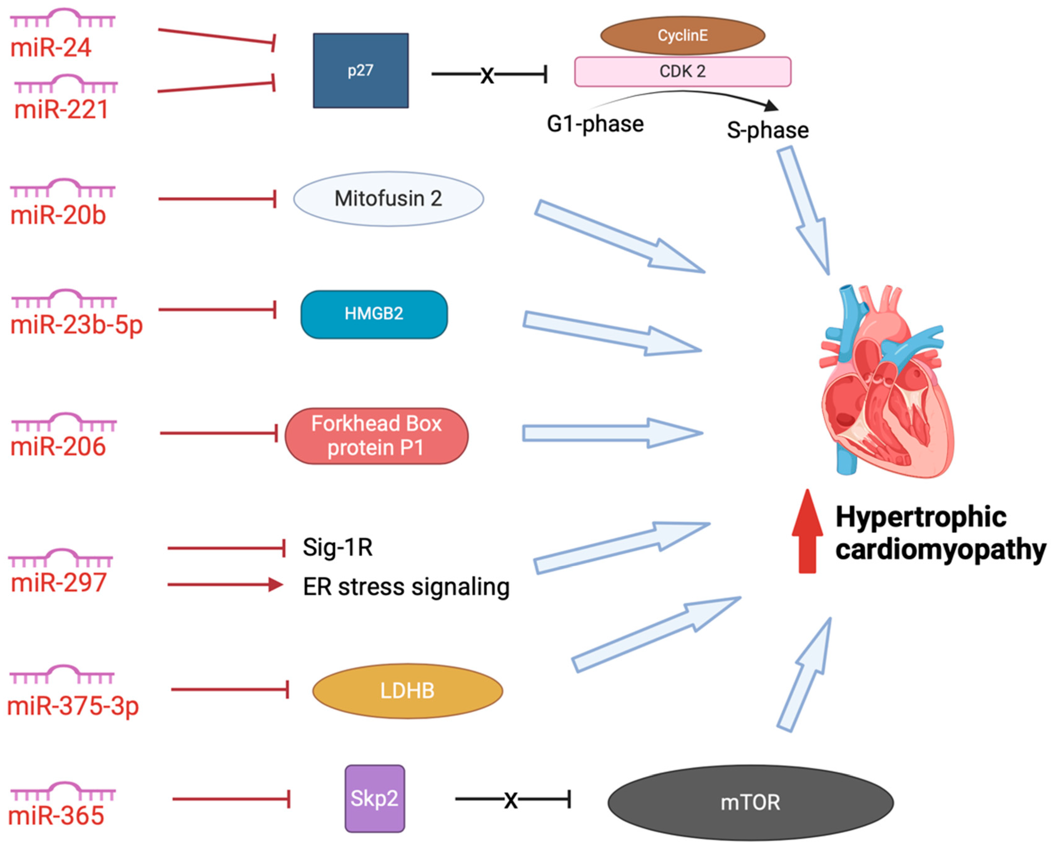
In addition to the exosomal miRNAs described above, other, less studied exosomal miRNAs promote HCM (Figure 5). These miRNAs promote dysfunction in cardiac structure, function, or metabolism [146,147,148,149,150,151,152,153]. MiR-24 and miR-221 both inhibit CDK inhibitor p27, thus disinhibiting CDK2 and allowing the progression of cell cycling from the G1 phase to the S phase [150,153]. This activity has led to exosomal miR-24 and miR-221 being investigated for their role in carcinomas and cancer metastasis, but limited studies have involved hypertrophic remodeling [154,155].
Another potentially important miRNA in the pathogenesis of HCM is miR-20b. MiR-20b inhibits mitofusin 2-mediated inter-organelle calcium crosstalk, promoting hypertrophic remodeling [152]. MiR-23b-5p blocks HMGB2, promoting cardiac dysfunction; however, the relationship between miR-23b-5p and remodeling in HCM has not yet been investigated [147]. MiR-206 plays an essential role in mediating cardiac hypertrophy by negatively regulating forkhead box protein P1 [149]. It has also been demonstrated that circulating exosomes, containing miR-206, are taken up by the heart, leading to sympathetic dysinnervation and increased arrhythmogenesis, an important feature of HCM pathogenesis [156]. MiR-297 inhibits the expression of sigma-1 receptor and activates ER stress signaling, promoting cardiac hypertrophy [146]. However, we are yet to delineate the role of exosomal miR-297 and differentiate this from serum miR-297. MiR-375-3p inhibits the expression of lactate dehydrogenase B chain (LDHB), promoting cardiac hypertrophy [151]. Again, although exosomal miR-375-3p has been implicated in the pathogenesis of carcinomas of the gastrointestinal tract, we are still yet to fully delineate the role of serum and exosomal miRNA-375-3p in HCM.
Another miRNA which shows promise as a potential marker and therapeutic target in cardiac disease is miRN-365. MiR-365 has been found abundantly in the exosomes extracted from pericardial fluid samples from patients with heart failure [157]. It has been identified as a circulatory cardiovascular-related miRNA, upregulated after exercise in humans [158]. MiR-365 blocks Skp2, uninhibiting mTOR, triggering autophagy and cardiac hypertrophy [148]. However, we are yet to understand the role that exosomal miRNA-365 plays in HCM in humans.

8. Exosomal MiRNAs That Both Attenuate and Promote HCM

8.1. MiR-29 from Cardiac Fibroblasts

MiR-29 family members, including miR-29a, miR-29b, and miR-29c, have garnered significant attention for their regulatory role in cardiac fibrosis and hypertrophy. Interestingly, early investigations have demonstrated that miR-29 family members show mixed activity that both promote and attenuate HCM. MiR-29a and miR-29b, in particular, have been investigated in mouse models of myocardial infarction and pressure overload, revealing that while they inhibit fibrosis via TGF-β suppression, they may also contribute to cardiomyocyte hypertrophy through Wnt activation [159,160].
MiR-29a is the most stable and most abundantly expressed member of the miR-29 family. MiR-29a and -29b-1 are encoded on human chromosome 7q32.3, and mir-29b-2 and -29c are found on chromosome 1q32.2. Cardiac fibroblasts have been identified as the primary cell type responsible for miR-29 expression in the heart, and the treatment of isolated mouse cardiac fibroblasts with TGF-β reduced the expression of miR-29a, miR-29b, and miR-29c [161].
A study by Liu and colleagues used mouse models and human tissue samples to identify a protective role of miR-29 family members in HCM [159]. This study found that endothelin-1 (ET1) signaling, known to be upregulated in early HCM, increases ROS in cardiomyocytes, stimulating the secretion of TGF-β and suppressing miR-29a expression. This suppression of miR-29a contributes to increased collagen production in fibroblasts, promoting fibrosis [159]. Therefore, the modulation of miR-29a to increase expression via exosomal transfer has been identified as a potential therapeutic target.
In addition, miR-29 has been shown to exhibit a cardioprotective effect in isoproterenol-induced cardiac hypertrophy by inhibiting a nuclear receptor, peroxisome proliferator-activated receptor δ [162]. Additionally, Li and colleagues suggested a potential role of miR-29a-3p in attenuating ET1-induced cardiomyocyte hypertrophy by inhibiting NFATc4 expression, although this finding was derived from an in vitro study using H9c2 cells [163].
However, despite these studies that support the concept that enhancing miR-29 would be a promising anti-fibrotic therapy for heart disease, there is also growing evidence that the miR-29 family is a potent inducer of cardiomyocyte hypertrophy [164]. Sassi and colleagues investigated the role of miR-29 in myocardial remodeling and identified that miR-29 promotes cardiomyocyte hypertrophy in vivo and increases cardiac fibrosis rather than decreasing it [160]. This was supported by their finding that the pharmacological inhibition or genetic deletion of miR-29 prevented both cardiac hypertrophy and fibrosis in mouse models. The study identified that the activity was carried out through the regulation of canonical and non-canonical Wnt signaling in cardiomyocytes, eliciting hypertrophic growth in these cells and the secretion of profibrotic proteins that signal towards cardiac fibroblasts. Hence, further investigations are warranted to elucidate the precise involvement of miR-29 in cardiac remodeling.

8.2. MiR-146a from Immune Cells

MiR-146a, expressed in various immune cell subsets, serves as a critical regulator of inflammatory signaling pathways in the myocardium and has been shown to exert a significant influence on cardiac hypertrophy. The genetic deletion or therapeutic inhibition of miR-146a during pressure overload diminishes the hypertrophic response and improves cardiac function [165]. This effect is attributed to miR-146a’s modulation of dihydrolipoamide succinyltransferase (DLST), a mitochondrial protein involved in energy metabolism. The overexpression of DLST counteracts pressure overload-induced hypertrophy, underscoring its role as a potential therapeutic target.
Research indicates that miR-146a plays a crucial role in fibrosis regulation in HCM. Studies in mouse models of pressure overload and patients with aortic stenosis revealed the upregulation of miR-146a. The inhibition of miR-146a during pressure overload attenuates fibrosis. Although the exact mechanisms are not fully elucidated, it is suggested that miR-146a may impact fibrosis indirectly via its effects on cardiac metabolism [165].
The impact of miR-146a extends to metabolic alterations in cardiomyocytes. MiR-146a knockout cells exhibit preserved glucose and fatty acid oxidation under pressure overload conditions, indicating a favorable metabolic profile [165]. DLST, targeted by miR-146a, plays a crucial role in regulating mitochondrial ATP production, thereby influencing cardiomyocyte metabolism. Therapeutic strategies targeting miR-146a may normalize metabolic changes associated with HCM, offering potential therapeutic benefits. A study on the role that exosomal-miR-146a-5p plays in HCM revealed that only a minor fraction of the plasma miR-146a was encapsulated in exosomes [166]. However, there are limitations in exosome isolation that affect our ability to quantify exosomal-miRNA, which will be discussed in the next section.

9. Challenges and Future Directions

9.1. Methodological Limitations and Technical Challenges

Despite significant advancements in our understanding of exosome-derived miRNAs in HCM, several methodological limitations and technical challenges hinder comprehensive research in this field. One major challenge lies in the isolation and characterization of exosomes from complex biological samples, such as blood or cardiac tissue. Current isolation methods often suffer from a low yield, contamination, and lack of standardization, leading to variability in the experimental results [167]. Moreover, the heterogeneity of exosomes, both in terms of size and cargo composition, poses a challenge for accurate characterization and functional analysis.
Animal models, such as TAC in rodents and cellular models of phenylephrine-induced hypertrophy, have provided insights into miRNA-mediated regulation in HCM. However, translating these findings to human HCM remains a challenge. Interspecies variability in miRNA expression and function complicates the direct extrapolation of results, as rodent miRNAs may not fully replicate the expression patterns, targets, or regulatory network of human miRNAs. Additionally, while murine models are helpful for understanding gene function and molecular mechanisms, the complexity of human HCM—especially in terms of genetic diversity and clinical heterogeneity—requires careful consideration when applying these insights to therapeutic approaches. Thus, while TAC and phenylephrine models provide foundational knowledge of miRNA-mediated regulation in cardiac hypertrophy, further studies in human-derived cells or humanized animal models are essential for validating these findings in the context of human HCM.
Furthermore, the identification and quantification of specific miRNAs within exosomes remain technically demanding. Existing techniques, such as next-generation sequencing and quantitative PCR, have inherent limitations, including sensitivity, specificity, and reproducibility issues. Improved methodologies for profiling exosomal miRNAs are necessary to accurately delineate their roles in HCM pathogenesis.

9.2. Unraveling Specific Signaling Pathways and Targets

A deeper understanding of the specific signaling pathways and molecular targets regulated by exosome-derived miRNAs is crucial for elucidating their roles in HCM. While studies have identified several miRNAs implicated in hypertrophic signaling cascades, the precise mechanisms underlying their effects remain incompletely understood. Unraveling the intricate interplay between exosomal miRNAs and target genes/pathways in cardiomyocytes, fibroblasts, endothelial cells, and immune cells will provide insights into the pathophysiology of HCM.
Moreover, deciphering the context-dependent effects of exosomal miRNAs in different stages of HCM progression is essential. It is likely that miRNA-mediated signaling networks vary dynamically during the disease, influenced by factors such as disease etiology, comorbidities, and therapeutic interventions. Integrative approaches, combining experimental models, omics technologies, and computational analyses, are needed to unravel the complexity of miRNA-mediated regulation in HCM.

9.3. Translational Potential for Diagnostic and Therapeutic Strategies

Despite the challenges, exosome-derived miRNAs hold significant promise as diagnostic biomarkers and therapeutic targets for HCM. The unique stability and cell-type specificity of exosomal miRNAs make them attractive candidates for non-invasive biomarker discovery and disease monitoring. However, the translation of exosomal miRNA-based diagnostic assays into clinical practice requires rigorous validation in large, well-characterized patient cohorts.
Furthermore, the exosome-mediated delivery of miRNA-based therapeutics offers a novel approach for targeting pathological pathways in HCM. Engineered exosomes loaded with specific miRNA mimics or inhibitors could be employed to modulate hypertrophic signaling cascades, attenuate fibrotic remodeling, and improve cardiac function in HCM patients. However, challenges related to targeted delivery, off-target effects, and safety must be addressed before clinical implementation.

10. Conclusions

In conclusion, the burgeoning field of exosome-derived miRNAs in HCM offers exciting avenues for understanding disease pathogenesis, identifying novel biomarkers, and developing targeted therapeutics. Despite methodological challenges and technical limitations, significant progress has been made in elucidating the roles of exosomal miRNAs in HCM. Studies have revealed specific miRNA signatures associated with hypertrophic signaling pathways, fibrotic remodeling, and inflammatory responses in the myocardium. Furthermore, emerging evidence suggests the translational potential of exosome-derived miRNAs as diagnostic biomarkers and therapeutic targets for HCM. By addressing existing challenges and leveraging the wealth of information offered by exosomal miRNAs, we can advance our understanding and management of HCM, ultimately improving outcomes for affected individuals.

Funding

This research received no external funding.

Institutional Review Board Statement

Not applicable.

Informed Consent Statement

Not applicable.

Data Availability Statement

Not applicable.

Conflicts of Interest

The authors declare no conflict of interest.

Abbreviations

α-MHCAlpha-myosin heavy chain
AKTProtein kinase B
ANPAtrial natriuretic peptide
ATPAdenosine triphosphate
β-MHCBeta-myosin heavy chain
CACNA1CCalcium voltage-gated channel subunit alpha1 C
CDKCyclin dependent kinase
CDK6-RbCyclin D kinase 6-retinoblastoma
DGCR8DiGeorge syndrome critical region 8
DLSTDihydrolipoamide succinyltransferase
DUSP-1Dual-specific phosphatase-1
EEEarly endosome
EHMTEuchromatic histone–lysine N-methyltransferase
EREndoplasmic reticulum
ESCRTEndosomal sorting complex required for transport
ET1Endothelin-1
EVExtracellular vesicle
H3K9me2Histone 3 lysine 9 dimethylation
HCMHypertrophic cardiomyopathy
HDAC4Histone deacetylase-4
HFpEFHeart failure with preserved ejection fraction
HMGB2High-mobility group box 2
HSPHeat shock protein
IGF-1Insulin-like growth factor-1
IGF-1RInsulin-like growth factor-1 receptor
ILVIntraluminal vesicle
JAKJanus kinase
LDHBLactate dehydrogenase B chain
LVOTLeft ventricular outflow tract obstruction
MAPKMitogen-activated protein kinase
MCUMitochondrial Ca2+ uniporter
MCUCMitochondrial Ca2+ uniporter complex
MEF2Myocyte enhancer factor 2
MiRNAMicroRNA
MLCKMyosin light chain kinase
mRNAMessenger RNA
mTORMammalian target of rapamycin
MVEMultivesicular endosomes
Myh6Alpha-myosin heavy chain gene
NFATCNuclear factor of activated T cells
PDCD4Programmed cell death 4
PI3KPhosphoinositide 3-kinase
PKCProtein kinase C
Pre-miRNAPrecursor miRNA
Pri-miRNAPrimary miRNA
PTENPhosphatase and tensin homolog deleted from chromosome 10
RISCRNA-induced silencing complex
ROSReactive oxygen species
SCDSudden cardiac death
SERCA2aSarcoplasmic/endoplasmic reticulum Ca2+-ATPase 2a
SGK1Serum- and glucocorticoid-responsive kinase-1
Sig-1RSigma-1 receptor
SIRT1Sirtuin 1
Skp2S-phase kinase-associated protein 2
SNARESoluble N-ethylmaleimide-sensitive factor attachment protein receptor
SOCS1Suppressor of cytokine signaling 1
SPRY1Sprout homolog 1
STAT3Signal transducer and activator of transcription 3
TACTransverse aortic constriction
TGFTransforming growth factor
TRBPTransactivation response RNA-binding protein
TWF1Twinfilin-1

References

  1. Marian, A.J.; Braunwald, E. Hypertrophic cardiomyopathy: Genetics, pathogenesis, clinical manifestations, diagnosis, and therapy. Circ. Res. 2017, 121, 749–770. [Google Scholar] [CrossRef] [PubMed]
  2. Van Der Velden, J.; Stienen, G.J.M. Cardiac Disorders and Pathophysiology of Sarco-meric Proteins. Physiol. Rev. 2019, 99, 381–426. [Google Scholar] [CrossRef] [PubMed]
  3. Jordà, P.; Oudit, G.Y.; Tadros, R. Unraveling the Genetic Substrate and Phenotypic Variability of Hypertrophic Cardiomyopathy: A Role for Desmosome Gene Variants? Can. J. Cardiol. 2022, 38, 3–5. [Google Scholar] [PubMed]
  4. Tudurachi, B.S.; Zăvoi, A.; Leonte, A.; Ureche, C.; Bîrgoan, S.G.; Chiuariu, T.; Anghel, L.; Radu, R.; Sascău, R.A.; Stătescu, C. An Update on MYBPC3 Gene Mutation in Hypertrophic Cardiomyopathy. Int. J. Mol. Sci. 2023, 24, 10510. [Google Scholar] [CrossRef] [PubMed]
  5. Teekakirikul, P.; Zhu, W.; Huang, H.C.; Fung, E. Hypertrophic cardiomyopathy: An overview of genetics and management. Biomolecules 2019, 9, 878. [Google Scholar] [CrossRef]
  6. Yang, K.C.; Breitbart, A.; De Lange, W.J.; Hofsteen, P.; Futakuchi-Tsuchida, A.; Xu, J.; Schopf, C.; Razumova, M.V.; Jiao, A.; Boucek, R.; et al. Novel Adult-Onset Systolic Cardiomyopathy Due to MYH7 E848G Mutation in Patient-Derived Induced Pluripotent Stem Cells. JACC Basic Transl. Sci. 2018, 3, 728–740. [Google Scholar] [CrossRef]
  7. Dobaczewski, M.; Chen, W.; Frangogiannis, N.G. Transforming growth factor (TGF)-β signaling in cardiac remodeling. J. Mol. Cell Cardiol. 2011, 51, 600–606. [Google Scholar]
  8. Dorn, G.W.; Force, T. Protein kinase cascades in the regulation of cardiac hypertrophy. J. Clin. Investig. 2005, 115, 527–537. [Google Scholar] [CrossRef]
  9. Di Bella, M.A. Overview and Update on Extracellular Vesicles: Considerations on Exosomes and Their Application in Modern Medicine. Biology 2022, 2022, 804. [Google Scholar] [CrossRef]
  10. Zwi-Dantsis, L.; Winter, C.W.; Kauscher, U.; Ferrini, A.; Wang, B.; Whittaker, T.E.; Hood, S.R.; Terracciano, C.M.; Stevens, M.M. Highly purified extracellular vesicles from human cardiomyocytes demonstrate preferential uptake by human endothelial cells. Nanoscale 2020, 12, 19844–19854. [Google Scholar] [CrossRef]
  11. Zhou, S.S.; Jin, J.P.; Wang, J.Q.; Zhang, Z.G.; Freedman, J.H.; Zheng, Y.; Cai, L. MiRNAS in cardiovascular diseases: Potential biomarkers, therapeutic targets and challenges review-article. Acta Pharmacol. Sin. 2018, 39, 1073–1084. [Google Scholar] [CrossRef] [PubMed]
  12. Maron, B.J.; Gardin, J.M.; Flack, J.M.; Gidding, S.S.; Kurosaki, T.T.; Bild, D.E. Prevalence of Hypertrophic Cardiomyopathy in a General Population of Young Adults. Circulation 1995, 92, 785–789. [Google Scholar] [CrossRef] [PubMed]
  13. Marian, A.J. Pathogenesis of diverse clinical and pathological phenotypes in hypertrophic cardiomyopathy. Lancet 2000, 355, 58–60. [Google Scholar] [CrossRef] [PubMed]
  14. Adamczak, D.M.; Oko-Sarnowska, Z. Sudden cardiac death in hypertrophic cardiomyopathy. Cardiol. Rev. 2018, 26, 145–151. [Google Scholar] [CrossRef]
  15. Link, M.S.; Bockstall, K.; Weinstock, J.; Alsheikh-Ali, A.A.; Semsarian, C.; Estes, N.A.M.; Spirito, P.; Haas, T.S.; Rowin, E.J.; Maron, M.S.; et al. Ventricular Tachyarrhythmias in Patients With Hypertrophic Cardiomyopathy and Defibrillators: Triggers, Treatment, and Implications. J. Cardiovasc. Electrophysiol. 2017, 28, 531–537. [Google Scholar] [CrossRef]
  16. Eduardo Carreño, J.; Apablaza, F.; Ocaranza, M.P.; Jalil, J.E. Cardiac Hypertrophy: Molecular and Cellular Events. Rev. Española de Cardiol. (Engl. Ed.) 2006, 59, 473–486. [Google Scholar] [CrossRef]
  17. Crocini, C.; Gotthardt, M. Cardiac sarcomere mechanics in health and disease. Biophys. Rev. 2021, 13, 637–652. [Google Scholar] [CrossRef]
  18. Schlittler, M.; Pramstaller, P.P.; Rossini, A.; De Bortoli, M. Myocardial Fibrosis in Hypertrophic Cardiomyopathy: A Perspective from Fibroblasts. Int. J. Mol. Sci. 2023, 24, 14845. [Google Scholar] [CrossRef]
  19. Frangogiannis, N.G. Cardiac fibrosis. Cardiovasc. Res. 2021, 117, 1450–1488. [Google Scholar] [CrossRef]
  20. Zile, M.R.; Baicu, C.F.; Ikonomidis, J.S.; Stroud, R.E.; Nietert, P.J.; Bradshaw, A.D.; Slater, R.; Palmer, B.M.; Van Buren, P.; Meyer, M.; et al. Myocardial stiffness in patients with heart failure and a preserved ejection fraction contributions of collagen and titin. Circulation 2015, 131, 1247–1259. [Google Scholar] [CrossRef]
  21. Frangogiannis, N.G. Transforming growth factor–ß in tissue fibrosis. J. Exp. Med. 2020, 217, e20190103. [Google Scholar] [CrossRef] [PubMed]
  22. Hartupee, J.; Mann, D.L. Role of inflammatory cells in fibroblast activation. J. Mol. Cell. Cardiol. 2016, 93, 143–148. [Google Scholar] [PubMed]
  23. Van Der Velden, J.; Tocchetti, C.G.; Varricchi, G.; Bianco, A.; Sequeira, V.; Hilfiker-Kleiner, D.; Hamdani, N.; Leite-Moreira, A.F.; Mayr, M.; Falcao-Pires, I.; et al. Metabolic changes in hypertrophic cardiomyopathies: Scientific update from the working group of myocardial function of the European Society of Cardiology. Cardiovasc. Res. 2018, 114, 1273–1280. [Google Scholar] [CrossRef] [PubMed]
  24. Ranjbarvaziri, S.; Kooiker, K.B.; Ellenberger, M.; Fajardo, G.; Zhao, M.; Vander Roest, A.S.; Woldeyes, R.A.; Koyano, T.T.; Fong, R.; Ma, N.; et al. Altered Cardiac Energetics and Mitochondrial Dysfunction in Hypertrophic Cardiomyopathy. Circulation 2021, 144, 1714–1731. [Google Scholar] [CrossRef] [PubMed]
  25. Wijnker, P.J.M.; Sequeira, V.; Kuster, D.i.W.D.; Van Der Velden, J. Hypertrophic Cardiomyopathy: A Vicious Cycle Triggered by Sarcomere Mutations and Secondary Disease Hits. Antioxid. Redox Signal. 2019, 31, 318–358. [Google Scholar] [CrossRef]
  26. Redza-Dutordoir, M.; Averill-Bates, D.A. Activation of apoptosis signalling pathways by reactive oxygen species. Biochim. Biophys. Acta Mol. Cell Res. 2016, 1863, 2977–2992. [Google Scholar]
  27. Periasamy, M.; Huke, S. SERCA Pump Level is a Critical Determinant of Ca2+Homeostasis and Cardiac Contractility. J. Mol. Cell. Cardiol. 2001, 33, 1053–1063. [Google Scholar] [CrossRef]
  28. Robinson, P.; Liu, X.; Sparrow, A.; Patel, S.; Zhang, Y.H.; Casadei, B.; Watkins, H.; Redwood, C. Hypertrophic cardiomyopathy mutations increase myofilament Ca2+ buffering, alter intracellular Ca2+ handling, and stimulate Ca2+-dependent signaling. J. Biol. Chem. 2018, 293, 10487–10499. [Google Scholar] [CrossRef]
  29. Coppini, R.; Ferrantini, C.; Mugelli, A.; Poggesi, C.; Cerbai, E. Altered Ca2+ and Na+ homeostasis in human hypertrophic cardiomyopathy: Implications for arrhythmogenesis. Front. Physiol. 2018, 9, 1391. [Google Scholar] [CrossRef]
  30. Maron, B.J.; Rowin, E.J.; Udelson, J.E.; Maron, M.S. Clinical Spectrum and Management of Heart Failure in Hypertrophic Cardiomyopathy. JACC Heart Fail. 2018, 6, 353–363. [Google Scholar]
  31. Beach, A.; Zhang, H.G.; Ratajczak, M.Z.; Kakar, S.S. Exosomes: An overview of biogenesis, composition and role in ovarian cancer. J. Ovarian Res. 2014, 7, 14. [Google Scholar] [CrossRef] [PubMed]
  32. Gurung, S.; Perocheau, D.; Touramanidou, L.; Baruteau, J. The exosome journey: From biogenesis to uptake and intracellular signalling. Cell Commun. Signal. 2021, 19, 47. [Google Scholar] [CrossRef]
  33. O’Brien, K.; Breyne, K.; Ughetto, S.; Laurent, L.C.; Breakefield, X.O. RNA delivery by extracellular vesicles in mammalian cells and its applications. Nat. Rev. Mol. Cell Biol. 2020, 21, 585–606. [Google Scholar] [CrossRef] [PubMed]
  34. Krylova, S.V.; Feng, D. The Machinery of Exosomes: Biogenesis, Release, and Uptake. Int. J. Mol. Sci. 2023, 24, 1337. [Google Scholar] [CrossRef] [PubMed]
  35. Ju, Y.; Bai, H.; Ren, L.; Zhang, L. The role of exosome and the escrt pathway on enveloped virus infection. Int. J. Mol. Sci. 2021, 22, 9060. [Google Scholar] [CrossRef] [PubMed]
  36. Tschuschke, M.; Kocherova, I.; Bryja, A.; Mozdziak, P.; Volponi, A.A.; Janowicz, K.; Sibiak, R.; Piotrowska-Kempisty, H.; Iżycki, D.; Bukowska, D.; et al. Inclusion biogenesis, methods of isolation and clinical application of human cellular exosomes. J. Clin. Med. 2020, 9, 436. [Google Scholar] [CrossRef]
  37. Xu, M.; Ji, J.; Jin, D.; Wu, Y.; Wu, T.; Lin, R.; Zhu, S.; Jiang, F.; Ji, Y.; Bao, B.; et al. The biogenesis and secretion of exosomes and multivesicular bodies (MVBs): Intercellular shuttles and implications in human diseases. Genes. Dis. 2023, 10, 1894–1907. [Google Scholar]
  38. Burtenshaw, D.; Regan, B.; Owen, K.; Collins, D.; McEneaney, D.; Megson, I.L.; Redmond, E.M.; Cahill, P.A. Exosomal Composition, Biogenesis and Profiling Using Point-of-Care Diagnostics—Implications for Cardiovascular Disease. Front. Cell Dev. Biol. 2022, 10, 853451. [Google Scholar] [CrossRef]
  39. Andreu, Z.; Yáñez-Mó, M. Tetraspanins in extracellular vesicle formation and function. Front. Immunol. 2014, 5, 442. [Google Scholar] [CrossRef]
  40. Doyle, L.M.; Wang, M.Z. Overview of extracellular vesicles, their origin, composition, purpose, and methods for exosome isolation and analysis. Cells 2019, 8, 727. [Google Scholar] [CrossRef]
  41. Zhang, Y.; Liu, Y.; Liu, H.; Tang, W.H. Exosomes: Biogenesis, biologic function and clinical potential. Cell Biosci. 2019, 9, 19. [Google Scholar] [CrossRef] [PubMed]
  42. Li, C.; Ni, Y.Q.; Xu, H.; Xiang, Q.Y.; Zhao, Y.; Zhan, J.K.; He, J.Y.; Li, S.; Liu, Y.S. Roles and mechanisms of exosomal non-coding RNAs in human health and diseases. Signal Transduct. Target. Ther. 2021, 6, 383. [Google Scholar] [CrossRef] [PubMed]
  43. Ranganathan, K.; Sivasankar, V. MicroRNAs—Biology and clinical applications. J. Oral Maxillofac. Pathol. 2014, 18, 229–234. [Google Scholar] [CrossRef] [PubMed]
  44. Macfarlane, L.-A.; Murphy, P.R. MicroRNA: Biogenesis, Function and Role in Cancer. Curr. Genom. 2010, 11, 537–561. [Google Scholar] [CrossRef] [PubMed]
  45. Han, J.; Lee, Y.; Yeom, K.H.; Kim, Y.K.; Jin, H.; Kim, V.N. The Drosha-DGCR8 complex in primary microRNA processing. Genes Dev. 2004, 18, 3016–3027. [Google Scholar] [CrossRef]
  46. Bohnsack, M.T.; Czaplinski, K.; Görlich, D. Exportin 5 is a RanGTP-dependent dsRNA-binding protein that mediates nuclear export of pre-miRNAs. RNA 2004, 10, 185–191. [Google Scholar] [CrossRef]
  47. Medley, J.C.; Panzade, G.; Zinovyeva, A.Y. microRNA strand selection: Unwinding the rules. Wiley Interdiscip. Rev. RNA 2021, 12, e1627. [Google Scholar] [CrossRef]
  48. Annese, T.; Tamma, R.; De Giorgis, M.; Ribatti, D. microRNAs Biogenesis, Functions and Role in Tumor Angiogenesis. Front. Oncol. 2020, 10, 581007. [Google Scholar] [CrossRef]
  49. Havens, M.A.; Reich, A.A.; Duelli, D.M.; Hastings, M.L. Biogenesis of mammalian microRNAs by a non-canonical processing pathway. Nucleic Acids Res. 2012, 40, 4626–4640. [Google Scholar] [CrossRef]
  50. Su, Z.; Wilson, B.; Kumar, P.; Dutta, A. Noncanonical Roles of tRNAs: TRNA Fragments and beyond. Annu. Rev. Genet. 2020, 54, 47–69. [Google Scholar] [CrossRef]
  51. Matkovich, S.J.; Wang, W.; Tu, Y.; Eschenbacher, W.H.; Dorn, L.E.; Condorelli, G.; Diwan, A.; Nerbonne, J.M.; Dorn, G.W. MicroRNA-133a protects against myocardial fibrosis and modulates electrical repolarization without affecting hypertrophy in pressure-overloaded adult hearts. Circ. Res. 2010, 106, 166–175. [Google Scholar] [CrossRef] [PubMed]
  52. Yang, B.; Lin, H.; Xiao, J.; Lu, Y.; Luo, X.; Li, B.; Zhang, Y.; Xu, C.; Bai, Y.; Wang, H.; et al. The muscle-specific microRNA miR-1 regulates cardiac arrhythmogenic potential by targeting GJA1 and KCNJ2. Nat. Med. 2007, 13, 486–491. [Google Scholar] [CrossRef] [PubMed]
  53. Sayed, D.; Hong, C.; Chen, I.Y.; Lypowy, J.; Abdellatif, M. MicroRNAs play an essential role in the development of cardiac hypertrophy. Circ. Res. 2007, 100, 416–424. [Google Scholar] [CrossRef] [PubMed]
  54. Zapata-Martínez, L.; Águila, S.; de los Reyes-García, A.M.; Carrillo-Tornel, S.; Lozano, M.L.; González-Conejero, R.; Martínez, C. Inflammatory microRNAs in cardiovascular pathology: Another brick in the wall. Front. Immunol. 2023, 14, 1196104. [Google Scholar] [CrossRef]
  55. Pang, J.K.S.; Phua, Q.H.; Soh, B.S. Applications of miRNAs in cardiac development, disease progression and regeneration. Stem Cell Res. Ther. 2019, 10, 336. [Google Scholar] [CrossRef]
  56. Ouyang, Z.; Wei, K. miRNA in cardiac development and regeneration. Cell Regen. 2021, 10, 14. [Google Scholar] [CrossRef]
  57. Wei, Z.; Bing, Z.; Shaohuan, Q.; Yanran, W.; Shuo, S.; Bi, T.; Feiyu, Z.; Heng, Z.; Qin, G.; Pinfang, K. Expression of miRNAs in plasma exosomes derived from patients with atrial fibrillation. Clin. Cardiol. 2020, 43, 1450–1459. [Google Scholar] [CrossRef]
  58. Fichtlscherer, S.; De Rosa, S.; Fox, H.; Schwietz, T.; Fischer, A.; Liebetrau, C.; Weber, M.; Hamm, C.W.; Röxe, T.; Müller-Ardogan, M.; et al. Circulating microRNAs in patients with coronary artery disease. Circ. Res. 2010, 107, 677–684. [Google Scholar] [CrossRef]
  59. Brown, C.; Mantzaris, M.; Nicolaou, E.; Karanasiou, G.; Papageorgiou, E.; Curigliano, G.; Cardinale, D.; Filippatos, G.; Memos, N.; Naka, K.K.; et al. A systematic review of miRNAs as biomarkers for chemotherapy-induced cardiotoxicity in breast cancer patients reveals potentially clinically informative panels as well as key challenges in miRNA research. Cardio-Oncology 2022, 8, 16. [Google Scholar] [CrossRef]
  60. Disney, M.D.; Yildirim, I.; Childs-Disney, J.L. Methods to enable the design of bioactive small molecules targeting RNA. Org. Biomol. Chem. 2014, 12, 1029–1039. [Google Scholar] [CrossRef]
  61. Henry, J.C.; Azevedo-Pouly, A.C.P.; Schmittgen, T.D. microRNA Replacement Therapy for Cancer. Pharm. Res. 2011, 28, 3030–3042. [Google Scholar] [CrossRef]
  62. Lennox, K.A.; Behlke, M.A. Chemical modification and design of anti-miRNA oligonucleotides. Gene Ther. 2011, 18, 1111–1120. [Google Scholar] [CrossRef]
  63. Liang, Y.; Duan, L.; Lu, J.; Xia, J. Engineering exosomes for targeted drug delivery. Theranostics 2021, 11, 3183–3195. [Google Scholar] [CrossRef]
  64. Fathi, M.; Gharakhanlou, R.; Rezaei, R. The changes of heart miR-1 and miR-133 expressions following physiological hypertrophy due to endurance training. Cell J. 2020, 22, 133–140. [Google Scholar] [CrossRef]
  65. Kalozoumi, G.; Yacoub, M.; Sanoudou, D. MicroRNAs in heart failure: Small molecules with major impact. Glob. Cardiol. Sci. Pract. 2014, 2014, 30. [Google Scholar] [CrossRef]
  66. de Gonzalo-Calvo, D.; van der Meer, R.W.; Rijzewijk, L.J.; Mit, J.W.A.; Revuelta-Lopez, E.; Nasarre, L.; Escola-Gil, J.C.; Lamb, H.J.; Llorente-Cortes, V. Serum microRNA-1 and microRNA-133a levels reflect myocardial steatosis in uncomplicated type 2 diabetes. Sci. Rep. 2017, 7, 47. [Google Scholar] [CrossRef]
  67. Roncarati, R.; Viviani Anselmi, C.; Losi, M.A.; Papa, L.; Cavarretta, E.; Da Costa Martins, P.; Contaldi, C.; Saccani Jotti, G.; Franzone, A.; Galastri, L.; et al. Circulating miR-29a, among other up-regulated microRNAs, is the only biomarker for both hypertrophy and fibrosis in patients with hypertrophic cardiomyopathy. J. Am. Coll. Cardiol. 2014, 63, 920–927. [Google Scholar] [CrossRef]
  68. Xinran Xu, F.D. The Role of miR-1 in the Heart: From Cardiac Morphogenesis to Physiological Function. Hum. Genet. Embryol. 2014, 4, 119. [Google Scholar] [CrossRef]
  69. Dewenter, M.; Von Der Lieth, A.; Katus, H.A.; Backs, J. Calcium signaling and transcriptional regulation in cardiomyocytes. Circ. Res. 2017, 121, 1000–1020. [Google Scholar] [CrossRef]
  70. Molkentin, J.D.; Lu, J.-R.; Antos, C.L.; Markham, B.; Richardson, J.; Robbins, J.; Grant, S.R.; Olson, E.N. A Calcineurin-Dependent Transcriptional Pathway for Cardiac Hypertrophy. Cell 1998, 93, 215–228. [Google Scholar] [CrossRef]
  71. Molkentin, J.D. Calcineurin-NFAT signaling regulates the cardiac hypertrophic response in coordination with the MAPKs. Cardiovasc. Res. 2004, 63, 467–475. [Google Scholar] [PubMed]
  72. Krenz, M.; Robbins, J. Impact of beta-myosin heavy chain expression on cardiac function during stress. J. Am. Coll. Cardiol. 2004, 44, 2390–2397. [Google Scholar] [CrossRef] [PubMed]
  73. Yin, H.; Zhao, L.; Zhang, S.; Zhang, Y.; Lei, S. MicroRNA-1 suppresses cardiac hypertrophy by targeting nuclear factor of activated T cells cytoplasmic 3. Mol. Med. Rep. 2015, 12, 8282–8288. [Google Scholar] [CrossRef] [PubMed]
  74. Zaglia, T.; Ceriotti, P.; Campo, A.; Borile, G.; Armani, A.; Carullo, P.; Prando, V.; Coppini, R.; Vida, V.; Stølen, T.O.; et al. Content of mitochondrial calcium uniporter (MCU) in cardiomyocytes is regulated by microRNA-1 in physiologic and pathologic hypertrophy. Proc. Natl. Acad. Sci. USA 2017, 114, E9006–E9015. [Google Scholar] [CrossRef] [PubMed]
  75. Yuan, W.; Tang, C.; Zhu, W.; Zhu, J.; Lin, Q.; Fu, Y.; Deng, C.; Xue, Y.; Yang, M.; Wu, S.; et al. CDK6 mediates the effect of attenuation of miR-1 on provoking cardiomyocyte hypertrophy. Mol. Cell. Biochem. 2016, 412, 289–296. [Google Scholar] [CrossRef]
  76. Diniz, G.P.; Lino, C.A.; Moreno, C.R.; Senger, N.; Barreto-Chaves, M.L.M. MicroRNA-1 overexpression blunts cardiomyocyte hypertrophy elicited by thyroid hormone. J. Cell Physiol. 2017, 232, 3360–3368. [Google Scholar] [CrossRef]
  77. Hinrichsen, R.; Hansen, A.H.; Haunsø, S.; Busk, P.K. Phosphorylation of pRb by cyclin D kinase is necessary for development of cardiac hypertrophy. Cell Prolif. 2008, 41, 813–829. [Google Scholar] [CrossRef]
  78. Li, Q.; Song, X.-W.; Zou, J.; Wang, G.-K.; Kremneva, E.; Li, X.-Q.; Zhu, N.; Sun, T.; Lappalainen, P.; Yuan, W.-J.; et al. Attenuation of microRNA-1 derepresses the cytoskeleton regulatory protein twinfilin-1 to provoke cardiac hypertrophy. J. Cell Sci. 2010, 123, 2444–2452. [Google Scholar] [CrossRef]
  79. Nevalainen, E.M.; Skwarek-Maruszewska, A.; Braun, A.; Moser, M.; Lappalainen, P. Two biochemically distinct and tissue-specific twinfilin isoforms are generated from the mouse Twf2 gene by alternative promoter usage. Biochem. J. 2008, 417, 593–600. [Google Scholar] [CrossRef]
  80. Elia, L.; Contu, R.; Quintavalle, M.; Varrone, F.; Chimenti, C.; Russo, M.A.; Cimino, V.; De Marinis, L.; Frustaci, A.; Catalucci, D.; et al. Reciprocal regulation of microrna-1 and insulin-like growth factor-1 signal transduction cascade in cardiac and skeletal muscle in physiological and pathological conditions. Circulation 2009, 120, 2377–2385. [Google Scholar] [CrossRef]
  81. Latronico, M.; Costinean, S.; Lavitrano, M.L.; Peschle, C.; Condorelli, G. Regulation of Cell Size and Contractile Function by AKT in Cardiomyocytes. Ann. N. Y. Acad. Sci. 2004, 1015, 250–260. [Google Scholar] [CrossRef] [PubMed]
  82. Condorelli, G.; Drusco, A.; Stassi, G.; Bellacosa, A.; Roncarati, R.; Iaccarino, G.; Russo, M.A.; Gu, Y.; Dalton, N.; Chung, C.; et al. Akt induces enhanced myocardial contractility and cell size in vivo in transgenic mice. Proc. Natl. Acad. Sci. USA 2002, 99, 12333–12338. [Google Scholar] [CrossRef] [PubMed]
  83. Dong, D.L.; Chen, C.; Huo, R.; Wang, N.; Li, Z.; Tu, Y.J.; Hu, J.T.; Chu, X.; Huang, W.; Yang, B.F. Reciprocal repression between MicroRNA-133 and calcineurin regulates cardiac hypertrophy: A novel mechanism for progressive cardiac hypertrophy. Hypertension 2010, 55, 946–952. [Google Scholar] [CrossRef] [PubMed]
  84. Singh, D.P.; Liu, L.H.; Øiseth, S.K.; Beloy, J.; Lundin, L.; Gidley, M.J.; Day, L. Influence of Boron on Carrot Cell Wall Structure and Its Resistance to Fracture. J. Agric. Food Chem. 2010, 58, 9181–9189. [Google Scholar] [CrossRef]
  85. Lee, S.-Y.; Lee, C.Y.; Ham, O.; Moon, J.Y.; Lee, J.; Seo, H.-H.; Shin, S.; Kim, S.W.; Lee, S.; Lim, S.; et al. microRNA-133a attenuates cardiomyocyte hypertrophy by targeting PKCδ and Gq. Mol. Cell. Biochem. 2018, 439, 105–115. [Google Scholar] [CrossRef]
  86. Liu, N.; Bezprozvannaya, S.; Williams, A.H.; Qi, X.; Richardson, J.A.; Bassel-Duby, R.; Olson, E.N. microRNA-133a regulates cardiomyocyte proliferation and suppresses smooth muscle gene expression in the heart. Genes Dev. 2008, 22, 3242–3254. [Google Scholar] [CrossRef]
  87. Diniz, G.P.; Lino, C.A.; Guedes, E.C.; do Nascimento Moreira, L.; Barreto-Chaves, M.L.M. Cardiac microRNA-133 is down-regulated in thyroid hormone-mediated cardiac hypertrophy partially via Type 1 Angiotensin II receptor. Basic Res. Cardiol. 2015, 110, 49. [Google Scholar] [CrossRef]
  88. Feng, B.; Chen, S.; George, B.; Feng, Q.; Chakrabarti, S. miR133a regulates cardiomyocyte hypertrophy in diabetes. Diabetes Metab. Res. Rev. 2010, 26, 40–49. [Google Scholar] [CrossRef]
  89. Duisters, R.F.; Tijsen, A.J.; Schroen, B.; Leenders, J.J.; Lentink, V.; Van Der Made, I.; Herias, V.; Van Leeuwen, R.E.; Schellings, M.W.; Barenbrug, P.; et al. MiR-133 and miR-30 Regulate connective tissue growth factor: Implications for a role of micrornas in myocardial matrix remodeling. Circ. Res. 2009, 104, 170–178. [Google Scholar] [CrossRef]
  90. Carè, A.; Catalucci, D.; Felicetti, F.; Bonci, D.; Addario, A.; Gallo, P.; Bang, M.-L.; Segnalini, P.; Gu, Y.; Dalton, N.D.; et al. MicroRNA-133 controls cardiac hypertrophy. Nat. Med. 2007, 13, 613–618. [Google Scholar] [CrossRef]
  91. Kuwabara, Y.; Ono, K.; Horie, T.; Nishi, H.; Nagao, K.; Kinoshita, M.; Watanabe, S.; Baba, O.; Kojima, Y.; Shizuta, S.; et al. Increased microRNA-1 and microRNA-133a levels in serum of patients with cardiovascular disease indicate myocardial damage. Circ. Cardiovasc. Genet. 2011, 4, 446–454. [Google Scholar] [CrossRef] [PubMed]
  92. Li, N.; Zhou, H.; Tang, Q. miR-133: A suppressor of cardiac remodeling? Front. Pharmacol. 2018, 9, 903. [Google Scholar] [CrossRef] [PubMed]
  93. Filtz, T.M.; Grubb, D.R.; McLeod-Dryden, T.J.; Luo, J.; Woodcock, E.A. Gq-initiated cardiomyocyte hypertrophy is mediated by phospholipase Cβ1b. FASEB J. 2009, 23, 3564–3570. [Google Scholar] [CrossRef] [PubMed]
  94. Yin, X.; Peng, C.; Ning, W.; Li, C.; Ren, Z.; Zhang, J.; Gao, H.; Zhao, K. miR-30a downregulation aggravates pressure overload-induced cardiomyocyte hypertrophy. Mol. Cell. Biochem. 2013, 379, 1–6. [Google Scholar] [CrossRef] [PubMed]
  95. Kuehbacher, A.; Urbich, C.; Zeiher, A.M.; Dimmeler, S. Role of Dicer and Drosha for endothelial microRNA expression and angiogenesis. Circ. Res. 2007, 101, 59–68. [Google Scholar] [CrossRef]
  96. Wang, P.; Li, C.; Deng, Y.; Yu, Q.; Meng, X.; Jiang, T.; Wang, Q.; Fu, Y. Effect of plasma-derived extracellular vesicles on angiogenesis and the ensuing proliferative diabetic retinopathy through a miR-30b-dependent mechanism. Diabetol. Metab. Syndr. 2022, 14, 188. [Google Scholar] [CrossRef]
  97. He, J.; Jiang, S.; Li, F.; Zhao, X.; Chu, E.; Sun, M.; Chen, M.; Li, H. MicroRNA-30b-5p is Involved in the Regulation of Cardiac Hypertrophy by Targeting CaMKIIδ. J. Investig. Med. 2013, 61, 604–612. [Google Scholar] [CrossRef]
  98. Raut, S.K.; Singh, G.B.; Rastogi, B.; Saikia, U.N.; Mittal, A.; Dogra, N.; Singh, S.; Prasad, R.; Khullar, M. miR-30c and miR-181a synergistically modulate p53–p21 pathway in diabetes induced cardiac hypertrophy. Mol. Cell. Biochem. 2016, 417, 191–203. [Google Scholar] [CrossRef]
  99. Li, J.; Salvador, A.M.; Li, G.; Valkov, N.; Ziegler, O.; Yeri, A.; Yang Xiao, C.; Meechoovet, B.; Alsop, E.; Rodosthenous, R.S.; et al. Mir-30d Regulates Cardiac Remodeling by Intracellular and Paracrine Signaling. Circ. Res. 2021, 128, E1–E23. [Google Scholar] [CrossRef]
  100. Li, J.; Sha, Z.; Zhu, X.; Xu, W.; Yuan, W.; Yang, T.; Jin, B.; Yan, Y.; Chen, R.; Wang, S.; et al. Targeting miR-30d reverses pathological cardiac hypertrophy. EBioMedicine 2022, 81, 104108. [Google Scholar] [CrossRef]
  101. Wang, J.X.; Jiao, J.Q.; Li, Q.; Long, B.; Wang, K.; Liu, J.P.; Li, Y.R.; Li, P.F. MiR-499 regulates mitochondrial dynamics by targeting calcineurin and dynamin-related protein-1. Nat. Med. 2011, 17, 71–78. [Google Scholar] [CrossRef] [PubMed]
  102. Wang, J.; Jia, Z.; Zhang, C.; Sun, M.; Wang, W.; Chen, P.; Ma, K.; Zhang, Y.; Li, X.; Zhou, C. miR-499 protects cardiomyocytes from H2O2-induced apoptosis via its effects on Pdcd4 and Pacs2. RNA Biol. 2014, 11, 339–350. [Google Scholar] [CrossRef] [PubMed]
  103. Wang, D.; Zhai, G.; Ji, Y.; Jing, H. microRNA-10a Targets T-box 5 to Inhibit the Development of Cardiac Hypertrophy. Int. Heart J. 2017, 58, 100–106. [Google Scholar] [CrossRef] [PubMed]
  104. Chu, Q.; Li, A.; Chen, X.; Qin, Y.; Sun, X.; Li, Y.; Yue, E.; Wang, C.; Ding, X.; Yan, Y.; et al. Overexpression of miR-135b attenuates pathological cardiac hypertrophy by targeting CACNA1C. Int. J. Cardiol. 2018, 269, 235–241. [Google Scholar] [CrossRef]
  105. Liu, B.L.; Cheng, M.; Hu, S.; Wang, S.; Wang, L.; Tu, X.; Huang, C.X.; Jiang, H.; Wu, G. Overexpression of miR-142-3p improves mitochondrial function in cardiac hypertrophy. Biomed. Pharmacother. 2018, 108, 1347–1356. [Google Scholar] [CrossRef]
  106. Ming, S.; Shui-yun, W.; Wei, Q.; Jian-hui, L.; Ru-tai, H.; Lei, S.; Mei, J.; Hui, W.; Ji-zheng, W. MIR-139-5p inhibits isoproterenol-induced cardiac hypertrophy by targetting c-Jun. Biosci. Rep. 2018, 38, BSR20171430. [Google Scholar] [CrossRef]
  107. Lu, Y.; Wu, F. A new miRNA regulator, miR-672, reduces cardiac hypertrophy by inhibiting JUN expression. Gene 2018, 648, 21–30. [Google Scholar] [CrossRef]
  108. Duan, Y.; Zhou, B.; Su, H.; Liu, Y.; Du, C. miR-150 regulates high glucose-induced cardiomyocyte hypertrophy by targeting the transcriptional co-activator p300. Exp. Cell Res. 2013, 319, 173–184. [Google Scholar] [CrossRef]
  109. Li, Q.; Xie, J.; Wang, B.; Li, R.; Bai, J.; Ding, L.; Gu, R.; Wang, L.; Xu, B. Overexpression of microRNA-99a attenuates cardiac hypertrophy. PLoS ONE 2016, 11, e0148480. [Google Scholar] [CrossRef]
  110. Ottaviani, L.; Juni, R.P.; de Abreu, R.C.; Sansonetti, M.; Sampaio-Pinto, V.; Halkein, J.; Hegenbarth, J.C.; Ring, N.; Knoops, K.; Kocken, J.M.M.; et al. Intercellular transfer of miR-200c-3p impairs the angiogenic capacity of cardiac endothelial cells. Mol. Ther. 2022, 30, 2257–2273. [Google Scholar] [CrossRef]
  111. Bueno, O.F.; De Windt, L.J.; Lim, H.W.; Tymitz, K.M.; Witt, S.A.; Kimball, T.R.; Molkentin, J.D. The Dual-Specificity Phosphatase MKP-1 Limits the Cardiac Hypertrophic Response In Vitro and In Vivo. Circ. Res. 2001, 88, 88–96. [Google Scholar] [CrossRef] [PubMed]
  112. Singh, G.B.; Raut, S.K.; Khanna, S.; Kumar, A.; Sharma, S.; Prasad, R.; Khullar, M. MicroRNA-200c modulates DUSP-1 expression in diabetes-induced cardiac hypertrophy. Mol. Cell. Biochem. 2017, 424, 1–11. [Google Scholar] [CrossRef] [PubMed]
  113. Chan, J.Y.; Takeda, M.; Briggs, L.E.; Graham, M.L.; Lu, J.T.; Horikoshi, N.; Weinberg, E.O.; Aoki, H.; Sato, N.; Chien, K.R.; et al. Identification of Cardiac-Specific Myosin Light Chain Kinase. Circ. Res. 2008, 102, 571–580. [Google Scholar] [CrossRef] [PubMed]
  114. Hu, S.; Cheng, M.; Guo, X.; Wang, S.; Liu, B.; Jiang, H.; Huang, C.; Wu, G. Down-regulation of miR-200c attenuates AngII-induced cardiac hypertrophy via targeting the MLCK-mediated pathway. J. Cell. Mol. Med. 2019, 23, 2505–2516. [Google Scholar] [CrossRef]
  115. Seok, H.Y.; Chen, J.; Kataoka, M.; Huang, Z.P.; Ding, J.; Yan, J.; Hu, X.; Wang, D.Z. Loss of MicroRNA-155 protects the heart from pathological cardiac hypertrophy. Circ. Res. 2014, 114, 1585–1595. [Google Scholar] [CrossRef]
  116. Yang, Y.; Zhou, Y.; Cao, Z.; Tong, X.Z.; Xie, H.Q.; Luo, T.; Hua, X.P.; Wang, H.Q. miR-155 functions downstream of angiotensin II receptor subtype 1 and calcineurin to regulate cardiac hypertrophy. Exp. Ther. Med. 2016, 12, 1556–1562. [Google Scholar] [CrossRef]
  117. Heymans, S.; Corsten, M.F.; Verhesen, W.; Carai, P.; Van Leeuwen, R.E.W.; Custers, K.; Peters, T.; Hazebroek, M.; Stöger, L.; Wijnands, E.; et al. Macrophage MicroRNA-155 promotes cardiac hypertrophy and failure. Circulation 2013, 128, 1420–1432. [Google Scholar] [CrossRef]
  118. Wang, B.; Wang, Z.M.; Ji, J.L.; Gan, W.; Zhang, A.; Shi, H.J.; Wang, H.; Lv, L.; Li, Z.; Tang, T.; et al. Macrophage-Derived Exosomal Mir-155 Regulating Cardiomyocyte Pyroptosis and Hypertrophy in Uremic Cardiomyopathy. JACC Basic Transl. Sci. 2020, 5, 148–166. [Google Scholar] [CrossRef]
  119. Heinrich, P.C.; Behrmann, I.; Müller-Newen, G.; Schaper, F.; Graeve, L. Interleukin-6-type cytokine signalling through the gp130/Jak/STAT pathway. Biochem. J. 1998, 334, 297–314. [Google Scholar] [CrossRef]
  120. Angelopoulos, A.; Oikonomou, E.; Antonopoulos, A.; Theofilis, P.; Zisimos, K.; Katsarou, O.; Gazouli, M.; Lazaros, G.; Papanikolaou, P.; Siasos, G.; et al. Expression of Circulating miR-21 and -29 and their Association with Myocardial Fibrosis in Hypertrophic Cardiomyopathy. Curr. Med. Chem. 2024, 31, 3987–3996. [Google Scholar] [CrossRef]
  121. Surina Fontanella, R.A.; Scisciola, L.; Marfella, R.; Paolisso, G.; Barbieri, M. miR-21 in Human Cardiomyopathies. Front. Cardiovasc. Med. 2021, 8, 767064. [Google Scholar]
  122. Cheng, Y.; Liu, X.; Zhang, S.; Lin, Y.; Yang, J.; Zhang, C. MicroRNA-21 protects against the H2O2-induced injury on cardiac myocytes via its target gene PDCD4. J. Mol. Cell Cardiol. 2009, 47, 5–14. [Google Scholar] [CrossRef] [PubMed]
  123. Liu, Z.; Wang, H.; Hou, G.; Cao, H.; Zhao, Y.; Yang, B. Notoginsenoside R1 protects oxygen and glucose deprivation-induced injury by upregulation of miR-21 in cardiomyocytes. J. Cell. Biochem. 2019, 120, 9181–9192. [Google Scholar] [CrossRef] [PubMed]
  124. Oudit, G.Y.; Penninger, J.M. Cardiac regulation by phosphoinositide 3-kinases and PTEN. Cardiovasc. Res. 2009, 82, 250–260. [Google Scholar] [CrossRef] [PubMed]
  125. Thum, T.; Gross, C.; Fiedler, J.; Fischer, T.; Kissler, S.; Bussen, M.; Galuppo, P.; Just, S.; Rottbauer, W.; Frantz, S.; et al. MicroRNA-21 contributes to myocardial disease by stimulating MAP kinase signalling in fibroblasts. Nature 2008, 456, 980–984. [Google Scholar] [CrossRef]
  126. Watanabe, K.; Narumi, T.; Watanabe, T.; Otaki, Y.; Takahashi, T.; Aono, T.; Goto, J.; Toshima, T.; Sugai, T.; Wanezaki, M.; et al. The association between microRNA-21 and hypertension-induced cardiac remodeling. PLoS ONE 2020, 15, e0226053. [Google Scholar] [CrossRef]
  127. Wang, W.; Liu, R.; Su, Y.; Li, H.; Xie, W.; Ning, B. MicroRNA-21-5p mediates TGF-β-regulated fibrogenic activation of spinal fibroblasts and the formation of fibrotic scars after spinal cord injury. Int. J. Biol. Sci. 2018, 14, 178–188. [Google Scholar] [CrossRef]
  128. Adam, O.; Löhfelm, B.; Thum, T.; Gupta, S.K.; Puhl, S.L.; Schäfers, H.J.; Böhm, M.; Laufs, U. Role of miR-21 in the pathogenesis of atrial fibrosis. Basic Res. Cardiol. 2012, 107, 278. [Google Scholar] [CrossRef]
  129. Sun, S.J.; Wei, R.; Li, F.; Liao, S.Y.; Tse, H.F. Mesenchymal stromal cell-derived exosomes in cardiac regeneration and repair. Stem Cell Rep. 2021, 16, 1662–1673. [Google Scholar]
  130. Huang, Z.P.; Chen, J.; Seok, H.Y.; Zhang, Z.; Kataoka, M.; Hu, X.; Wang, D.Z. MicroRNA-22 regulates cardiac hypertrophy and remodeling in response to stress. Circ. Res. 2013, 112, 1234–1243. [Google Scholar] [CrossRef]
  131. Matsushima, S.; Sadoshima, J. The role of sirtuins in cardiac disease. Am. J. Physiol. Heart Circ. Physiol. 2015, 309, 1375–1389. [Google Scholar] [CrossRef] [PubMed]
  132. Kook, H.; Kee, H.J. Roles and targets of class I and IIa histone deacetylases in cardiac hypertrophy. J. Biomed. Biotechnol. 2011, 2011, 928326. [Google Scholar]
  133. Xu, X.-D.; Song, X.-W.; Li, Q.; Wang, G.-K.; Jing, Q.; Qin, Y.-W. Attenuation of MicroRNA-22 derepressed PTEN to effectively protect rat cardiomyocytes from hypertrophy. J. Cell. Physiol. 2012, 227, 1391–1398. [Google Scholar] [CrossRef] [PubMed]
  134. Tu, Y.; Wan, L.; Bu, L.; Zhao, D.; Dong, D.; Huang, T.; Cheng, Z.; Shen, B. MicroRNA-22 downregulation by atorvastatin in a mouse model of cardiac hypertrophy: A new mechanism for antihypertrophic intervention. Cell. Physiol. Biochem. 2013, 31, 997–1008. [Google Scholar] [CrossRef] [PubMed]
  135. Nie, X.; Fan, J.; Li, H.; Yin, Z.; Zhao, Y.; Dai, B.; Dong, N.; Chen, C.; Wang, D.W. miR-217 Promotes Cardiac Hypertrophy and Dysfunction by Targeting PTEN. Mol. Ther. Nucleic Acids 2018, 12, 254–266. [Google Scholar] [CrossRef]
  136. Thienpont, B.; Aronsen, J.M.; Robinson, E.L.; Okkenhaug, H.; Loche, E.; Ferrini, A.; Brien, P.; Alkass, K.; Tomasso, A.; Agrawal, A.; et al. The H3K9 dimethyltransferases EHMT1/2 protect against pathological cardiac hypertrophy. J. Clin. Investig. 2017, 127, 335–348. [Google Scholar] [CrossRef]
  137. Inagawa, M.; Nakajima, K.; Makino, T.; Ogawa, S.; Kojima, M.; Ito, S.; Ikenishi, A.; Hayashi, T.; Schwartz, R.J.; Nakamura, K.; et al. Histone H3 lysine 9 methyltransferases, G9a and GLP are essential for cardiac morphogenesis. Mech. Dev. 2013, 130, 519–531. [Google Scholar] [CrossRef]
  138. Bi, S.; Wang, C.; Jin, Y.; Lv, Z.; Xing, X.; Lu, Q. Correlation between serum exosome derived miR-208a and acute coronary syndrome. Int. J. Clin. Exp. Med. 2015, 8, 4275. [Google Scholar]
  139. Rawal, S.; Nagesh, P.T.; Coffey, S.; Van Hout, I.; Galvin, I.F.; Bunton, R.W.; Davis, P.; Williams, M.J.A.; Katare, R. Early dysregulation of cardiac-specific microRNA-208a is linked to maladaptive cardiac remodelling in diabetic myocardium. Cardiovasc. Diabetol. 2019, 18, 13. [Google Scholar] [CrossRef]
  140. Amin, M.M.J.; Trevelyan, C.J.; Turner, N.A. MicroRNA-214 in health and disease. Cells 2021, 10, 3274. [Google Scholar] [CrossRef]
  141. Arrighi, N.; Moratal, C.; Savary, G.; Fassy, J.; Nottet, N.; Pons, N.; Clément, N.; Fellah, S.; Larrue, R.; Magnone, V.; et al. The fibromir mir-214-3p is upregulated in duchenne muscular dystrophy and promotes differentiation of human fibro-adipogenic muscle progenitors. Cells 2021, 10, 1832. [Google Scholar] [CrossRef] [PubMed]
  142. Ma, L.; Yang, X.; Wei, R.; Ye, T.; Zhou, J.K.; Wen, M.; Men, R.; Li, P.; Dong, B.; Liu, L.; et al. MicroRNA-214 promotes hepatic stellate cell activation and liver fibrosis by suppressing Sufu expression article. Cell Death Dis. 2018, 9, 718. [Google Scholar] [CrossRef] [PubMed]
  143. Gluba-Sagr, A.; Franczyk, B.; Rysz-Górzyńska, M.; Ławiński, J.; Rysz, J. The Role of miRNA in Renal Fibrosis Leading to Chronic Kidney Disease. Biomedicines 2023, 11, 2358. [Google Scholar] [CrossRef] [PubMed]
  144. Aurora, A.B.; Mahmoud, A.I.; Luo, X.; Johnson, B.A.; Van Rooij, E.; Matsuzaki, S.; Humphries, K.M.; Hill, J.A.; Bassel-Duby, R.; Sadek, H.A.; et al. MicroRNA-214 protects the mouse heart from ischemic injury by controlling Ca2+ overload and cell death. J. Clin. Investig. 2012, 122, 1222–1232. [Google Scholar] [CrossRef] [PubMed]
  145. Yang, T.; Zhang, G.F.; Chen, X.F.; Gu, H.H.; Fu, S.Z.; Xu, H.F.; Feng, Q.; Ni, Y.M. MicroRNA-214 provokes cardiac hypertrophy via repression of EZH2. Biochem. Biophys. Res. Commun. 2013, 436, 578–584. [Google Scholar] [CrossRef]
  146. Bao, Q.; Zhao, M.; Chen, L.; Wang, Y.; Wu, S.; Wu, W.; Liu, X. MicroRNA-297 promotes cardiomyocyte hypertrophy via targeting sigma-1 receptor. Life Sci. 2017, 175, 1–10. [Google Scholar] [CrossRef]
  147. Boureima Oumarou, D.; Ji, H.; Xu, J.; Li, S.; Ruan, W.; Xiao, F.; Yu, F. Involvement of microRNA-23b-5p in the promotion of cardiac hypertrophy and dysfunction via the HMGB2 signaling pathway. Biomed. Pharmacother. 2019, 116, 108977. [Google Scholar] [CrossRef]
  148. Wu, H.; Wang, Y.; Wang, X.; Li, R.; Yin, D. MicroRNA-365 accelerates cardiac hypertrophy by inhibiting autophagy via the modulation of Skp2 expression. Biochem. Biophys. Res. Commun. 2017, 484, 304–310. [Google Scholar] [CrossRef]
  149. Yang, Y.; Del Re, D.P.; Nakano, N.; Sciarretta, S.; Zhai, P.; Park, J.; Sayed, D.; Shirakabe, A.; Matsushima, S.; Park, Y.; et al. MIR-206 Mediates YAP-Induced Cardiac Hypertrophy and Survival. Circ. Res. 2015, 117, 891–904. [Google Scholar] [CrossRef]
  150. Gao, J.; Zhu, M.; Liu, R.F.; Zhang, J.S.; Xu, M. Cardiac Hypertrophy is Positively Regulated by MicroRNA-24 in RatsmicroRNA-24. Chin. Med. J. 2018, 131, 1333–1341. [Google Scholar] [CrossRef]
  151. Feng, H.; Wu, J.; Chen, P.; Wang, J.; Deng, Y.; Zhu, G.; Xian, J.; Huang, L.; Ouyang, W. MicroRNA-375-3p inhibitor suppresses angiotensin II-induced cardiomyocyte hypertrophy by promoting lactate dehydrogenase B expression. J. Cell Physiol. 2019, 234, 14198–14209. [Google Scholar] [CrossRef] [PubMed]
  152. Qiu, Y.; Cheng, R.; Liang, C.; Yao, Y.; Zhang, W.; Zhang, J.; Zhang, M.; Li, B.; Xu, C.; Zhang, R. MicroRNA-20b Promotes Cardiac Hypertrophy by the Inhibition of Mitofusin 2-Mediated Inter-organelle Ca2+ Cross-Talk. Mol. Ther. Nucleic Acids 2020, 19, 1343–1356. [Google Scholar] [CrossRef] [PubMed]
  153. Su, M.; Wang, J.; Wang, C.; Wang, X.; Dong, W.; Qiu, W.; Wang, Y.; Zhao, X.; Zou, Y.; Song, L.; et al. MicroRNA-221 inhibits autophagy and promotes heart failure by modulating the p27/CDK2/mTOR axis. Cell Death Differ. 2015, 22, 986–999. [Google Scholar] [CrossRef] [PubMed]
  154. Tian, F.; Wang, P.; Lin, D.; Dai, J.; Liu, Q.; Guan, Y.; Zhan, Y.; Yang, Y.; Wang, W.; Wang, J.; et al. Exosome-delivered miR-221/222 exacerbates tumor liver metastasis by targeting SPINT1 in colorectal cancer. Cancer Sci. 2021, 112, 3744–3755. [Google Scholar] [CrossRef]
  155. Otmani, K.; Rouas, R.; Lagneaux, L.; Krayem, M.; Duvillier, H.; Berehab, M.; Lewalle, P. Acute myeloid leukemia-derived exosomes deliver miR-24-3p to hinder the T-cell immune response through DENN/MADD targeting in the NF-κB signaling pathways. Cell Commun. Signal. 2023, 21, 253. [Google Scholar] [CrossRef]
  156. Zaglia, T.; Prando, V.; Bertoli, S.; Favaro, G.; Di Mauro, V.; Guescini, M.; Di Bona, A.; Lo Verso, F.; Soares, R.; Da Costa Martins, P.; et al. Circulating muscle-derived mir-206 links skeletal muscle dysfunction to cardiac autonomic denervation. Vasc. Pharmacol. 2020, 132, 106742. [Google Scholar]
  157. Kuosmanen, S.M.; Hartikainen, J.; Hippeläinen, M.; Kokki, H.; Levonen, A.L.; Tavi, P. MicroRNA profiling of pericardial fluid samples from patients with heart failure. PLoS ONE 2015, 10, e0119646. [Google Scholar] [CrossRef]
  158. Barber, J.L.; Zellars, K.N.; Barringhaus, K.G.; Bouchard, C.; Spinale, F.G.; Sarzynski, M.A. The Effects of Regular Exercise on Circulating Cardiovascular-related MicroRNAs. Sci. Rep. 2019, 9, 7527. [Google Scholar] [CrossRef]
  159. Liu, Y.; Afzal, J.; Vakrou, S.; Greenland, G.V.; Talbot, C.C.; Hebl, V.B.; Guan, Y.; Karmali, R.; Tardiff, J.C.; Leinwand, L.A.; et al. Differences in microRNA-29 and Pro-fibrotic Gene Expression in Mouse and Human Hypertrophic Cardiomyopathy. Front. Cardiovasc. Med. 2019, 6, 170. [Google Scholar] [CrossRef]
  160. Sassi, Y.; Avramopoulos, P.; Ramanujam, D.; Grüter, L.; Werfel, S.; Giosele, S.; Brunner, A.D.; Esfandyari, D.; Papadopoulou, A.S.; De Strooper, B.; et al. Cardiac myocyte miR-29 promotes pathological remodeling of the heart by activating Wnt signaling. Nat. Commun. 2017, 8, 1614. [Google Scholar] [CrossRef]
  161. Van Rooij, E.; Sutherland, L.B.; Thatcher, J.E.; Dimaio, J.M.; Haris Naseem, R.; Marshall, W.S.; Hill, J.A.; Olson, E.N. Dysregulation of microRNAs after myocardial infarction reveals a role of miR-29 in cardiac fibrosis. Proc. Natl. Acad. Sci. USA 2008, 105, 13027–13032. [Google Scholar] [CrossRef] [PubMed]
  162. Zhang, S.; Yin, Z.; Dai, F.-F.; Wang, H.; Zhou, M.-J.; Yang, M.-H.; Zhang, S.-F.; Fu, Z.-F.; Mei, Y.-W.; Zang, M.-X.; et al. miR-29a attenuates cardiac hypertrophy through inhibition of PPARδ expression. J. Cell. Physiol. 2019, 234, 13252–13262. [Google Scholar] [CrossRef] [PubMed]
  163. Li, M.; Wang, N.; Zhang, J.; He, H.-P.; Gong, H.-Q.; Zhang, R.; Song, T.-F.; Zhang, L.-N.; Guo, Z.-X.; Cao, D.-S.; et al. MicroRNA-29a-3p attenuates ET-1-induced hypertrophic responses in H9c2 cardiomyocytes. Gene 2016, 585, 44–50. [Google Scholar] [CrossRef]
  164. Jentzsch, C.; Leierseder, S.; Loyer, X.; Flohrschütz, I.; Sassi, Y.; Hartmann, D.; Thum, T.; Laggerbauer, B.; Engelhardt, S. A phenotypic screen to identify hypertrophy-modulating microRNAs in primary cardiomyocytes. J. Mol. Cell. Cardiol. 2012, 52, 13–20. [Google Scholar] [CrossRef] [PubMed]
  165. Heggermont, W.A.; Papageorgiou, A.P.; Quaegebeur, A.; Deckx, S.; Carai, P.; Verhesen, W.; Eelen, G.; Schoors, S.; Van Leeuwen, R.; Alekseev, S.; et al. Inhibition of MicroRNA-146a and overexpression of its target dihydrolipoyl succinyltransferase protect against pressure overload-induced cardiac hypertrophy and dysfunction. Circulation 2017, 136, 747–761. [Google Scholar] [CrossRef] [PubMed]
  166. Ntelios, D.; Efthimiadis, G.; Zegkos, T.; Didagelos, M.; Katopodi, T.; Meditskou, S.; Parcharidou, D.; Karvounis, H.; Tzimagiorgis, G. Correlation of miR-146a-5p plasma levels and rs2910164 polymorphism with left ventricle outflow tract obstruction in hypertrophic cardiomyopathy. Hell. J. Cardiol. 2021, 62, 349–354. [Google Scholar] [CrossRef] [PubMed]
  167. Bhadra, M.; Sachan, M. An overview of challenges associated with exosomal miRNA isolation toward liquid biopsy-based ovarian cancer detection. Heliyon 2024, 10, e30328. [Google Scholar]
Figure 1. The pathogenesis of hypertrophic cardiomyopathy, highlighting the progression from early myocardial hypertrophy to end-stage heart failure. The key pathological changes over time include transitions from hypertrophy to diastolic dysfunction, arrhythmias, myocardial fibrosis, atrial dilatation, and eventual systolic dysfunction. This sequence emphasizes the progressive structural and functional deterioration in HCM, underscoring the critical role of early detection and intervention in altering the disease trajectory and improving patient outcomes. The image was created with Biorender.
Cardiogenetics 14 00019 g001
Figure 2. Production and structure of exosomes. EE = early endosome, ILV = intraluminal vesicle, MVE = multivesicular endosome. Created with Biorender.
Cardiogenetics 14 00019 g002
Figure 3. Micro-RNA biogenesis. Created with Biorender.
Cardiogenetics 14 00019 g003
Figure 4. Exosomal miRNAs attenuate pathogenic changes in hypertrophic cardiomyopathy by targeting different pathways. MiRNA-10a inhibits T-box 5; miR-99a inhibits mTOR to attenuate the hypertrophy of cardiomyocytes; miR-135b inhibits CACNA1C which encodes the L-type voltage-gated calcium channels; miR-139-5p and miR-672-5p inhibit c-Jun; miR-142-3p inhibits Src homology 2 (SH2) B adaptor protein 1 to improve mitochondrial function; and miR-150 inhibits transcriptional co-activator p300. CACNA1C = calcium voltage-gated channel subunit alpha1 C; miRNA = microRNA; mTOR = mammalian target of rapamycin; SH2B1 = Src homology 2 B adaptor protein 1.
Cardiogenetics 14 00019 g004
Figure 5. Pro-hypertrophic miRNAs augment cardiac hypertrophy by targeting different pathways. miR-24 and miR-221 inhibit p27, promoting cell cycling; miR-20b inhibits mitofusin 2-mediated inter-organelle calcium crosstalk; miR-23b-5p blocks HMGB2; miR-206 inhibits forkhead box protein P1; miR-297 inhibits the expression of Sig-1R and activates ER stress signaling; miR-375-3p inhibits the expression of LDHB; and miR-365 downregulates the expression of Skp2. ER: endoplasmic reticulum; HMGB2: high-mobility group box 2; LDHB: lactate dehydrogenase B chain; Sig-1R: sigma-1 receptor; Skp2: S-phase kinase-associated protein 2.
Cardiogenetics 14 00019 g005
Disclaimer/Publisher’s Note: The statements, opinions and data contained in all publications are solely those of the individual author(s) and contributor(s) and not of MDPI and/or the editor(s). MDPI and/or the editor(s) disclaim responsibility for any injury to people or property resulting from any ideas, methods, instructions or products referred to in the content.