Article

First Report of Streptococcus ruminantium in Wildlife: Phenotypic Differences with a Spanish Domestic Ruminant Isolate

by
Carlos Neila-Ibáñez
1,2,
Ester Pintado
3,
Roser Velarde
4,
Xavier Fernández Aguilar
4,†,
Enric Vidal
1,2,
Virginia Aragon
1,2,‡ and
M. Lourdes Abarca
5,*,‡
1
IRTA, Centre de Recerca en Sanitat Animal (CReSA, IRTA-UAB), Campus de la Universitat Autònoma de Barcelona, Bellaterra, 08193 Barcelona, Spain
2
OIE Collaborating Centre for the Research and Control of Emerging and Re-Emerging Swine Diseases in Europe (IRTA-CReSA), Campus de la Universitat Autònoma de Barcelona, Bellaterra, 08193 Barcelona, Spain
3
Servei de Diagnòstic de Patologia Veterinària, Facultat de Veterinària, Universitat Autònoma de Barcelona, Bellaterra, 08193 Barcelona, Spain
4
Wildlife Ecology & Health Group (WEH) and Servei d’Ecopatologia de Fauna Salvatge (SEFaS), Departament de Medicina i Cirurgia Animals, Universitat Autònoma de Barcelona, Bellaterra, 08193 Barcelona, Spain
5
Grup de Micologia Veterinària, Departament de Sanitat i d’Anatomia Animals, Universitat Autònoma de Barcelona, Bellaterra, 08193 Barcelona, Spain
*
Author to whom correspondence should be addressed.
Present address: Department of Ecosystem and Public Health, Faculty of Veterinary Medicine, University of Calgary, 3280 Hospital Dr. NW, Calgary, AB T2N 4Z6, Canada.
These authors contributed equally to this work.
Microbiol. Res. 2022, 13(1), 102-113; https://doi.org/10.3390/microbiolres13010008
Submission received: 13 January 2022 / Revised: 2 February 2022 / Accepted: 4 February 2022 / Published: 8 February 2022

Abstract

:
Streptococcus ruminantium is a recent reclassification of the former Streptococcus suis serovar 33. Although knowledge about S. suis is extensive, information on S. ruminantium host range and pathogenic potential is still scarce. This bacterium has been isolated from lesions in domestic ruminants, but there are no reports in wild animals. Here, we provide information on lesions associated with S. ruminantium in Pyrenean chamois (Rupicapra pyrenaica) and domestic sheep from NE Spain, as well as phenotypic biopatterns and antimicrobial resistance (AMR) of the isolates. Overall, lesions caused by S. ruminantium were similar to those caused by S. suis, excluding polyserositis. Heterogeneity of the phenotypic profiles was observed within the S. ruminantium strains by VITEK-2, resulting in only two tests common to all S. ruminantium isolates and different from S. suis: Alpha-Galactosidase and Methyl-B-D-Glucopyranoside, both positive for S. suis and negative for S. ruminantium strains. Isolates from Pyrenean chamois were susceptible to all antimicrobials tested, except danofloxacin, whereas the domestic sheep isolate was resistant to tetracycline. In conclusion, S. ruminantium can cause infection and be associated with pathology in both wild and domestic ruminants. Due to its phenotypic diversity, a specific PCR is optimal for identification in routine diagnosis.

1. Introduction

Nowadays, novel bacteria are continually discovered or renamed due to the use of genomic information. Within the Streptococcus genus, four new species were proposed in 2020 [1]. Streptococcus comprises species that are naturally found in the microbiota of mucosae, especially in the upper respiratory tract, but can also cause disease in humans and animals [2].
Serovar 33 of Streptococcus suis, one of the most important swine bacterial pathogens worldwide [3], has recently been reclassified as a species nova, Streptococcus ruminantium [4]. With the development of a specific PCR for the identification of S. ruminantium by Okura et al. [5], many S. suis-like strains, including serovar 33 and non-typeable S. suis strains, together with strains from other species, such as Streptococcus oralis and Streptococcus mitis, have been reclassified as S. ruminantium [5,6]. Despite capsule switching being demonstrated for S. suis among serotypes 2, 3, and 7 [7], it is unknown if S. ruminantium could be involved in this interchange with other S. suis serotypes. Thus far, all S. ruminantium isolates, except two that were recovered from a milk-feeding robot and a bulk tank milk [5,6], have been isolated from domestic ruminants (cattle, sheep, and goat), but none from wildlife species.
Due to its recent description, information about lesions associated with S. ruminantium is scarce. In cattle, S. ruminantium has been associated with endocarditis, arthritis, pneumonia, mastitis, and abscesses in the liver, lung, and tympanic cavity, whereas in sheep, it has been associated with pneumonia, endocarditis, and arthritis [5,6]. No clinical information is available on the only strain isolated from goats [5,6]. S. ruminantium, like its relative S. suis, has also been found as commensal in tonsils or oral cavities in ruminants [5,6]. Despite the lack of epidemiological information, S. ruminantium could be a colonizer of the upper respiratory tract of ruminants, similar to S. suis in piglets.
To prevent the development of bacterial infections, livestock is often treated prophylactically with antimicrobial agents. The widespread use of antimicrobials creates a selective environmental pressure, which favors selecting bacterial genes that confer antimicrobial resistance. In high-altitude alpine environments, where Pyrenean chamois (Rupicapra pyrenaica) lives, the presence of free-ranging livestock during summer seems to be the main source of AMR for wild animals [8]. Domestic sheep are closely monitored, and antimicrobials are applied when an infectious process is detected. Therefore, the closer the animals are to humans, the greater the exposure to antimicrobials and possibly to resistant bacteria [9].
The present study gives the first information about the lesions produced by the biochemical characterization and antimicrobial susceptibility of four clinical isolates from Pyrenean chamois and one from domestic sheep from Catalonia, Spain, classified as S. ruminantium by partial 16S rRNA gene sequence and specific PCR. These strains represent the first report of S. ruminantium in wildlife worldwide and the first report in domestic ruminants in Spain.

2. Materials and Methods

2.1. Bacterial Identification and Genotyping

Five S. ruminantium isolates were included in the present study, four from Pyrenean chamois and one from domestic sheep. Information on hosts is included in Table 1.
Bacterial isolates were recovered from the lesions after plating onto Columbia agar and chocolate agar (Biomérieux, Marcy l’Étoile, France) and incubation 24 h in a 5% CO2 atmosphere at 37 °C.
DNA extraction was performed using Chelex based Instagene™ Matrix (Bio-Rad Laboratories, Hercules, CA, USA) following the manufacturer’s instructions. S. ruminantium identification was performed with the species-specific PCR described by Okura et al. [5], based on the 16S rRNA gene sequence. To discriminate from S. suis, the recN-PCR described by Ishida et al. [10] was also performed. S. suis reference strain P1/7 was used as the negative and positive control, respectively, in the described PCRs. To confirm that the five isolates were not epidemiologically associated and constituted different S. ruminantium strains, fingerprinting by ERIC-PCR was performed [11]. Table 2 shows the primers used in the PCRs.
The identification of the isolates was further confirmed by partially sequencing the 16S rRNA gene, using the primers 358F and 907R (Table 2). The resulting sequences were compared to the GenBank database using the BLAST algorithm at NCBI.

2.2. Isolates Characterization

Biochemical characterization was performed using the VITEK-2 system with the compact Gram-positive card (Biomérieux) for the five S. ruminantium isolates and S. suis serovar 2 virulence reference strain P1/7.
Antimicrobial susceptibility testing to determine minimal inhibitory concentrations (MIC) was performed using Sensititre Vet Bovine BOPO7F Plate (Sensititre® Susceptibility plates, Thermo Fisher Scientific, West Sussex, UK) following manufacturer’s instructions. The antimicrobials tested were: ampicillin (AMP, 0.25–16 μg/mL), ceftiofur (XNL, 0.25–8 μg/mL), clindamycin (CLI, 0.25–16 μg/mL), danofloxacin (DANO, 0.12–1 μg/mL), enrofloxacin (ENRO, 0.12–2 μg/mL), florfenicol (FFN, 0.25–8 μg/mL), gamithromycin (GAM, 1–8 μg/mL), gentamicin (GEN, 1–16 μg/mL), neomycin (NEO, 4–32 μg/mL), penicillin (PEN, 0.12–8 μg/mL), spectinomycin (SPE, 8–64 μg/mL), sulfadimethoxine (SDM, 256 μg/mL), tetracycline (TET, 0.5–8 μg/mL), tiamulin (TIA, 0.5–32 μg/mL), tildipirosin (TIP, 1–16 μg/mL), tilmicosin (TIL, 2–16 μg/mL), trimethoprim-sulfamethoxazole (SXT, 2/38 μg/mL), tulathromycin (TUL, 8–64 μg/mL) and tylosin tartrate (TYLT, 0.5–32 μg/mL). Plates were incubated at 37 °C for 24 h and the reading was performed manually.
When available, results were interpreted using VET01S and VET08 CLSI resistance breakpoints for cattle [13,14].

2.3. Histopathology

Histopathological examinations were performed for the cases from 2019 and 2020 (isolates CCGMV928, CCGMV933, and CCGMV935, Table 1). For histopathological examination, representative tissue samples were collected and preserved in 10% neutral buffered formalin. After 48 h of fixation, tissues were embedded in paraffin and routinely stained with hematoxylin-eosin and Gram staining.

3. Results

3.1. Specific PCR Is a Suitable Method for Identification of S. ruminantium

All samples presented colonies with alpha-hemolysis and macroscopically compatible with Streptococcus spp. after 24 h of incubation on Columbia agar.
The sequence from the 16s rRNA gene fragment from all the isolates showed the highest homology to S. ruminantium in NCBI (Table S1).
All isolates were negative for the recN-PCR, specific for S. suis, and presented a PCR product of approximately 240 bp in the S. ruminantium-PCR, described by Okura et al. [5] as the specific PCR product. Different fingerprinting profiles were observed in the ERIC-PCR (Figure S1), confirming that the five isolates may be considered different strains.

3.2. S. ruminantium Isolates Presented Phenotypic Differences

Forty-three different biochemical reactions were performed using the VITEK-2 instrument. Bacterial identification of each isolate using the phenotypic biopatterns is shown in Table S2. S. ruminantium strains showed a heterogeneous pattern, with only 65.1% of the tests (28 out of 43, Table S2) with identical results for all the five strains. These results led to a different species identification among the S. ruminantium isolates, but all were identified within the Streptococcus genus. Moreover, all strains except CCGMV933 were identified as possible S. suis (50–95% identification, Table S2). The strains did not share an identical biochemical profile. Only two tests were common to all the S. ruminantium isolates and different to the S. suis reference strain P1/7: Alpha-Galactosidase and Methyl-B-D-Glucopyranoside, both positive for S. suis and negative for S. ruminantium.
All the strains were susceptible to ampicillin, ceftiofur, clindamycin, gamithromycin, penicillin, sulfadimethoxine, trimethoprim-sulfamethoxazole, and tulathromycin, as no growth was observed at the lowest concentration of antimicrobial tested (Table 3). The strain CCGMV928, isolated from domestic sheep, was resistant to tetracycline since it grew at all concentrations tested. This strain also showed the highest MIC value for gentamicin, which could be considered as intermediate susceptibility. In the case of danofloxacin, only a susceptible breakpoint has been established (≤0.25 µg/mL), and therefore only strains CCGMV935 and CCGMV928 can be considered susceptible. The strains showed similar patterns for the remaining antimicrobials tested, and according to the guidelines described in material and methods, they could be considered susceptible.

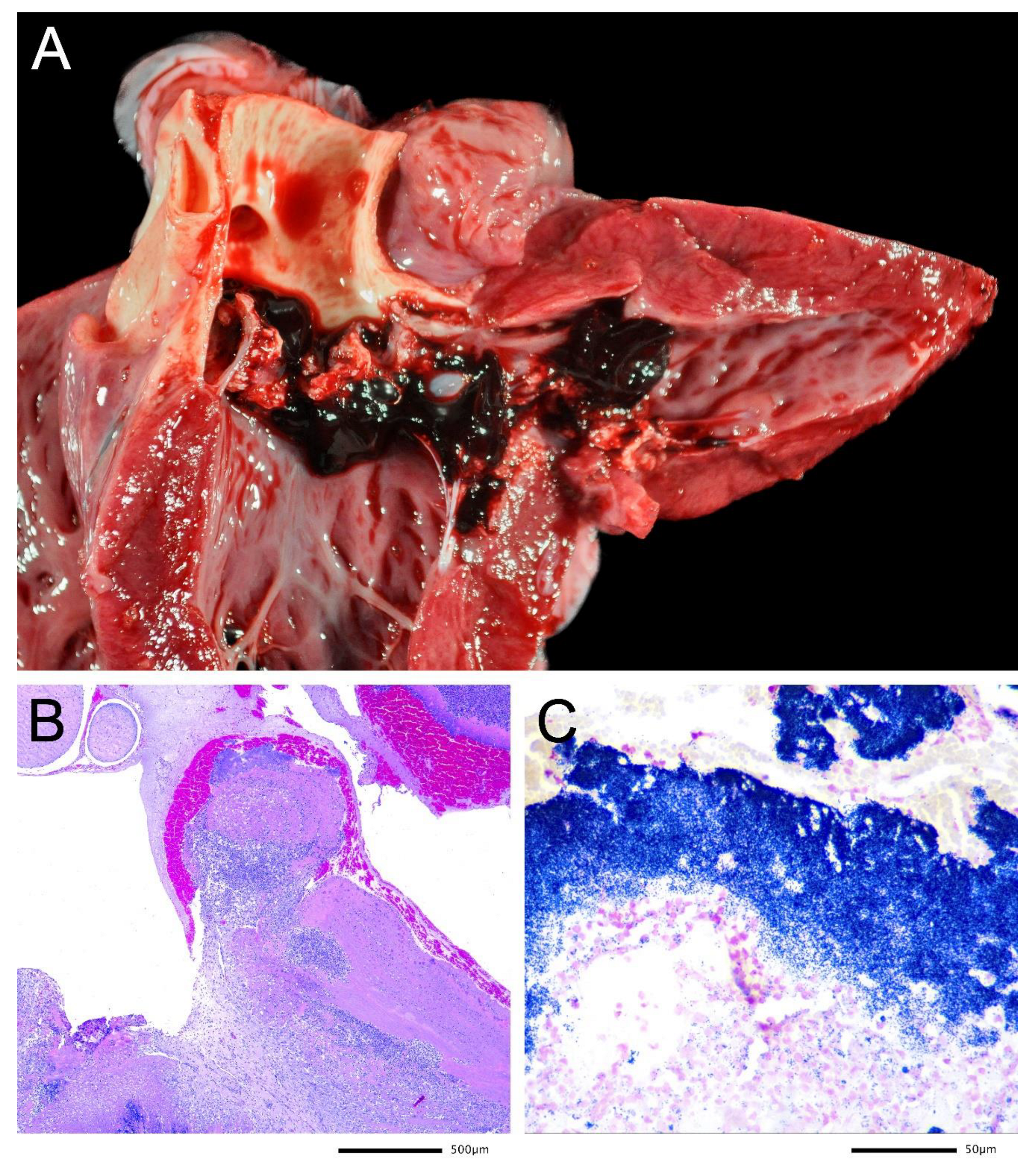
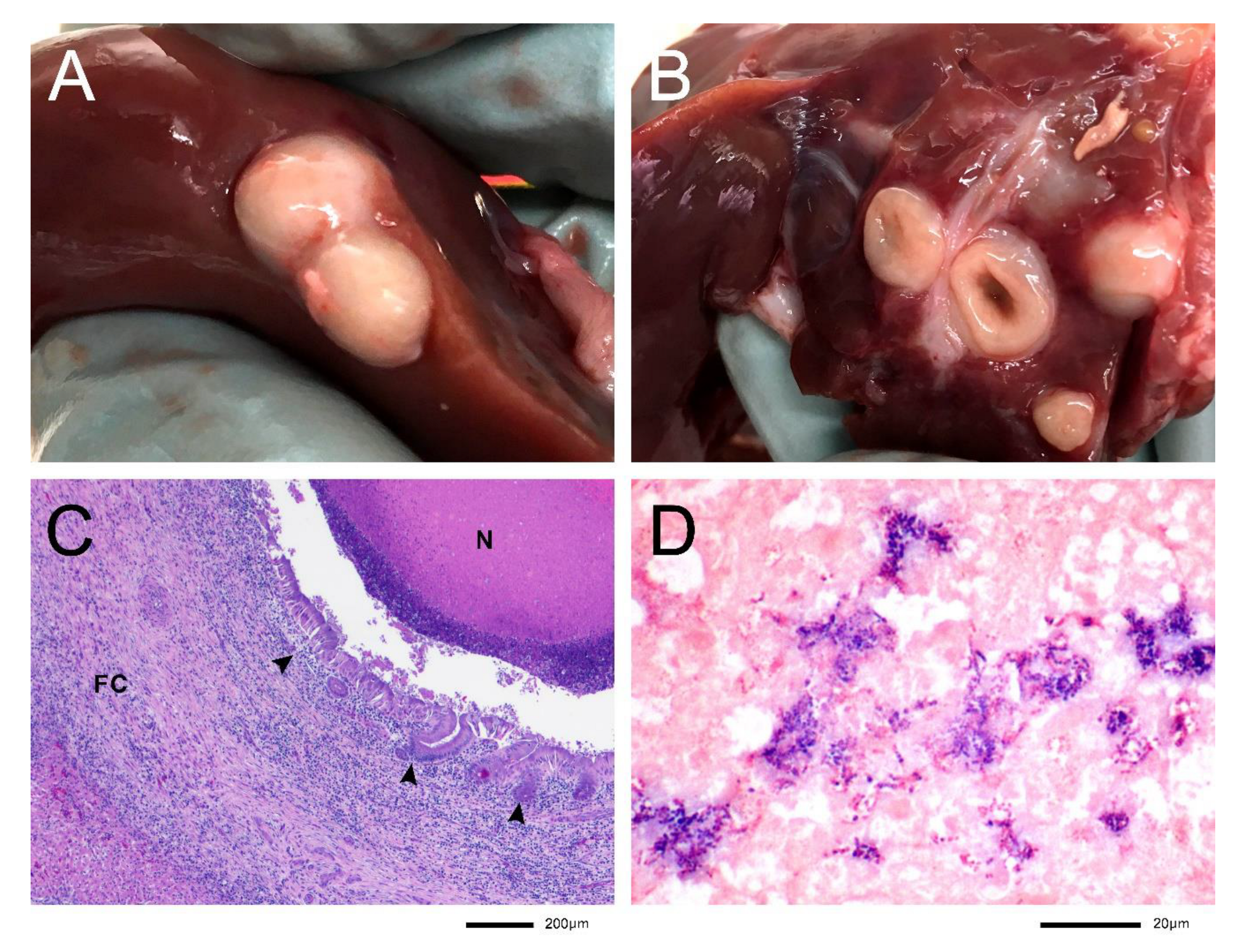
3.3. Lesions Associated with S. ruminantium Isolation

The Pyrenean chamois from which the CCGMV933 strain was isolated was found with severe depression and poor body condition and died during transportation to the Faculty of Veterinary Medicine of the Universitat Autònoma de Barcelona. At the post-mortem and microscopic examinations, the main findings were nonsuppurative encephalitis consistent with a viral infection, mild to moderate and chronic suppurative bacterial cranioventral bronchopneumonia, and acute multifocal suppurative hepatitis. S. ruminantium was isolated from the lung, along with Pasteurella multocida. Pestivirus infection (Border disease virus) was confirmed through PCR in spleen and brain tissues following previously published methods [15].
The Pyrenean chamois of the CCGMV935 strain was found dead with poor body condition and mild alopecic and hyperkeratotic lesions around the coronary band of the hooves in all four legs. At necropsy, irregular yellow vegetations covered with clotted blood were observed on the left atrioventricular valve, aortic, and right atrioventricular (Figure 1A). Histological examinations showed valve leaflets expanded by fibroconnective tissue, fibrin, inflammatory cells, and numerous coccus-shaped Gram-positive bacteria (Figure 1B,C). Other findings were microscopic multifocal/embolic septic fibrin thrombi and necrosis in the brain, lungs, liver, kidney (infarcts and embolic glomerulitis), spleen and bone marrow, and multifocal fibrinoid arteritis consistent with septicemia. In all these tissues, Gram-positive cocci were identified within the lesions. There were lesions of a preexistent suppurative cranioventral bacterial bronchopneumonia, from where numerous colonies of Mannheimia haemolytica were isolated, and caudal verminous pneumonia was also present. Skin lesions were highly consistent with mild chronic lesions of contagious ecthyma (parapoxvirus infection, endemic in Pyrenean chamois).
In the case of the domestic sheep, where S. ruminantium CCGMV928 was isolated from, multiple white to yellowish nodular suppurative lesions of 0.5 to 2 cm in diameter were detected in the liver (Figure 2A,B). These lesions were randomly distributed on the surface and throughout the parenchyma. Adjacent lymph nodes appeared to increase in size but without evident macroscopic lesions. Additionally, there were consolidation and reddening of cranio-ventral lung lobes consistent with suppurative bronchopneumonia and multifocal areas of grey to whitish discoloration and consolidation in caudo-dorsal lobes consistent with verminous pneumonia. At histology, multiple extensive abscesses were detected, distorting the hepatic parenchymal architecture. These were characterized by a large necrotic core with abundant cellular debris and eosinophilic amorphous material with multifocal areas of mineralization (Figure 2C). Numerous Gram-positive cocci were observed mixed with the necrotic material (Figure 2D). The periphery of this core was lined by numerous viable and degenerated neutrophils, surrounded by a thick band of fibroconnective tissue (capsule), highly infiltrated by lymphocytes, plasma cells, histiocytes with lesser neutrophils, and scattered eosinophils. Occasionally, remnants of biliary epithelium could be seen surrounding these abscesses (Figure 2B). The vast majority of the portal spaces were markedly expanded by abundant lymphocytes, plasma cells, some histiocytes, and numerous eosinophils.

4. Discussion

The presence and associated pathology of the new pathogen S. ruminantium, previously classified as S. suis serovar 33, has been assessed for the first time in wildlife. The diverse ERIC-PCR profiles and biochemical characteristics among strains isolated in Pyrenean chamois in different years suggest a high S. ruminantium diversity in NE Spain.
Wildlife ecosystems, such as the alpine mountains where the Pyrenean chamois live, could be a reservoir of resistance genes as well as resistant microorganisms [16,17]. In order to monitor the health of an ecosystem in the face of man-enhanced hazards, wildlife inhabiting these ecosystems has the potential to serve as sentinels [18]. Despite the major concern that AMR represents for human health and the environment, knowledge of the bacteria associated with disease and their AMR profiles is relatively sparse in wildlife, and considerable knowledge gaps remain [9,19,20]. The S. ruminantium isolated from domestic sheep was the only strain resistant to tetracycline (grew in the presence of concentrations of up to 8 μg/mL). Even though it was not possible to obtain information on the use of tetracyclines in the farm of origin, this antimicrobial belongs to the class of veterinary antimicrobials with the largest amount of sales, expressed in mg/PCU, in the UE in the 2005–2018 period (30.7% in 2018) [21]. Consequently, the presence of tetracycline-resistant strains is common in food-producing animals [22,23]. Moreover, within the streptococci of bovine or ovine origin, tetracycline was reported as the antibiotic with the highest prevalence of resistance [24].
Due to limited bibliography, little is known about AMR in S. ruminantium. Okura et al. (2019) found genes that can confer resistance to different antimicrobials in 13 S. ruminantium isolates, including aminoglycosides, macrolides, phenicols, streptomycin, streptothricin, and tetracyclines, as well as high MIC values for chloramphenicol, erythromycin, kanamycin, streptomycin, and tetracycline in some of their tested strains. In agreement with our results, Gottschalk et al. reported that S. ruminantium strains were susceptible to penicillin, ampicillin, ceftiofur, enrofloxacin, trimethoprim-sulfamethoxazole, and florfenicol but highly resistant to chlortetracycline and oxytetracycline [6]. Thus, the possible role of S. ruminantium as a reservoir of AMR genes for other streptococci should be studied.
AMR profiles of bacteria from wild mammals are influenced by the proximity to human activities, being the use of antimicrobials in agriculture is one of the main sources of AMR [25]. Pyrenean chamois may be a good indicator of the environmental contamination of alpine ecosystems with resistance genes or microorganisms, as demonstrated for respiratory pathogens such as Pasteurella multocida or Mannheimia haemolytica [17]. In the present study, due to the low number of Pyrenean chamois strains, this species’ suitability for studying resistances in S. ruminantium cannot be stated, and further research on this bacterium is needed to confirm these observations.
Overall, lesions caused by S. ruminantium reported in the literature are similar to those caused by S. suis. However, ruminants did not present polyserositis, which is a typical clinical sign in S. suis infection in piglets [3,26,27]. The lesions observed in the Pyrenean chamois in our study, both pneumonic and heart lesions, are consistent with previously described lesions for S. ruminantium in livestock [5,6]. Additionally, the liver abscesses produced by the CCGMV928 strain in sheep coincide with the described pathology in cattle [5]. The biliary epithelium surrounding the abscesses suggests an ascendant infection as a plausible origin in this sheep case. Unfortunately, it has not been possible to obtain additional data on the incidence of these lesions from the farm of origin. This particular case was submitted to the slaughterhouse support service (SESC) as a suspected tuberculosis case. Still, liver abscesses are a common finding rarely submitted for further investigations. Thus, it is probable that S. ruminantium cases are underreported. In the Pyrenean chamois, the origins of the S. ruminantium infections were not clear. The most chronic lesion for the CCGMV935 animal was the valve vegetation, evidenced by a severe proliferation of fibrous tissue. Despite the severity of the lesions from which S. ruminantium was isolated in these cases, S. ruminantium does not seem to be a pathogen with high clinical prevalence, according to the low number of global reports in livestock. However, misidentification is likely contributing to this low reporting. The epidemiological information recovered in the present study suggests that S. ruminantium may be the cause of secondary infections produced by a weakened immune system by other pathogens or stressors. As a result, it may be considered an opportunistic pathogen. In this sense, three Pyrenean chamois had significant viral (pestivirus or parapoxvirus infection) and/or bacterial concomitant infections. Pestivirus in chamois is commonly a chronic infection that has particularly been associated with immunosuppression and opportunistic secondary infections [28]. On the other hand, Pasteurellaceae are primary agents for pneumonia and were isolated in all pneumonic lesions from which S. ruminantium was also isolated. Altogether, it suggests that S. ruminantium may also be an opportunistic pathogen for chamois. In the domestic sheep case, despite no parasitic infections being recorded by the slaughterhouse veterinary authorities, numerous eosinophils were observed in the portal spaces, suggesting that a parasitic component was also present.
In the case of the closely related S. suis, higher susceptibility to infection has been demonstrated in piglets previously infected with porcine reproductive and respiratory syndrome virus [29,30,31], swine influenza virus [32], or porcine circovirus type 2 [33], but also in piglets inoculated with S. suis and pseudorabies virus [34].
The proper identification of S. ruminantium is key to improving this pathogen’s scientific knowledge. Our results indicate that phenotypic characterization is insufficient to distinguish S. ruminantium from S. suis, and it could lead to a misidentification problem. In our opinion, the best approach for the identification of S. ruminantium is the specific PCR described by Okura et al. [5], which we recommend as a first step for the identification of Streptococcus-like isolates from ruminants. Isolates that are negative for this PCR should also be tested using the recN-PCR for S. suis as suggested by Gottschalk et al. [6] since there are other species described that can lead to a misidentification, as is the case of S. rupicapreae in chamois, negative for both PCR (data not shown). The identification of S. ruminantium using VITEK-2 is not recommended due to the high heterogeneity shown by the strains of this study. The 16S rRNA gene sequencing is also useful for identifying isolates negative to S. ruminantium or S. suis PCRs. Still, it is necessary to keep databases up to date because new species are continuously described, as was the case of S. ruminantium.
In the present study, S. ruminantium was isolated from pneumonic and heart lesions in Pyrenean chamois for the first time and from a liver lesion in sheep, which was previously reported. Identification of S. ruminantium was facilitated due to the use of a specific PCR, useful for the correct identification of this bacterium. However, phenotypic/biochemical profiles led to the misidentification of the isolates. AMR of this pathogen is an issue that should be further studied when more strains are available, both from clinical and non-clinical isolates. In our case, the isolates from wildlife showed a low antimicrobial resistance. However, it is necessary to establish veterinary-specific breakpoints for antimicrobials and improve testing methods since the information available is not fully applicable, and results can lead to controversy [35]. To conclude, more research, such as the sequencing of clinical isolates and isolates recovered from healthy animals, is needed to further assess the host range and pathogenic potential of S. ruminantium in livestock and wildlife.

Supplementary Materials

The following are available online at https://www.mdpi.com/article/10.3390/microbiolres13010008/s1, Table S1: 16S rRNA query coverage and percent identity at NCBI for S. ruminantium strains. Table S2: Biochemical characterization by Vitek 2 GP (Biomérieux) for Gram-positive cocci of S. ruminantium isolates from domestic and wild ruminants in Spain and reference S. suis serovar 2 strain P1/7. Figure S1: ERIC-PCR of the Pyrenean chamois isolates. Columns 1 and 7, loading buffer. 2 strain RP15178-A2, 3 strain RP16030-M1, 4 strain CCGMV933, 5 strain CCGMV935, 6 negative control (water).

Author Contributions

C.N.-I., V.A., and M.L.A. designed the study, performed the bacterial isolation and identification, and drafted the manuscript. R.V., X.F.A., and E.V. collected field samples and participated in the pathological diagnosis. E.P. and R.V. performed the histopathology. All authors have read and agreed to the published version of the manuscript.

Funding

This project has received funding from the European Union Horizon 2020 research and innovation programme under grant agreement No 727966. Carlos Neila-Ibáñez was funded by this project. Also, financial support came from Servei Veterinari de Bacteriologia i Micologia of the Universitat Autònoma de Barcelona.

Institutional Review Board Statement

Ethical approval was not sought or required since all strains were recovered from animals that died in hunting, natural death, or slaughterhouse, and diagnostic tests were performed as part of standard clinical investigations.

Data Availability Statement

The data that support the findings of this study may be available on request from the corresponding author.

Acknowledgments

The General (Passive) Surveillance Program for game species is funded by the Sub-direcció General d’Activitats Cinegètiques i Pesca Continental of the Ministry of Climate Action, Food and Rural Agenda of the Government of Catalonia. We are grateful to the gamekeepers and hunters from the Freser-Setcases National Game Reserve. The liver sheep where the strain was isolated was provided by SESC, the slaughterhouse support network (www.cresa.cat/blogs/sesc, accessed on 01 December 2022) an IRTA-CReSA service funded by the Public Health Agency of Catalonia (ASPCAT, Agència de Salut Pública de Catalunya), Health Department, Government of Catalonia. The authors are also grateful to the CERCA program from the Generalitat de Catalunya.

Conflicts of Interest

The authors declare no conflict of interest.

References

  1. Parte, A.C.; Carbasse, J.S.; Meier-Kolthoff, J.P.; Reimer, L.C.; Göker, M. List of Prokaryotic names with Standing in Nomenclature (LPSN) moves to the DSMZ. Int. J. Syst. Evol. Microbiol. 2020, 70, 5607–5612. [Google Scholar] [CrossRef] [PubMed]
  2. Krzyściak, W.; Pluskwa, K.K.; Jurczak, A.; Kościelniak, D. The pathogenicity of the Streptococcus genus. Eur. J. Clin. Microbiol. 2013, 32, 1361–1376. [Google Scholar] [CrossRef] [Green Version]
  3. Gottschalk, M.; Segura, M. Streptococci. In Diseases of Swine, 11th ed.; Zimmerman, J.J., Karriker, L.A., Ramirez, A., Schwartz, K.J., Stevenson, G.W., Zhang, J., Eds.; John Wiley & Sons: Hoboken, NJ, USA, 2019; pp. 934–950. [Google Scholar]
  4. Tohya, M.; Arai, S.; Tomida, J.; Watanabe, T.; Kawamura, Y.; Katsumi, M.; Ushimizu, M.; Ishida-Kuroki, K.; Yoshizumi, M.; Uzawa, Y.; et al. Defining the taxonomic status of Streptococcus suis serotype 33: The proposal for Streptococcus ruminantium sp. nov. Int. J. Syst. Evol. Microbiol. 2017, 67, 3660–3665. [Google Scholar] [CrossRef] [PubMed]
  5. Okura, M.; Maruyama, F.; Ota, A.; Tanaka, T.; Matoba, Y.; Osawa, A.; Sadaat, S.M.; Osaki, M.; Toyoda, A.; Ogura, Y.; et al. Genotypic diversity of Streptococcus suis and the S. suis-like bacterium Streptococcus ruminantium in ruminants. Veter. Res. 2019, 50, 94. [Google Scholar] [CrossRef] [PubMed] [Green Version]
  6. Gottschalk, M.; Lacouture, S.; Fecteau, G.; DesRochers, A.; Boa, A.; Saab, M.E.; Okura, M. Canada: Isolation of Streptococcus ruminantium (Streptococcus suis-like) from diseased ruminants in Canada. Can. Vet. J. 2020, 61, 473–475. [Google Scholar]
  7. Zhu, Y.; Dong, W.; Ma, J.; Zhang, Y.; Zhong, X.; Pan, Z.; Liu, G.; Wu, Z.; Yao, H. Comparative genetic analyses provide clues about capsule switching in Streptococcus suis 2 strains with different virulence levels and genetic backgrounds. Microbiol. Res. 2021, 250, 126814. [Google Scholar] [CrossRef] [PubMed]
  8. Espunyes, J.; Cabezón, O.; Dias-Alves, A.; Miralles, P.; Ayats, T.; Cerdà-Cuéllar, M. Assessing the role of livestock and sympatric wild ruminants in spreading antimicrobial resistant Campylobacter and Salmonella in alpine ecosystems. BMC Veter. Res. 2021, 17, 79. [Google Scholar] [CrossRef]
  9. Ramey, A.M.; Ahlstrom, C.A. Antibiotic Resistant Bacteria in Wildlife: Perspectives on Trends, Acquisition and Dissemination, Data Gaps, and Future Directions. J. Wildl. Dis. 2020, 56, 1–15. [Google Scholar] [CrossRef]
  10. Ishida, S.; Tien, L.H.T.; Osawa, R.; Tohya, M.; Nomoto, R.; Kawamura, Y.; Takahashi, T.; Kikuchi, N.; Kikuchi, K.; Sekizaki, T. Development of an appropriate PCR system for the reclassification of Streptococcus suis. J. Microbiol. Methods 2014, 107, 66–70. [Google Scholar] [CrossRef]
  11. Versalovic, J.; Koeuth, T.; Lupski, R. Distribution of repetitive DNA sequences in eubacteria and application to finerpriting of bacterial enomes. Nucleic Acids Res. 1991, 19, 6823–6831. [Google Scholar] [CrossRef]
  12. Lane, D.J. 16S/23S rRNA Sequencing. In Nucleic Acid Techniques in Bacterial Systematics, 1st ed.; Stackebrandt, E., Goodfellow, M., Eds.; John Wiley & Sons: New York, NY, USA, 1991; pp. 115–175. [Google Scholar]
  13. CLSI. Performance Standards for Antimicrobial Disk and Dilution Susceptibility Tests for Bacteria Isolated from Animals, 4th ed.; Clinical and Laboratory Standards Institute: Malvern, PA, USA, 2018. [Google Scholar]
  14. CLSI. Performance Standards for Antimicrobial Disk and Dilution Susceptibility Tests for Bacteria Isolated from Animals, 5th ed.; Clinical and Laboratory Standards Institute: Malvern, PA, USA, 2020. [Google Scholar]
  15. Vilček, Š.; Herring, A.J.; Herring, J.A.; Nettleton, P.F.; Lowings, J.P.; Paton, D.J. Pestiviruses isolated from pigs, cattle and sheep can be allocated into at least three genogroups using polymerase chain reaction and restriction endonuclease analysis. Arch. Virol. 1994, 136, 309–323. [Google Scholar] [CrossRef] [PubMed]
  16. White, A.; Hughes, J.M. Critical Importance of a One Health Approach to Antimicrobial Resistance. EcoHealth 2019, 16, 404–409. [Google Scholar] [CrossRef] [Green Version]
  17. Torres-Blas, I.; Aguilar, X.F.; Cabezón, O.; Aragon, V.; Migura-García, L. Antimicrobial Resistance in Pasteurellaceae Isolates from Pyrenean Chamois (Rupicapra pyrenaica) and Domestic Sheep in an Alpine Ecosystem. Animals 2021, 11, 1686. [Google Scholar] [CrossRef] [PubMed]
  18. Conrad, P.A.; Meek, L.A.; Dumit, J. Operationalizing a One Health approach to global health challenges. Comp. Immunol. Microbiol. Infect. Dis. 2013, 36, 211–216. [Google Scholar] [CrossRef] [PubMed]
  19. Vittecoq, M.; Godreuil, S.; Prugnolle, F.; Durand, P.; Brazier, L.; Renaud, N.; Arnal, A.; Aberkane, S.; Jean-Pierre, H.; Gauthier-Clerc, M.; et al. Antimicrobial resistance in wildlife. J. Appl. Ecol. 2016, 53, 519–529. [Google Scholar] [CrossRef] [Green Version]
  20. Mühldorfer, K.; Rau, J.; Fawzy, A.; Heydel, C.; Glaeser, S.P.; Van Der Linden, M.; Kutzer, P.; Knauf-Witzens, T.; Hanczaruk, M.; Eckert, A.S.; et al. Streptococcus castoreus, an uncommon group A Streptococcus in beavers. Antonie van Leeuwenhoek. Int. J. Gen. Mol. Microbiol. 2019, 112, 1663–1673. [Google Scholar] [CrossRef]
  21. EMA. Sales of Veterinary Antimicrobial Agents in 31 European Countries in 2018—EMA/24309/2020. 2020. Available online: https://www.ema.europa.eu/en/documents/report/sales-veterinary-antimicrobial-agents-31-european-countries-2018-trends-2010-2018-tenth-esvac-report_en.pdf (accessed on 12 January 2022).
  22. Liegeon, G.; Delory, T.; Picardeau, M. Antibiotic susceptibilities of livestock isolates of leptospira. Int. J. Antimicrob. Agents 2018, 51, 693–699. [Google Scholar] [CrossRef]
  23. Bourély, C.; Cazeau, G.; Jouy, E.; Haenni, M.; Madec, J.-Y.; Jarrige, N.; Leblond, A.; Gay, E. Antimicrobial resistance of Pasteurella multocida isolated from diseased food-producing animals and pets. Veter. Microbiol. 2019, 235, 280–284. [Google Scholar] [CrossRef]
  24. Haenni, M.; Lupo, A.; Madec, J.-Y. Antimicrobial Resistance in Streptococcus spp. In Antimicrobial Resistance in Bacteria from Livestock and Companion Animals, 1st ed.; Schwarz, S., Cavaco, L.M., Jianzhong, S., Aarestrup, F.M., Eds.; American Society of Microbiology: Washington, DC, USA, 2018. [Google Scholar] [CrossRef]
  25. Allen, H.K.; Donato, J.; Wang, H.H.; Cloud-Hansen, K.A.; Davies, J.; Handelsman, J. Call of the Wild: Antibiotic resistance genes in natural environments. Nat. Rev. Microbiol. 2010, 8, 251–259. [Google Scholar] [CrossRef]
  26. Staats, J.J.; Feder, I.; Okwumabua, O.; Chengappa, M.M. Streptococcus Suis: Past and Present. Vet. Res. Commun. 1997, 21, 381–407. [Google Scholar] [CrossRef]
  27. Okwumabua, O.; Peterson, H.; Hsu, H.-M.; Bochsler, P.; Behr, M. Isolation and partial characterization of Streptococcus suis from clinical cases in cattle. J. Veter. Diagn. Investig. 2017, 29, 160–168. [Google Scholar] [CrossRef] [Green Version]
  28. Marco, I.; Cabezón, O.; Velarde, R.; Fernández-Sirera, L.; Colom-Cadena, A.; Serrano, E.; Rosell, R.; Casas-Díaz, E.; Lavín, S.; Ferron, E.S. The two sides of border disease in Pyrenean chamois (Rupicapra pyrenaica): Silent persistence and population collapse. Anim. Health Res. Rev. 2015, 16, 70–77. [Google Scholar] [CrossRef]
  29. Galina, L.; Pijoan, C.; Sitjar, M.; Christianson, W.; Rossow, K.; Collins, J. Interaction between Streptococcus suis serotype 2 and porcine reproductive and respiratory syndrome virus in specific pathogen-free piglets. Veter. Rec. 1994, 134, 60–64. [Google Scholar] [CrossRef]
  30. Thanawongnuwech, R.; Brown, G.B.; Halbur, P.G.; Roth, J.A.; Royer, R.L.; Thacker, B.J. Pathogenesis of Porcine Reproductive and Respiratory Syndrome Virus-induced Increase in Susceptibility to Streptococcus suis Infection. Veter. Pathol. 2000, 37, 143–152. [Google Scholar] [CrossRef] [PubMed] [Green Version]
  31. Feng, W.-H.; Laster, S.M.; Tompkins, M.; Brown, T.; Xu, J.-S.; Altier, C.; Gomez, W.; Benfield, D.; McCaw, M.B. In Utero Infection by Porcine Reproductive and Respiratory Syndrome Virus Is Sufficient to Increase Susceptibility of Piglets to Challenge by Streptococcus suis Type II. J. Virol. 2001, 75, 4889–4895. [Google Scholar] [CrossRef] [Green Version]
  32. Lin, X.; Huang, C.; Shi, J.; Wang, R.; Sun, X.; Liu, X.; Zhao, L.; Jin, M. Investigation of Pathogenesis of H1N1 Influenza Virus and Swine Streptococcus suis Serotype 2 Co-Infection in Pigs by Microarray Analysis. PLoS ONE 2015, 10, e0124086. [Google Scholar] [CrossRef] [Green Version]
  33. Wang, Q.; Zhou, H.; Hao, Q.; Li, M.; Liu, J.; Fan, H. Coinfection with porcine circovirus type 2 and Streptococcus suis serotype 2 enhances pathogenicity by dysregulation of the immune responses in piglets. Veter. Microbiol. 2020, 243, 108653. [Google Scholar] [CrossRef] [PubMed]
  34. Iglesias, J.G.; Trujano, M.; Xu, J. Inoculation of pigs with Streptococcus suis type 2 alone or in combination with pseudorabies virus. Am. J. Veter. Res. 1992, 53, 364–367. [Google Scholar]
  35. Sanchez Armengol, E.; Harmanci, M.; Laffleur, F. Current strategies to determine antifungal and antimicrobial activity of natural compounds. Microbiol. Res. 2021, 252, 126867. [Google Scholar] [CrossRef] [PubMed]
Figure 1. Pyrenean chamois heart from which Streptococcus ruminantium was isolated, strain CCGMV935. (A) Vegetative endocarditis of the left atrioventricular valve in a chamois (Rupicapra pyrenaica) caused by S. ruminantium. (B) H&E staining shows that the valve leaflet is expanded by fibrin, inflammatory cells, and numerous bacterial colonies. (C) Gram staining showing coccus-shaped Gram-positive (dark blue) bacteria in the vegetation.
Microbiolres 13 00008 g001
Figure 2. Sheep liver from which Streptococcus ruminantium was isolated, strain CCGMV928. (A) White nodular lesions on a liver surface measuring 0.5 to 2 cm in diameter. (B) Detail of a sectioned intraparenchymal nodule. (C) H&E staining showing a necrotic core (N) surrounded by a suppurative inflammatory infiltrate and a fibrous capsule (FC). Notice the remains of the biliary epithelium (arrowheads). (D) Gram staining. Showing multiple coccus-shaped Gram-positive bacteria within the necrotic core.
Microbiolres 13 00008 g002
Table 1. Main characteristics of the Streptococcus ruminantium isolates included in this study.
IsolateOrgan of IsolationHost SpeciesAge and SexCause of DeathMain Necropsy FindingsPlaceDate
RP15178-A2Lung lesionChamois
(Rupicapra pyrenaica)
4 years, maleSeasonal controlled huntingInfectious queratoconjunctivitis, diarrhea, and bacterial cranioventral suppurative bronchopneumoniaFreser-Setcases National Game Reserve (Queralbs, Catalonia, Spain)December 2015
RP16030-M1Lung lesionChamois
(Rupicapra pyrenaica)
4 years, femaleSeasonal controlled huntingOld fibrous pleural adhesions from previous pneumonia and cysticercosis in peritoneum and diaphragmFreser-Setcases National Game Reserve (Queralbs, Catalonia, Spain) May 2016
CCGMV933Lung lesionChamois
(Rupicapra pyrenaica)
14 months, maleSevere pestivirus infection *Cachexia, bacterial cranioventral suppurative bronchopneumonia, multifocal acute suppurative hepatitis, and nonsuppurative viral meningoencephalitisAigüestortes i Estany de Sant Maurici National Park (La Vall de Boí, Catalonia, Spain)August 2020
CCGMV935 Heart valves vegetationChamois
(Rupicapra pyrenaica)
5 months, maleBacterial septicemia *Bacterial suppurative endocarditis, meningoencephalitis, embolic pneumonia, hepatitis, nephritis, splenitis, lymphadenitis, myelitis, contagious ecthyma, verminous pneumonia, and bronchopneumoniaFreser-Setcases National Game Reserve (Queralbs, Catalonia, Spain)October 2020
CCGMV928 Liver abscessDomestic sheep
(Ovis aries)
3 months, maleSlaughterhouseLiver abscesses and cranioventral suppurative bronchopneumoniaMunicipal slaughterhouse (Farm located in Catalonia, Spain)September 2019
* Probable cause of death.
Table 2. Primers used for bacteria identification.
PCRPrimersPrimer sequenceReference
16S358F
907R
CTACGGGAGGCAGCAGT
CCGTCWATTCMTTTGAGTTT
Amplification and sequencing of the V2-V5 region of the 16S rRNA gene [12].
recN-S. suisSSrecN-F
SSrecN-R
CTACAAACAGCTCTCTTCT
ACAACAGCCAATTCATGGCGTGATT
PCR based on the S. suis recombination/repair protein (recN) [10].
S. ruminantiumForward
Reverse
GCAAGTGGAACGCAACTTTTCA
CTATGTATCGTTGCCTTGGTAG
PCR designed to discriminate S. ruminantium from S. suis. [5].
ERICERIC-1F
ERIC-2R
ATGTAAGCTCCTGGGGATTCAC
AAGTAAGTGACTGGGGTGAGCG
Enterobacterial repetitive intergenic consensus PCR [11].
Table 3. Minimal Inhibitory Concentrations (μg/mL) obtained using Sensititre Vet Bovine BOPO7F Plate.
AntimicrobialRP15178-A2RP16030-M1CCGMV933CCGMV935CCGMV928
Ampicillin≤0.25≤0.25≤0.25≤0.25≤0.25
Ceftiofur≤0.25≤0.25≤0.25≤0.25≤0.25
Clindamycin≤0.25≤0.25≤0.25≤0.25≤0.25
Danofloxacin0.50 *0.50 *1 *≤0.120.25
Enrofloxacin0.500.501≤0.120.25
Florfenicol21211
Gamithromycin≤1≤1≤1≤1≤1
Gentamicin44448
Neomycin168161616
Penicillin≤0.12≤0.12≤0.12≤0.12≤0.12
Spectinomycin1616161616
Sulfadimethoxine≤256≤256≤256≤256≤256
Tetracycline111≤0.5>8 *
Tiamulin4481616
Tildipirosin16881616
Tilmicosin888168
Trimethoprim-sulfamethoxazole≤2/38≤2/38≤2/38≤2/38≤2/38
Tulathromycin≤8≤8≤8≤8≤8
Tylosin tartrate1≤0.5≤0.5≤0.5≤0.5
* Resistant.
Publisher’s Note: MDPI stays neutral with regard to jurisdictional claims in published maps and institutional affiliations.

