Article

The Morphological and Functional Properties of Lactiplantibacillus plantarum B411 Subjected to Acid, Bile and Heat Multi-Stress Adaptation Process and Subsequent Long-Term Freezing

by
Thobeka N. Dlangalala
,
Moloko G. Mathipa-Mdakane
and
Mapitsi S. Thantsha
*
Department of Biochemistry, Genetics and Microbiology, University of Pretoria, Pretoria 0002, South Africa
*
Author to whom correspondence should be addressed.
Microbiol. Res. 2022, 13(4), 909-927; https://doi.org/10.3390/microbiolres13040064
Submission received: 25 October 2022 / Revised: 28 October 2022 / Accepted: 2 November 2022 / Published: 7 November 2022

Abstract

:
The preadaptation of probiotics to sub-lethal levels of multiple stress factors boosts their survival and stability. However, little is known about how long-term cold storage affects the properties of such preadapted probiotics. This study examined the impact of long-term freezing on structural and functional properties of multi-stress (acid, bile and heat) adapted Lactiplantibacillus plantarum B411. Cell morphology was investigated using scanning electron microscopy, and then their selected functional (bile salt hydrolase (BSH) activity, surface hydrophobicity, auto-aggregative and antimicrobial) properties were evaluated. Furthermore, the survival of L. plantarum B411 cells in yoghurt and juices during storage and under simulated gastrointestinal (GIT) conditions was evaluated. Long-term freezing negatively affected the morphology, auto-aggregation ability, BSH and antimicrobial activities of L. plantarum B411. The viability of freshly adapted and old adapted L. plantarum B411 cells in foods was similar. Under simulated GIT conditions, the viability of the stress adapted cells from the freezer diminished more than that of freshly adapted cells. Prolonged freezing compromised some functional properties of stress adapted cells and their stability under simulated GIT conditions. Care should thus be taken to ensure that a method used to preserve stress adapted cells does not cause them to lose beneficial properties, nor revert to their pre-adaptation status.

1. Introduction

Lactiplantibacillus plantarum, formerly known as Lactobacillus plantarum [1], is a lactic acid bacterium used as a starter culture for the production of many fermented foods, as well as a probiotic [2]. It is recommended that probiotics maintain viability levels at or above 107 CFU/mL or grams at the time of consumption [3] in order for them to confer beneficial effects. However, the viability and stability of probiotic strains are threatened by stressors such as, among others, low and alkaline pH, high and low temperatures, the concentration of chemicals, osmotic pressure, oxidative conditions and starvation, encountered during production (culture handling), processing, storage and transition through the GIT [2,4,5,6,7,8,9]. These stress factors can significantly affect growth, metabolism, biological and the physiological functions of probiotics, which consequently alter their functional properties [7,10]. It is thus crucial then, that viability and functional properties are sufficiently conserved within the product following these phases [2,8,10,11].
Various methods, such as screening for resistant strains, microencapsulation, the addition of extra nutrients, prebiotics and protectants, the manipulation of the physiological state or the exploitation of cellular stress response, have been explored for the production of robust probiotic strains [2,4]. The manipulation of the physiological state of the probiotics is achieved by exploiting stress responses present in probiotics, through a stress pre-treatment process [12]. This process allows stress responses to be elicited by exposing the microorganisms to sub-lethal doses of the stress factor so that they are able to overcome the subsequent lethal stresses encountered during manufacture and within the GIT [5,13,14,15]. Exposure to one type of stress can offer resilience to a different type of stress downstream, a mechanism known as cross-protection [9,14]. Much of the early research into stress adaptation focused on a single stress factor at a time [16]. Our previous study reported that the adaptation of probiotics to multiple stresses can offer superior protection [17]. However, following stress adaptation, strains must be reassessed for functional properties as this process can negatively or positively impact probiotic traits [18]. Kulkarni et al. [14] investigated the robustness of lyophilized heat adapted Lactobacillus acidophilus during storage at 4 °C for 15 days. There is limited knowledge on how multi-stress adapted probiotics are affected by extended periods of cold storage. This is an important aspect to probe into as freezing and freeze-drying are methods commonly used by the food industry for storage and the preservation of probiotics [14]. These methods expose probiotics to cold stress known to decrease growth and the proliferation of bacteria through changing membrane fluidity, lowering enzyme activities and the stability of the RNA structure [6].
Probiotics must be formulated in a way that will be conducive for their oral consumption [19]. They are incorporated into a number of functional foods (mostly dairy based) [20]. The choice of food carrier has been implicated in enhancing the GIT survival of probiotics. Fermented milks were the first food to which probiotics were added, and they still enjoy commercial success as the leading probiotic food product, accounting for 78% of probiotic sales worldwide [21,22]. However, these products do not suit the dietary habits of vegans or vegetarians, moreover, a proportion of the global population suffers from lactose intolerance [21]. This has resulted in the use of alternate delivery vehicles for probiotics, with fruit and vegetable juices being example of such alternatives. Understanding the relationship between viability and a food matrix can go a long way towards the development of effective probiotic products that can withstand stressful GIT conditions [23]. This is especially so since the commercial success of these products depends on their viability and ability to retain functionality [24]. Taking all these into consideration, the aim of this study was to determine the effect of long-term freezer storage on the morphology and functional properties of multi-stress adapted L. plantarum B411, and its subsequent survival in food matrices during storage, as well as under simulated GIT conditions.

2. Materials and Methods

2.1. Bacterial Strains and Culture Conditions

Lactiplantibacillus plantarum B411 glycerol stock culture (MRS broth with 50% (v/v) glycerol) from the Probiotics Research Group, Department of Biochemistry, Genetics and Microbiology, University of Pretoria, Pretoria, South Africa, was used as the test probiotic strain. Non-adapted L. plantarum cells (50% glycerol stock stored at −20 °C for 24 months), were subcultured twice in de Man Rogosa Sharpe (MRS) broth (Merck, Darmstadt, Germany). They were incubated at 37 °C for 72 h in anaerobic jars containing Anaerocult® A gaspacks with Anaerotest® strip (Merck, Darmstadt, Germany). All anaerobic incubations in all subsequent experiments were performed in this same way. The freshly adapted cells were prepared using a subsample withdrawn from the subcultured non-adapted cells. The old adapted L. plantarum cells were the cells previously adapted to multiple stress factors and then stored in 50% glycerol (v/v) at −20 °C for 24 months. Before each experiment the cultures were normalized to an optical density of 0.2 at 600 nm which equates to approximately 108 CFU/mL. Escherichia coli ATCC 25922 and Staphylococcus aureus subsp. aureus ATCC 33591 obtained as 20% glycerol stock cultures were obtained from the Department of Consumer and Food Sciences, University of Pretoria, were used as the indicator bacteria and test pathogen, respectively. They were revived by growing at 37 °C overnight in Luria Bertani (LB) broth incubated in an orbital shaking incubator, LM- 530R at 100 rpm. They were then plated onto Mannitol salt agar (Merck, Darmstadt, Germany) and MacConkey agar (Merck, Darmstadt, Germany) plates for S. aureus ATCC 33591 and E. coli ATCC 25922, respectively. All the plates were incubated at 37 °C for 48 h. The pathogens were then subcultured twice before their use in experiments.

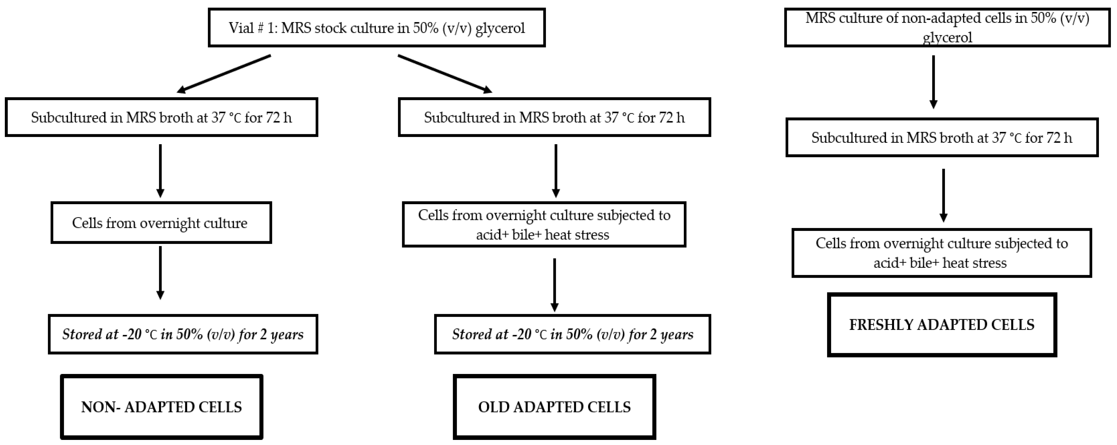
2.2. Acid, Bile and Heat Stress Adaptation of L. plantarum B411

All stress adaptation tests of L. plantarum B411 were carried out according to the methods previously described by Ma et al. [10] and Mathipa and Thantsha [17], with minor modifications. Cells from the overnight culture were harvested by centrifugation at 604× g for 15 min and the pellets were re-suspended in 1 mL ¼ strength Ringer’s solution (Merck, Darmstadt, Germany). Then, 1 mL aliquot of the suspension was added to 9 mL of MRS broth at pH 2 (adjusted using 1 M HCl), and then incubated at 37 °C for 2 h, for acid adaptation. The acid adapted cells were then harvested by centrifuging at 604× g for 15 min and then suspended in 1 mL MRS broth. Subsequently, 100 µL aliquot of these acid adapted cells was set aside while the 900 µL was aseptically transferred to 9 mL of MRS containing 2% bile solution (w/v), for bile stress adaptation. The resultant cell suspension was incubated in a shaking incubator (100 rpm) for 60 min at 37 °C. The acid-bile adapted cells were then harvested, 100 µL aliquot set aside and the 900 µL aseptically transferred to 9 mL of MRS broth and then incubated at 55 °C in a dry bath (AccuBlockTM, Labnet, Edison, NJ, USA) for 120 min, for heat stress adaptation. The acid-bile-heat adapted cells were harvested by centrifugation and then suspended in 1 mL MRS broth. An aliquot (100 µL) of these cells was also set aside while the remaining 900 µL was used in subsequent experiments. All the 100 µL cell aliquots that were collected after the different stress adaptation steps were serially diluted separately up to 10−7 using ¼ strength Ringer’s solution and then each dilution plated onto MRS agar plates in triplicate to enumerate numbers of viable cells. The plates were incubated anaerobically for 72 h at 37 °C.
The L. plantarum B411 cells that were previously subjected to these stress factors (acid, bile and heat), using the same process described, followed by their storage at −20 °C in 50% (v/v) glycerol for 24 months were used as old-adapted cells, while the cells that were not subjected to the stress adaptation process (MRS broth culture in 50% (v/v) glycerol, stored at −20 °C for 24 months) were used as non-adapted cells. Figure 1 is a summarized scheme used for production of the different L. plantarum B411 cells used.

2.3. Tolerance of L. plantarum B411 to Different Stress Factors

2.3.1. Acid Tolerance

The acid tolerance of the non-adapted, freshly adapted, and old adapted L. plantarum B411 cells was carried out using the protocol of Mathipa and Thantsha [17]. L. plantarum B411 were suspended into MRS broth at an optical density of 0.2 at 600 nm (approximately 108 CFU/mL). Then 1 mL of each was separately subcultured into 9 mL of fresh MRS broth adjusted to pH (2, 2.5 and 3) with 1 M HCl and incubated at 37 °C for 3 h. Subsamples (1 mL) were drawn hourly and serially diluted up to 10−7 using ¼ strength Ringer’s solution, and then 100 µL from each dilution was plated onto MRS agar plates in triplicate. The plates were incubated anaerobically in anaerobic jars with Anaerocult® A gaspacks and Anaerotest® strips at 37 °C for 72 h.

2.3.2. Bile Tolerance

The tolerance of L. plantarum B411 cells to bile salts was carried out according to the method of Mathipa and Thantsha [17] with minor modifications. Briefly, 1 mL of non-adapted, freshly adapted, and old adapted L. plantarum B411 cells were was added to separate 9 mL MRS broths containing 0.3, 0.5 and 2% (w/v) bile salts. The cells suspensions were incubated and subsamples drawn hourly for plating as was done in Section 2.3.1.

2.4. Bile Salt Hydrolase Activity

Bile salt hydrolase (BSH) activity was performed using a method described by Sedláčková et al. [25], with minor modifications. Overnight cultures of non-adapted, freshly adapted and old adapted L. plantarum B411 were grown and used to assess BSH activity. Soft agar was prepared as follows: MRS broth (50 g/L), bile salts (0.3% v/w; Sigma, South Africa), bacteriological agar (15 g/L; Merck, Germany) and CaCl2 (0.375 g/L; Sigma- Aldrich, USA). Sterile pipette tips were used to puncture holes into agar. L. plantarum B411 cells (200 µL) were placed into the holes. The plates were then left in the laminar flow for 10 min before they were incubated under anaerobic conditions at 37 °C for 72 h. Plates that did not contain bile salts were used as a negative control. The presence of translucent halos around holes indicated bile salt hydrolase activity. The experiment was performed in triplicate in three independent trials.

2.5. Antimicrobial Activity Assay

The antimicrobial tests were performed using the method of Mohankumar and Murugalatha [26], with modifications. Cell free supernatants (CFS) of the non-adapted, freshly adapted and old adapted L. plantarum B411 cultures were obtained by centrifuging overnight cultures at 14,691× g for 20 min at 4 °C and then filtering the culture medium through a 0.22 µm membrane filter. Lawns of the test pathogens (E. coli ATCC 25922 or S. aureus ATCC 33591) were prepared by spread-plating 100 µL of their overnight cultures adjusted to OD600 = 0.2 onto separate Mueller Hinton agar plates. Subsequently, the back of a sterile 1000 µL pipette tip was used to puncture holes into the agar plates and then l00 µL of CFS of the different L. plantarum B411 cultures were added to the wells. The plates were incubated overnight at 37 °C in an upright position. Zones of inhibition around the wells indicated antimicrobial activity. The experiment was performed in triplicate in three independent trials.

2.6. Scanning Electron Microscopy (SEM)

The preparation of the samples for microscopy was carried out according to a method described by Booyens et al. [27], with minor modifications. L. plantarum B411 (non-adapted, freshly adapted and old adapted) overnight MRS broth cultures were harvested using centrifugation at 604× g for 2 min. The cells were then fixed using 2.5% glutaraldehyde in 0.075 mol−1 phosphate buffer (pH 7.4) for 1 h. They were subsequently washed three times for 15 min in 0.15 mol−1 phosphate buffer solution (PBS) before being dehydrated in a series of graded alcohol concentrations (25%, 50%, 75% and 100% ethanol, for 15 min each wash). Finally, the cells were critically dried for 24 h before being coated with carbon. The resulting cells were viewed using a JSM-840 and JSM-5800 LV scanning electron microscopes.

2.7. Auto-Aggregation

The auto-aggregation assay was carried out according to the method described by Li et al. [28], with slight alterations. Briefly, cells from the overnight non-adapted, freshly adapted and old adapted L. plantarum B411 cultures were harvested using centrifugation (1152× g, 10 min, 4 °C), washed twice in 0.9% saline, and then re-suspended in 10 mL of the same saline. The suspensions were adjusted to an absorbance reading of 0.3 (±0.05) at 600 nm. Each bacterial suspension was then incubated at room temperature and spectrophotometric readings (600 nm) using 1 mL of suspension were taken at time interval 0, 3, 6 h. Auto-aggregation experiments were performed in triplicate in three independent trials. The following equation was used to measure auto-aggregation percentage:
A u t o a g g r e g a t i o n   % = [ 1 A t A 0 ] × 100
At represents absorbance at different time intervals and A0 represents the initial absorbance reading.

2.8. Cell Surface Hydrophobicity

The non-adapted, freshly adapted and old adapted L. plantarum B411 cells were grown in MRS broth for 24 h at 37 °C. They were harvested by centrifugation at 1677× g for 10 min and then washed twice in PBS (pH 7). The L. plantarum cells were suspended into PBS and adjusted to an optical density (OD 500 nm) of 1. Subsequently, 1 mL of the culture was added to 1 mL of chloroform and mixed vigorously by vortexing for 30 s. Following phase separation (30 min) the OD540 of the aqueous phase was taken. Cell surface hydrophobicity assays were performed in triplicate in three independent trials. The following equation was used to determine hydrophobicity percentage:
H y d r o p h o b i c i t y   % = [ A 540   i n i t i a l A 540   a q u e o s   p h a s e A 540   i n i t i a l ] × 100

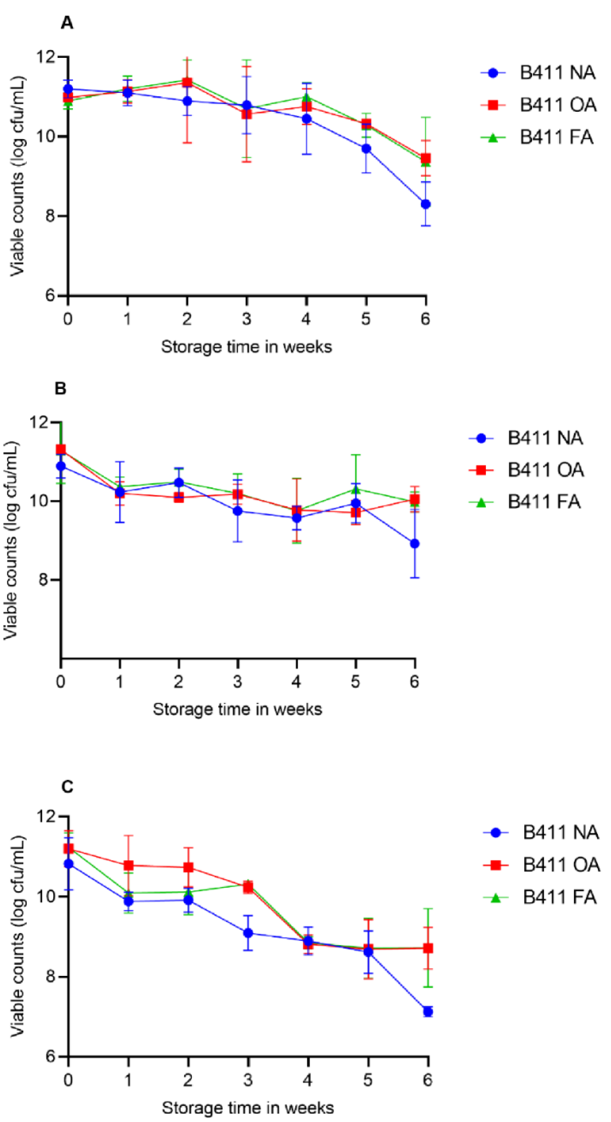
2.9. Survival of L. plantarum B411 in Food during Storage

2.9.1. Preparation of Yoghurt

Yoghurt was prepared in the laboratory according to the method described by Amakiri and Thantsha [29]. Briefly, 8 g of powdered full cream milk was added to 250 mL of skimmed milk in three separate flasks. The flasks were swirled to homogenize the mixture and then placed in a water bath at 72 °C for 3 min to pasteurize the milk. The pasteurized milk was cooled to 42 °C and then inoculated with a spoon of a commercial plain yoghurt containing starter cultures (Streptococcus thermophilus and Lactobacillus delbrueckii subsp. bulgaricus). The freshly inoculated milk was then incubated at 37 °C until the pH dropped to 4.53. The yoghurt was then pasteurized for 30 min at 72 °C to kill or inactivate the starter cultures. A tenfold serial dilution was performed for the yoghurt using ¼ strength Ringer’s solution and 100 µL of each dilution were plated onto M-17 and MRS agar plates for S. thermophilus and L. delbruecki subsp. bulgaricus, respectively. All the plates were incubated for 72 h at 37 °C in anaerobic jars, as described before. The colonies counted were reported as CFU/mL.

2.9.2. Selection of Fruit and Vegetable Juice

One hundred percent pasteurized cranberry and carrot, juices free from preservatives, were obtained from a retail store in Pretoria, South Africa. Cranberry was used as a fruit-based matrix and carrot juice served as a vegetable-based matrix. The same brand of juice was used in all the three independent trials. The juices were bought before their best before dates, and stored overnight at 4 °C before their use in experiments.

2.9.3. Preparation of L. plantarum B411

L. plantarum cells were grown according to a method described by Ding and Shah [30]. Briefly, 1.5 mL of the non-adapted, freshly adapted and old adapted cultures of L. plantarum were separately grown in 50 mL of MRS broth for 18 h for them to reach the early logarithm phase. The cells were then concentrated by centrifugation at 15,000× g for 30 min at 4 °C. They were subsequently washed twice with sterile phosphate buffered saline (PBS) pH 7 by centrifuging at 604× g for 5 min. The viable counts of cells were determined by pour plating onto MRS agar plates.

2.9.4. Assessment of L. plantarum B411 Viability in Yoghurt and Juices during Storage

For determining the viability in food products, 30 mL of yoghurt or juice was aseptically added into separate sterile glass bottles. Each food matrix was then separately inoculated with L. plantarum B411 cells (non-adapted, freshly adapted and old adapted) to a final concentration of 1011 CFU/mL in triplicate. The inoculated food samples were stored at 4 °C for 6 weeks. A new bottle of juice or yoghurt was used each week to perform viable plate counts. The first assessments were done in week 0, a day after storage at 4 °C. The viable counts of L. plantarum B411 present in the food samples were analyzed weekly over the storage period. On each day of sampling, a 1 mL subsample of the yoghurt or juice was drawn and serially diluted into 9 mL ¼ strength Ringer’s solution up to 10−9 dilution. One hundred microliters of each dilution were spread plated onto MRS agar plates in triplicate. The plates were then incubated at 37 °C for 72 h under anaerobic conditions as previously described.

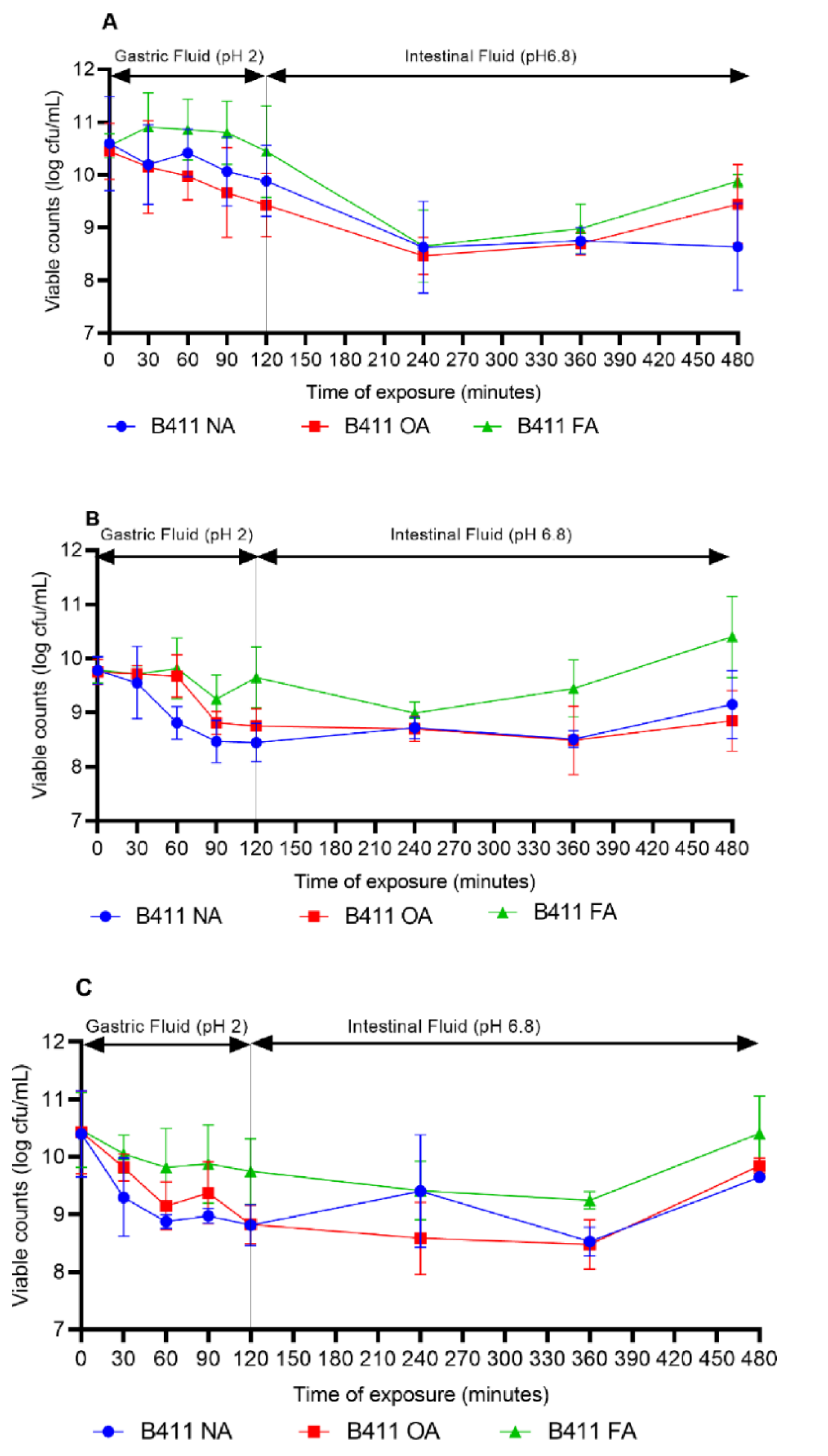
2.10. Effect of Food Matrixes on the Survival of L. plantarum B411 under Simulated GIT Conditions

L. plantarum B411 cells were prepared as described in Section 2.9.3 and then re-supended in the juices and yoghurt at a final concentration of approximately 1010 CFU/mL. Simulated gastrointestinal fluids were prepared according to methods described by Amakiri and Thantsha [29]. To make simulated gastric fluid (SGF), pepsin (P7000, 1:10000, ICN) (3 g/L) was suspended into NaCl (0.5% w/v) and the pH was adjusted to 2 using 12 M HCl. The solution was then filter sterilized using a 0.45 µm membrane (Millipore, Burlington, MA, USA) followed by a 0.22 µm filter membrane. Simulated intestinal fluid (SIF) was prepared by dissolving 6.8 g of monobasic potassium phosphate (Sigma, St. Louis, MO, USA) into 250 mL of distilled water, after which 77 mL of 0.2 M NaOH was added, followed by 500 mL of distilled water. The solution was then vortexed for 30 min and then 10 g of pancreatin (P-1500, Sigma, St. Louis, MO, USA) was added and mixed. The pH of the solution was adjusted to 6.8 using 0.2 M NaOH and 0.2 M HCL before being made up to a final volume of 1000 mL. The solution was filter sterilized by passing it through a 0.45 µm filter membrane (Millipore, Burlington, MA, USA) followed by 0.22 µm filter membrane (Millipore, Burlington, MA, USA).
One milliliter of yoghurt, carrot or cranberry juice containing L. plantarum B411 cells was added to 9 mL of SGF (pH 7) and vortexed for 30 s. These were then incubated with shaking (50 rpm) at 37 °C for 2 h. Then, 1 mL subsamples were drawn at 30 min intervals from the tubes containing L. plantarum B411 and sequentially diluted in ¼ strength Ringer’s solution and plated onto MRS agar plates to determine the number of surviving L. plantarum cells. After 2 h, the remaining bacteria were pelleted by centrifuging at 7000× g for 5 min before being re-suspended in 5 mL of SIF (pH 6.8) and then incubated with shaking (50 rpm) at 37 °C for 6 h. To enumerate the bacteria, 1 mL subsamples were then withdrawn every 2 h, serially diluted up to 10−7 in ¼ strength Ringer’s solution, and then each dilution plated onto MRS agar plates. The plates were incubated anaerobically as previously described. Each experiment was performed in triplicate in three independent trials.

2.11. Statistical Analysis

Data captured from three independent trials were used to calculate means and standard deviations, the differences in the means were calculated using least significant difference, (LSD). Data were analyzed using the GraphPad Prism 7.01 software (GraphPad Software Inc., San Diego, CA, USA), and any p value less than or equal to 0.05 was taken as statistically significant. Where appropriate, Tukey’s multiple comparisons test, with p < 0.05 as a significant difference, was used to identify statistically significant differences.

3. Results

3.1. Adaptation of L. plantarum B411 to Acid, Bile and Heat

It is imperative that probiotic strains are capable of withstanding the harsh assaults of production, storage and the GIT in order to maintain viability, functionality and stability. Figure 2A shows viable counts of L. plantarum B411 during the multi-step stress adaptation process, while Figure 2B shows reduction in viability of L. plantarum at each step. As expected for a sub-lethal process there was a reduction in viable counts during exposure to all stress factors, but no complete cessation of growth. The viability of L. plantarum at each step remained at or above 90% of its initial viability during exposure to the different stress factors (Figure 2A). When the cells were exposed to pH 2 for 120 min, their viable count decreased by 0.18 log CFU/mL from an original count of 8 log CFU/mL (Figure 2B). The surviving cells were used as the acid adapted cells. When these acid adapted cells were subsequently exposed to bile stress (2% bile) for 120 min, their viability decreased by 0.71 log CFU/mL from 7.82 log CFU/mL (Figure 2B). Survivors were used as acid-bile adapted cells. Interestingly, there was a 0.6 log CFU/mL increase in viability when the acid-bile adapted cells were successively exposed to heat stress (55 °C) for 120 min. The surviving cells were then used in subsequent experiments as the freshly adapted (acid- bile- heat adapted) cells. The L. plantarum B411 cells that survived a similar acid-bile-heat multi-stress adaptation process as described for freshly adapted cells, followed by storage at −20 °C for 2 years, were used as old adapted cells.

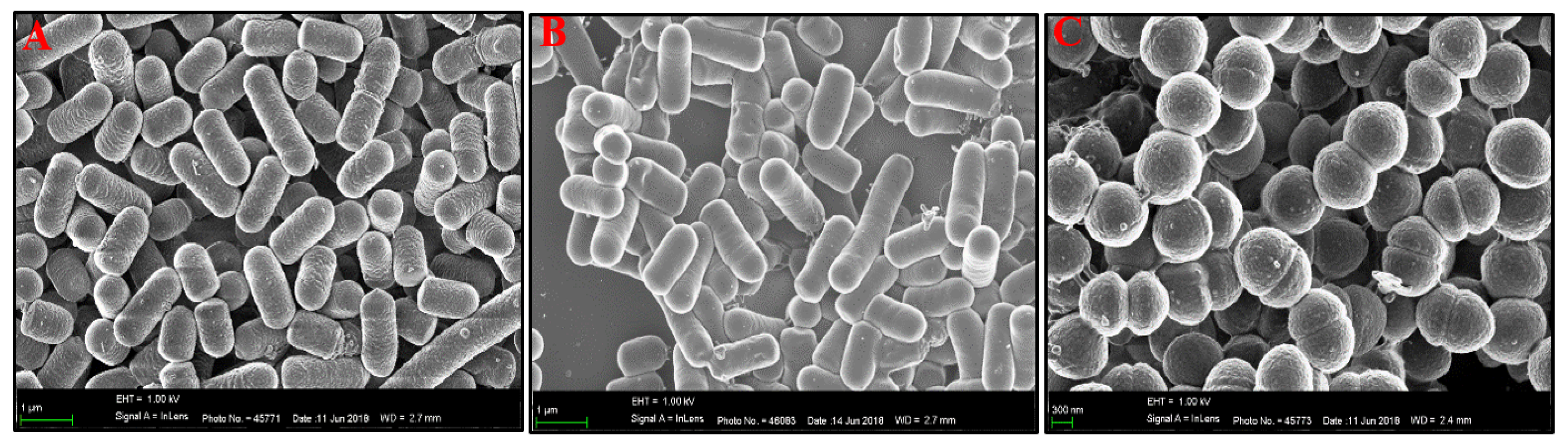
3.2. Scanning Electron Microscopy (SEM) of L. plantarum B411 Cells

Probiotics are able to competitively inhibit pathogens through, amongst other mechanisms, competition for adherent receptors. It is therefore crucial that during the stress adaptation of probiotics, they do not lose this beneficial property. The shape of probiotics plays a role in their ability to adhere to different surfaces. We thus examined whether there were any discernible differences in the shapes of the non-adapted, freshly adapted and the old adapted L. plantarum cells (Figure 3). SEM revealed no differences in the shape of the non-adapted (those subjected to long-term freezing, but not stress adapted) and freshly adapted (those subjected to stress adaptation but not subsequent long-term freezing) cells. They were both rod shaped, with short and long cells (Figure 3A,B). These results indicated that individually, the stress adaptation process and long-term freezing did not change the morphology of L. plantarum B411 cells. On the contrary, the morphology of the old adapted cells (those subjected to stress adaptation and subsequent long-term freezing) was altered, with cells appearing coccoid, with cross-walls (Figure 3C).

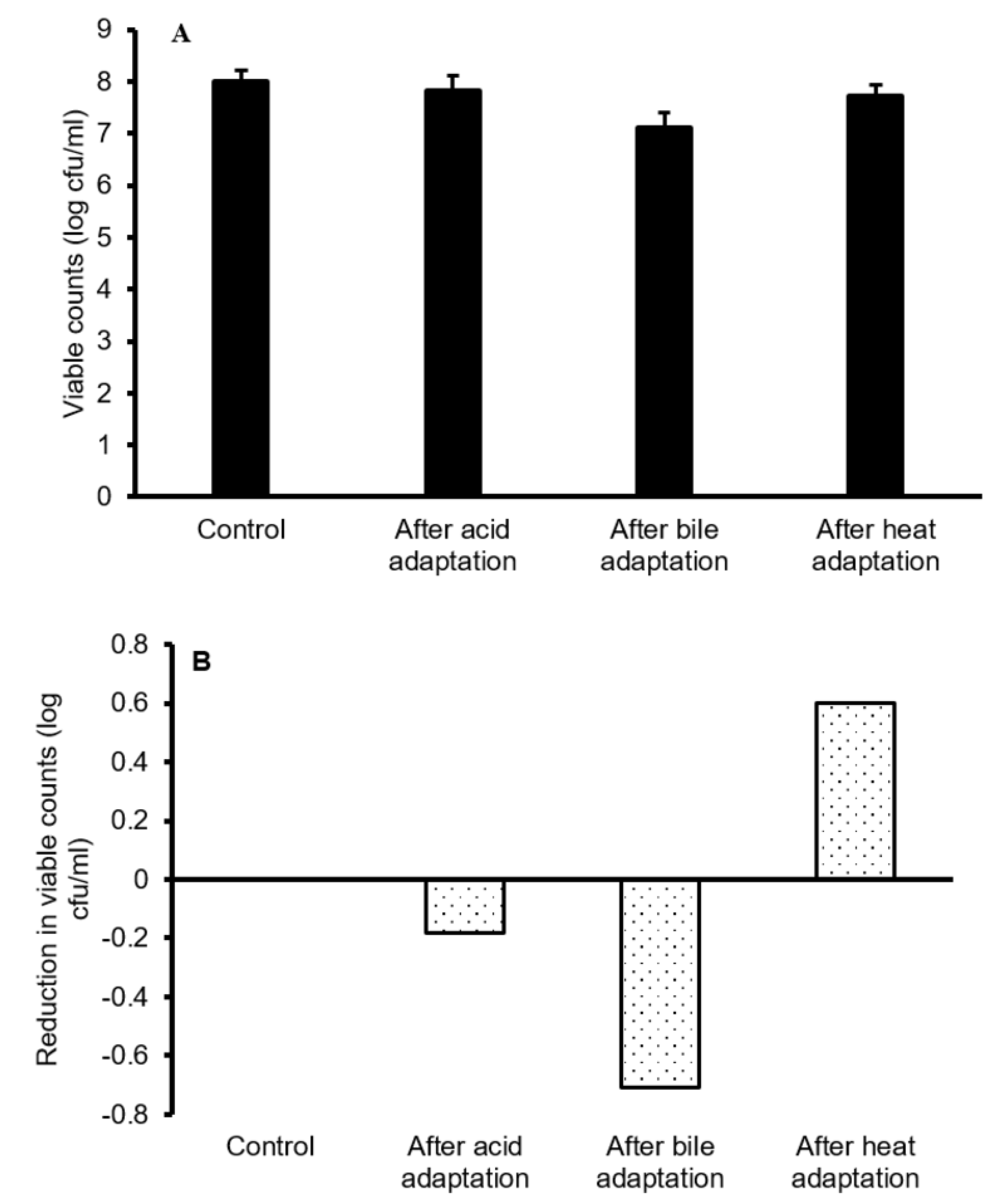
3.3. Acid and Bile Tolerance by Non-Adapted, Freshly Adapted and Old Adapted L. plantarum B411 Cells

Table 1 shows the tolerance of non-adapted, freshly adapted and old adapted cells of L. plantarum B411 to acid and bile. When all cells were exposed to pH 2, 2.5 and 3 over a period of 180 min, their viability ranged from 6.60 to 8.29 log CFU/mL, with the non-adapted adapted cells surviving slightly better at all pH values than both the freshly adapted and the old adapted cells. It was worth noting that at pH 2, viable counts of non-adapted cells increased, while those of the other cells decreased. The old adapted cells were less acid tolerant than the freshly adapted cells as their viability decreased more both at pH 2.5 and 3. Similar to what was observed in the acid tolerance assays, when the cells were exposed to 0.3, 0.5 and 2% bile concentrations for 180 min, a decrease in viability was observed (Table 1). The numbers of surviving cells ranged from 4.55 to 7.85 log CFU/mL where the non-adapted cells tolerated 0.5% bile better than both the stress adapted cells. Bile tolerance was marginally higher for the freshly adapted than for the old adapted cells. For all the cells, the number of survivors decreased with an increase of bile concentration, ultimately resulting in a total loss of viable cells due to exposure to 2% bile salt. Overall, there were no significant differences in acid and bile tolerance between the non-adapted, freshly adapted and old-adapted cells (p > 0.05).

3.4. Bile Salt Hydrolase Activity

The results of the BSH activity assays were positive for the freshly adapted cells, indicated by opaque halos around the punctured holes containing the cells. The BSH activity results were negative for both the non-adapted and the old adapted cells.

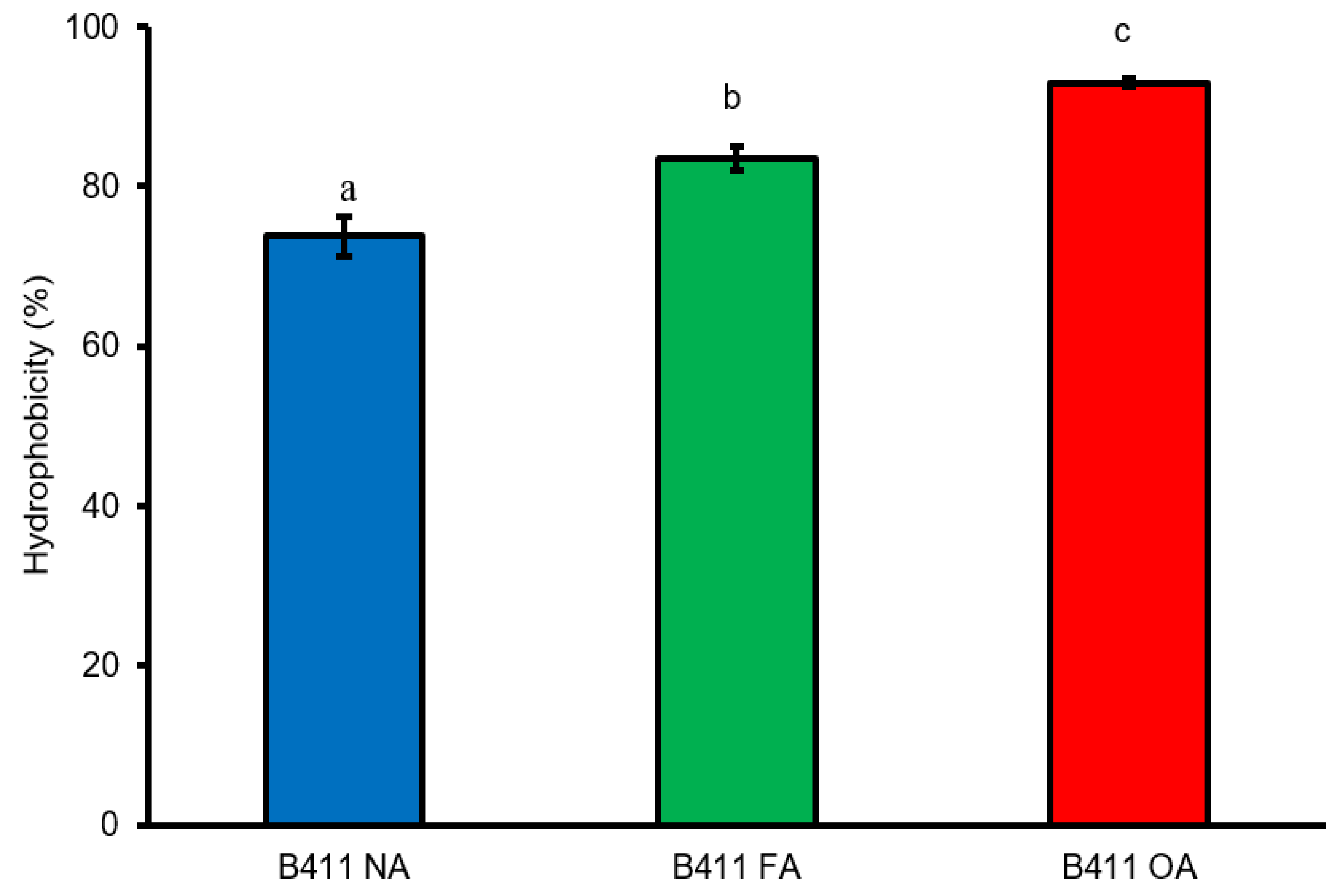
3.5. Auto-Aggregation and Cell Surface Hydrophobicity

The non-adapted cells displayed superior aggregation abilities than both the freshly adapted and old adapted cells (Table 2). The auto-aggregation of the non-adapted cells was 1.3 and 3.3 percentage points higher than was recorded for the freshly adapted and the old adapted cells, respectively. These results indicate that the multi-stress adaptation process did not improve the auto-aggregative properties of L. plantarum B411 cells. When comparing the stress adapted cells, freshly adapted cells auto-aggregated better than old adapted cells by 2%.
Figure 4 depicts the cell surface hydrophobicity of the different L. plantarum B411 cells. All cells had high cell surface hydrophobicity, ranging between 73.2 to 92.7%. The order of cell surface hydrophobicity based on percentages was old adapted cells > freshly adapted cells > non-adapted cells.

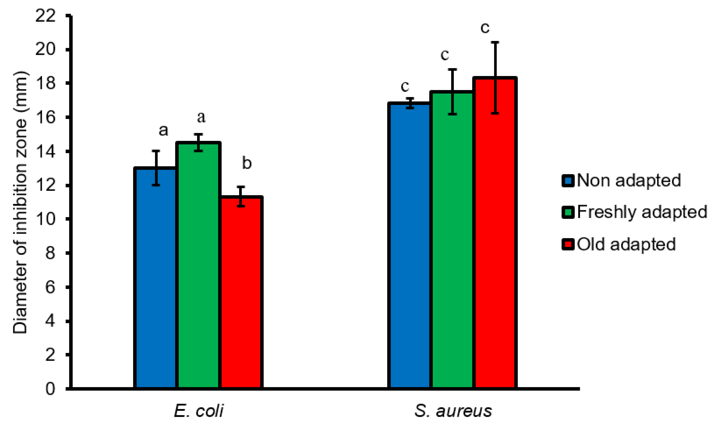
3.6. Antimicrobial Activity Assay of L. plantarum B411 Cells against E. coli ATCC 25922 and S. aureus ATCC 33591

One of the properties that make probiotics more favorable is their ability to inhibit pathogens. The current study looked at the antimicrobial ability of L. plantarum B411 cells against E. coli ATCC 25922 and S. aureus ATCC 33591. The zones of inhibition produced by non-adapted, freshly adapted and old adapted cells showed that the antimicrobial activity against S. aureus ATCC 33591 (17.5–17.83 mm) was notably higher than against E. coli ATCC 25922 (11.33–14.5 mm) (p = 0.023) (Figure 5). For E. coli ATCC 25922, the freshly adapted cells were more potent than both the non-adapted and old adapted cells. Additionally, both the non-adapted and freshly adapted cells significantly inhibited growth of E. coli ATCC 25922 better than old adapted cells (p < 0.001). Multi-stress adaptation initially increased the antagonistic activity of L. plantarum B411 against E. coli ATCC 25922 as observed for freshly adapted cells, however the inhibition ability was reduced as a result of long-term freezing of stress adapted cells. Similarly, freshly adapted cells exhibited enhanced antagonistic activity against S. aureus ATCC 33591 than the non-adapted and old adapted cells. However, there were no significant differences in the inhibition of S. aureus ATCC 33591 by the different L. plantarum B411 cells (p = 0.097). Contrary to what was observed for E. coli ATCC 25922, stress adaptation coupled with long-term freezing did not diminish the potency of stress adapted cells against S. aureus ATCC 33591.

3.7. Survival of L. plantarum B411 in Food Matrixes during Storage and under Simulated GIT Conditions

The viability of the different L. plantarum B411 cells when incorporated into different food matrices and stored for a period of 6 weeks is shown in Figure 6. During storage there were significant reductions in viability of all the cells in yoghurt (Figure 6A), where viable numbers of non-adapted cells decreased by 3.02 log (p < 0.0001), while those of the freshly and old-adapted cells decreased by 1.7 log (p = 0.0037) and 1.93 log (p = 0.0003), respectively. In carrot juice (Figure 6B), there were reductions of 2.06 log CFU/mL (p < 0.0001), 1.31 log CFU/mL (p = 0.0037) and 1.28 log CFU/mL (p = 0.0003) for the non-adapted, freshly adapted and old adapted cells, respectively. Similarly, in cranberry juice (Figure 6C), there were significant decreases (p < 0.0001) of 3.21 log CFU/mL, 2.59 log CFU/mL and 2.55 log CFU/mL for the non-adapted, old adapted and freshly adapted cells, respectively. From the results of all three food matrices, the non-adapted cells showed significantly higher reductions in viability during storage than both the freshly adapted and old adapted cells (p = 0.0025). However, there were no significant differences in reductions between the freshly adapted and the old adapted cells (p = 0.95).
In addition to investigating the viability of L. plantarum B411 cells during the shelf life of the foods into which they were incorporated, it was further investigated how these cells survived in the simulated gastrointestinal conditions. All the cells in all the different food matrices showed a decrease in viability when exposed to simulated gastric fluid (SGF) but their numbers increased during subsequent exposure to simulated intestinal fluid (SIF). However, when comparing the initial viable numbers to those that were present at the end of the exposure to the simulated GIT conditions, there was, overall, a net decrease in the viability of all L. plantarum B411 cells. In yoghurt (Figure 7A), there were decreases from 10.59 to 8.73 log CFU/mL (a 1.86 log reduction), 10.74 and 8.73 log CFU/mL (a 0.63 log reduction) and 10.37 to 9.34 log CFU/mL (a 1.03 log reduction) for non-, old- and freshly adapted cells. In carrot juice (Figure 7B), decreases from 9.75 to 9.29 log CFU/mL (a 0.47 log reduction) and 9.74 to 9.05 log CFU/mL (a 0.23 log reduction) were recorded for non-adapted and old adapted cells, respectively. However, there was an increase in viability from 9.59 to 10.36 log CFU/mL (a 0.62 log increase) for the freshly adapted cells. In cranberry juice (Figure 7C), the viability of non-adapted cells decreased from 10.29 to 9.59 log CFU/mL (a 0.7 log reduction). Viable counts of stress adapted cells decreased from 10.31 to 9.8 log CFU/mL (a 0.33 log reduction) and 10.46 to 10.22 log CFU/mL (a 0.24 log reduction) for old adapted and freshly adapted cells, respectively. Overall, at the end of exposure to simulated gastrointestinal fluids, viable numbers of freshly stress adapted cells were significantly higher than those of non-adapted (p = 0035) and old adapted cells (p = 0.021). Furthermore, there were no significant differences in viable numbers of non-adapted and old adapted cells (p = 0.72).

4. Discussion

Bacterial morphology plays an important role in cell surface attachment, hence any variations in their shape can potentially influence their adhesion ability [31]. It is known that bacteria alter their morphology in response to changing environments [31,32]. The shape of the freshly adapted L. plantarum B411 cells remained the same as that of non-adapted cells, while that of old adapted cells changed from rod to coccoid. Therefore, the altered shape of the old adapted cells could be attributed to the coupled effects of multi- stress adaptation and long-term freezing as the non-adapted and freshly adapted cells were identical, indicating that, separately, these processes did not alter the morphology of the cells. The treatment of bifidobacteria with garlic extracts transformed them from their original long rods to either shorter rods or completely cocci [27]. The exposure of L. plantarum ATCC 14197 cells to alkali changed them from long rods with slender thalli to short rods with thickset thalli [33]. The shrinking of rod-shaped bacteria to coccoid during periods of stress is hypothesized to be a protective mechanism that limits cell maintenance requirements [34]. The similarly observed altered shape for L. plantarum B411 cells in this study in response to long-term freezing could impede their attachment abilities as cocci cells attach to surfaces with a smaller area than rods that use the length of their cell to do so [31]. The change of the cell shape could also influence their aggregation and hydrophobicity properties, which are involved in interaction between the bacteria and epithelial cells [35].
Tolerance of the non-adapted L. plantarum B411 cells to low acidity (pH 2) was not startling as Lactobacilli are known to be intrinsically acid tolerant [36]. The weaker acid tolerance of the stress adapted cells when compared to their non-adapted counterparts was not envisaged, as previous studies reported enhanced subsequent survival of probiotics due to their pre-exposure to stressful conditions [2,17]. A closely related study reported that acid tolerance of the heat adapted L. plantarum KLDS 1.0328 variant was higher than that of the non-adapted strain [10]. Similarly, Kulkarni et al. [14] reported improved pH tolerance for the lyophilized heat adapted Lactobacillus acidophilus NCFM strain. However, in these previous studies, relatively short-term storage was investigated, which may explain the contradictory results reported between these studies. It was worth noting that in the current study, the freshly adapted cells slightly tolerated the acid better than the old adapted cells. Thus, although the stress adaptation process did not enhance the acid tolerance of L. plantarum B411, long-term freezing of multi-stress adapted cells further diminished its acid tolerance, an effect which is undesirable.
Bile is a biological detergent, which at high concentrations dissolves membrane lipids resulting in leakage of cytoplasmic contents and ultimately cell death [37]. Sahadeva et al. [38] reported that stress adapted lactobacilli survived 2% bile concentrations at levels as high as 8.51 log units. On the contrary, in this study none of the L. plantarum B411 cells survived this bile concentration. Furthermore, their survival at the other bile concentrations tested (0.3% and 0.5%) were similar. Thus, the currently reported stress adaptation process alone, as well as in conjunction with long-term freezing, did not improve bile tolerance of the L. plantarum B411 cells. Similarly, Ma et al. [2] reported significant reduction in viable counts of L. plantarum KLDS 1.0628 pre-adapted to 0.2% or 0.3% bile salt for 1 h. The de-conjugation of bile salts through the activity of the inducible bsh gene is regarded as one of the ways by which bacteria acquire bile stress tolerance [18,39]. The presence of a putative BSH activity was linked to bile salt resistance and human intestinal survival of Lactobacillus gasseri JCM1131T as its purified protein showed enhanced deconjugation activity for taurocholic and taurochenodeoxycholic acids [40]. The expression of BSH activity improved bile tolerance of Lactococcus lactis [18]. Thus, it was expected that cells exhibiting BSH activity would tolerate bile better than those without this enzyme activity. In contrast, current findings suggest that, although non-adapted cells showed better bile tolerance than all the stress adapted cells, this could not be attributed to BSH activity as these cells tested negative for this enzyme. This observation was not unique to our study, as previously, Moser and Savage [41] reported in their study on BSH activity and lactobacilli that there was no link between BSH activity and the lethal effects of conjugated bile salts. Lv et al. [42] demonstrated how under bile stress Ligilactobacillus salivarius LI01 used a strengthened bile efflux and general stress response systems, rather than BSH activity to combat the stress. Possibly, the superior bile tolerance of non-adapted cells without BSH activity could be attributed to these systems. Although BSH activity did not result in increased bile salt resistance, this trait is still desirable because bile salt hydrolysis can reduce serum cholesterol levels [40,43] and is involved in regulation of lipid absorption, glucose metabolism and energy homeostasis [40]. Unfortunately, the stress adaptation process induced transient BSH expression by L. plantarum B411 cells as long-term freezing of the stress adapted cells allowed them to revert to their original phenotype of negative BSH activity.
Environmental stresses can affect bacterial cell surface properties and thus their ability to attach to intestinal cells [44,45,46]. This was true in the current study as non-adapted cells auto-aggregated more than stress adapted cells. Freshly adapted cells displayed superior aggregation abilities than old adapted cells, indicating that stress adaptation and subsequent long-term freezing further reduced the ability of cells to auto-aggregate. Thus, the adversity of stress adaptation on the auto-aggregative ability of L. plantarum B411 cells was aggravated by long-term freezing of stress adapted cells. This agrees with the concept that cell surface properties involved in aggregation are negatively influenced by certain types of stress conditions [46]. Contrary to the auto-aggregation results, the stress adaptation process increased cell surface hydrophobicity, a property that was even enhanced in stress adapted cells subjected to long-term freezing, as indicated by better hydrophobicity of the old adapted cells in comparison with the freshly adapted cells. Haddaji et al. [45] showed that two strains of Lacticaseibacillus casei under osmotic stress exhibited significantly lower hydrophobicity than non-stressed strains. In another study, Haddaji et al. [19] reported that L. plantarum exposed to acid and heat stresses exhibited a decrease in surface hydrophobicity at low pH and high temperature. Thus, the results of the current study were opposing to those of the abovementioned studies as they portrayed pronounced hydrophobicity for both the freshly adapted and old adapted cells. However, the increased hydrophobicity may not translate to better adhesion properties of these cells as Ma et al. [10] reported that alkali treated L. plantarum KLDS 1.0328 displaying high surface hydrophobicity adhered weakly to Caco-2 cells.
All L. plantarum B411 cells were inhibitory to both E. coli ATCC 25922 and S. aureus ATCC 33591, with the latter being more susceptible. These results correlated with those of Tareb et al. [47], who tested the antimicrobial activity of lactobacilli against several enteropathogens and reported high susceptibility of the Gram positive than Gram negative cells. Our findings indicate that although stress adaption improved the antibacterial effects of L. plantarum B411 cells, synergistically, stress adaptation and long-term freezing diminished this much sought-after attribute, specifically against E. coli ATCC 25922. It has been suggested that the viability of probiotics should not fall below >106 CFU by the end of storage. The results of the current study showed significant declines (p < 0.05) in the viability of all L. plantarum B411 cells (non-adapted, freshly adapted and old adapted) incorporated into all three food matrices throughout their storage time. However, all of these cells still survived at levels recommended for beneficial effects. The viability of the non-adapted cells dropped more than that of the old adapted and freshly adapted cells, which showed similar levels of survival. The decrease in viability in the food products could be attributed to the prolonged exposure of L. plantarum B411 to the somewhat acidic nature of all three food matrices. The pH of yoghurt drops to around 4.5 after fermentation, while carrot and cranberry juices usually have pH of 4 [23]. The ability of both the stress adapted L. plantarum B411 cells to maintain their high numbers despite significant viability losses is attributed to their pre-adaptation to sub-lethal stresses as previously reported for Lactobacilli and Bifidobacteria subsequent to stress pre-adaptation treatment [11,35,48]. The long-term freezing of multi-stress adapted cells did not have any noticeable effect on their stability when incorporated into different food matrices.
Following survival in products during storage, probiotics are ingested and must survive the GIT conditions to reach their target site still in large quantities [49]. It is recommended that gastric and intestinal conditions should not be separated when assessing probiotic survival in the GIT as they could have a synergistic effect on probiotics’ viability [50]. Therefore, the current study investigated the viability of the different L. plantarum B411 cells incorporated into different food matrices when exposed sequentially to SGF and SIF. The type of food matrix affected the survival of L. plantarum B411 during simulated digestion. Ranadheera et al. [51] suggested that using low pH fruit juices as carriers could make probiotics more resilient to subsequent gastric acidity. This was in agreement with the results obtained in the current study as cells incorporated into carrot and cranberry juice survived more than those in yoghurt under simulated GIT conditions. However, these results contradicted those of Champagne and Gardner [13], who reported that incorporating probiotics into a dairy rather than a juice matrix resulted in better GIT survival due to the lower buffering capacity of juices compared to dairy products. Fruit and vegetable juices are high in carbohydrates and dietary fiber, which act as natural prebiotics, allowing survival in the adverse conditions of the stomach and intestines [52]. Although survival in yoghurt decreased, viability levels were still acceptable, meaning that the dairy proteins and fats also offered sufficient protection to the L. plantarum cells [49]. Overall, during exposure to simulated GIT conditions, there was a significant decrease (p < 0.05) in viable counts of the non-adapted and old adapted cells, while the decrease in viable counts of freshly adapted cells was non-significant (p > 0.05). The results obtained for freshly adapted cells are similar to those reported by Mathipa and Thantsha [17], where the survival of multi-stress adapted L. plantarum cells under simulated GIT conditions was significantly higher than that of their non-adapted counterparts. Since the reduction in viable counts of freshly adapted was insignificant, it can be deduced that multi-stress adaptation together with long-term freezing hindered the GIT survival of L. plantarum B411 cells even when incorporated into foods.

5. Conclusions

The current findings showed that long-term freezing of multi-stress adapted cells negatively impact some of the functional properties of the L. plantarum B411 cells. The affected attributes include the cell morphology, BSH activity, antimicrobial activity and cell surface properties. Therefore, it is important that the storage method used for the stress adapted cells should be chosen cautiously to avoid those that can have counteracting effects on the positive effects initially attained through the stress adaptation process. Multi-stress adaptation can be effective in enhancing the survival of probiotics incorporated into different foods, both during the shelf life of the foods and upon their subsequent GIT journey. However, the use of freezing as a long-term preservation method for stress adapted probiotics can negatively affect acquired stress tolerance attributes. Additionally, when developing probiotic products, the type of food matrix should be taken into consideration as this also has a profound influence on survival of probiotic cultures during storage and subsequent exposure to GIT conditions. Further research is needed to determine the suitability of other storage methods for the long-term preservation of multi-stress adapted cells.

Author Contributions

T.N.D.: investigation, data curation, formal analysis, wrote first draft of manuscript and reviewed the final manuscript. M.G.M.-M.: methodology, formal analysis, supervision and writing (review and editing). M.S.T.: conceptualization, methodology, project administration, funding acquisition, supervision, and writing (review and editing). All authors have read and agreed to the published version of the manuscript.

Funding

This research was funded by National Research Foundation (NRF) of South Africa, Competitive Programme for Rated Researchers, grant number UID 118531 and the University of Pretoria.

Institutional Review Board Statement

Not applicable.

Informed Consent Statement

Not applicable.

Data Availability Statement

Not applicable.

Acknowledgments

This work was supported in part by the National Research Foundation (NRF) of South Africa and the University of Pretoria research funds. The authors are grateful to Eudri Venter and Erna van Wilpe of the Laboratory for Microscopy and Microanalysis (University of Pretoria) for technical assistance with SEM.

Conflicts of Interest

The authors declare no conflict of interest. The funders had no role in the design of the study; in the collection, analyses, or interpretation of data; in the writing of the manuscript, or in the decision to publish the results.

References

  1. Zheng, J.; Wittouck, S.; Salvetti, E.; Franz, C.M.; Harris, H.; Mattarelli, P.; O’Toole, P.W.; Pot, B.; Vandamme, P.; Walter, J.; et al. A taxonomic note on the genus Lactobacillus: Description of 23 novel genera, emended description of the genus Lactobacillus Beijerinck 1901, and union of Lactobacillaceae and Leuconostocaceae. Int. J. Syst. Evol. Microbiol. 2020, 70, 2782–2858. [Google Scholar] [CrossRef] [PubMed]
  2. Ma, J.; Xu, C.; Liu, F.; Hou, J.; Shao, H.; Yu, W. Stress adaptation and cross-protection of Lactobacillus plantarum KLDS 1.0628. CyTA-J. Food. 2021, 19, 72–80. [Google Scholar] [CrossRef]
  3. Champagne, C.P.; Ross, R.P.; Saarela, M.; Hansen, K.F.; Charalampopoulos, D. Recommendations for the viability assessment of probiotics as concentrated cultures and in food matrices. Int. J. Food Microbiol. 2011, 149, 185–193. [Google Scholar] [CrossRef]
  4. Chen, M.-J.; Tang, H.-Y.; Chiang, M.-L. Effects of heat, cold, acod and bile salt adaptations on the stress tolerance and protein expression of kefir-isolated probiotic Lactobacillus kefiranofaciens M1. Food Microbiol. 2017, 66, 20–27. [Google Scholar] [CrossRef]
  5. Gaucher, F.; Bonnasie, S.; Rabah, H.; Marchand, P.; Blanc, P.; Jeantet, R.; Jan, G. Review: Adaptation of beneficial Propionibacteria, lactobacilli, and bifidobacteria improves tolerance toward technological and digestive stresses. Front. Microbiol. 2019, 10, 841. [Google Scholar] [CrossRef]
  6. Liu, S.; Ma, Y.; Zheng, Y.; Zhao, W.; Luo, T.; Zhang, J.; Yang, Z. Cold-stress response of probiotic Lactobacillus plantarum K25 by iTRAQ proteomic analysis. J. Microbiol. Biotechnol. 2020, 30, 187–195. [Google Scholar] [CrossRef] [PubMed]
  7. Mbye, M.; Baig, M.F.; AbuQamar, S.F.; El-Tarabily, K.A.; Obaid, R.S.; Osaili, T.M.; Al-Nabulsi, A.A.; Turner, M.S.; Shah, N.P.; Ayyash, M.M. Updates on understanding of probiotic lactic acid bacteria responses to environmental stresses and highlights on proteomic analyses. Compr. Rev. Food Sci. Food Saf. 2020, 19, 1110–1124. [Google Scholar] [CrossRef] [Green Version]
  8. Min, M.; Bunt, C.R.; Mason, S.L.; Hussain, M.A. Non-dairy probiotic food products: An emerging group of functional foods. Crit. Rev. Food Sci. Nutr. 2018, 59, 2626–2641. [Google Scholar] [CrossRef]
  9. Yang, H.; He, M.; Wu, C. Cross protection of lactic acid bacteria during environmetal stresses: Stress responses and underlying mechanisms. LWT-Food Sci. Technol. 2021, 144, 111203. [Google Scholar] [CrossRef]
  10. Ma, J.; Wang, W.; Sun, C.; Gu, L.; Liu, Z.; Yu, W.; Chen, L.; Jing, Z.; Hou, J. Effects of environmental stresses on the physiological characteristics, adhesion ability and pathogen adhesion inhibition of Lactobacillus plantarum KLDS 1.0328. Process Biochem. 2020, 92, 426–436. [Google Scholar] [CrossRef]
  11. Corcoran, B.; Stanton, C.; Fitzgerald, G.; Ross, R. Life under stress: The probiotic stress response and how it may be manipulated. Curr. Pharm. Des. 2008, 14, 1382–1399. [Google Scholar] [CrossRef] [PubMed]
  12. Sanz, Y. Ecological and functional implications of the acid-adaptation ability of Bifidobacterium: A way of selecting improved probiotic strains. Int. Dairy J. 2007, 17, 1284–1289. [Google Scholar] [CrossRef]
  13. Champagne, C.P.; Gardner, N. Effect of storage in a fruit drink on subsequent survival of probiotic lactobacilli to gastro-intestinal stresses. Food Res. Int. 2008, 41, 539–543. [Google Scholar] [CrossRef]
  14. Kulkarni, S.; Haq, S.F.; Samant, S.; Sukumaran, S. Adaptation of Lactobacillus acidophilus to thermal stress yields a thermotolerant variant which also exhibits improved survival at pH 2. Probiotics Antimicro. Prot. 2018, 10, 717–727. [Google Scholar] [CrossRef]
  15. Sánchez, B.; Ruiz, L.; Gueimonde, M.; Ruas-Madiedo, P.; Margolles, A. Toward improving technological and functional properties of probiotics in foods. Trends Food Sci. Technol. 2012, 26, 56–63. [Google Scholar] [CrossRef]
  16. Sánchez, B.; Champomier-Vergès, M.C.; Collado, M.; Anglade, P.; Baraige, F.; Sanz, Y.; de los Reyes-Gavilán, C.G.; Margolles, A.; Zagorec, M. Low-pH adaptation and the acid tolerance response of Bifidobacterium longum biotype longum. Appl. Environ. Microbiol. 2007, 73, 6450–6459. [Google Scholar] [CrossRef] [Green Version]
  17. Mathipa, M.G.; Thantsha, M.S. Cocktails of probiotics pre-adapted to multiple stress factors are more robust under simulated gastrointestinal conditions than their parental counterparts and exhibit enhanced antagonistic capabilities against Escherichia coli and Staphylococcus aureus. Gut Pathog. 2015, 7, 5. [Google Scholar] [CrossRef] [Green Version]
  18. Ruiz, L.; Margolles, A.; Sánchez, B. Bile resistance mechanisms in Lactobacillus and Bifidobacterium. Front. Microbiol. 2013, 4, 396. [Google Scholar] [CrossRef] [Green Version]
  19. Haddaji, N.; Mahdhi, A.B.; Ismaiil, M.B.; Bakhrouf, A. Effect of environmental stress on cell surface and membrane fatty acids of Lactobacillus plantarum. Arch. Microbiol. 2017, 199, 1243–1250. [Google Scholar] [CrossRef]
  20. da Cruz, A.G.; Faria, D.A.F.; Van Dender, A.G.F. Packaging system and probiotic dairy foods. Food Res. Int. 2007, 40, 951–956. [Google Scholar] [CrossRef]
  21. Granato, D.; Branco, G.F.; Nazzaro, F.; Cruz, A.G.; Faria, J.A.F. Functional foods and nondairy probiotic food development: Trends, concepts, and products. Compr. Rev. Food Sci. Food Saf. 2010, 9, 292–302. [Google Scholar] [CrossRef] [PubMed]
  22. Homayoni, R.A.; Vaghef, M.E.; Alipoor, B.; Vaghef, M.L. The comparison of food and supplement as probiotic delivery vehicles. Crit. Rev. Food Sci. Nutr. 2016, 56, 896–909. [Google Scholar] [CrossRef] [PubMed]
  23. Shori, A.B. Influence of food matrix on the viability of probiotic bacteria: A review based on dairy and non-dairy beverages. Food Biosci. 2016, 13, 1–8. [Google Scholar] [CrossRef]
  24. Kumar, V.B.; Vijayendra, S.V.N.; Reddy, O.V.S. Trends in dairy and non-dairy probiotic products—A review. J. Food Sci. Technol. 2015, 52, 6112–6124. [Google Scholar] [CrossRef] [PubMed] [Green Version]
  25. Sedláčková, P.; Horáčková, S.; Shi, T.; Kosová, M.; Plocková, M. Two different methods for screening of bile salt hydrolase activity in Lactobacillus Strains. Czech. J. Food Sci. 2015, 33, 13–18. [Google Scholar] [CrossRef] [Green Version]
  26. Mohankumar, A.; Murugalatha, N. Characterization and antibacterial activity of bacteriocin producing Lactobacillus isolated from raw cattle milk sample. Int. J. Biol. 2011, 3, 128. [Google Scholar] [CrossRef]
  27. Booyens, J.; Labuschagne, M.C.; Thantsha, M.S. In vitro antibacterial mechanism of action of crude garlic (Allium sativum) clove extract on selected probiotic Bifidobacterium species as revealed by SEM, TEM, and SDS-PAGE analysis. Probiotics Antimicrob. Proteins 2014, 6, 82–87. [Google Scholar] [CrossRef] [Green Version]
  28. Li, Q.; Liu, X.; Dong, M.; Zhou, J.; Wang, Y. Aggregation and adhesion abilities of 18 Lactic acid bacteria strains isolated from traditional fermented food. Int. J. Agric. Policy Res. 2015, 3, 84–92. [Google Scholar]
  29. Amakiri, A.C.; Thantsha, M.S. Survival of Bifidobacterium longum LMG 13197 microencapsulated in vegetal or vegetal-inulin matrix in simulated gastrointestinal fluids and yoghurt. Springerplus 2016, 5, 1343. [Google Scholar] [CrossRef] [Green Version]
  30. Ding, W.K.; Shah, N.P. Survival of free and microencapsulated probiotic bacteria in orange and apple juices. Int. Food Res. J. 2008, 15, 219–232. [Google Scholar] [CrossRef]
  31. Young, K.D. Bacterial morphology: Why have different shapes? Curr. Opin. Microbiol. 2007, 10, 596–600. [Google Scholar] [CrossRef] [PubMed]
  32. Oh, E.; McMullen, L.; Jeon, B. Impact of oxidative stress defense on bacterial survival and morphological change in Campylobacter jejuni under aerobic conditions. Front. Microbiol. 2015, 6, 295. [Google Scholar] [CrossRef] [PubMed]
  33. Wang, W.; He, J.; Pan, D.; Wu, Z.; Guo, Y.; Zeng, X.; Lian, L. Metabolomics analysis of Lactobacillus plantarum ATCC 14917 adhesion activity under initial acid and alkali stress. PLoS ONE 2018, 13, e0196231. [Google Scholar] [CrossRef] [PubMed] [Green Version]
  34. Chaiyanan, S.; Chaiyanan, S.; Grim, C.; Maugel, T.; Huq, A.; Colwell, R.R. Ultrastructure of coccoid viable but non-culturable Vibrio cholerae. Environ. Microbiol. 2007, 9, 393–402. [Google Scholar] [CrossRef]
  35. Zhang, H.; Wang, Q.; Liu, H.; Kong, B.; Chen, Q. In vitro growth performance, antioxidant activity and cell surface physiological characteristics of Pediococcus pentosaceus R1 and Lactobacillus fermentum R6 stressed at different NaCl concentration. Food Funct. 2020, 11, 6376–6386. [Google Scholar] [CrossRef]
  36. Mills, S.; Stanton, C.; Fitzgerald, G.F.; Ross, R.P. Enhancing the stress responses of probiotics for a lifestyle from gut to product and back again. Microb. Cell Fact. 2011, 10, S19. [Google Scholar] [CrossRef] [Green Version]
  37. Urdaneta, V.; Casadesús, J. Interactions between bacteria and bile salts in the gastrointestinal and hepatobiliary tracts. Front. Med. 2017, 4, 163. [Google Scholar] [CrossRef] [Green Version]
  38. Sahadeva, R.P.K.; Leong, S.F.; Chua, K.H.; Tan, C.H.; Chan, H.Y.; Tong, E.V.; Wong, S.Y.; Chan, H.K. Survival of commercial probiotic strains to pH and bile. Int. Food Res. J. 2011, 18, 1515–1522. [Google Scholar]
  39. Bi, J.; Liu, S.; Du, G.; Chen, J. Bile tolerance of Lactococcus lactis is enhanced by expresssion of bile salt hydrolase threby producing less bile acid in the cells. Biotechnol. Lett. 2016, 38, 659–665. [Google Scholar] [CrossRef]
  40. Kusada, H.; Morinaga, K.; Tamaki, H. Identification of bile salt hydrolase and bile salt resistance in a probiotic bacterium Lactobacillus gasseri JCM1131T. Microorganisms 2021, 9, 1011. [Google Scholar] [CrossRef]
  41. Moser, S.A.; Savage, D.C. Bile salt hydrolase activity and resistance to toxicity of conjugated bile salts are unrelated properties in Lactobacilli. Appl. Environ. Microbiol. 2001, 67, 3476–3480. [Google Scholar] [CrossRef] [PubMed] [Green Version]
  42. Lv, L.-X.; Yan, R.; Shi, H.-Y.; Shi, D.; Fang, D.-Q.; Jiang, H.-Y.; Wu, W.-R.; Guo, F.-F.; Jiang, X.-W.; Gu, S.-L.; et al. Integrated transcriptomic and proteomic analysis of the bile stress response in probiotic Lactobacillus salivarius LI01. J. Proteomics 2017, 150, 216–229. [Google Scholar] [CrossRef] [PubMed]
  43. Sridevi, N.; Vishwe, P.; Prabhune, A. Hypocholesteremic effect of bile salt hydrolase from Lactobacillus buchneri ATCC 4005. Food Res. Int. 2009, 42, 516–520. [Google Scholar] [CrossRef]
  44. Gong, X.; Yu, H.; Chen, J.; Han, B. Cell surface properties of Lactobacillus salivarius under osmotic stress. Eur. Food Res. Technol. 2012, 234, 671–678. [Google Scholar] [CrossRef]
  45. Haddaji, N.; Boubaker, K.; Lagha, R.; Khouadja, S.; Bakhrouf, A. Effect of high temperature on viability of Lactobacillus casei and analysis of secreted and GroEL proteins profiles. African J. Bacteriol. Res. 2015, 7, 29–34. [Google Scholar] [CrossRef]
  46. Shakirova, L.; Grube, M.; Gavare, M.; Auzina, L.; Zikmanis, P. Lactobacillus acidophilus La5 and Bifidobacterium lactis Bb12 cell surface hydrophobicity and survival of the cells under adverse environmental conditions. J. Ind. Microbiol. Biotechnol. 2013, 40, 85–93. [Google Scholar] [CrossRef]
  47. Tareb, R.; Bernardeau, M.; Gueguen, M.; Vernoux, J.-P. In vitro characterization of aggregation and adhesion properties of viable and heat-killed forms of two probiotic Lactobacillus strains and interaction with foodborne zoonotic bacteria, especially Campylobacter jejuni. J. Med. Microbiol. 2013, 62, 637–649. [Google Scholar] [CrossRef]
  48. Gueimonde, M.; Sánchez, B. Enhancing probiotic stability in industrial processes. Microb. Ecol. Health Dis. 2012, 23, 18562. [Google Scholar] [CrossRef] [Green Version]
  49. Amund, O.D. Exploring the relationship between exposure to technological and gastrointestinal stress and probiotic functional properties of lactobacilli and bifidobacteria. Can. J. Microbiol. 2016, 62, 715–725. [Google Scholar] [CrossRef]
  50. Vizoso Pinto, M.G.; Holzapfel, W.H.; Franz, C.; Schillinger, U. Lactobacillus spp. with in vitro probiotic properties from human faeces and traditional fermented products. Int. J. Food Microbiol. 2006, 109, 205–214. [Google Scholar] [CrossRef]
  51. Ranadheera, C.S.; Prasanna, P.H.B.; Vidanarachchi, J.K. Fruit juices as probiotic carriers. In Fruit Juices: Types, Nutritional Composition and Health Benefits; Nova Science: New York, NY, USA, 2014; pp. 253–267. [Google Scholar]
  52. Patel, A.R. Probiotic fruit and vegetable juices—Recent advances and future perspective. Int. Food Res. J. 2017, 24, 1850–1857. [Google Scholar]
Figure 1. A summarized scheme for production of the different L. plantarum B411 cells used.
Microbiolres 13 00064 g001
Figure 2. Viability of L. plantarum B411 during sequential exposure to multiple sub-lethal stress factors: acid (pH 2), bile (2%) and heat (55 °C). (A) Viable counts of L. plantarum B411 during the multi-step stress adaptation process and (B) Reduction in viability of L. plantarum at each step. Each bar represents the mean of three replicates, error bars are standard deviations.
Microbiolres 13 00064 g002
Figure 3. Scanning electron micrographs of non-adapted (A), freshly adapted (B) and old adapted (C) L. plantarum B411 cells.
Microbiolres 13 00064 g003
Figure 4. Cell surface hydrophobicity of non-adapted (NA), freshly adapted (FA) and old adapted (OA) L. plantarum B411 cells. Each bar represents the mean of three replicates, error bars are standard deviation and bars marked with different letters (a, b, c) indicate significant difference at p < 0.05.
Microbiolres 13 00064 g004
Figure 5. Antimicrobial activity of cell free supernatants of L. plantarum B411 against E. coli ATCC 25922 and S. aureus ATCC 33591 depicted as zones of inhibition in millimeters. Each bar represents mean of triplicates from three separate trials. The error bars are standard deviations. Bars marked with different letters (a, b, c) indicate significant difference at p < 0.05.
Microbiolres 13 00064 g005
Figure 6. Survival of the different L. plantarum B411 in food matrices: yoghurt (A), carrot juice (B) and cranberry juice (C) over 6 weeks. NA = non-adapted, OA = old adapted, FA = freshly adapted. Error bars are standard deviation of means (n = 3).
Microbiolres 13 00064 g006
Figure 7. Survival of different L. plantarum B411 cells incorporated into yoghurt (A), carrot juice (B) and cranberry juice (C) during exposure to simulated gastrointestinal conditions for 8 h. NA = non- adapted, OA = old adapted, FA = freshly adapted. Error bars represent standard deviation of mean (n = 3).
Microbiolres 13 00064 g007
Table 1. Survival of the different L. plantarum B411 cells at pH 2, 2.5 and 3 as well as in bile concentrations of 0.3, 0.5 and 2% over 180 min.
L. plantarum CellspH 2pH 2.5pH 3
Time (min)
601201806012018060120180
Viable Counts Log (cfu/mL)
Non-adapted 7.88 ± 1.117.71 ± 1.258.11 ± 1.238.21 ± 0.787.77 ± 1.788.07 ± 1.698.16 ± 1.158.28 ± 1.117.94 ± 1.98
Freshly adapted 8.22 ± 1.548.26 ± 1.717.90 ± 1.897.60 ± 1.237.90 ± 1.456.99 ± 1.788.22 ± 2.428.18 ± 1.238.02 ± 1.87
Old adapted7.89 ± 0.996.60 ± 1.327.86 ± 1.208.01 ± 1.477.86 ± 1.247.28 ± 2.368.29 ± 1.898.25 ± 1.747.83 ± 1.28
Bile concentrations (%)
0.30.52
Time (min)
601201806012018060120180
Viable counts log (cfu/mL)
Non-adapted 7.85 ± 2.07.33 ± 1.117.31 ± 1.696.51 ± 0.895.87 ± 1.785.55 ± 1.47---
Freshly adapted 6.81 ± 1.16.53 ± 1.506.51 ± 1.105.91 ± 1.255.98 ± 1.964.55 ± 1.91---
Old adapted 7.10 ± 1.966.90 ± 2.366.81 ± 2.795.77 ± 1.685.20 ± 2.144.69 ± 1.23---
Each value is a mean of 3 triplicates from 3 separate experiments, ± standard deviation (SD). The time 0 count for all the cells was 8 log (CFU/mL). There were no significant differences between the means of the different cells for both acid and bile tolerance (p > 0.05).
Table 2. Auto-aggregation percentages of L. plantarum B411 cells over a 6 h period.
L. plantarum B411 CellsAuto-Aggregation (%)
0 h3 h6 h
Non-adapted0.00 a15.80 a ± 1.0610.33 a ± 2.88
Freshly adapted0.00 a8.73 b ± 4.039.04 a ± 1.41
Old adapted0.00 a9.33 b ± 3.097.06 b ± 1.73
Each value is a mean of 3 replicates from 3 independent trials ± standard deviation. Means with different superscripts in the same column are significantly different at p < 0.05.
Publisher’s Note: MDPI stays neutral with regard to jurisdictional claims in published maps and institutional affiliations.

