Article

Effects of Fructooligosaccharides (FOS) on the Immune Response of the Shrimp Penaeus vannamei and on the Reduction in Vibrio spp. and Pseudomonas spp. in Cultures of Post-Larvae

by
Yulaine Corrales Barrios
1,
Alessandra Roncarati
2,
Leonardo Davier Martín Ríos
1,
Maikelis Rodríguez González
1,
Marbelys González Salotén
1,
Yeidel López Zaldívar
1 and
Amilcar Arenal
3,*
1
Department of Morphophysiology, Faculty of Agricultural Sciences, University of Camagüey, Camagüey 70100, Cuba
2
School of Biosciences and Veterinary Medicine, University of Camerino, 62032 Matelica, Italy
3
School of Veterinary Medicine, Saint Nicholas’ University, Morne Daniel, Roseau 00152, Dominica
*
Author to whom correspondence should be addressed.
Microbiol. Res. 2023, 14(3), 870-882; https://doi.org/10.3390/microbiolres14030060
Submission received: 16 May 2023 / Revised: 3 July 2023 / Accepted: 4 July 2023 / Published: 8 July 2023

Abstract

:
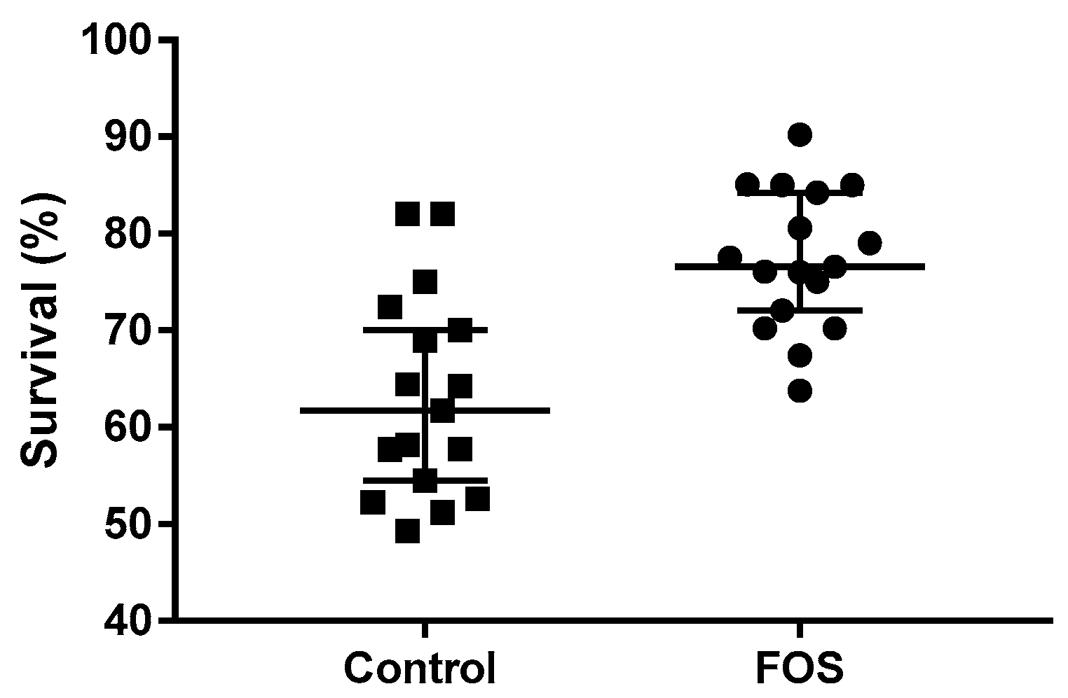
Penaeus spp. are the most cultivated type of shrimp because they have rapid growth and good adaptation to farming conditions. Due to diseases that result in high mortality and a decreased product quality, the cultivation of these shrimp globally, and in Cuba in particular, comes with the risk of significant financial losses. This study examined the effect of fructooligosaccharide (FOS, 1-kestose) on the growth and immune response of shrimp, as well as the multitude of Vibrio spp. and Pseudomonas spp. in Penaeus vannamei post-larvae under culture conditions. Fructooligosaccharide was applied in a completely randomized manner at a concentration of 0.4%, in both experimental groups with seventeen tanks each. In the results of this investigation, animals of greater weight (control 6.8 ± 0.2 mg; FOS 9.5 ± 0.3 mg; p < 0.001), length (control 1.1 ± 0.1 mm; FOS 1.3 ± 0.1 mm; p < 0.001), and survival (control 61.7% (95% CI of median 54.2–70.0); FOS 76.6% (95% CI of median 72.1–84.2); p < 0.001) were obtained when administered FOS relative to the control. An increase in the activity of proteases (p < 0.001), enzymes of the innate immune system such as phenoloxidase (p < 0.001), and lysozymes (p < 0.001) was observed, as well as an increase in the number of lectins (p < 0.001). Changes in the microbiota could be observed, with a reduction in Vibrio spp. and Pseudomonas spp. (control 2.4 × 10³ ± 0.5 × 10³; FOS 1.1 × 10² ± 0.3 × 10²; p < 0.001). FOS improves the quality of the post-larvae of P. vannamei as reflected in the length, weight, and survival of the animals. Moreover, FOS stimulates the P. vannamei immune system through the enzymatic activities of phenoloxidase, lysozyme, and a number of lectins. The reduction in the population load of Vibrio spp. and Pseudomonas spp. might be a consequence of the improvement in the quality and immune system of P. vannamei.

1. Introduction

It is estimated that aquaculture represents 49% of the global production of aquatic animals. Crustaceans represent 9.7% of aquaculture production, preceded by freshwater fish and mollusks, but in terms of commercial value, they occupy the second position [1]. Due to the high demand from importing nations, shrimp aquaculture is becoming increasingly popular worldwide [2]. Shrimp in the genus Penaeus are the most cultivated due to the rapid growth and aquaculture adaptation of species such as Penaeus vannamei and Penaeus monodon [1].
Shrimp farming, both in Cuba and globally, faces the risk of large financial losses due to diseases that cause high mortality, and a decrease in the production quality. Moreover, crustaceans do not have specific immune responses against these infectious agents; their defensive function consists of a developed, non-specific, defense system, which is based on circulating hemocytes and various defense proteins [3]. Due to the continuous outbreaks and spread of diseases in shrimp farming systems, it is urgent to identify the effect of environmental factors on the immune response of penaeid shrimp [4]. The application of antibiotics as prophylactics was an effective strategy for controlling bacterial diseases in shrimp, but their indiscriminate use fosters the development of bacterial resistance [5]. Prebiotics are supplements that are selectively metabolized by beneficial bacteria in the digestive tract and promote the dietary modulation of the intestinal microbiota, thereby improving animal health, primarily by stimulating the growth and activity of Bifidobacteria and Lactobacilli. In addition, prebiotics do not generate dependency or negative impacts on culture organisms or the environment [6]. Immunosaccharides stimulate the immune system directly. Immunosaccharide prebiotics include fructooligosaccharide (FOS), mannan oligosaccharide, inulin, or β-glucan [7].
The activation of enzymes such as peroxidase, superoxide dismutase, and the pro-phenoloxidase (proPO) system can initiate several immunological responses. In addition, other types of cytotoxic and cellular recognition mechanisms, such as coagulation proteins, lectins, peptides, antimicrobials, peroxynectins, the generation of reactive oxygen species, quinones, opsonization, phagocytosis, and nodulation, can be stimulated to eliminate the pathogen [4]. The shrimp immune system is currently described by how certain immunological parameters vary, for example, the enzymatic activities of crucial enzymes linked to the immune system such as lysozyme, phenoloxidase, peroxidase, superoxide dismutase, and lectin. The current study determined the effects of the administration of FOS under aquaculture conditions on the growth and survival, as well as on the immune response of post-larvae of Penaeus vannamei.

2. Materials and Methods

2.1. Location

The post-larvae (PL) were obtained from the Yagüacam hatchery center in Cumanayagüa, Cienfuegos, Cuba. The research was carried out in the Cultisur post-larval nursery area in Santa Cruz del Sur, Camagüey, Cuba. P. vannamei post-larvae at substage 8 days (PL 8) were stocked at 1.5 million per rectangular cement tank, having a water volume of 60 m3.

2.2. Experimental Design

A completely randomized design was used for 34 tanks (17 with FOS treatment and 17 as control group). In the experimental group, the prebiotic fructooligosaccharide (FOS) was homogenized with sterile distilled water, then mixed with commercial shrimp feed (35% protein) using the sprayer method (40 g of FOS per kg of feed).
FOS was also added to the pond water 1 mL/m3 every day. The control group (without FOS treatment) received the standard aquaculture treatment previously established in the center.

2.3. Culture Parameters

The temperature was maintained between 28 and 30 °C throughout the experiment, salinity at 30 ppt, pH 7.8, and dissolved oxygen at 5.00 mg/L from the third day of the PL stocking. For water exchange, 50% of each tank was replaced using a filter (1 mm2 of mesh) attached to the outlet of the tanks to eliminate solid and organic wastes. The animals were kept for 15 days under these conditions (PL 23 substage). Commercial food (Malta Cleyton® 35% protein, Mexico City, Mexico) was used for feeding. Feed was offered 12 times a day in amounts recommended by the manufacturer. The feed composition was: protein (35.03%), fat (9.04%), fiber (2.84%), water (8.84%), and ash (6.09%).

2.4. Shrimp Sample Collection and Preservation

For sample collection, 15 days after starting the treatment, three shrimp samples were taken randomly from each tank after filtering with a 1 mm nylon mesh. Shrimp samples were washed twice in phosphate-buffered saline (PBS) solution at pH 7.4. Sample preservation was carried out in vials without preservation solution at −20 °C until its evaluation in the Molecular Biology Laboratory of the Faculty of Agricultural Sciences at the University of Camagüey.

2.5. Evaluation of the Quality of Post-Larvae

The quality of the post-larvae was evaluated by determining the development parameters: length and weight. Twenty post-larvae per tank were washed with water, dried with filter paper, and weighed on an analytical balance (Sartorius, model 1205) with a precision of ±0.1 mg. The length of the post-larvae was determined by measuring them from the rostral base to the end of the telson under a microscope (±0.1 mm). At the end of each bioassay, survival values per tank were recorded.

2.6. Processing of Samples

Approximately 100 PL 23 were taken from each tank per 1.5 mL vial. The post-larvae were macerated in 1.5 mL tubes with 200 µL PBS. Then, 200 µL PBS was added and homogenized in a vortex. The homogenates of the samples were centrifuged at 8000× g for 10 min, and the supernatant was then stored at −20 °C in new 1.5 mL vials. The concentration of total proteins was determined by the Bradford MM [8] method. The supernatant was also used to determine the presence of lectins in the larvae.

2.7. Evaluation of Immune Response Parameters

2.7.1. Phenoloxidase-Specific Enzymatic Activity

Phenoloxidase activity was determined by measuring the dopachrome produced from dihydroxyphenylalanine (L-DOPA), pH 7.6 [9]. A negative control was used to measure the spontaneous oxidation of L-DOPA. Per sample, 20 μL of supernatant was used. The assay was carried out in Nunc® 96-well flat-bottom polystyrene plates with a total volume of 400 μL. Results were expressed in U/mg protein, where U was defined as a 0.001 increase in absorbance at 490 nm per min.

2.7.2. Superoxide Dismutase-Specific Enzymatic Activity

The specific activity of SOD was determined according to the oxidation of nitroblue tetrazolium (NBT) in the presence of riboflavin [10]. The measurement sample was exposed to 8000–9000 lux, with the light-emitting source 30 cm from the reaction site. The specific enzymatic activity was obtained by dividing the enzymatic activity value by the protein concentration value in each sample. The assay was carried out in Nunc ® 96-well flat-bottom polystyrene plates with a total volume of 400 μL, using 20 µL per sample. Results were expressed in U μg−1 protein, where U was defined as a 0.001 increase at 630 nm per minute.

2.7.3. Peroxidase-Specific Enzymatic Activity

Peroxidase activity was quantified using a standard curve made with horseradish peroxidase (HRP) coupled to a mouse anti-polyhistidine (Sigma-Aldrich, St. Louis, MO, USA) antibody [11]. Peroxidase assessments were performed in Nunc ® 96-well polystyrene plates (Denmark) by adding 20 μL of sample and 50 μL of a solution containing 0.15% H2O2 (v/v) and 0.10% o-phenylenediamine (weight/volume) in phosphate buffer, pH 5, (0.05 M Na2HPO4, 0.02 M citric acid). The mixtures were incubated at 25 °C for 30 min. The reactions were stopped with 50 μL of sulfuric acid (2 M) and the chromogen formation was quantified after two hours at 490 nm. The results were expressed in U/mg of protein, where U was defined as the formation of 1 μmol of 2,3-diaminophenazine by oxidation of o-phenylenediamine per minute. Per sample, 20 μL of supernatant was used. A molar extinction coefficient of 16,700 M−1 cm−1 was assumed for 2,3-diaminophenazine (Fornera and Walde, 2010).

2.7.4. Lysozyme-Specific Enzymatic Activity

The lysozyme-specific enzymatic activity was determined by the absorbance variation over time of a lyophilized solution of Microccocus lysodeikticus [12]. The final solution, with a final volume of 180 µL, included the lyophilized M. lysodeikticus (0.4 mg/mL) dissolved in phosphate buffer (NaH2PO4/Na2 HPO4) at a concentration of 100 mM, with adjustment to pH 8 with orthophosphoric acid (H3PO4) (85%). The solution had the desired absorbance of 0.3. Per sample, 20 µL was added, with absorbance at 490 nm measured over time. The absorbance was measured every 5 min during the incubation. One unit of enzymatic activity is equivalent to a 0.001 decrease at 490 nm per min and was reported as specific enzymatic activity by dividing the enzymatic activity value by the protein concentration value for each sample.

2.7.5. Lectin Expression

Rabbit blood was used to determine the presence of lectins and agglutinins. The blood was collected in a 15 mL centrifuge tube (Corning, Corning, NY, USA) with ethylenediaminetetraacetic acid (EDTA 50 mmol L−1) and stored at 4 °C until the time of the test. The blood was centrifuged at 1500× g for 10 min and the serum was discarded. The erythrocyte pellet was washed twice with PBS. Then, it was centrifuged at 2000× g for 10 min. Next, a suspension of erythrocytes (1% (v/v)) was prepared in PBS pH 7.2. Then, 25 µL of each of the samples was mixed with 25 µL of PBS pH 7.2 in 96-well concave-bottom plates (Costar®, Corning, NY, USA), and 25 µL of the erythrocyte suspension was added to each sample. The reactions were incubated for 20 min at 28 °C, and the presence of lectins was determined by observing hemagglutination. The assay was carried out in triplicate. A suspension of erythrocytes in PBS, pH 7.2, was used as a negative control [13].

2.7.6. Bacteriological Analysis

For the microbiological evaluation of bacteria, presumptively of the genus Vibrio spp. and Pseudomonas spp., the number of colony-forming units (cfu) mL−1 (that grew on thiosulfate–citrate–bile salts–sucrose (TCBS) agar) was determined in the samples at the end of the experiment. To determine the number of cfu mL−1 of bacteria in the larvae, 3 samples were taken per tank with an average of 50 individuals in each sample. The larvae were disinfested with 0.1% benzalkonium chloride [13] and rinsed with sterile H2Od to eliminate external bacteria. Whole organisms were macerated with a homogenizer (Wiggenhauser, D-130, Selangor, Malaysia) in 500 µL of sterile PBS. Serial dilutions of 1:10 were made for the macerated samples. A 10 µL subsample of each dilution was plated on the TCBS plates. All plates were incubated at 28 °C for 24 to 48 h.

2.8. Statistical Analysis

The statistical analysis of the experimental data was carried out with the help of the GraphPad Prism software version 6.01 (2012, GraphPad Software, Boston, MA, USA). The data obtained for weight, height, survival, and specific enzymatic activities were verified to have a normal distribution (Kolmogórov–Smirnov). Fisher’s F-test was then applied to the sample to determine the homogeneity between the variances. The difference between the means was determined using the Student’s t-test. In the case of survival and lectin titers, the non-parametric Mann–Whitney U-test was used to detect differences between medians.

3. Results and Discussion

3.1. Length, Weight, and Survival

Prebiotics are classified as organic additives because they are derived from natural ingredients such as alginate, inulin, and various oligosaccharides [14,15]. In aquaculture, both dietary FOS and short-chain FOS (scFOS) have gained much interest due to their growth-promoting factors in several aquatic species, including P. vannamei [16]. In addition, they have been used as sole carbon sources to evaluate the growth, length, and weight of shrimp of similar age, relating to their intestinal bacterial composition [17]. The authors confirmed that weight, length, and survival are related to the administration of prebiotics and the change in the composition of the intestinal microbiota.
Prebiotics can be metabolized by beneficial bacteria in the genus Lactobacillus and by Bifidobacterium, producing short-chain fatty acids (scFA) and lactate [14]. However, the effect of prebiotics on shrimp growth and weight gain is controversial throughout the literature. While Zhou Z, Ding Z and Huiyuan L [16] reported an increased growth in P. vannamei, other reports did not find a significant increase in the growth and survival of P. vannamei fed with fructooligosaccharides included in the diet [18,19]. Thus, the effect of prebiotics on the growth performance of aquatic species may be ambiguous because some studies with fish have not observed positive improvements in growth performance with the dietary supplements of prebiotics [20,21,22,23].
In studies on the effect of scFOS supplementation on the beneficial microbiota of the white shrimp digestive tract [24], they did not improve weight gain, feed conversion, or shrimp survival. On the other hand, the analysis by Zhou Z, Ding Z and Huiyuan L [16] showed that scFOS supplementation in the diet at concentrations of 0.4% improved growth rate and survival, stimulated bacterial growth, and stimulated immune enzymes.
Similar results were obtained in this study, with the only difference being that FOS was also administered directly into the water over 21 days (Figure 1). Significant differences were reported in tanks treated with FOS compared to the control without FOS. The animals treated with FOS were heavier and larger than the control. However, there are no reports of the effect of FOS stimulating growth or immune response when administered directly into the water. Perhaps one of the possible effects of this prebiotic is indirectly on the intestinal microbiota due to microorganisms stimulated in the water by FOS.
When analyzing the results obtained (Figure 2), there is evidence of improved survival in the group where FOS was administered, compared to the control group. However, Ref. [25] applied this prebiotic to P. vannamei over 35 days of culture. No differences were observed between the experimental groups regarding survival; the same outcome occurred when these researchers [26] used this experimental design but with galactooligosaccharides (GOS). In contrast, Fuandila NN, Widanarni W and Yuhana M [27] observed an increased survival rate when using honey as a prebiotic in P. vannamei.

3.2. Innate Immune System

Phenoloxidase is an essential enzyme for the immune response of invertebrates. Its activity is not only limited to the production of compounds with bactericidal action but also to a wide range of immune functions, which makes it a central component of the innate immune response [28]. Phenoloxidase is the main enzyme in shrimp that enables wound repair and cuticle sclerotization due to its microbicidal actions [29]. Immunostimulants, as molecules and/or whole cells of bacteria and yeasts [30], can be used to increase the stimulation of the immune response and resistance to stress and diseases, through direct interaction with the cells of the immune system and their activation [31].
High phenoloxidase activity improved the ability of Pacific white shrimp to recognize foreign entities entering their bodies [32]. A better immune response would protect shrimp against pathogen infections [33]. Dong C and Wang J [34] reported that FOS increased the activity of phenoloxidase or superoxide dismutase, and can positively influence the survival rate of P. vannamei.
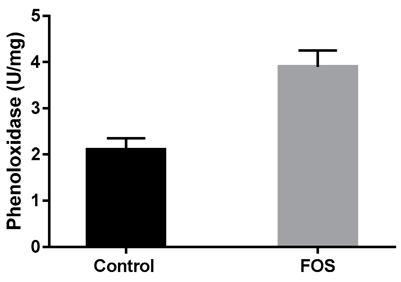
In the experiment, the animals treated with FOS showed phenoloxidase activity values higher than the untreated control group (Figure 3). Similarly, Safari O, Shahsavani D, Paolucci M and Atash MMS [35] reported that the administration of FOS can stimulate the rate of hemocyte proliferation, because an increase in the number of hemocytes explains the high activity of phenoloxidase, as they are the main producers of this enzyme.
Peroxidase activity is closely related to the generation and removal of reactive oxygen species (ROS) generated by oxidative metabolism during phagocytosis of foreign particles. It may have cytotoxic activity against foreign cell types that invade the organism, which would provide it with an additional immune function [36]. However, it is unclear whether there are distinct mechanisms for shrimp glutathione peroxidase to participate in immunity other than their antioxidant activity [37].
Peroxidase activity is related to environmental stressors such as exposure to viral and bacterial pathogens [38]. A decrease in total peroxidase activity has been reported under stress conditions due to high cadmium levels and pH stress in P. vannamei [39].
The total peroxidase activity after two hours did not show any significant differences between the tanks treated with FOS and the control tanks (control group 1.84 × 10⁴ ± 0.08 × 10⁴ U/mg and FOS group 1.75 × 10⁴ ± 0.06 × 10⁴ U/mg). The lack of any difference between the two groups may be due to similarities in the oxidative status of shrimp larvae in the tanks. The result suggests that the effect of FOS on the immune response does not follow a stimulation product of oxidative stress in shrimp larvae since the stimulation of immune parameters is evident without altering their oxidative status.
From the current results, the SOD activity between the experimental groups also did not show significant differences (control group 0.88 ± 0.06 U/mg and FOS group 0.95 ± 0.08 U/mg). It may be because, in both groups, the physiological states were not exposed to stress. This result coincides with the one obtained by Varela-Granados Y, Frías-Gómez SA, Hernández-Hernández LH, Powell MS and Vega-Villasante F [40], with no differentiation of SOD activity between treated (fructo- and mannan oligosaccharides) and untreated groups. However, it does not coincide with that obtained by Hu X, Yang HL, Yan YY, Zhang CX, Ye Jd, Lu KL, Hu LH, Zhang JJ, Ruan L and Sun YZ [41], showing that the form of administration of FOS within the diet can have effects dependent on the concentration of FOS and other environmental factors. It has been discussed that SOD activities improved by prebiotics seem to be dose-dependent [40]. However, mannan and fructooligosaccharides were able to stimulate SOD in crayfish, with higher SOD activities observed for the combination of both prebiotics [35]. Therefore, more research is needed to resolve the effect of FOS on the antioxidant response of cultured species.
Two members of the superoxide dismutase family exist in invertebrates Cu/Zn (SOD 1) and Mn (SOD 2). In addition, it is known that environmental factors such as hypoxia and high temperatures can stimulate the expression of SOD 2 in shrimp. However, SOD 1 expression is continuous, because its expression is constitutive [42]. Even though the SOD 2 expression increases faster against pathogens, both enzymes (SOD 1 and SOD 2) increase their expression upon challenge against pathogens [43].
Superoxide dismutase possesses the ability to decrease superoxide anions, which occur in water and hydrogen peroxide. Extracellular SOD co-operates in the destruction of ingested or encapsulated parasites during the respiratory burst generated with phagocytosis [44]. The antioxidant system is frequently used as a potential indicator of oxidative stress in marine organisms [45]. Discoveries related to the cellular activity of SOD show that it plays a crucial role as a modulator of the immune response [46]. This distribution is closely related to the antioxidant function and is found at higher levels in the places where the respiratory burst occurs, confirming its role in preventing and eliminating oxidative damage [47].
Lysozymes are proteins that are specifically synthesized by hemocytes, mainly granular hemocytes [48]. They are well-characterized in penaeid shrimp, where they have lytic activity against Gram-negative and Gram-positive bacterial species, including pathogenic Vibrio spp. [12]. There are three types of lysozymes, c-lysozyme, g-lysozyme, and i-lysozyme, of which c-lysozyme and i-lysozyme have been reported in P. vannamei, both with potent antimicrobial activity [49].
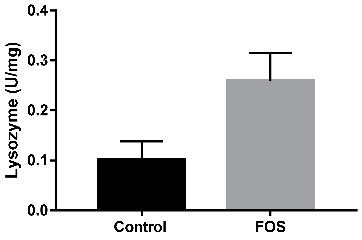
FOS administration in the freshwater prawn Macrobrachium acanthurus does not affect lysozyme activity, compared to the control [40]. In contrast, the results of the current study showed a stimulation of lysozyme activity in the group treated with FOS as compared to the untreated control group (Figure 4). On the other hand, research by Elshopakey GE, Risha EF, Abdalla OA, Okamura Y, Hanh VD, Ibuki M, Sudhakaran R and Itami T [50] showed similar results to this study with the prebiotic β-1,4-mannobiose increasing lysozyme expression. Within their research, the increase in the number of circulating hemocytes in the hemolymph, specifically granular hemocytes, was also verified. This increase in the number of granular hemocytes, the main producers of lysozyme in shrimp, could explain the increase in lysozyme activity in the current experiment. Previous studies have shown that supplementing mannan oligosaccharide and FOS in a juvenile narrow-clawed crayfish leads to a higher activity of lysozymes [35]. These authors observed a higher lysozyme activity when both oligosaccharides were combined, compared to the individual supplementations.
However, it is worth highlighting the differences in the methodology used here, which constitutes the first determination of the effect of FOS administered in water on lysozyme activity. The induction of lysozyme expression is linked to several biotic and abiotic factors. It is known that exposure to Vibrio anguillarum or Micrococcus lysodeikticus induces a significant upregulation of lysozyme gene expression, with no change in total hemocyte counts. Furthermore, even when temperature stress does not change the lysozyme expression profile [51], it is affected by culture parameters such as diet composition and pH [52]. Therefore, thorough research is needed to study the number of hemocytes under these culture conditions, as well as their expression of lysozyme.
Lectins are proteins that play a role in the pattern recognition receptor (PRR) reaction in invertebrates and recognize all or parts of sugar molecules [53]. According to Song SK, Beck BR, Kim D, Park J, Kim J, Kim HD and Ringø E [54], prebiotics such as FOS, mannan oligosaccharides, inulin, and β-glucan are also classified as immunosaccharides, which could directly enhance non-specific immune responses through interactions with the pattern recognition receptors. Furthermore, beneficial bacteria in the intestines may interact with gut-associated lymphoid tissue (GALT), which could stimulate the immune system and improve the host’s ability to prevent infection [55].
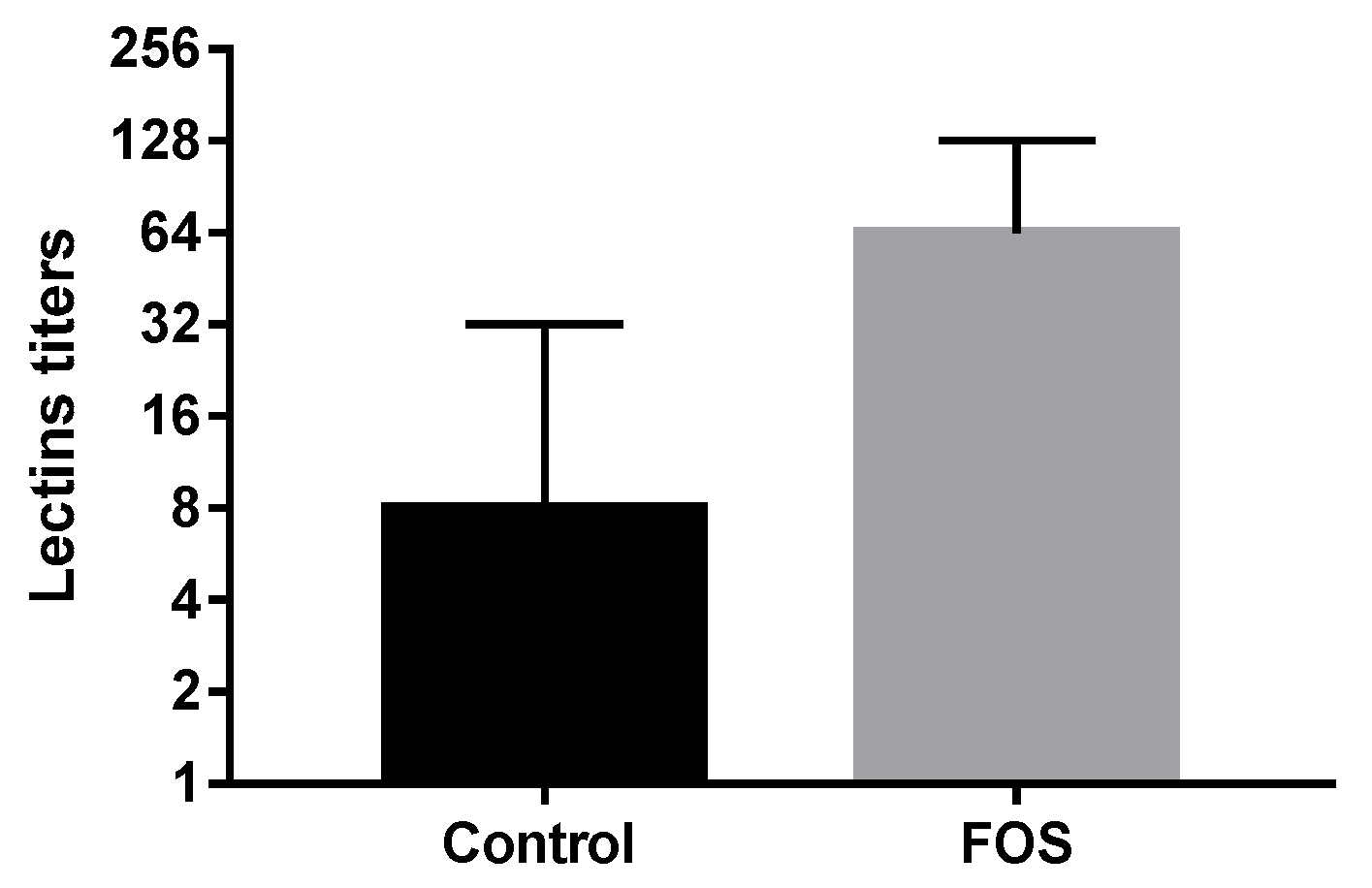
The hemagglutination capacity of the group treated with FOS was higher (p < 0.001) compared to the control group (Figure 5). Thus, the results of the study show that FOS addition directly in the water improves the immune response parameters in the bacterial recognition of P. vannamei. This might be because the carbohydrate components of the bacterial cell wall and membrane attach to lectin receptors in shrimp, which could directly enhance shrimp immune system parameters such as phenoloxidase and lysozyme. These enzymes were found to undergo a significant increase in their activity in the tanks treated with FOS, compared to the untreated ones. Both are synthesized in hemocytes upon pathogen recognition patterns through an interaction with lectins on the surface of the hemocytes (Liu et al., 2020). In addition, a greater stimulation of the defense cells, due to an increase in the growth of beneficial bacteria in the culture media, could induce overexpression of lectins in the tanks treated with FOS [56].

3.3. Protease and Amylase-Specific Enzymatic Activity

When comparing the behavior of the specific enzymatic activity of proteases in this study (Figure 6), it is evident that the response obtained in the group treated with FOS was superior, with statistically significant differences (p < 0.001). Fuandila NN, Widanarni W and Yuhana M [27] found that using bee honey stimulates digestive enzymes, such as proteases, due to the concentration of prebiotic oligosaccharides in honey. Although some studies [27,57] support the ability of some oligosaccharides to stimulate amylases, in this study, there was no stimulation of amylase activity using the prebiotic FOS (control group 0.74 ± 0.09 U/mg and FOS group 0.65 ± 0.07 U/mg). The supplementation with mannan oligosaccharides and FOS in diets of the juvenile narrow-clawed crayfish results in a higher activity of amylases [35]. These authors demonstrated that the combination of both oligosaccharides results in an even higher amylase activity. Recently, amylase activity was stimulated in P. vannamei using a diet containing 0.5% FOS. In that study, however, FOS appeared to lead to some hepatopancreatic abnormalities, such as hypertrophied B-cells [58].

3.4. Bacteriological Analysis

Figure 7 shows the values obtained in cfu/mL on TCBS agar for potential opportunistic pathogens. Changes in the microbiota were observed when applying the FOS prebiotic, with a reduction in Vibrio spp. and Pseudomonas spp., compared to the control group (control 2.4 × 10³ ± 0.5 103 cfu/mL; FOS 1.1 × 10² ± 0.3 × 10² cfu/mL). The results obtained by Mustafa A, Buentello A, Gatlin D, Lightner D, Hume M and Lawrence A [26] support the ability of fructooligosaccharides to modify microbial communities and that they can suppress or improve the growth of specific bacteria to favor a healthy physiology of P. vannamei shrimp post-larvae.
The results of the current study suggest that FOS can not only change the populations of the intestinal microbiota, in particular, a significant reduction in Vibrio spp. and Pseudomonas spp., but can also create a positive association with the gut microbiota and digestive tract cells. Therefore, it can improve the physiological performance and stimulate the innate immune system of P. vannamei shrimp post-larvae.
Previous studies have shown that oligosaccharide supplementation in the culture water of the animals confers health benefits [59,60]. Similarly, other authors have shown that small molecules (GHRP-6) added to the culture water of shrimp and tilapia can improve their immune systems and growth [61,62]. The authors demonstrated that, in one hour, the molecules reach the highest levels in the target animal. According to those results, one can hypothesize that FOS can follow the same route after their supplementation in the water. However, further studies are necessary to corroborate their mechanism of entrance into the host and their effect on the whole microbiota of the host and the pond water.

4. Conclusions

The administration of the prebiotic FOS during the culture of post-larvae of P. vannamei augments their length, weight, and survival, which indicates an overall improvement in the quality of the post-larvae. Moreover, FOS enhanced the innate immune system in P. vannamei post-larvae by stimulating the activity of the phenoloxidase and lysozyme enzymes, as well as the ability to recognize pathogens by increasing the lectin titers, which demonstrates a better health condition. Referring to P. vannamei metabolism, FOS increases the activity of proteases in the post-larvae of P. vannamei, which consequently improves their digestive capacity to process shrimp feed. Also, FOS reduces the populations of Vibrio spp. and Pseudomonas spp. in P. vannamei shrimp post-larvae.

Author Contributions

Conceptualization, Y.C.B. and A.A.; methodology, Y.C.B., A.R. and A.A.; software, L.D.M.R. and A.A.; validation, Y.C.B., A.R. and A.A.; formal analysis, Y.C.B., A.R., L.D.M.R. and A.A.; investigation, Y.C.B., A.R., L.D.M.R., M.R.G., M.G.S. and Y.L.Z.; resources, A.A.; data curation, Y.C.B., A.R. and A.A.; writing—original draft preparation, Y.C.B., A.R. and A.A.; writing—review and editing, Y.C.B., A.R. and A.A.; visualization, Y.C.B. and A.R.; supervision, A.A.; project administration, Y.C.B. and A.A.; funding acquisition, Y.C.B., A.R. and A.A. All authors have read and agreed to the published version of the manuscript.

Funding

Shrimp biofloc project, CITMA, Ministry of Science, Technology and Environment, Cuba. PT 223CM002_002.

Institutional Review Board Statement

The experiments performed in this research are part of the shrimp Biofloc project approved by an Ethics Committee. Name: Facultad de Ciencias Agropecuarias, Universidad de Camagüey; approval code: UCFACA 1-2019; approval date: 1 February 2023.

Informed Consent Statement

Following Cuban Legislation, No. 180/07 (Gaceta Oficial 084, 19 December 2007), this field study on commercial farms did not require ethical approval. Consent to use farm production data was given by the National Shrimp Organization company.

Data Availability Statement

The data presented in this study are openly available in Zenodo at [doi: 10.5281/zenodo.8122374] (3 July 2023), reference number [8122374].

Acknowledgments

We thank John van der Meer (Pan-American Marine Biotechnology Association) for advice on English improvement for the manuscript.

Conflicts of Interest

The authors declare no conflict of interest.

References

  1. Abdel-Tawwab, M. Feed Supplementation to Freshwater Fish: Experimental Approaches; LAP Lambert Academic Publishing: Saarbrücken, Germany, 2016. [Google Scholar]
  2. Aguirre-Guzman, G.; Sanchez-Martinez, J.G.; Campa-Cordova, A.I.; Luna-Gonzalez, A.; Ascencio, F. Penaeid shrimp immune system. Thai J. Vet. Med. 2009, 39, 205–215. [Google Scholar]
  3. Akhter, N.; Wu, B.; Memon, A.M.; Mohsin, M. Probiotics and prebiotics associated with aquaculture: A review. Fish Shellfish Immunol. 2015, 45, 733–741. [Google Scholar] [CrossRef] [PubMed]
  4. Anadón, A.; Ares, I.; Martínez-Larrañaga, M.R.; Martínez, M.A. Prebiotics and probiotics in feed and animal health. In Nutraceuticals in Veterinary Medicine; Springer: Berlin/Heidelberg, Germany, 2019; pp. 261–285. [Google Scholar]
  5. Beauchamp, C.; Fridovich, I. Superoxide dismutase: Improved assays and an assay applicable to acrylamide gels. Anal. Biochem. 1971, 44, 276–287. [Google Scholar] [CrossRef]
  6. Bi, J.; Ning, M.; Xie, X.; Fan, W.; Huang, Y.; Gu, W.; Wang, W.; Wang, L.; Meng, Q. A typical C-type lectin, perlucin-like protein, is involved in the innate immune defense of whiteleg shrimp Litopenaeus vannamei. Fish Shellfish Immunol. 2020, 103, 293–301. [Google Scholar] [CrossRef]
  7. Bradford, M.M. A rapid and sensitive method for the quantitation of microgram quantities of protein utilizing the principle of protein-dye binding. Anal. Biochem. 1976, 72, 248–254. [Google Scholar] [CrossRef]
  8. Burge, E.J.; Madigan, D.J.; Burnett, L.E.; Burnett, K.G. Lysozyme gene expression by hemocytes of Pacific white shrimp, Litopenaeus vannamei, after injection with Vibrio. Fish Shellfish Immunol. 2007, 22, 327–339. [Google Scholar] [CrossRef]
  9. Burr, G.; Gatlin, D.M., III; Hume, M. Effects of the prebiotics GroBiotic®-A and inulin on the intestinal microbiota of red drum, Sciaenops ocellatus. J. World Aquac. Soc. 2009, 40, 440–449. [Google Scholar] [CrossRef]
  10. Bush, S.R.; Belton, B.; Little, D.C.; Islam, M.S. Emerging trends in aquaculture value chain research. Aquaculture 2019, 498, 428–434. [Google Scholar] [CrossRef] [Green Version]
  11. Campa-Córdova, A.; Hernández-Saavedra, N.; Aguirre-Guzmán, G.; Ascencio, F. Immunomodulatory response of superoxide dismutase in juvenile American white shrimp (Litopenaeus vannamei) exposed to immunostimulants. Cienc. Mar. 2005, 31, 661–669. [Google Scholar] [CrossRef] [Green Version]
  12. Cerenius, L.; Söderhäll, K. Immune properties of invertebrate phenoloxidases. Dev. Comp. Immunol. 2021, 122, 104098. [Google Scholar] [CrossRef]
  13. Chang, Y.-H.; Kumar, R.; Ng, T.H.; Wang, H.-C. What vaccination studies tell us about immunological memory within the innate immune system of cultured shrimp and crayfish. Dev. Comp. Immunol. 2018, 80, 53–66. [Google Scholar] [CrossRef] [PubMed]
  14. Chen, W.W.; Romano, N.; Ebrahimi, M.; Natrah, I. The effects of dietary fructooligosaccharide on growth, intestinal short chain fatty acids level and hepatopancreatic condition of the giant freshwater prawn (Macrobrachium rosenbergii) post-larvae. Aquaculture 2017, 100, 95–101. [Google Scholar] [CrossRef]
  15. Chen, Y.-Y.; Chen, J.-C.; Tseng, K.-C.; Lin, Y.-C.; Huang, C.-L. Activation of immunity, immune response, antioxidant ability, and resistance against Vibrio alginolyticus in white shrimp Litopenaeus vannamei decrease under long-term culture at low pH. Fish Shellfish Immunol. 2015, 46, 192–199. [Google Scholar] [CrossRef]
  16. De-La-Re-Vega, E.; García-Galaz, A.; Díaz-Cinco, M.E.; Sotelo-Mundo, R.R. White shrimp (Litopenaeus vannamei) recombinant lysozyme has antibacterial activity against Gram negative bacteria: Vibrio alginolyticus, Vibrio parahemolyticus and Vibrio cholerae. Fish Shellfish Immunol. 2006, 20, 405–408. [Google Scholar] [CrossRef] [PubMed]
  17. Denis, M.; Thayappan, K.; Ramasamy, S.; Munusamy, A. Lectin in innate immunity of Crustacea. Austin Biol 2016, 1, 1001. [Google Scholar]
  18. Dimitroglou, A.; Merrifield, D.L.; Spring, P.; Sweetman, J.; Moate, R.; Davies, S.J. Effects of mannan oligosaccharide (MOS) supplementation on growth performance, feed utilisation, intestinal histology and gut microbiota of gilthead sea bream (Sparus aurata). Aquaculture 2010, 300, 182–188. [Google Scholar] [CrossRef]
  19. Divya, M.; Vaseeharan, B.; Anjugam, M.; Iswarya, A.; Karthikeyan, S.; Velusamy, P.; Govindarajan, M.; Alharbi, N.S.; Kadaikunnan, S.; Khaled, J.M.; et al. Phenoloxidase activation, antimicrobial, and antibiofilm properties of β-glucan binding protein from Scylla serrata crab hemolymph. Int. J. Biol. Macromol. 2018, 114, 864–873. [Google Scholar] [CrossRef] [PubMed]
  20. Dong, C.; Wang, J. Immunostimulatory effects of dietary fructooligosaccharides on red swamp crayfish, Procambarus clarkii (Girard). Aquac. Res. 2013, 44, 1416–1424. [Google Scholar] [CrossRef]
  21. Elshopakey, G.E.; Risha, E.F.; Abdalla, O.A.; Okamura, Y.; Hanh, V.D.; Ibuki, M.; Sudhakaran, R.; Itami, T. Enhancement of immune response and resistance against Vibrio parahaemolyticus in kuruma shrimp (Marsupenaeus japonicus) by dietary supplementation of β-1, 4-mannobiose. Fish Shellfish Immunol. 2018, 74, 26–34. [Google Scholar] [CrossRef]
  22. Fan, J.; Li, B.; Hong, Q.; Yan, Z.; Yang, X.; Lu, K.; Chen, G.; Wang, L.; Chen, Y. A glutathione peroxidase gene from Litopenaeus vannamei is involved in oxidative stress responses and pathogen infection resistance. Int. J. Mol. Sci. 2022, 23, 567. [Google Scholar] [CrossRef]
  23. FAO. The State of World Fisheries and Aquaculture 2022. Towards Blue Transformation; FAO: Rome, Italy, 2022. [Google Scholar]
  24. Franco, R.; Martín, L.; Arenal, A.; Santiesteban, D.; Sotolongo, J.; Cabrera, H.; Mejías, J.; Rodríguez, G.; Moreno, A.G.; Pimentel, E.; et al. Evaluation of two probiotics used during farm production of white shrimp Litopenaeus vannamei (Crustacea: Decapoda). Aquac. Res. 2017, 48, 1936–1950. [Google Scholar] [CrossRef]
  25. Fuandila, N.N.; Widanarni, W.; Yuhana, M. Growth performance and immune response of prebiotic honey fed pacific white shrimp Litopenaeus vannamei to Vibrio parahaemolyticus infection. J. Appl. Aquac. 2020, 32, 221–235. [Google Scholar] [CrossRef]
  26. García-Carreño, F.L.; Cota, K.; Navarrete Del Toro, M.A. Phenoloxidase activity of hemocyanin in whiteleg shrimp Penaeus vannamei: Conversion, characterization of catalytic properties, and role in postmortem melanosis. J. Agric. Food Chem. 2008, 56, 6454–6459. [Google Scholar] [CrossRef] [PubMed]
  27. Genc, M.; Aktas, M.; Genc, E.; Yilmaz, E. Effects of dietary mannan oligosaccharide on growth, body composition and hepatopancreas histology of Penaeus semisulcatus (de Haan 1844). Aquac. Nutr. 2007, 13, 156–161. [Google Scholar] [CrossRef]
  28. Hasyimi, W.; Widanarni, W.; Yuhana, M. Growth Performance and Intestinal Microbiota Diversity in Pacific White Shrimp Litopenaeus vannamei Fed with a Probiotic Bacterium, Honey Prebiotic, and Synbiotic. Curr. Microbiol. 2020, 77, 2982–2990. [Google Scholar] [CrossRef]
  29. Hernández-López, J.; Gollas-Galván, T.; Vargas-Albores, F. Activation of the prophenoloxidase system of the brown shrimp Penaeus californiensis Holmes. Comp. Biochem. Physiol. Part C Pharmacol. 1996 Toxicol. Endocrinol. 1996, 113, 61–66. [Google Scholar] [CrossRef]
  30. Hu, F.; Wang, Y.; Hu, J.; Bao, Z.; Wang, M. A novel c-type lysozyme from Litopenaeus vannamei exhibits potent antimicrobial activity. Fish Shellfish Immunol. 2022, 131, 729–735. [Google Scholar] [CrossRef]
  31. Hu, X.; Yang, H.L.; Yan, Y.Y.; Zhang, C.X.; Ye, J.D.; Lu, K.L.; Hu, L.H.; Zhang, J.J.; Ruan, L.; Sun, Y.Z. Effects of fructooligosaccharide on growth, immunity and intestinal microbiota of shrimp (Litopenaeus vannamei) fed diets with fish meal partially replaced by soybean meal. Aquac. Nutr. 2019, 25, 194–204. [Google Scholar] [CrossRef] [Green Version]
  32. Li, H.; Parisi, M.-G.; Toubiana, M.; Cammarata, M.; Roch, P. Lysozyme gene expression and hemocyte behaviour in the Mediterranean mussel, Mytilus galloprovincialis, after injection of various bacteria or temperature stresses. Fish Shellfish Immunol. 2008, 25, 143–152. [Google Scholar] [CrossRef] [Green Version]
  33. Li, P.; Burr, G.S.; Gatlin, D.M., III; Hume, M.E.; Patnaik, S.; Castille, F.L.; Lawrence, A.L. Dietary supplementation of short-chain fructooligosaccharides influences gastrointestinal microbiota composition and immunity characteristics of Pacific white shrimp, Litopenaeus vannamei, cultured in a recirculating system. J. Nutr. 2007, 137, 2763–2768. [Google Scholar] [CrossRef] [Green Version]
  34. Lin, Y.-C.; Lee, F.F.; Wu, C.-L.; Chen, J.-C. Molecular cloning and characterization of a cytosolic manganese superoxide dismutase (cytMnSOD) and mitochondrial manganese superoxide dismutase (mtMnSOD) from the kuruma shrimp Marsupenaeus japonicus. Fish Shellfish Immunol. 2010, 28, 143–150. [Google Scholar] [CrossRef]
  35. Liu, C.-H.; Tseng, M.-C.; Cheng, W. Identification and cloning of the antioxidant enzyme, glutathione peroxidase, of white shrimp, Litopenaeus vannamei, and its expression following Vibrio alginolyticus infection. Fish Shellfish Immunol. 2007, 23, 34–45. [Google Scholar] [CrossRef]
  36. Lu, X.; Wang, C.; Liu, B. The role of Cu/Zn-SOD and Mn-SOD in the immune response to oxidative stress and pathogen challenge in the clam Meretrix meretrix. Fish Shellfish Immunol. 2015, 42, 58–65. [Google Scholar] [CrossRef]
  37. Lulijwa, R.; Rupia, E.J.; Alfaro, A.C. Antibiotic use in aquaculture, policies and regulation, health and environmental risks: A review of the top 15 major producers. Rev. Aquac. 2020, 12, 640–663. [Google Scholar] [CrossRef]
  38. Mahious, A.S.; Gatesoupe, F.J.; Hervi, M.; Metailler, R.; Ollevier, F. Effect of dietary inulin and oligosaccharides as prebiotics for weaning turbot, Psetta maxima (Linnaeus, C. 1758). Aquac. Int. 2006, 14, 219–229. [Google Scholar] [CrossRef] [Green Version]
  39. Martín, L.; Castillo, N.M.; Arenal, A.; Rodríguez, G.; Franco, R.; Santiesteban, D.; Sotolongo, J.; Forrellat, A.; Espinosa, G.; Carrillo, O.; et al. Ontogenetic changes of innate immune parameters from eggs to early postlarvae of white shrimp Litopenaeus vannamei (Crustacea: Decapoda). Aquaculture 2012, 358, 234–239. [Google Scholar] [CrossRef]
  40. Martínez, R.; Carpio, Y.; Arenal, A.; Lugo, J.M.; Morales, R.; Martín, L.; Rodríguez, R.F.; Acosta, J.; Morales, A.; Duconge, J.; et al. Significant improvement of shrimp growth performance by growth hormone-releasing peptide-6 immersion treatments. Aquac. Res. 2017, 48, 4632–4645. [Google Scholar] [CrossRef]
  41. Martinez, R.; Carpio, Y.; Morales, A.; Lugo, J.M.; Herrera, F.; Zaldívar, C.; Carrillo, O.; Arenal, A.; Pimentel, E.; Estrada, M.P. Oral administration of the growth hormone secretagogue-6 (GHRP-6) enhances growth and non-specific immune responses in tilapia (Oreochromis sp.). Aquaculture 2016, 452, 304–310. [Google Scholar] [CrossRef]
  42. Mastan, S. Use of immunostimulants in aquaculture disease management. Int. J. Fish. Aquat. Stud. 2015, 2, 277–280. [Google Scholar]
  43. Mustafa, A.; Buentello, A.; Gatlin, D.; Lightner, D.; Hume, M.; Lawrence, A. Dietary supplementation of galactooligosaccharides (GOS) in Pacific white shrimp, Litopenaeus vannamei, cultured in a recirculating system and its effects on gut microflora, growth, stress, and immune response. J. Immunoass. Immunochem. 2019, 40, 662–675. [Google Scholar] [CrossRef]
  44. Mustafa, A.; Buentello, A.; Gatlin, D., III; Lightner, D.; Hume, M.; Lawrence, A. Effects of fructooligosaccharides (FOS) on growth, survival, gut microflora, stress, and immune response in Pacific white shrimp, Litopenaeus vannamei, cultured in a recirculating system. J. Immunoass. Immunochem. 2020, 41, 45–59. [Google Scholar] [CrossRef]
  45. Reznichenko, A.; Reznichenko, L.; Dorozhkin, V.; Noskov, S.; Vodianitskaia, S. Prospects of the use of prebiotics in broiler poultry farming as an alternative to antibiotics. In Proceedings of the International Scientific-Practical Conference “Agriculture and Food Security: Technology, Innovation, Markets, Human Resources” (FIES 2021), Kazan, Russia, 26–28 May 2021. [Google Scholar]
  46. Ringø, E.; Olsen, R.; Gifstad, T.; Dalmo, R.; Amlund, H.; Hemre, G.-I.; Bakke, A. Prebiotics in aquaculture: A review. Aquac. Nutr. 2010, 16, 117–136. [Google Scholar] [CrossRef]
  47. Ringø, E.; Song, S. Application of dietary supplements (synbiotics and probiotics in combination with plant products and β-glucans) in aquaculture. Aquac. Nutr. 2016, 22, 4–24. [Google Scholar] [CrossRef]
  48. Ríos, L.D.M.; Barrios, Y.C.; Salotén, G.; Olimpia, M.C.F.; Alarcón, H.C.; Arenal, A. Principales factores que modifican el sistema inmune en camarones peneidos estrategias para un cultivo sostenible. Rev. Prod. Anim. 2022, 34, 103–126. [Google Scholar]
  49. Safari, O.; Shahsavani, D.; Paolucci, M.; Atash, M.M.S. Single or combined effects of fructo-and mannan oligosaccharide supplements on the growth performance, nutrient digestibility, immune responses and stress resistance of juvenile narrow clawed crayfish, Astacus leptodactylus leptodactylus Eschscholtz, 1823. Aquaculture 2014, 432, 192–203. [Google Scholar] [CrossRef]
  50. Salze, G.; McLean, E.; Schwarz, M.; Craig, S. Dietary mannan oligosaccharide enhances salinity tolerance and gut development of larval cobia. Aquaculture 2008, 274, 148–152. [Google Scholar] [CrossRef]
  51. Song, S.K.; Beck, B.R.; Kim, D.; Park, J.; Kim, J.; Kim, H.D.; Ringø, E. Prebiotics as immunostimulants in aquaculture: A review. Fish Shellfish Immunol. 2014, 40, 40–48. [Google Scholar] [CrossRef]
  52. Sritunyalucksana, K.; Söderhäll, K. The proPO and clotting system in crustaceans. Aquaculture 2000, 191, 53–70. [Google Scholar] [CrossRef]
  53. Varela-Granados, Y.; Frías-Gómez, S.A.; Hernández-Hernández, L.H.; Powell, M.S.; Vega-Villasante, F. Effects of mannan oligosaccharides and fructooligosaccharides on the growth and nonspecific immune responses of juvenile freshwater prawn Macrobrachium acanthurus. Lat. Am. J. Aquat. Res. 2021, 49, 299–306. [Google Scholar] [CrossRef]
  54. Vaseeharan, B.; Thaya, R. Medicinal plant derivatives as immunostimulants: An alternative to chemotherapeutics and antibiotics in aquaculture. Aquac. Int. 2014, 22, 1079–1091. [Google Scholar] [CrossRef]
  55. Wang, L.; Wang, W.-N.; Liu, Y.; Cai, D.-X.; Li, J.-Z.; Wang, A.-L. Two types of ATPases from the Pacific white shrimp, Litopenaeus vannamei in response to environmental stress. Mol. Biol. Rep. 2012, 39, 6427–6438. [Google Scholar] [CrossRef] [PubMed]
  56. Wang, W.-N.; Zhou, J.; Wang, P.; Tian, T.-T.; Zheng, Y.; Liu, Y.; Mai, W.-J.; Wang, A.-L. Oxidative stress, DNA damage and antioxidant enzyme gene expression in the Pacific white shrimp, Litopenaeus vannamei when exposed to acute pH stress. Comp. Biochem. Physiol. Part C Toxicol. Pharmacol. 2009, 150, 428–435. [Google Scholar] [CrossRef] [PubMed]
  57. Wang, W.; Sun, J.; Liu, C.; Xue, Z. Application of immunostimulants in aquaculture: Current knowledge and future perspectives. Aquac. Res. 2017, 48, 1–23. [Google Scholar] [CrossRef]
  58. Wee, W.; Hamid, N.K.A.; Mat, K.; Khalif, R.I.A.R.; Rusli, N.D.; Rahman, M.M.; Kabir, M.A.; Wei, L.S. The effects of mixed prebiotics in aquaculture: A review. Aquac. Fish. 2022. [Google Scholar] [CrossRef]
  59. Włodarczyk, A.; Wilczek, G.; Wilczek, P.; Student, S.; Ostróżka, A.; Tarnawska, M.; Rost-Roszkowska, M. Relationship between ROS production, MnSOD activation and periods of fasting and re-feeding in freshwater shrimp Neocaridina davidi (Crustacea, Malacostraca). PeerJ 2019, 7, e7399. [Google Scholar] [CrossRef] [Green Version]
  60. Xiong, J.; Dai, W.; Zhu, J.; Liu, K.; Dong, C.; Qiu, Q. The underlying ecological processes of gut microbiota among cohabitating retarded, overgrown and normal shrimp. Microb. Ecol. 2017, 73, 988–999. [Google Scholar] [CrossRef] [PubMed]
  61. Zhou, Z.; Ding, Z.; Huiyuan, L. Effects of dietary short-chain fructooligosaccharides on intestinal microflora, survival, and growth performance of juvenile white shrimp, Litopenaeus vannamei. J. World Aquac. Soc. 2007, 38, 296–301. [Google Scholar] [CrossRef]
  62. Zhu, F.; Zhang, X. Protection of shrimp against white spot syndrome virus (WSSV) with β-1, 3-D-glucan-encapsulated vp28-siRNA particles. Mar. Biotechnol. 2012, 14, 63–68. [Google Scholar] [CrossRef]
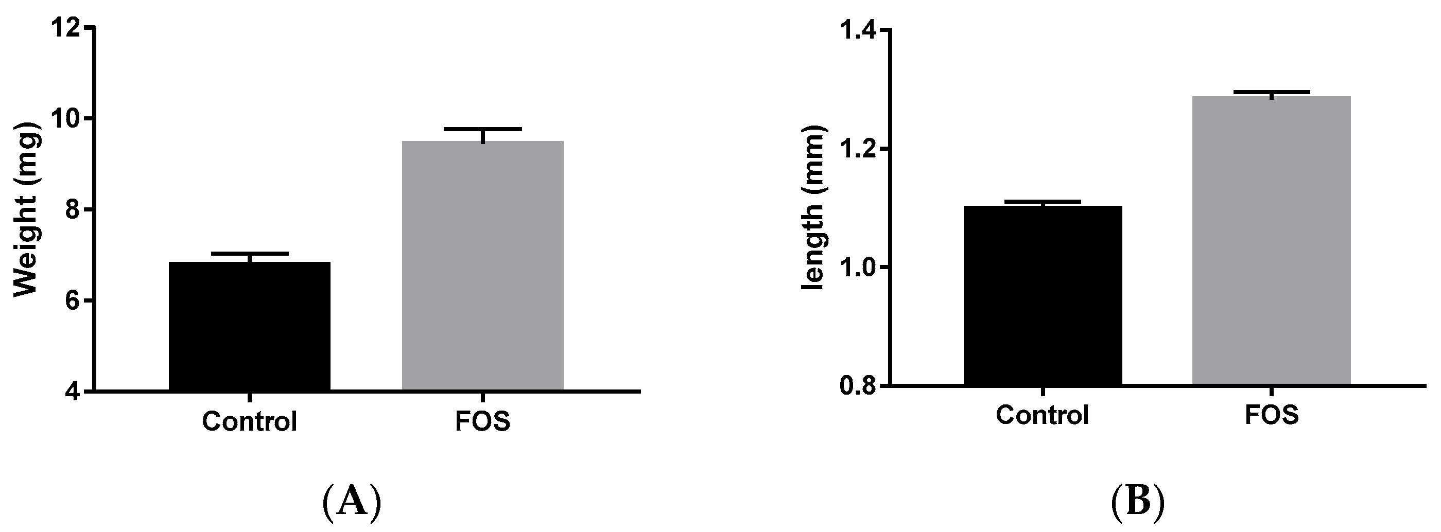
Figure 1. Quality evaluation of Penaeus vannamei post-larvae treated with FOS, (fructooligosaccharide, 1-kestose) (n = 340) and the control (n = 340) groups. Weight (A) and length (B) are given. Data represent mean ± SEM (standard error of the mean), and T bars indicate the SEM (p < 0.001, Student’s t-test).
Microbiolres 14 00060 g001
Figure 2. Survival of P. vannamei post-larvae. The bars represent the median in the control (n = 17) and the FOS-treated groups (n = 17) (p < 0.001, Mann–Whitney U-test). The T bars indicate the range of reported survival values.
Microbiolres 14 00060 g002
Figure 3. Specific activity of phenoloxidase (U mg−1) in Penaeus vannamei post-larvae treated with FOS (fructooligosaccharide, 1-kestose) (n = 17) and the control (n = 17) groups. Data represent mean ± standard deviation (SD). T bars indicates the SD (p < 0.001, Student’s t-test).
Microbiolres 14 00060 g003
Figure 4. Specific activity of lysozyme in Penaeus vannamei post-larvae treated with FOS (fructooligosaccharide, 1-kestose) (n = 17) and the control (n = 17) groups. Data represent mean ± standard deviation (SD). T bars indicate the SD (p < 0.001, Student’s t-test).
Microbiolres 14 00060 g004
Figure 5. Rabbit erythrocyte hemagglutination titers measuring lectins from P. vannamei post-larvae (Log2, scale). The bars represent the median in the control (n = 17) and the FOS-treated (fructooligosaccharide, 1-kestose) groups (n = 17) (p < 0.001, Mann–Whitney U-test). The T bars indicate the range of reported hemagglutination titer values.
Microbiolres 14 00060 g005
Figure 6. Specific activity of protease in Penaeus vannamei post-larvae treated with FOS (fructooligosaccharide, 1-kestose) (n = 17) and the control (n = 17) groups. Data represent mean ± standard deviation (SD). T bars indicate the SD (p < 0.001, Student’s t-test).
Microbiolres 14 00060 g006
Figure 7. Bacterial count (Vibrio spp. and Pseudomonas spp.) in P. vannamei post-larvae (colony-forming units (cfu/mL); Log10, scale). The bars represent the median in the control (n = 17) and the FOS-treated (fructooligosaccharide, 1-kestose) groups (n = 17). Data represent the mean and T bars indicate the SD (p < 0.001, Student’s t-test).
Microbiolres 14 00060 g007
Disclaimer/Publisher’s Note: The statements, opinions and data contained in all publications are solely those of the individual author(s) and contributor(s) and not of MDPI and/or the editor(s). MDPI and/or the editor(s) disclaim responsibility for any injury to people or property resulting from any ideas, methods, instructions or products referred to in the content.