Communication

Baicalin Attenuated PANX-1/P2X7 Axis, P2Y6, and NLRP3/Caspase-1 Signaling Pathways in Peritonitis Induced by Glaesserella parasuis

1
Hubei Key Laboratory of Animal Nutrition and Feed Science, Wuhan Polytechnic University, Wuhan 430023, China
2
Hubei Collaborative Innovation Center for Animal Nutrition and Feed Safety, Wuhan 430023, China
*
Author to whom correspondence should be addressed.
These authors contributed equally to this work.
Microbiol. Res. 2023, 14(3), 1114-1123; https://doi.org/10.3390/microbiolres14030074
Submission received: 1 July 2023 / Revised: 5 August 2023 / Accepted: 9 August 2023 / Published: 9 August 2023

Abstract

:
Glaesserella parasuis (G. parasuis) can cause peritonitis in piglets. However, the pathogenesis of peritonitis remains unclear. Baicalin has been shown to possess anti-inflammatory and anti-oxidant functions. The aim of this study was to investigate the role of the PANX-1/P2X7 axis and the P2Y6 signaling pathway in peritonitis induced by G. parasuis and the effect of baicain on the PANX-1/P2X7 axis and P2Y6 pathway activation triggered by G. parasuis. A G. parasuis serovar 5 isolate SH0165 strain was obtained from the lungs of commercially produced pigs which had the typical symptoms of Glässer’s disease, namely arthritis, fibrinous polyserositis, hemorrhagic pneumonia, and meningitis. Then, 35 piglets were randomly divided into five groups, each group containing seven piglets. The groups consisted of a negative control group, an infection group, a 25 mg/kg baicalin group, a 50 mg/kg baicalin group, and a 100 mg/kg baicalin group. The results showed that G. parasuis could promote PANX-1/P2X7 axis and P2Y6 activation; induce NLRP3/caspase-1, IL-1β and IL-18 expression; trigger PLC/PKC and MLCK/MLC signaling activation; attenuate the expression of tight junction proteins ZO-1, E-cadherin, Occludins, and claudin 1; and stimulate CD14, CD24, CD36, CD47, and CD91 expression in the peritoneum as measured via Western blot (p < 0.01; PLC, p < 0.05). Baicalin could significantly inhibit PANX-1/P2X7 axis, P2Y6, and NLRP3/caspase-1 activation; reduce IL-1β and IL-18 expression; attenuate PLC/PKC and MLCK/MLC activation; promote ZO-1, E-cadherin, occludins, and claudin 1 expression; and reduce CD14, CD24, CD36, CD47, and CD91 expression in the peritoneum induced by G. parasuis as measured via Western blot. Our results deepen the understanding of the mechanism of peritonitis triggered by G. parasuis and provide some novel potential methods of controlling G. parasuis infection.

1. Introduction

Porcine Glässer’s disease, which is caused by the pathogen Glaesserella parasuis (G. parasuis), is a worldwide epidemic, leading to huge economic losses in the swine industry [1]. G. parasuis is widely prevalent on almost every pig farm. The typical clinical signs of Glässer’s disease are fibrinous polyserositis, polyarthritis, and meningitis [2]. G. parasuis is considered a common bacteria colonizing the upper respiratory tract of pigs [3]. Fifteen serovars of G. parasuis have been confirmed via the immunodiffusion method by using heat-stable antigen extracts [4]. Some important related virulent factors, PliA and HtrA, were found to be associated with the pathogenicity of G. parasuis [5,6]. Serovar 5 is thought to be a highly virulent isolate and has the potential to cause peritoneal injury contributing to peritonitis [7]. However, the pathogenic mechanism of peritonitis induced by G. parasuis remains unclear.
Pannexins (PANXs) are composed of large-pore membrane channels and secrete ATP and signal molecules [8]. It is well known that PANX-1 is the most characteristic protein of the PANX family (PANX-1, PANX-2, and PANX-3) and is expressed in many kinds of cells [9]. PANX-1 is central to many distinct inflammatory responses and injury responses [10]. Previous reports have showed that extracellular ATP activates multiple purinergic receptors (P2X and P2Y) which promote inflammation responses in a variety of diseases [11]. However, whether the PANX-1/P2X7 axis and P2Y6 are involved in inflammation during peritonitis induced by G. parasuis has not been investigated.
Multidrug resistance has been increasing all over the world and is considered a public health threat [12]. Several recent investigations have reported the emergence from different origins of virulent multidrug-resistant bacterial pathogens that increase the necessity of the proper use of antibiotics as well as the application of safe, new, potent antimicrobial agents [13,14]. Baicalin is a flavonoid isolated from the rhizome of Scutellaria baicalensis [15]. Baicalin displays important anti-microbial, anti-oxidative, and anti-inflammatory functions [16]. Our previous study has showed that baicalin alleviates porcine peritoneal mesothelial cells apoptosis induced by G. parasuis via the protein kinase C- mitogen-activated protein kinase (PKC-MAPK) pathway [17]. In addition, baicalin could reduce vascular endothelial cell apoptosis via the receptors of advanced glycation endproducts (RAGE), MAPK, and activator protein-1 (AP-1) during G. parasuis invasion [18]. Baicalin inhibits NF-κB and NLRP3 inflammasome signaling pathway activation in porcine aortic vascular endothelial cells triggered by G. parasuis [19]. However, whether baicalin has this effect on PANX-1/P2X7 axis and P2Y6 activation in the peritonitis elicited by G. parasuis has not been reported.
In this study, the aim was to investigate the role of the PANX-1/P2X7 axis and the P2Y6 signaling pathway in the inflammation responses during peritonitis triggered by G. parasuis and whether baicalin is infective in inhibiting PANX-1/P2X7 axis and P2Y6 activation in peritonitis induced by G. parasuis. Our findings deepened the understanding of the mechanism of peritonitis triggered by G. parasuis, which might provide new strategies to control G. parasuis infection in clinical practice.

2. Meterials and Methods

2.1. Bacterial Strains

A strain of G. parasuis serovar 5 isolate, SH0165, was obtained from the lungs of commercially produced pigs which exhibited the arthritis, fibrinous polyserositis, haemorrhagic pneumonia, and meningitis typical of Glässer’s disease [20]. The SH0165 isolate was grown in tryptic soy broth (TSB) (Difco Laboratories, Detroit, MI, USA) for 12 h at 37 °C or cultured on tryptic soy agar (TSA) (Difco Laboratories, Detroit, MI, USA) for 24 h at 37 °C. Meanwhile, 10% newborn bovine serum (Sijiqing, Hangzhou, China) and 10 μg/mL nicotinamide adeninedinucleotide (NAD) (Sigma, St.Louis, MO, USA) were added to the culture medium.

2.2. Drugs

Baicalin was purchased from the National Institutes for Food and Drug Control (Beijing, B110715-201318). Baicalin was dissolved inRPMI-1640 medium (Gibco, Grand Island, NY, USA) in the study.

2.3. Experiment Design

Thirty-five 30-day-old, naturally farrowed, early-weaned piglets (Duroc × Landrace × large white) weighing between 8 and 10 kg were obtained from Wuhan Wannianqing Animal Husbandry Co., Ltd. (Wuhan, China) for the animal experiments.
The selection of pig sample size is explained in our previous research [21]. The 35 piglets were randomly divided into five groups, and each group contained seven piglets. The groups were a negative control group, an infection group, a 25 mg/kg baicalin group, a 50 mg/kg baicalin group, and a 100 mg/kg baicalin gorup. Prior to G. parasuis challenge, the piglets in the baicalin group were injected intramuscularly with baicalin at 25, 50, and 100 mg/kg BW, respectively. After 30 min, the infection group, 25 mg/kg baicalin group, 50 mg/kg baicalin group, and 100 mg/kg baicalin group were intraperitoneally challenged with 1 × 10⁹ CFU G. parasuis in 2 mL normal saline. The piglets from the negative control group were injected intraperitoneally with an equivalent volume of saline. The piglets were monitored for 7 days after the G. parasuis challenge, after which the peritoneum of piglets from all groups was collected and used for protein function study.

2.4. Western Blot

In the study, at least three independent samples in each group were repeated to obtain the results. The proteins from the peritoneum of the piglets were isolated via the whole protein extraction kit (Kaiji, Nanjing, China), and the protein concentrations were determined using the Enhanced BCA Protein Assay Kit (Beyotime Biotechnology, Shanghai, China). The effects of baicalin on protein expression levels in the peritoneum tissue were investigated according to our previous study, with some minor modifications [17]. Briefly, the isolated proteins were extracted with 12% SDS-PAGE. After being transferred onto polyvinylidene difluoride membranes, the membranes were blocked with 5% skim milk for 2 h and then were washed with Tris-buffered saline containing Tween 20 (TBST) five times. After incubation with the corresponding antibodies and five washes with TBST, the membranes were incubated with horseradish-peroxidase-linked goat anti-rabbit antibody (Proteintech, Chicago, USA) for 25 min at37 °C and visualized using ECL solution (Thermo Pierce ECL, Waltham, MA, USA). The protein expression levels were estimated via the FluorChem™ FC2 AIC system (Alpha Innotech, San Leandro, CA, USA).

2.5. Statistical Analysis

The experimental data is presented as mean ± SD. The difference between two groups was determined two-tailed Student’s t test. p values of <0.05 were considered significant.* indicates p < 0.05 and **, p < 0.01.

3. Results

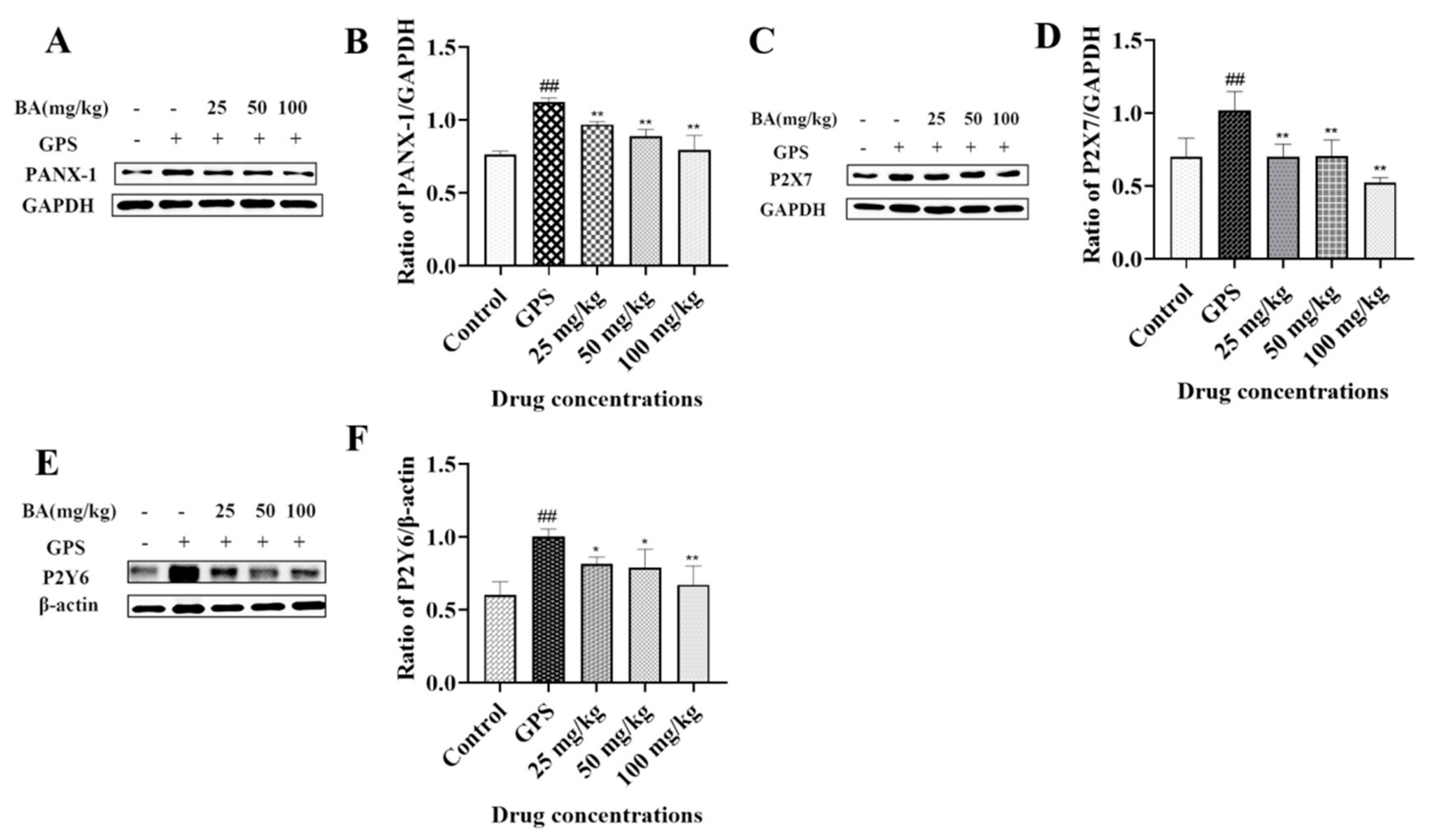
3.1. The Effects of Baicalin on PANX-1, P2X7, and P2Y6 Expression in Peritoneum Induced by G. parasuis

In the piglets challenged with G. parasuis, PANX-1, P2X7, and P2Y6 expression levels were significantly upregulated in the peritoneum compared with the control group (p < 0.01) (Figure 1). However, when the piglets were pretreated with 25–100 mg/kg baicalin, the levels of PANX-1, P2X7, and P2Y6 expression were decreased compared with the infection group (p < 0.01) (P2Y6, 25-50 mg/kg, p < 0.05) (Figure 1). The levels of PANX-1, P2X7, and P2Y6 expression in the baicalin groups were not changed compared to the control group (Figure 1).

3.2. The Effects of Baicalin on NOD-like Receptor Thermal Protein Domain-Associated Protein3 (NLRP3) Inflammasome Activation Triggered by G. parasuis in Peritoneum

The results showed that G. parasuis could activate higher levels of NLRP3 and Cleaved-caspase-1 expression in the peritoneum compared with the control group (p < 0.01) (Figure 2). When the piglets were pretreated with 25–100 mg/kg baicalin, the NLRP3 and Cleaved-caspase-1 expression levels in the peritoneum were significantly inhibited compared with the infection group (p < 0.01) (Figure 2). The levels of NLRP3 and Cleaved-caspase-1 expression in the baicalin groups did not differ from those of the control group (Figure 2).

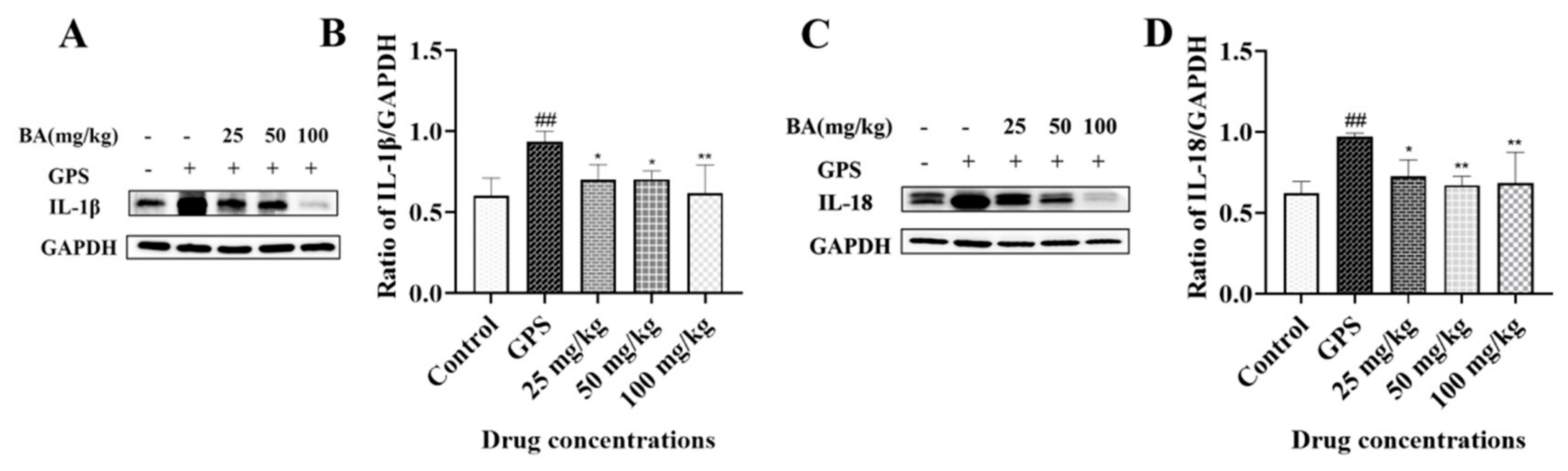
3.3. The Effects of Baicalin on IL-1β and IL-18 Release in Peritoneum Elicited by G. parasuis

In the piglets infected with G. parasuis, cytokine IL-1β and IL-18 expression were significantly increased in the peritoneum compared with the control group (p < 0.01) (Figure 3). A 25–100 mg/kg dose of baicalin could significantly reduce IL-1β and IL-18 production in the peritoneum compared with the infection group (p < 0.01) (25–50 mg/kg, IL-1β and 25 mg/kg, IL-18, p < 0.05) (Figure 3). The levels of IL-1β and IL-18 expression in the baicalin groups were not changed compared to the control group (Figure 3).

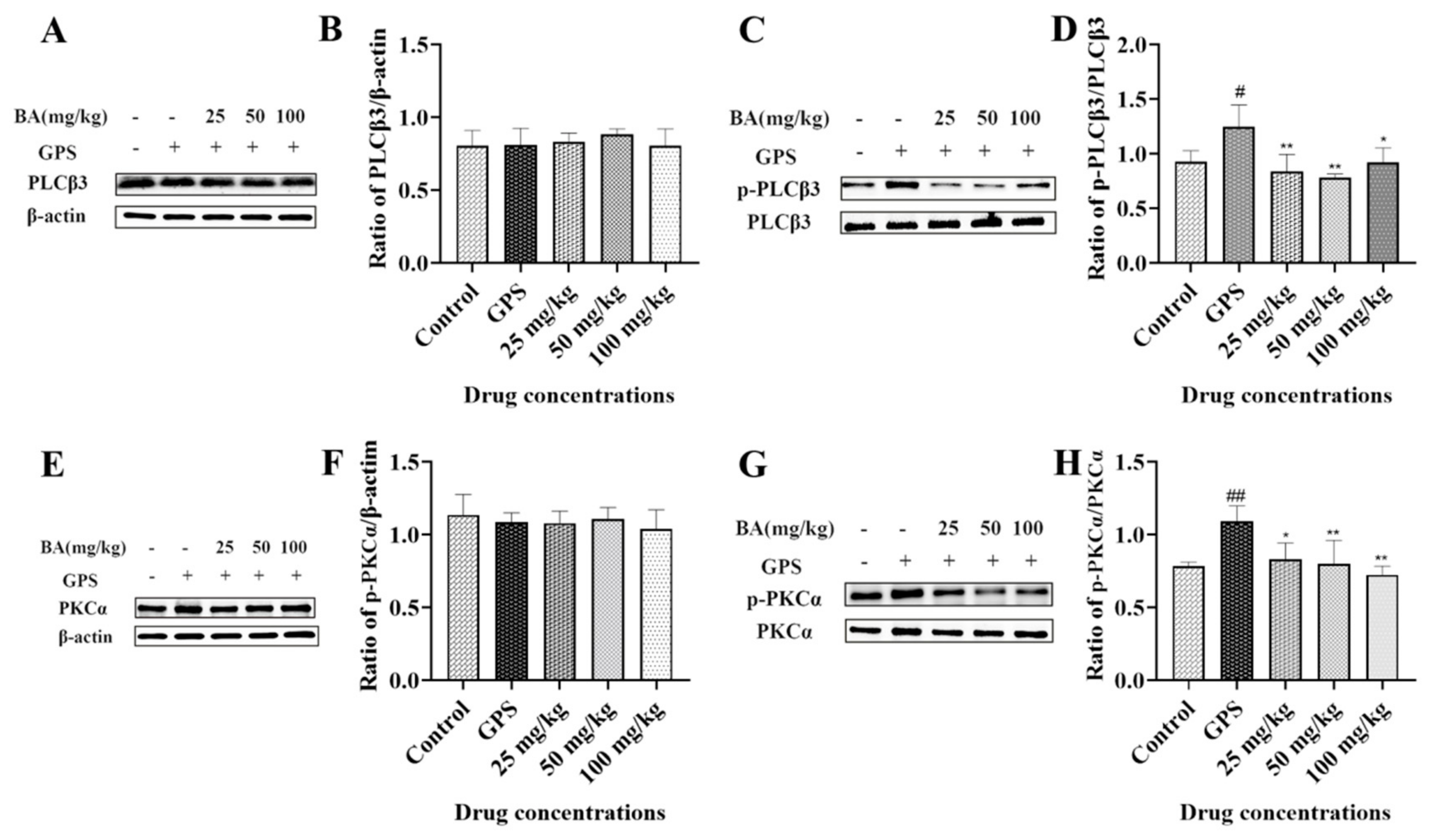
3.4. The Effects of Baicalin on Phospholipase C (PLC) and PKC Activation in Peritoneum Induced by G. parasuis

The results showed that the expression levels of PLCβ3 and PKCα in the peritoneum did not differ between the baicalin treatment group, the infection group, and the control group (Figure 4). When the piglets were challenged with G. parasuis, the expression levels of p-PLCβ3 and p-PKCα in the peritoneum were significantly upregulated compared to the control group (p-PLCβ3, p < 0.05; p-PKCα, p < 0.01) (Figure 4). A 25–100 mg/kg dose of baicalin could significantly inhibit the p-PLCβ3 and p-PKCα expression levels in the peritoneum compared to the infection group (p < 0.01) (100 mg/kg, p-PLCβ3, p < 0.05; 25 mg/kg, p-PKCα, p < 0.05) (Figure 4). The levels of p-PLCβ3 and p-PKCα expression in the baicalin groups did not differ from the control group (Figure 4).

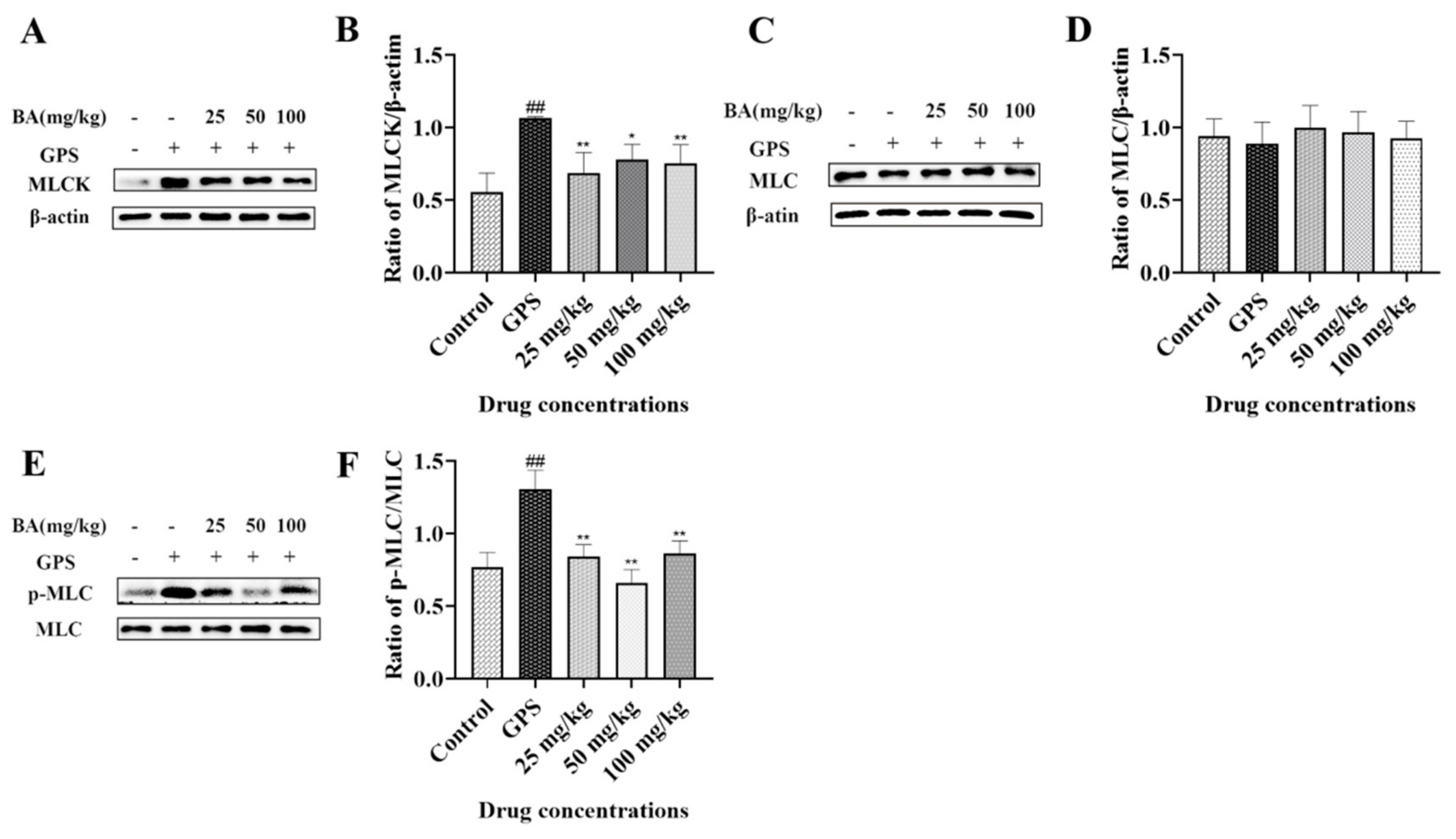
3.5. The Effects of Baicalin on Myosin-Light-Chain Kinase (MLCK) and Myosin-Light-Chain (MLC) Expression Triggered by G. parasuis in Peritoneum

The results showed that G. parasuis did not induce MLC expression in the peritoneum compared to the control group (Figure 5). A 25–100 mg/kg dose of baicalin did not inhibit the MLC expression level in the peritoneum compared to the infection group. However, when the piglets were challenged with G. parasuis, the p-MLC and MLCK expression levels in the peritoneum were significantly increased compared to the control group (p < 0.01) (Figure 5). A 25–100 mg/kg dose of baicalin could downregulate the expression levels of p-MLC and MLCK in the peritoneum compared to the infection group (p < 0.01) (50 mg/kg, p < 0.05) (Figure 5). The levels of p-MLC and MLCK expression in the baicalin groups did not differ from those of the control group (Figure 5).

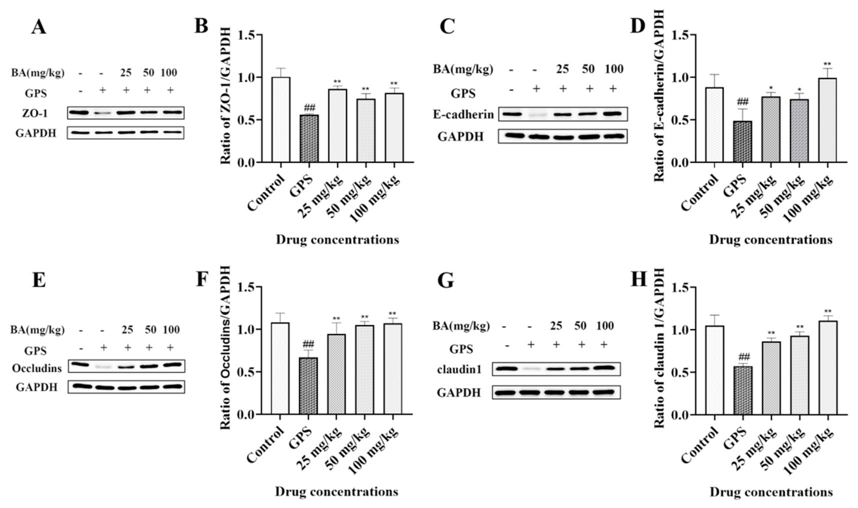
3.6. The Effects of Baicalin on ZO-1, E-Cadherin, Occludins, and Claudin 1 Expression in Peritoneum Elicited by G. parasuis

The results demonstrated that G. parasuis could significantly attenuate ZO-1, E-cadherin, Occludins, and claudin 1 expression in the peritoneum compared to the control group (p < 0.01) (Figure 6). When the piglets were pretreated with 25–100 mg/kg baicalin, the protein expression levels of ZO-1, E-cadherin, Occludins, and claudin 1 in the peritoneum were increased compared to the infection group (p < 0.01) (E-cadherin, 25–50 mg/kg, p < 0.05) (Figure 6). The levels of ZO-1, E-cadherin, Occludins, and claudin 1expression in the baicalin groups did not differ from those of the control group (Figure 6).

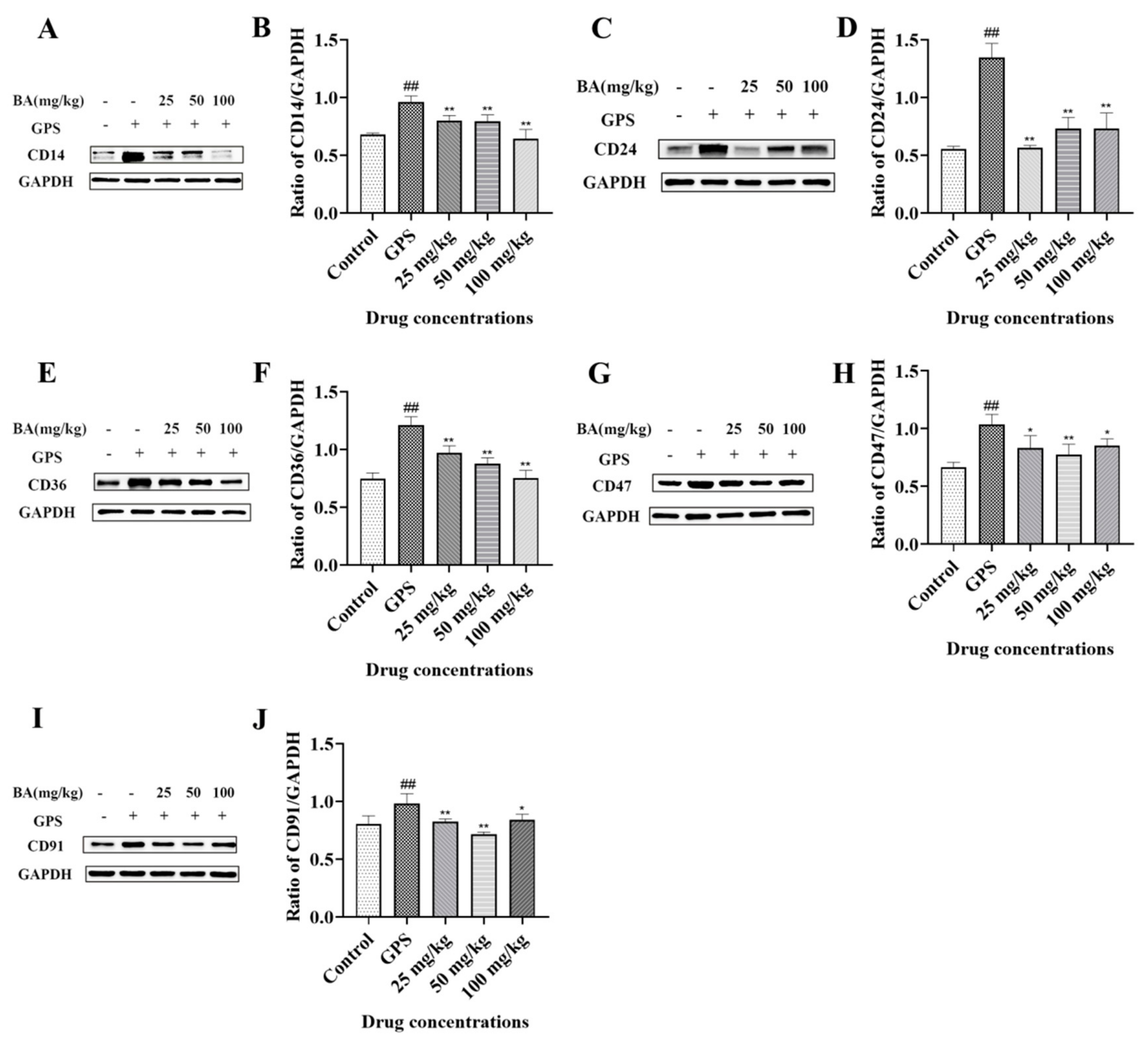
3.7. The Effects of Baicalin on Cluster of Differentiation (CD)14, CD24, CD36, CD47, and CD91 Expression Induced by G. parasuis in Peritoneum

We also explored CD14, CD24, CD36, CD47, and CD91 expression levels in the peritoneum. The results showed that G. parasuis could promote CD14, CD24, CD36, CD47, and CD91 expression in the peritoneum compared to the control group (p < 0.01) (Figure 7). A 25–100 mg/kg dose of baicalin reduced the CD14, CD24, CD36, CD47, and CD91 expression levels in the peritoneum compared to the infection group (p < 0.01) (CD47, 25 and 100 mg/kg, p < 0.05; CD91, 100 mg/kg, p < 0.05) (Figure 7). The levels of CD14, CD24, CD36, CD47, and CD91expression in the baicalin groups did not differ from those of the control group (Figure 7).

4. Discussion

G. parasuis has become one of the most epidemic bacterial respiratory disease pathogens in the pig industry, and the pathogenesis of Glässer’s disease is still unclear. In this study, we first described the activation of the PANX-1/P2X7 and P2Y6 signaling pathway in the peritoneum during the peritonitis processes induced by G. parasuis in a piglet model, which enhanced our knowledge of the G. parasuis pathogenesis in piglets.
The PANX-1 channel is a cellular material exchange pore in the body which can release ATP from the intracellular environment into the extracellular environment when the channel is open [22]. It has been documented that PANX-1 channel can release ATP to initiate signal transduction and activate purinergic P2X7 receptors during bacterial infection [23]. Extracellular ATP elevation could activate P2X7 during cell injury or inflammation, leading to the formation of active NLRP3 inflammasome [24]. Previous studies have showed that P2Y6 is specifically activated by UDP, leading to PKC and PLC signaling pathway activation [25]. Blocking the opening of PANX-1 channels could reduce infection and inflammation in acute Pseudomonas aeruginosa pneumonia [26]; thus, it is thought to be an important drug target [27]. G. parasuis can cause huge economic losses in the pig industry. Our results indicate that baicalin could inhibit PANX-1/P2X7 and P2Y6 activation in the peritoneum, leading to the alleviation of inflammation responses caused by G. parasuis; this might provide some novel methods of controlling peritonitis induced by G. parasuis and the economic losses caused by G. parasuis.
Researchers have reported that tight junctions and their related endothelial cells can form a unique, dynamic, and multi-functional interface [28]. Abnormal expression of tight junction proteins in endothelial cells increases intracellular space and barrier leakage [29]. Inflammatory damage might be related to the destruction of tight junction proteins [30]. Previous studies reported that inflammatory factor production activates the NLRP3-Caspase-1 and IL-1β signaling pathways, which contribute to inflammatory damage and disrupt tight junctions [31]. In this study, we found that G. parasuis activates the NLRP3/caspase-1 signaling pathway in the peritoneum, and the expression levels of the tight junction-related proteins ZO-1, E-cadherin, Occludins, and claudin 1 were downregulated by G. parasuis. However, baicalin could enhance tight junction-related protein expression in the peritoneum triggered by G. parasuis. Our results partially explain the anti-inflammatory mechanism of baicalin, which inhibits peritonitis by improving peritoneum tight junction-related protein expression, but the specific mechanism needs further study.
In this study, we investigated the expression levels of CD14, CD24, CD36, CD47, and CD91 in the peritoneum, and the results indicate that baicalin attenuates CD14, CD24, CD36, CD47, and CD91 expression. Researchers have reported that chemokines, termed “find-me” signals, attract phagocytes toward apoptotic cells, which express an ionic phospholipid phosphatidylserine, or “eat-me” signals and “Don’t eat me” signals, to distinguish healthy cells from apoptotic cells for phagocytosis [32]. CD14, CD36, and CD91 were considered as the “eat-me” signals, and CD24 and CD47 were considered as the “Don’t eat me” signals [33]. CD14 was reported to be involved in skin burn-induced myocardial injury via the MAPK signaling pathway [34]. CD24 in tumor cells could interact with Siglec-10 on the surface of immune cells to induce immune escape by tumor cells [35]. CD36 participates in almitic acid-induced ferroptosis contributing to calcium-iron imbalance in colon cancer cells [36]. CD47 plays an important role in endothelial cell proliferation, apoptosis, inflammation, and atherosclerotic response [37]. Extracellular HMGB1 impairs macrophage-mediated efferocytosis by suppressing CD91 transport [38]. Although these CD molecules are involved in peritonitis induced by G. parasuis, the function needs further investigation.
Taken together, our results showed that G. parasuis could stimulate PANX-1/P2X7 axis and P2Y6 expression; trigger NLRP3/caspase-1 activation; elicit IL-1β and IL-18 expression, induce PLC/PKC and MLCK/MLC signaling activation; attenuate tight junction protein expression; and promote CD14, CD24, CD36, CD47, and CD91 expression in the peritoneum, thereby contributing to peritonitis. Baicalin could attenuate such protein expression and signaling pathway activation to relieve peritonitis induced by G. parasuis, which might provide us with some important novel potential methods of reducing inflammation responses during G. parasuis infection.

Author Contributions

Y.Q. conceived and designed the experiments; S.F., X.T., J.L. and Y.Y. performed the experiments; S.F., X.T., J.L., Y.Y., X.L., M.R., L.G., C.Y., B.Z., Y.L. and Q.L. analyzed the data; S.F. wrote the paper. All authors have read and agreed to the published version of the manuscript.

Funding

This work was supported by the National Natural Science Foundation of China (grant no. 32072917), Key Research and Development Plan of Hubei Province, China (2022BBA0055),and the Natural Science Foundation of Hubei Province, China (grant no. 2022CFB418).

Institutional Review Board Statement

Animal studies were approved by the Animal Care and Use Committee of Wuhan Polytechnic University, Hubei Province, China (WPU202209003).

Informed Consent Statement

Not applicable.

Data Availability Statement

The datasets used and/or analyzed during the current study are available from the corresponding author on reasonable request.

Conflicts of Interest

The authors declare no conflict of interest.

References

  1. Liu, M.; Wang, Q.; Wu, W.; Chen, M.; Zhang, P.; Guo, M.; Lin, H.; Ma, Z.; Zhou, H.; Fan, H. Glaesserella parasuis serotype 5 breaches the porcine respiratory epithelial barrier by inducing autophagy and blocking the cell membrane Claudin-1 replenishment. PLoS Pathog. 2022, 18, e1010912. [Google Scholar] [CrossRef] [PubMed]
  2. Guo, Z.; Jia, Y.; Huang, C.; Zhou, Y.; Chen, X.; Yin, R.; Guo, Y.; Wang, L.; Yuan, J.; Wang, J.; et al. Immunogenicity and protection against Glaesserella parasuis serotype 13 infection after vaccination with recombinant protein LolA in mice. J. Vet. Med. Sci. 2022, 84, 1527–1535. [Google Scholar] [CrossRef] [PubMed]
  3. Jiang, C.; Ma, N.; Cao, H.; Zeng, W.; Ren, J.; Hu, Y.; Zhou, J.; Zhang, M.; Li, C.; Lang, Y.; et al. TurboID Screening of the OmpP2 Protein Reveals Host Proteins Involved in Recognition and Phagocytosis of Glaesserella parasuis by iPAM Cells. Microbiol. Spectr. 2022, 10, e0230722. [Google Scholar] [CrossRef] [PubMed]
  4. Kielstein, P.; Rapp-Gabrielson, V.J. Designation of 15 serovars of Haemophilus parasuis on the basis of immunodiffusion using heat-stable antigen extracts. J. Clin. Microbiol. 1992, 30, 862–865. [Google Scholar] [CrossRef]
  5. An, J.; Cai, J.; Zhang, B.; Li, Y. Pili Subunit PilA Contributes to the Cytoadhesion of Glaesserella Parasuis to Host Cells and Provides Immunoprotection. Appl. Environ. Microbiol. 2023, 89, e0200222. [Google Scholar] [CrossRef]
  6. Zhang, X.; Lin, Y.; Xu, X.; Wen, S.; Wang, Z.; Gu, J.; He, Q.; Cai, X. HtrA is involved in stress response and adhesion in Glaesserella parasuis serovar 5 strain Nagasaki. Vet. Microbiol. 2023, 282, 109748. [Google Scholar] [CrossRef]
  7. Zhang, J.; Zhang, Z.; Xu, J.; Ye, C.; Fu, S.; Hu, C.A.; Qiu, Y.; Liu, Y. Protective Effects of Baicalin on Peritoneal Tight Junctions in Piglets Challenged with Glaesserella parasuis. Molecules 2021, 26, 1268. [Google Scholar] [CrossRef]
  8. Wei, Z.Y.; Qu, H.L.; Dai, Y.J.; Wang, Q.; Ling, Z.M.; Su, W.F.; Zhao, Y.Y.; Shen, W.X.; Chen, G. Pannexin 1, a large-pore membrane channel, contributes to hypotonicity-induced ATP release in Schwann cells. Neural Regen. Res. 2021, 16, 899–904. [Google Scholar] [CrossRef]
  9. Wang, Q.; Li, H.Y.; Ling, Z.M.; Chen, G.; Wei, Z.Y. Inhibition of Schwann cell pannexin 1 attenuates neuropathic pain through the suppression of inflammatory responses. J. Neuroinflamm. 2022, 19, 244. [Google Scholar] [CrossRef]
  10. Koval, M.; Cwiek, A.; Carr, T.; Good, M.E.; Lohman, A.W.; Isakson, B.E. Pannexin 1 as a driver of inflammation and ischemia-reperfusion injury. Purinergic Signal. 2021, 17, 521–531. [Google Scholar] [CrossRef]
  11. Seo, J.H.; Dalal, M.S.; Contreras, J.E. Pannexin-1 Channels as Mediators of Neuroinflammation. Int. J. Mol. Sci. 2021, 22, 5189. [Google Scholar] [CrossRef] [PubMed]
  12. Qi, B.; Li, F.; Chen, K.; Ding, W.; Xue, Y.; Wang, Y.; Wang, H.; Ding, K.; Zhao, Z. Comparison of the Glaesserella parasuis Virulence in Mice and Piglets. Front. Vet. Sci. 2021, 8, 659244. [Google Scholar] [CrossRef] [PubMed]
  13. Algammal, A.M.; Abo Hashem, M.E.; Alfifi, K.J.; Al-Otaibi, A.S.; Alatawy, M.; ElTarabili, R.M.; Abd El-Ghany, W.A.; Hetta, H.F.; Hamouda, A.M.; Elewa, A.A.; et al. Sequence Analysis, Antibiogram Profile, Virulence and Antibiotic Resistance Genes of XDR and MDR Gallibacterium anatis Isolated from Layer Chickens in Egypt. Infect. Drug Resist. 2022, 15, 4321–4334. [Google Scholar] [CrossRef]
  14. Algammal, A.M.; Ibrahim, R.A.; Alfifi, K.J.; Ghabban, H.; Alghamdi, S.; Kabrah, A.; Khafagy, A.R.; Abou-Elela, G.M.; Abu-Elala, N.M.; Donadu, M.G.; et al. A First Report of Molecular Typing, Virulence Traits, and Phenotypic and Genotypic Resistance Patterns of Newly Emerging XDR and MDR Aeromonas veronii in Mugil seheli. Pathogens 2022, 11, 1262. [Google Scholar] [CrossRef] [PubMed]
  15. Li, X.; Li, S.; Ma, C.; Li, T.; Yang, L. Preparation of baicalin-loaded ligand-modified nanoparticles for nose-to-brain delivery for neuroprotection in cerebral ischemia. Drug Deliv. 2022, 29, 1282–1298. [Google Scholar] [CrossRef]
  16. Bao, M.; Ma, Y.; Liang, M.; Sun, X.; Ju, X.; Yong, Y.; Liu, X. Research progress on pharmacological effects and new dosage forms of baicalin. Vet. Med. Sci. 2022, 8, 2773–2784. [Google Scholar] [CrossRef]
  17. Lu, Q.; Zhou, L.; Wang, Z.; Li, X.; Ding, L.; Qiu, Y.; Guo, P.; Ye, C.; Fu, S.; Wu, Z.; et al. Baicalin Alleviate Apoptosis via PKC-MAPK Pathway in Porcine Peritoneal Mesothelial Cells Induced by Glaesserella parasuis. Molecules 2022, 27, 5083. [Google Scholar] [CrossRef]
  18. Fu, S.; Zhao, W.; Xiong, C.; Guo, L.; Guo, J.; Qiu, Y.; Hu, C.A.; Ye, C.; Liu, Y.; Wu, Z.; et al. Baicalin modulates apoptosis via RAGE, MAPK, and AP-1 in vascular endothelial cells during Haemophilus parasuis invasion. Innate Immun. 2019, 25, 420–432. [Google Scholar] [CrossRef] [Green Version]
  19. Fu, S.; Liu, H.; Xu, L.; Qiu, Y.; Liu, Y.; Wu, Z.; Ye, C.; Hou, Y.; Hu, C.A. Baicalin modulates NF-κB and NLRP3 inflammasome signaling in porcine aortic vascular endothelial cells Infected by Haemophilus parasuis Causing Glässer’s disease. Sci. Rep. 2018, 8, 807. [Google Scholar] [CrossRef] [Green Version]
  20. Cai, X.; Chen, H.; Blackall, P.J.; Yin, Z.; Wang, L.; Liu, Z.; Jin, M. Serological characterization of Haemophilus parasuis isolates from China. Vet. Microbiol. 2005, 111, 231–236. [Google Scholar] [CrossRef]
  21. Fu, S.; Yin, R.; Zuo, S.; Liu, J.; Zhang, Y.; Guo, L.; Qiu, Y.; Ye, C.; Liu, Y.; Wu, Z.; et al. The effects of baicalin on piglets challenged with Glaesserella parasuis. Vet. Res. 2020, 51, 102. [Google Scholar] [CrossRef]
  22. Ohyama, S.; Ouchi, T.; Kimura, M.; Kurashima, R.; Yasumatsu, K.; Nishida, D.; Hitomi, S.; Ubaidus, S.; Kuroda, H.; Ito, S.; et al. Piezo1-pannexin-1-P2X(3) axis in odontoblasts and neurons mediates sensory transduction in dentinal sensitivity. Front. Physiol. 2022, 13, 891759. [Google Scholar] [CrossRef] [PubMed]
  23. Loureiro, A.V.; Moura-Neto, L.I.; Martins, C.S.; Silva, P.I.M.; Lopes, M.B.S.; Leitão, R.F.C.; Coelho-Aguiar, J.M.; Moura-Neto, V.; Warren, C.A.; Costa, D.V.S.; et al. Role of Pannexin-1-P2X7R signaling on cell death and pro-inflammatory mediator expression induced by Clostridioides difficile toxins in enteric glia. Front. Immunol. 2022, 13, 956340. [Google Scholar] [CrossRef] [PubMed]
  24. Higashikuni, Y.; Liu, W.; Numata, G.; Tanaka, K.; Fukuda, D.; Tanaka, Y.; Hirata, Y.; Imamura, T.; Takimoto, E.; Komuro, I.; et al. NLRP3 Inflammasome Activation Through Heart-Brain Interaction Initiates Cardiac Inflammation and Hypertrophy During Pressure Overload. Circulation 2023, 147, 338–355. [Google Scholar] [CrossRef]
  25. Wang, Y.; Pei, Y.; Yao, Y.; Cheng, W.; Li, Y.; Duan, Y. P2Y6R: A promising new target in inflammatory diseases and the advance of its antagonists. Curr. Med. Chem. 2023, 30, 2209–2224. [Google Scholar] [PubMed]
  26. Wonnenberg, B.; Tschernig, T.; Voss, M.; Bischoff, M.; Meier, C.; Schirmer, S.H.; Langer, F.; Bals, R.; Beisswenger, C. Probenecid reduces infection and inflammation in acute Pseudomonas aeruginosa pneumonia. Int. J. Med. Microbiol. IJMM 2014, 304, 725–729. [Google Scholar] [CrossRef] [PubMed]
  27. Onódi, Z.; Koch, S.; Rubinstein, J.; Ferdinandy, P.; Varga, Z.V. Drug repurposing for cardiovascular diseases: New targets and indications for probenecid. Br. J. Pharmacol. 2023, 180, 685–700. [Google Scholar] [CrossRef] [PubMed]
  28. Andjelkovic, A.V.; Situ, M.; Citalan-Madrid, A.F.; Stamatovic, S.M.; Xiang, J.; Keep, R.F. Blood-Brain Barrier Dysfunction in Normal Aging and Neurodegeneration: Mechanisms, Impact, and Treatments. Stroke 2023, 54, 661–672. [Google Scholar] [CrossRef] [PubMed]
  29. Qin, X.D.; Yang, T.Q.; Zeng, J.H.; Cai, H.B.; Qi, S.H.; Jiang, J.J.; Cheng, Y.; Xu, L.S.; Bu, F. Overexpression of mitogen-activated protein kinase phosphatase-1 in endothelial cells reduces blood-brain barrier injury in a mouse model of ischemic stroke. Neural Regen. Res. 2023, 18, 1743–1749. [Google Scholar]
  30. Dou, Y.; Liu, P.; Ding, Z.; Zhou, Y.; Jing, H.; Ren, Y.; Heger, Z.; Adam, V.; Li, N. Orally Administrable H(2) S-Scavenging Metal-Organic Framework Prepared by Co-Flow Microfluidics for Comprehensive Restoration of Intestinal Milieu. Adv. Mater. 2023, 35, e2210047. [Google Scholar] [CrossRef]
  31. Xue, Y.; Wang, H.; Tian, B.; Wang, S.; Gao, X.J. Selenium Deficiency Promotes the Expression of LncRNA-MORC3, Activating NLRP3-Caspase-1/IL-1β Signaling to Induce Inflammatory Damage and Disrupt Tight Junctions in Piglets. Biol. Trace Elem. Res. 2023, 201, 2365–2376. [Google Scholar] [CrossRef] [PubMed]
  32. Pontejo, S.M.; Murphy, P.M. Chemokines act as phosphatidylserine-bound “find-me” signals in apoptotic cell clearance. PLoS Biol. 2021, 19, e3001259. [Google Scholar] [CrossRef] [PubMed]
  33. Baraniecki, Ł.; Tokarz-Deptuła, B.; Syrenicz, A.; Deptuła, W. Macrophage efferocytosis in atherosclerosis. Scand. J. Immunol. 2022, 97, e13251. [Google Scholar] [CrossRef] [PubMed]
  34. Zhu, Z.; Zou, B.; Gao, S.; Zhang, D.; Guo, J.; Chen, B.; Hou, H.; Zhu, X. CD14 Involvement in Third-degree Skin Burn-induced Myocardial Injury via the MAPK Signaling Pathway. Cell Biochem. Biophys. 2022, 80, 139–150. [Google Scholar] [CrossRef] [PubMed]
  35. Yin, S.S.; Gao, F.H. Molecular Mechanism of Tumor Cell Immune Escape Mediated by CD24/Siglec-10. Front. Immunol. 2020, 11, 1324. [Google Scholar] [CrossRef] [PubMed]
  36. Kuang, H.; Sun, X.; Liu, Y.; Tang, M.; Wei, Y.; Shi, Y.; Li, R.; Xiao, G.; Kang, J.; Wang, F.; et al. Palmitic acid-induced ferroptosis via CD36 activates ER stress to break calcium-iron balance in colon cancer cells. FEBS J. 2023, 290, 3664–3687. [Google Scholar] [CrossRef]
  37. Zhao, W.; Shen, B.; Cheng, Q.; Zhou, Y.; Chen, K. Roles of TSP1-CD47 signaling pathway in senescence of endothelial cells: Cell cycle, inflammation and metabolism. Mol. Biol. Rep. 2023, 50, 4579–4585. [Google Scholar] [CrossRef] [PubMed]
  38. Wang, Y.; Zhang, W.; Xu, Y.; Wu, D.; Gao, Z.; Zhou, J.; Qian, H.; He, B.; Wang, G. Extracellular HMGB1 Impairs Macrophage-Mediated Efferocytosis by Suppressing the Rab43-Controlled Cell Surface Transport of CD91. Front. Immunol. 2022, 13, 767630. [Google Scholar] [CrossRef]
Figure 1. Detection of PANX-1, P2X7, and P2Y6 expression induced by G. parasuis in the peritoneum. The expression levels of PANX-1 (A,B), P2X7(C,D) and P2Y6 (E,F) were measured via Western blot. BA: Baicalin. GPS: G. parasuis. ## p < 0.01 vs. control. * p < 0.05 and ** p < 0.01.
Microbiolres 14 00074 g001
Figure 2. Effect of baicalin on NLRP3 and Cleaved-caspase-1 activation triggered by G. parasuis in the peritoneum. The NLRP3 (A,B) and Cleaved-caspase-1 (C,D) expression were determined via Western blot. BA: Baicalin. GPS: G. parasuis. ## p < 0.01 vs. control. ** p < 0.01.
Microbiolres 14 00074 g002
Figure 3. Effects of baicalin on IL-1β (A,B) and IL-18 (C,D) expression induced by G. parasuis in the peritoneum. IL-1β and IL-18 expressions were explored by Western blot. BA: Baicalin. GPS: G. parasuis. ## p < 0.01vs. control. * p < 0.05 and ** p < 0.01.
Microbiolres 14 00074 g003
Figure 4. Effects of baicalin on PLC/PKC signaling pathway activation triggered by G. parasuis in the peritoneum. PLCβ3 (A,B), p-PLCβ3 (C,D), PKCα (E,F), and p-PKCα (G,H) expression levels were determined via Western blot. BA: Baicalin. GPS: G. parasuis. # p < 0.05, ## p < 0.01 vs. control. * p < 0.05 and ** p < 0.01.
Microbiolres 14 00074 g004
Figure 5. Effects of baicalin on MLCK/MLC pathway activation triggered by G. parasuis in the peritoneum. MLCK (A,B), MLC (C,D) and p-MLC (E,F) levels were determined via Western blot. BA: Baicalin. GPS: G. parasuis. ## p < 0.01 vs. control. * p < 0.05 and ** p < 0.01.
Microbiolres 14 00074 g005
Figure 6. Effects of baicalin on ZO-1, E-cadherin, Occludins, and claudin 1 expression induced by G. parasuis in the peritoneum. The levels of ZO-1 (A,B), E-cadherin (C,D), Occludins (E,F), and claudin 1 (G,H) were measured via Western blot. BA: Baicalin. GPS: G. parasuis. ## p < 0.01 vs. control. * p < 0.05 and ** p < 0.01.
Microbiolres 14 00074 g006
Figure 7. Effects of baicalin on CD14, CD24, CD36, CD47, and CD91 expression triggered by G. parasuis in the peritoneum.CD14 (A,B), CD24 (C,D), CD36 (E,F), CD47 (G,H), and CD91 (I,J) expression levels were determined via Western blot. BA: Baicalin. GPS: G. parasuis. ## p < 0.01 vs. control. * p < 0.05 and ** p < 0.01.
Microbiolres 14 00074 g007
Disclaimer/Publisher’s Note: The statements, opinions and data contained in all publications are solely those of the individual author(s) and contributor(s) and not of MDPI and/or the editor(s). MDPI and/or the editor(s) disclaim responsibility for any injury to people or property resulting from any ideas, methods, instructions or products referred to in the content.

