Article

The Influence of Pre-Existing Immunity against Human Common Cold Coronaviruses on COVID-19 Susceptibility and Severity

by
Erick De La Torre Tarazona
1,2,*,†,
Daniel Jiménez
1,†,‡,
Daniel Marcos-Mencía
3,
Alejandro Mendieta-Baro
1,
Alejandro Rivera-Delgado
1,
Beatriz Romero-Hernández
3,4,
Alfonso Muriel
4,5,
Mario Rodríguez-Domínguez
3,4,
Sergio Serrano-Villar
1,2 and
Santiago Moreno
1,2,6
1
Infectious Diseases Department, Hospital Universitario Ramón y Cajal, Instituto Ramón y Cajal de Investigación Sanitaria (IRYCIS), 28034 Madrid, Spain
2
Centro de Investigación Biomédica en Red de Enfermedades Infecciosas (CIBERINFEC), Instituto de Salud Carlos III, 28029 Madrid, Spain
3
Microbiology Department, Hospital Universitario Ramón y Cajal, Instituto Ramón y Cajal de Investigación Sanitaria (IRYCIS), 28034 Madrid, Spain
4
Consorcio de Investigación Biomédica en Red de Epidemiología y Salud Pública (CIBERESP), Instituto de Salud Carlos III, 28029 Madrid, Spain
5
Biostatistics Unit, Hospital Universitario Ramón y Cajal, Instituto Ramón y Cajal de Investigación Sanitaria (IRYCIS), 28034 Madrid, Spain
6
Department of Medicine, Universidad de Alcalá, 28054 Madrid, Spain
*
Author to whom correspondence should be addressed.
These authors contributed equally to this work.
Deceased.
Microbiol. Res. 2023, 14(3), 1364-1375; https://doi.org/10.3390/microbiolres14030093
Submission received: 29 August 2023 / Revised: 10 September 2023 / Accepted: 11 September 2023 / Published: 14 September 2023

Abstract

:
The susceptibility to SARS-CoV-2 infection and the severity of COVID-19 manifestations vary significantly among individuals, prompting the need for a deeper understanding of the disease. Our objective in this study was to investigate whether previous infections with human common cold coronaviruses (hCCCoV) might impact susceptibility to and the progression of SARS-CoV-2 infections. We assessed the serum antibody levels against SARS-CoV-2 and four hCCCoV (H-CoV-OC43, -NL63, -HKU1, and -229E) in three distinct populations: 95 uninfected individuals (COVID-19-negative), 83 individuals with mild or asymptomatic COVID-19 (COVID-19-mild), and 45 patients who died due to COVID-19 (COVID-19-severe). The first two groups were matched in terms of their exposure to SARS-CoV-2. We did not observe any differences in the mean antibody levels between the COVID-19-mild and the COVID-19-negative participants. However, individuals in the COVID-19-mild group exhibited a higher frequency of antibody levels (sample/control) > 0.5 against H-CoV-HKU1, and >1 against H-CoV-229E and -OC43 (p < 0.05). In terms of severity, we noted significantly elevated H-CoV-NL63 IgG levels in the COVID-19-severe group compared to the other groups (p < 0.01). Our findings suggest a potential mild influence of hCCCoV antibody levels on the susceptibility to SARS-CoV-2 infection and the severity of COVID-19. These observations could aid in the development of strategies for predicting and mitigating the severity of COVID-19.

1. Introduction

The emergence of severe acute respiratory syndrome coronavirus 2 (SARS-CoV-2), first reported in late 2019 in Wuhan (China), and its subsequent rapid and uncontrolled global spread led to an unprecedented pandemic and a monumental public health crisis. As of the latest available data, there have been over 767 million reported cases of SARS-CoV-2 infections and more than 6.9 million deaths due to COVID-19 worldwide [1]. The clinical manifestation of COVID-19 is extraordinarily diverse, spanning a spectrum that encompasses a wide range of scenarios. These include asymptomatic cases, mild flu-like symptoms, moderate respiratory distress and severe disease, which can potentially result in death due to fatal pulmonary failure [2]. This intricate and multifaceted nature of COVID-19 underscores the need for a comprehensive and exhaustive understanding of its different clinical expressions. Moreover, the underlying factors that govern the manifestation of these clinical phenotypes need to be deciphered in greater depth, as this understanding could potentially guide targeted therapeutic strategies and improve the prediction of outcomes, necessitating a multidimensional approach to unravel the heterogeneity intrinsic to this viral disease [3].
Among the myriad factors that have been postulated to wield influence over susceptibility to SARS-CoV-2 infection and/or the progression of COVID-19, the role of pre-existing immunity developed against other human coronaviruses (H-CoV) has emerged as a noteworthy and potentially pivotal determinant. Prior to the emergence of SARS-CoV-2, the scientific community was familiar with the existence of four human common cold coronaviruses (hCCCoV): H-CoV-NL63, H-CoV-229E, H-CoV-OC43, and H-CoV-HKU1. These four viruses, categorized into the alpha (H-CoV-229E and -NL63) and beta-coronavirus (H-CoV-OC43 and -HKU1) groups, are commonly responsible for mild upper respiratory tract diseases, often exhibiting a seasonal pattern with a higher prevalence during the colder months of winter [4,5]. Taken together, it has been estimated that these hCCCoV account for a substantial 15–30% of global common cold cases [6]. Given their recurrent circulation, a significant proportion of the global population has developed antibodies against these coronaviruses. Notably, the seroprevalence of these antibodies exceeds a remarkable 90% or more among the adult population for all four hCCCoV [7,8].
The landscape of studies aimed at elucidating the alterations in hCCCoV-specific IgG levels following SARS-CoV-2 infection yields a perplexing array of results. While some studies suggest that SARS-CoV-2 infection does not enhance specific antibody titers against hCCCoV [9,10], other works have reported an increase in specific antibodies targeting betacoronaviruses, such as H-CoV-OC43 and/or -HKU1 [11,12,13,14,15,16,17,18], as well as specific antibodies against alphacoronaviruses [19,20]. Additionally, there have been reports indicating the elevated antibody responses against all four hCCCoV [21,22].
The quest to unravel the potential influence of pre-existing hCCCoV immunity on susceptibility to SARS-CoV-2 infection has also yielded a plethora of diverse findings [23]. While certain investigations found no discernible correlation between levels of hCCCoV antibodies and either the severity of COVID-19 or susceptibility to SARS-CoV-2 infections [10,11,24], there are compelling counterarguments. A notable subset of studies has proposed that higher levels of antibodies against hCCCoV are intricately associated with milder COVID-19 disease outcomes [9,19,20,22,25,26], a shorter duration of symptoms [17], or even a reduced risk of contracting the virus. However, it is imperative to note that certain studies have associated higher levels of hCCCoV antibodies with increased SARS-CoV-2 disease severity [12,14,15,18]. This inherent variability in findings can be attributed, at least in part, to the differences in methodologies employed across these previous studies, alongside the substantial diversity in the health statuses of the populations that have been evaluated.
Given the potential influence of pre-existing immunity against hCCCoV on the outcome of SARS-CoV-2 infection, it is crucial to provide novel insights into the interplay between hCCCoV antibodies and both the acquisition of SARS-CoV-2 and the progression of COVID-19 through complementary strategies. To evaluate SARS-CoV-2 acquisition, the present study analyzed antibody levels against hCCCoV and SARS-CoV-2 in two distinct populations: individuals with either asymptomatic or mild COVID-19, and uninfected individuals. Careful matching of these groups was carried out, considering variables such the degree of viral exposure, age and gender. Furthermore, a third group of individuals who developed severe COVID-19 was also analyzed, in order to evaluate the impact of hCCCoV antibodies on the course of COVID-19 progression. The ultimate aim of this research is to contribute with novel insights into the panorama of susceptibility to SARS-CoV-2 infection and the gradient severity that characterizes COVID-19, all in the context of the intricate interplay with immunity against hCCCoV.

2. Materials and Methods

2.1. Study Design and Categorization of the Study Population

We categorized each participant into three distinct groups: COVID-19-negative, COVID-19-mild, and COVID-19-severe. The COVID-19-mild group consisted of healthcare workers who either experienced mild COVID-19 symptoms or were asymptomatic to the disease. These individuals were randomly selected from those healthcare workers who had tested positive for SARS-CoV-2 through PCR analysis, as recorded in the archives of the Microbiology Laboratory. Likewise, the COVID-19-negative group consisted of healthcare workers who exhibited no clinical signs of symptoms suggestive of SARS-CoV-2 infection. These individuals consistently tested negative for SARS-CoV-2 through PCR testing and lack of detectable SARS-CoV-2 IgG. Like their COVID-19-mild counterparts, participants in this group were also selected from the same archives. COVID-19-mild and COVID-19-negative groups were matched based on age (±5 years), gender, and estimated degree of SARS-CoV-2 exposure determined by occupational position within the healthcare sector. On the other hand, to assess the influence on disease severity, the inclusion of the COVID-19-severe group allowed us to evaluate H-CoV IgG levels in severe COVID-19 scenarios, although the clinical characteristics of this group were not matched with those of the COVID-19-mild and COVID-19-negative groups.
The occupational positions of participants from the COVID-19-mild and COVID-19-negative groups were classified into three risk levels based on the potential for exposure to SARS-CoV-2. The high-risk category encompassed roles such as resident medical interns, medical specialists, service and section heads, emergency physicians, physiotherapists, caretakers, and nurses. Intermediate-risk roles included social workers, radio-diagnosis technicians, optometrists, resident intern biologists, administrative assistants, and hairdressers. Finally, the low-risk category included job positions such as pathological anatomy and clinical diagnosis technicians, cooks, plumbers, mechanics, and kitchen helpers.
We enrolled participants who were not vaccinated against SARS-CoV-2 during the first wave of the pandemic (March to June 2020). Serum samples were collected from each participant at the time of inclusion into the study. Ethical considerations were upheld, as the study was approved by the Ethics Committee for Clinical Research of the same hospital. Furthermore, participants provided their written informed consent before being included in this study.

2.2. Determination of SARS-CoV-2 Infection

To confirm the classification of participants into their respective groups, we employed both qPCR analysis and evaluation of anti-SARS-CoV-2 IgG levels. The detection of N, S, and ORF1ab genes from SARS-CoV-2 was performed using multiplex qPCR on nasopharyngeal samples, following the manufacturer’s guidelines of the TaqPath COVID-19 kit (Thermofisher, Waltham, MA, USA). The presence of SARS-CoV-2 antibodies in the serum was assessed using an indirect chemiluminescence immunoassay (Vircell, Granada, Spain).

2.3. Preparation of hCCCoV Antigens

For the preparation of hCCCoV antigens, we reconstituted the lyophilized hCCCoV nucleocapsid (N) antigen (Creative Diagnostics, New York, NY, USA) using phosphate-buffered saline (PBS) (Sigma-Aldrich, Saint Louis, MO, USA) to achieve a final concentration of 2 μg/mL. Additionally, we prepared a wash buffer by adding 0.1% Tween-20 (Sigma-Aldrich, Saint Louis, MO, USA) to PBS, resulting in PBST.

2.4. Antibody Quantification by ELISA

The quantification of IgG against the N protein of H-CoV-NL63, -HKU1, -229E, and -OC43 (Creative Diagnostics, New York, NY, USA) was performed using an indirect and manual ELISA technique. Serum samples were heat-inactivated at 56 °C for 30 min. Subsequently, 96-well plates (Sigma-Aldrich, Saint Louis, MO, USA) were coated overnight at 4 °C with 50 μL of hCCCoV nucleocapsid antigen per well. Following this, the plates were blocked with 200 µL of a blocking buffer (PBST supplemented with 3% nonfat milk) for 1 h at 37 °C. After the blocking step, the plates were washed three times with 200 μL of PBST. Then, 50 μL serum samples, diluted at a ratio of 1/20, were added to each well, and this was followed by a 2 h incubation period. Subsequent to incubation, the plates were washed again, and 100 μL of a 1/3000 diluted conjugated anti-human IgG solution (Streptavidin/HRP, Agilent, Santa Clara, CA, USA) was added. The plates were then incubated for an additional hour at 37 °C, followed by an additional three further washes. Subsequently, 100 μL of substrate solution (3,3′,5,5′-Tetramethylbenzidine (TMB) Liquid Substrate System for ELISA, Sigma-Aldrich, Saint Louis, MO, USA) was added to the plates, and they were incubated at room temperature for 10 min. The reaction was stopped by adding 50 μL of stop solution (hydrochloric acid 1N, Sigma-Aldrich, Saint Louis, MO, USA). The absorbance levels were measured at 405 nm, with a blank well used to correct for background reactivity for each serum sample. A positive control (anti-IgG F2 polyclonal antibody, Creative Diagnostics, New York, NY, USA) and a negative control were systematically included in each assay. IgG values against hCCCoV are expressed as sample/control (s/co), which is the difference in the absorbances obtained from the sample and the background control. For the determination of IgG targeting the S protein of SARS-CoV-2, we followed the manufacturer’s guidelines of the Abbott Alinity SARS-CoV-2 assay (Abbot, North Chicago, IL, USA). IgG values against SARS-CoV-2 are expressed as UA/mL.

2.5. Statistical Analysis

Descriptive statistics were used to summarize the obtained data. The data of hCCCoV IgG levels were tested for normality using either the Kolmogorov–Smirnov or Shapiro–Wilk tests, ensuring the validity of subsequent analyses. Non-parametric tests, namely the Mann–Whitney U and Kruskal–Wallis tests, were used to compare H-CoV IgG titers across the analyzed groups. Also, a Pearson correlation analysis was performed to analyze the correlation patterns in H-CoV IgG titers across all studied individuals included in the study. Logistic regression analysis was conducted to assess the relationship between hCCCoV IgG levels and susceptibility to SARS-CoV-2 infection. IBM SPSS Statistics 26.6 was employed for the statistical analysis.

3. Results

3.1. Characteristics of the Study Population

Our study included a total of 223 participants. Among these participants, we categorized them into three groups: the COVID-19-negative group, consisting of 83 individuals who had not been infected by SARS-CoV-2; the COVID-19-mild group, comprising 95 individuals who experienced asymptomatic or mild COVID-19; and the COVID-19-severe group, which included 45 individuals who died as a result of severe COVID-19 cases. Individuals from the COVID-19-negative and COVID-19-mild groups were carefully matched for sex, age (within a narrow range of ±5 years), and their degree of exposure to SARS-CoV-2 based on their job position. The median age of these participants was 41 years, and a majority of them, accounting for 88%, were female. Also, in both the COVID-19-negative and COVID-19- mild groups, 87% of participants had a high degree of exposure to SARS-CoV-2 due to their occupational roles.

3.2. Examination of hCCCoV IgG Levels in the Study Population

Antibody titers against the N protein of the four hCCCoV and the S protein of SARS-CoV-2 were quantified within each group of participants. Our analysis of antibody levels revealed that the COVID-19-mild group exhibited slightly elevated hCCCoV IgG titers when compared to the COVID-19-negative group. However, these differences did not reach statistical significance (p > 0.1), as illustrated in Figure 1 and detailed in Table S1.
Furthermore, our investigation delved into the correlation between IgG titers against hCCCoV and SARS-CoV-2 across all participants. Performing a Pearson correlation analysis, we observed a positive correlation among the alphacoronaviruses (H-CoV-NL63 and -229E, r = 0.19; p < 0.01) and a similar correlation among the betacoronaviruses (H-CoV-HKU1 and -OC43, r = 0.24; p < 0.001). Interestingly, we also observed a positive correlation between H-CoV-NL63 and SARS-CoV-2 IgG titers (r = 0.20, p < 0.01), as shown Figure S1.

3.3. Evaluation of the Influence of hCCCoV IgG Levels on the Susceptibility to SARS-CoV-2 Infection

To explore the potential impact of hCCCoV IgG levels on susceptibility to SARS-CoV-2, we considered the COVID-19-negative group as having lower susceptibility to SARS-CoV-2, and the COVID-19-mild group as having high susceptibility to this viral infection. In these groups, we examined seropositivity to hCCCoV infections based on IgG levels, considering concentrations above 0.5 or 1 as positive (as detailed in Table 1). We performed logistic regression analysis to examine the potential relationship between hCCCoV IgG levels and susceptibility to SARS-CoV-2 infection. Our observations indicate a significant association between a higher proportion of individuals in the COVID-19-mild group with antibody levels exceeding 0.5 against H-CoV-HKU1, and levels higher than 1 against H-CoV-229E and -OC43 (p < 0.05). These findings suggest a potential role of the influence of higher hCCCoV IgG levels on the susceptibility to SARS-CoV-2 infections.

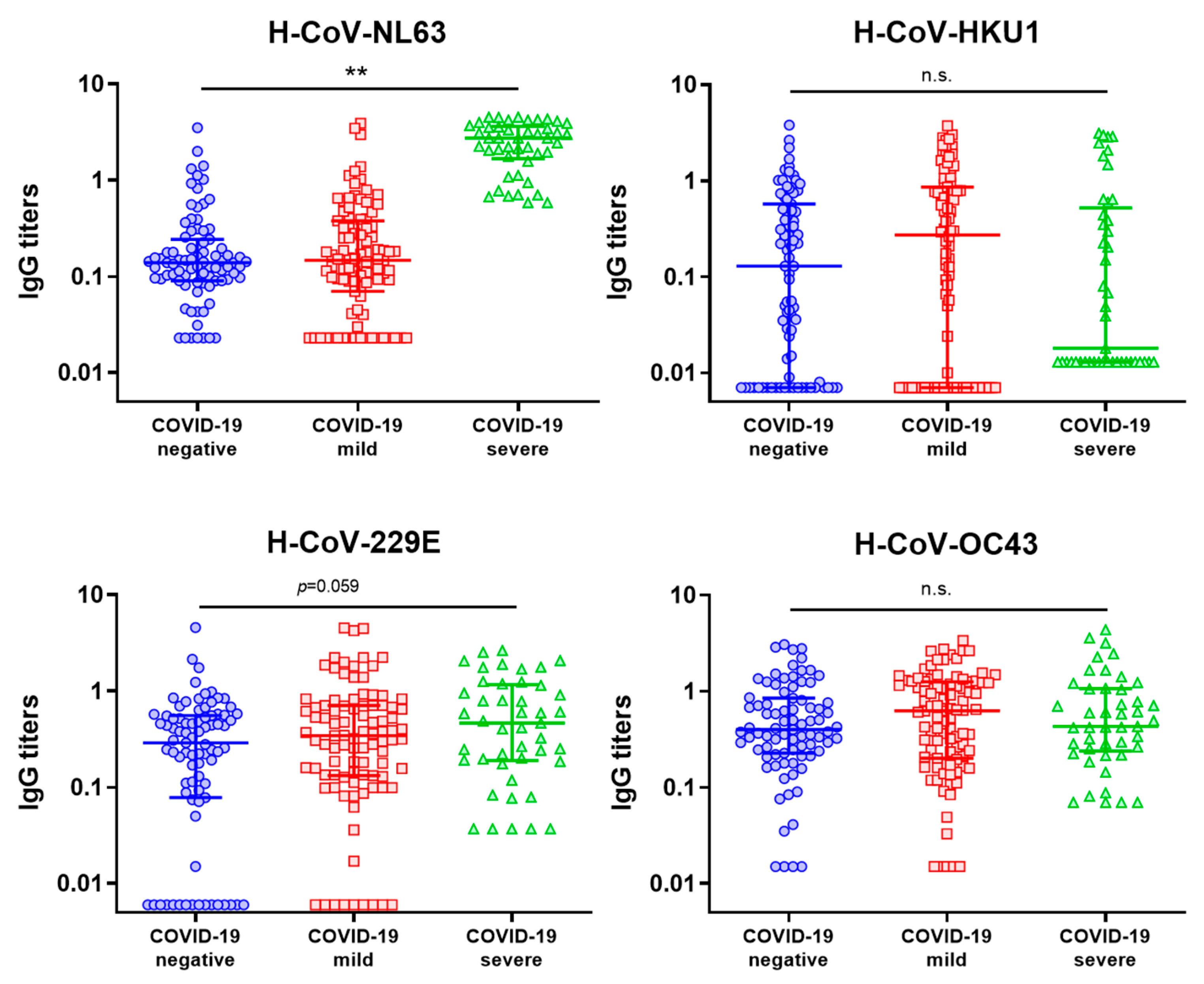
3.4. Evaluation of the Influence of hCCCoV IgG Levels on the Severity of COVID-19

In order to comprehensively assess the impact of hCCCoV IgG titers on COVID-19 severity, we included the COVID-19-severe group in our analysis. A significant increase in H-CoV-NL63 IgG levels (median of 2.739; IQR: 1.673–3.628, p < 0.01) was observed in comparison to the other groups (median of 0.136 (IQR: 0.09–0.238) and 0.149 (IQR: 0.079–0.388) for the COVID-19-negative and COVID-19-mild groups, respectively). Likewise, the IgG levels against the other three hCCCoV remained relatively similar across all three groups (as illustrated in Figure 1).
In addition to this, we also analyzed SARS-CoV-2 IgG levels across the participants, which exhibited significant variations among the analyzed groups (p < 0.01). The COVID-19-mild group showed a median IgG level of 470.95 UA/mL (IQR: 211–1027), while the COVID-19-severe group displayed SARS-CoV-2 IgG levels that were approximately 2.7 times higher than those in the COVID-19-mild group, with a median of 1256.3 UA/mL (IQR: 31.0–13776.2, p < 0.01), as detailed in Figure 1 and Table S1. These findings align with previous studies that have also reported elevated SARS-CoV-2 antibody levels in severe cases of COVID-19 [27].

4. Discussion

The emergence of SARS-CoV-2 and the subsequent global pandemic have prompted rigorous investigation aimed at elucidating the intricate factors influencing susceptibility to SARS-CoV-2 infection and the severity of COVID-19 cases. The different populations evaluated have exhibited varying degrees of susceptibility to SARS-CoV-2 infection, as well as diverse manifestations of COVID-19 severity. These disparities may arise from a complex interplay of several host and viral factors. Understanding the factors influencing these variabilities is of paramount importance to improve clinical management and devise more effective public health strategies. In this study, our primary objective was to explore the correlation between hCCCoV IgG levels and susceptibility to SARS-CoV-2 among healthcare workers who had comparable degrees of exposure to the virus but differing SARS-CoV-2 infection statuses.
Our initial focus lay in the assessment of hCCCoV IgG levels in two groups: the COVID-19-negative group, consisting of uninfected individuals, and the COVID-19-mild group, consisting of individuals who experienced asymptomatic or mild COVID-19. These two groups were matched based on sex, age, and degrees of SARS-CoV-2 exposure. This matching strategy between both groups was adopted to minimize the impact of these potential confounding factors, which could influence the acquisition and/or progression of COVID-19. Some investigations have revealed age and gender disparities in COVID-19 susceptibility and outcomes. Older adults, particularly those aged 65 and older, are at a higher risk of developing severe illness and complications if they contract the virus [28]. Likewise, some studies have suggested that men are more likely to experience severe cases and higher mortality rates compared to women [29]. To our knowledge, few works have considered this matching approach in order to reduce the variability of demographic and clinical characteristics among the analyzed groups of individuals infected or not with SARS-CoV-2.
Our initial analysis revealed only a slight, albeit statistically non-significant, increase in the median IgG levels for the four hCCCoV evaluated among individuals in the COVID-19-mild group compared to the COVID-19-negative group. These observations align with previous research that has reported slightly elevated antibody levels against H-CoV-HKU1 and -229E in individuals with COVID-19 compared to their uninfected counterparts [30]. Additionally, several studies have suggested that H-CoV-HKU1 and -OC43 IgG levels may increase during mild SARS-CoV-2 infection, and hCCCoV antibodies could be boosted following infection with this virus [15,31,32]. The elevated hCCCoV antibody levels in SARS-CoV-2-infected individuals might be attributed to cross-reactivity among H-CoV antibodies or elevated IgG levels pre-existing prior to SARS-CoV-2 infection.
Several studies have reported cross-reactivity among hCCCoV antibodies [21,33,34], often attributed to homology found in their antigenic regions. For instance, the protein homology between SARS-CoV-2 and seasonal hCCCoV antigens is estimated to be approximately 19.8% for S1, 39.9% for S2, and 33.0% for N [35]. Thus, it is plausible that antibodies generated in response to hCCCoV infections in the pre-pandemic period may recognize epitopes on the N protein of SARS-CoV-2 [36]. Interestingly, despite lower sequence identity, cross-reactivity with SARS-CoV-2 appears to be more pronounced for alpha hCCCoV compared to beta hCCCoV, underscoring the importance of the conformational recognition of antigens [36]. This observation aligns with our data, as we observed a positive correlation among IgG levels between SARS-CoV-2 and H-CoV-NL63 (Figure S1).
However, it is important to emphasize that while cross-reactivity among hCCCoV antibodies is evident, studies have shown that this cross-reaction with SARS-CoV-2 antigens in sera from the pre-COVID-19 era does not necessarily neutralize the SARS-CoV-2 infection in vitro [37]. Moreover, other studies have suggested that baseline hCCCoV antibodies do not confer protection against SARS-CoV-2 infection [32], and hCCCoV neutralizing antibodies tend to be species-specific without exhibiting cross-reactivity against SARS-CoV-2 RBD [38]. This phenomenon could be potentially attributed to the high homology of most structural proteins among hCCCoV, with the notable exception of the more species-specific S protein [39,40,41]. The RBD region, in particular, is poorly conserved among these coronaviruses [38]. Moreover, it is important to underscore the strong correlation between the levels of RBD-binding antibodies and SARS-CoV-2 neutralizing antibodies, and immune sera from individuals recently exposed to hCCCoV infections do not exhibit cross-reactivity with SARS-CoV-2 RBD [38]. Previous studies have also indicated that hCCCoV antibodies are present at varying levels prior to SARS-CoV-2 infection and continue to be detectable after infection. Some research suggests that SARS-CoV-2 infection does not induce specific antibodies against hCCCoV [9,10]. Furthermore, pre-existing anti-N antibodies targeting endemic alphacoronaviruses and S2 domain-specific anti-spike antibodies against betacoronavirus H-CoV-OC43 have been found to be elevated in COVID-19 patients, while hCCCoV antibody concentrations remain relatively unchanged throughout the course of the disease [42].
Hence, while cross-reactivity among hCCCoV antibodies can be observed, it does not necessarily provide protection against SARS-CoV-2. In fact, one report suggests that pre-existing antibodies targeting the spike antigen of H-CoV-OC43 might actually enhance susceptibility to SARS-CoV-2 [42]. To evaluate the potential impact of hCCCoV IgG levels on susceptibility to SARS-CoV-2, we considered the COVID-19-negative and COVID-19-mild groups as having lower and high susceptibility to this viral infection, respectively. In these groups, we examined seropositivity to hCCCoV infections based on IgG levels, considering concentrations above 0.5 or 1 as positive (Table 1). Some previous works have also considered similar cut-off values to determine the seropositivity to hCCCoV infections [7,31]. Our results show an association between a higher proportion of individuals in the COVID-19-mild group with antibody levels higher than 0.5 against H-CoV-HKU1, or higher than 1 against H-CoV-229E and -OC43 (p < 0.05). These results suggest a potential influence of higher hCCCoV IgG levels on an increased susceptibility to SARS-CoV-2 infection, due to these groups being matched by degrees of exposition to this virus.
On the other hand, a definitive connection between the levels of hCCCoV antibodies and the severity of COVID-19 remains elusive [30]. In our investigation of hCCCoV antibody levels in severe COVID-19 cases, we included individuals who were SARS-CoV-2-positive and had died due to severe COVID-19. Notably, we observed higher levels of antibodies against H-CoV-NL63 in this group compared to the other groups (Figure 1, p < 0.01). Similarly, other studies have found elevated IgG levels against H-CoV-NL63 and -229E in severe COVID-19 cases, or that levels of H-CoV-HKU1 and -OC43 antibodies may be increased during convalescence in severe cases compared to mild cases [31]. Another study reported significantly higher H-CoV-OC43 S IgG titers in patients with severe disease compared to those with mild symptoms within the first 21 days after symptom onset, establishing a correlation with disease severity in individuals aged over 60 [12].
Furthermore, some reports suggest that prior infections with hCCCoV could impact the quality of the antibody response against SARS-CoV-2. A study analyzing hCCCoV antibodies before and after SARS-CoV-2 infection found that elevated H-CoV-OC43 IgG levels prior to infection were associated with a stronger IgG response to SARS-CoV-2 after infection. This raises the question of whether high baseline H-CoV-OC43 antibody levels might have an adverse effect on the immune response against SARS-CoV-2 [32], considering that higher IgG levels are associated with more severe disease in our study (Table S1) and previous reports [27,32]. In addition, the presence of pre-existing hCCCoV antibodies can also influence the type and magnitude of responses against SARS-CoV-2, which could affect COVID-19 progression and/or severity. For instance, a report indicates that some COVID-19 patients with lower titers of beta hCCCoV antibodies developed anti-SARS-CoV-2 IgM before IgG, and the ability of their IgG to react to beta hCCCoV was detected in the early sera of most patients before the appearance of anti-SARS-CoV-2 IgG [37]. In other cases, the induction of IgG and IgM against S and N proteins of SARS-CoV-2 negatively correlated with a strong back-boosting effect to conserved S protein regions of H-CoV-OC43 and -HKU1 in hospitalized COVID-19 individuals [15], indicating that previously generated immune responses could impede the establishment of a more specific immune response due to the structural similarities among different coronaviruses. This phenomenon is known as the Hoskins effect, or immunological imprinting [43].
Furthermore, the relationship between pre-existing immunity and vaccine responses has not been clearly deciphered, representing an interesting area for investigation. For instance, previous research revealed that immunization with hCCCoV spike proteins before SARS-CoV-2 immunization impedes the generation of SARS-CoV-2-neutralizing antibodies in a mouse model, suggesting that pre-existing hCCCoV antibodies could hinder SARS-CoV-2 antibody-based immunity following infection, negatively impacting the immune response against SARS-CoV-2 [32]. However, another work indicates that despite the lack of protection from SARS-CoV-2 infection by the immunity targeting hCCCoV spike proteins, it has no detrimental effect on protection mediated by COVID-19 mRNA vaccination [44]. In human population, some reports indicate a positive correlation between S-IgG antibodies of HCoV-OC43 and S-RBD antibodies to SARS-CoV-2 after vaccination [45], and that SARS-CoV-2 vaccine-induced antibody levels is not affected by the levels of pre-existing hCCCoV antibodies [46]. Further research is necessary to explore in greater detail the potential functional consequences of pre-existing immunological states on protective immune responses post-infection and post-long-term exposure to epidemic viruses. As vaccination campaigns continue worldwide, especially in high-risk populations such as the elderly and immunocompromised individuals, evaluating how hCCCoV antibodies may impact the efficacy of SARS-CoV-2 vaccines could inform vaccination strategies and contribute to the global effort to control the severity of SARS-CoV-2 infection.
While our study offers valuable insights, it is important to indicate its limitations. Due to the challenges and constraints faced by hospitals during the early stages of the SARS-CoV-2 pandemic, we were unable to conduct a longitudinal study to compare hCCCoV antibodies before SARS-CoV-2 infection or track the dynamics of these antibodies over time after this viral infection. Additionally, limited access to clinical data hindered our ability to consider other clinical factors, such as obesity, diabetes, and hypertension, among others, which could potentially also influence the course or acquisition of SARS-CoV-2 infection. Despite these limitations, the findings from our paired case–control study, characterized by group matching based on sex, age and levels of exposure to SARS-CoV-2, suggest a link between higher levels of hCCCoV antibodies and an increased susceptibility to SARS-CoV-2 infection, which aligns with findings from some previous studies. Likewise, we observed that higher levels of alpha hCCCoV antibodies, such as H-CoV-NL63, may play a role in the severity of COVID-19. As research in this field continues to advance, our findings underscore the importance of adopting a multidimensional approach to unravel the intricate interactions between coronaviruses and human immune responses, potentially informing more effective strategies for disease management and prevention. Future research endeavors should explore other potential determinants, including host genetics, underlying health conditions, and cellular and humoral immune response dynamics, in order to identify stronger associations among host determinants and outcomes in emerging viral diseases.

Supplementary Materials

The following supporting information can be downloaded at: https://www.mdpi.com/article/10.3390/microbiolres14030093/s1, Table S1: Antibody levels against hCCCoV and SARS-CoV-2 in uninfected and COVID-19 individuals; Figure S1: Correlation between hCCCoV and SARS-CoV-2 IgG titers in the study population.

Author Contributions

Conceptualization, S.M. and S.S.-V.; methodology, D.J., D.M.-M., B.R.-H., M.R.-D. and S.M.; validation, E.D.L.T.T., M.R.-D. and S.M.; formal analysis, E.D.L.T.T., A.M. and S.M.; investigation, E.D.L.T.T., D.J., D.M.-M., A.M.-B., A.R.-D., B.R.-H., A.M., M.R.-D. and S.M.; resources, S.M.; data curation, E.D.L.T.T., A.M., S.S.-V. and S.M.; writing—original draft preparation, E.D.L.T.T.; writing—review and editing, E.D.L.T.T., S.S.-V. and S.M.; visualization, E.D.L.T.T., D.J. and S.M.; supervision, S.M.; funding acquisition, S.M. All authors have read and agreed to the published version of the manuscript.

Funding

This research was funded by the Fundación para la Investigación Biomédica del Hospital Universitario Ramón y Cajal (Reference 2020/0187). E.D.L.T.T. was supported by the Ministry of Science and Innovation (Sara Borrell program) from the Spanish Government.

Institutional Review Board Statement

This study was conducted in accordance with the Declaration of Helsinki, and approved by Ethics Committee of Hospital Ramón y Cajal (protocol code 244/20, approved on 2 July 2020).

Informed Consent Statement

Informed consent was obtained from all alive subjects involved in the study.

Data Availability Statement

The datasets generated and/or analyzed in the present study are available from the corresponding author upon reasonable request.

Conflicts of Interest

The authors declare no conflict of interest.

References

  1. World Health Organization. WHO Coronavirus Disease (COVID-19) Dashboard. Available online: https://covid19.who.int (accessed on 29 June 2023).
  2. Chen, Y.; Klein, S.L.; Garibaldi, B.T.; Li, H.; Wu, C.; Osevala, N.M.; Li, T.; Margolick, J.B.; Pawelec, G.; Leng, S.X. Aging in COVID-19: Vulnerability, immunity and intervention. Ageing Res. Rev. 2021, 65, 101205. [Google Scholar] [CrossRef] [PubMed]
  3. Lusczek, E.R.; Ingraham, N.E.; Karam, B.S.; Proper, J.; Siegel, L.; Helgeson, E.S.; Tignanelli, C.J. Characterizing COVID-19 clinical phenotypes and associated comorbidities and complication profiles. PLoS ONE 2021, 16, e0248956. [Google Scholar] [CrossRef] [PubMed]
  4. Esper, F.; Weibel, C.; Ferguson, D.; Landry, M.L.; Kahn, J.S. Evidence of a novel human coronavirus that is associated with respiratory tract disease in infants and young children. J. Infect. Dis. 2005, 191, 492–498. [Google Scholar] [CrossRef] [PubMed]
  5. Pyrc, K.; Berkhout, B.; van der Hoek, L. Identification of new human coronaviruses. Expert Rev. Anti Infect. Ther. 2007, 5, 245–253. [Google Scholar] [CrossRef]
  6. Liu, D.X.; Liang, J.Q.; Fung, T.S. Human Coronavirus-229E, -OC43, -NL63, and -HKU1 (Coronaviridae). Encycl. Virol. 2021, 2, 428–440. [Google Scholar] [CrossRef]
  7. Gorse, G.J.; Patel, G.B.; Vitale, J.N.; O’Connor, T.Z. Prevalence of antibodies to four human coronaviruses is lower in nasal secretions than in serum. Clin. Vaccine Immunol. 2010, 17, 1875–1880. [Google Scholar] [CrossRef]
  8. Gao, X.; Zhou, H.; Wu, C.; Xiao, Y.; Ren, L.; Paranhos-Baccalà, G.; Guo, L.; Wang, J. Antibody against nucleocapsid protein predicts susceptibility to human coronavirus infection. J. Infect. 2015, 71, 599–602. [Google Scholar] [CrossRef]
  9. Dugas, M.; Grote-Westrick, T.; Merle, U.; Fontenay, M.; Kremer, A.E.; Hanses, F.; Vollenberg, R.; Lorentzen, E.; Tiwari-Heckler, S.; Duchemin, J.; et al. Lack of antibodies against seasonal coronavirus OC43 nucleocapsid protein identifies patients at risk of critical COVID-19. J. Clin. Virol. 2021, 139, 104847. [Google Scholar] [CrossRef]
  10. Loos, C.; Atyeo, C.; Fischinger, S.; Burke, J.; Slein, M.D.; Streeck, H.; Lauffenburger, D.; Ryan, E.T.; Charles, R.C.; Alter, G. Evolution of Early SARS-CoV-2 and Cross-Coronavirus Immunity. mSphere 2020, 5, e00622-20. [Google Scholar] [CrossRef]
  11. Anderson, E.M.; Li, S.H.; Awofolaju, M.; Eilola, T.; Goodwin, E.; Bolton, M.J.; Gouma, S.; Manzoni, T.B.; Hicks, P.; Goel, R.R.; et al. SARS-CoV-2 infections elicit higher levels of original antigenic sin antibodies compared with SARS-CoV-2 mRNA vaccinations. Cell Rep. 2022, 41, 111496. [Google Scholar] [CrossRef]
  12. Guo, L.; Wang, Y.; Kang, L.; Hu, Y.; Wang, L.; Zhong, J.; Chen, H.; Ren, L.; Gu, X.; Wang, G.; et al. Cross-reactive antibody against human coronavirus OC43 spike protein correlates with disease severity in COVID-19 patients: A retrospective study. Emerg. Microbes Infect. 2021, 10, 664–676. [Google Scholar] [CrossRef] [PubMed]
  13. Nguyen-Contant, P.; Embong, A.K.; Kanagaiah, P.; Chaves, F.A.; Yang, H.; Branche, A.R.; Topham, D.J.; Sangster, M.Y. S Protein-Reactive IgG and Memory B Cell Production after Human SARS-CoV-2 Infection Includes Broad Reactivity to the S2 Subunit. mBio 2020, 11, e01991-20. [Google Scholar] [CrossRef] [PubMed]
  14. Prévost, J.; Gasser, R.; Beaudoin-Bussières, G.; Richard, J.; Duerr, R.; Laumaea, A.; Anand, S.P.; Goyette, G.; Benlarbi, M.; Ding, S.; et al. Cross-Sectional Evaluation of Humoral Responses against SARS-CoV-2 Spike. Cell Rep. Med. 2020, 1, 100126. [Google Scholar] [CrossRef] [PubMed]
  15. Aydillo, T.; Rombauts, A.; Stadlbauer, D.; Aslam, S.; Abelenda-Alonso, G.; Escalera, A.; Amanat, F.; Jiang, K.; Krammer, F.; Carratala, J.; et al. Immunological imprinting of the antibody response in COVID-19 patients. Nat. Commun. 2021, 12, 3781. [Google Scholar] [CrossRef]
  16. Cohen, K.W.; Linderman, S.L.; Moodie, Z.; Czartoski, J.; Lai, L.; Mantus, G.; Norwood, C.; Nyhoff, L.E.; Edara, V.V.; Floyd, K.; et al. Longitudinal analysis shows durable and broad immune memory after SARS-CoV-2 infection with persisting antibody responses and memory B and T cells. Cell Rep. Med. 2021, 2, 100354. [Google Scholar] [CrossRef]
  17. Gouma, S.; Weirick, M.E.; Bolton, M.J.; Arevalo, C.P.; Goodwin, E.C.; Anderson, E.M.; McAllister, C.M.; Christensen, S.R.; Dunbar, D.; Fiore, D.; et al. Health care worker seromonitoring reveals complex relationships between common coronavirus antibodies and COVID-19 symptom duration. JCI Insight 2021, 6, e150449. [Google Scholar] [CrossRef]
  18. Aguilar-Bretones, M.; Westerhuis, B.M.; Raadsen, M.P.; de Bruin, E.; Chandler, F.D.; Okba, N.M.; Haagmans, B.L.; Langerak, T.; Endeman, H.; Akker, J.P.v.D.; et al. Seasonal coronavirus-specific B cells with limited SARS-CoV-2 cross-reactivity dominate the IgG response in severe COVID-19. J. Clin. Investig. 2021, 131, e150613. [Google Scholar] [CrossRef]
  19. Becker, M.; Strengert, M.; Junker, D.; Kaiser, P.D.; Kerrinnes, T.; Traenkle, B.; Dinter, H.; Häring, J.; Ghozzi, S.; Zeck, A.; et al. Exploring beyond clinical routine SARS-CoV-2 serology using MultiCoV-Ab to evaluate endemic coronavirus cross-reactivity. Nat. Commun. 2021, 12, 1152. [Google Scholar] [CrossRef]
  20. Ortega, N.; Ribes, M.; Vidal, M.; Rubio, R.; Aguilar, R.; Williams, S.; Barrios, D.; Alonso, S.; Hernández-Luis, P.; Mitchell, R.A.; et al. Seven-month kinetics of SARS-CoV-2 antibodies and role of pre-existing antibodies to human coronaviruses. Nat. Commun. 2021, 12, 4740. [Google Scholar] [CrossRef]
  21. Ng, K.W.; Faulkner, N.; Cornish, G.H.; Rosa, A.; Harvey, R.; Hussain, S.; Ulferts, R.; Earl, C.; Wrobel, A.G.; Benton, D.J.; et al. Preexisting and de novo humoral immunity to SARS-CoV-2 in humans. Science 2020, 370, 1339–1343. [Google Scholar] [CrossRef]
  22. Shrock, E.; Fujimura, E.; Kula, T.; Timms, R.T.; Lee, I.-H.; Leng, Y.; Robinson, M.L.; Sie, B.M.; Li, M.Z.; Chen, Y.; et al. Viral epitope profiling of COVID-19 patients reveals cross-reactivity and correlates of severity. Science 2020, 370, eabd4250. [Google Scholar] [CrossRef] [PubMed]
  23. Sealy, R.E.; Hurwitz, J.L. Cross-Reactive Immune Responses toward the Common Cold Human Coronaviruses and Severe Acute Respiratory Syndrome Coronavirus 2 (SARS-CoV-2): Mini-Review and a Murine Study. Microorganisms 2021, 9, 1643. [Google Scholar] [CrossRef] [PubMed]
  24. Gombar, S.; Bergquist, T.; Pejaver, V.; Hammarlund, N.E.; Murugesan, K.; Mooney, S.; Shah, N.; Pinsky, B.A.; Banaei, N. SARS-CoV-2 infection and COVID-19 severity in individuals with prior seasonal coronavirus infection. Diagn. Microbiol. Infect. Dis. 2021, 100, 115338. [Google Scholar] [CrossRef] [PubMed]
  25. Henss, L.; Scholz, T.; von Rhein, C.; Wieters, I.; Borgans, F.; Eberhardt, F.J.; Zacharowski, K.; Ciesek, S.; Rohde, G.; Vehreschild, M.; et al. Analysis of Humoral Immune Responses in Patients with Severe Acute Respiratory Syndrome Coronavirus 2 Infection. J. Infect. Dis. 2021, 223, 56–61. [Google Scholar] [CrossRef]
  26. Sagar, M.; Reifler, K.; Rossi, M.; Miller, N.S.; Sinha, P.; White, L.F.; Mizgerd, J.P. Recent endemic coronavirus infection is associated with less-severe COVID-19. J. Clin. Investig. 2021, 131, e143380. [Google Scholar] [CrossRef]
  27. Garcia-Beltran, W.F.; Lam, E.C.; Astudillo, M.G.; Yang, D.; Miller, T.E.; Feldman, J.; Hauser, B.M.; Caradonna, T.M.; Clayton, K.L.; Nitido, A.D.; et al. COVID-19-neutralizing antibodies predict disease severity and survival. Cell 2021, 184, 476–488.e11. [Google Scholar] [CrossRef]
  28. Zhou, F.; Yu, T.; Du, R.; Fan, G.; Liu, Y.; Liu, Z.; Xiang, J.; Wang, Y.; Song, B.; Gu, X.; et al. Clinical course and risk factors for mortality of adult inpatients with COVID-19 in Wuhan, China: A retrospective cohort study. Lancet 2020, 395, 1054–1062. [Google Scholar] [CrossRef]
  29. Jin, J.M.; Bai, P.; He, W.; Wu, F.; Liu, X.F.; Han, D.M.; Liu, S.; Yang, J.K. Gender Differences in Patients with COVID-19: Focus on Severity and Mortality. Front. Public Health 2020, 8, 152. [Google Scholar] [CrossRef]
  30. Adams, O.; Andrée, M.; Rabl, D.; Ostermann, P.N.; Schaal, H.; Lehnert, E.; Ackerstaff, S.; Müller, L.; Fischer, J.C. Humoral response to SARS-CoV-2 and seasonal coronaviruses in COVID-19 patients. J. Med. Virol. 2022, 94, 1096–1103. [Google Scholar] [CrossRef]
  31. Imai, K.; Matsuoka, M.; Tabata, S.; Kitagawa, Y.; Nagura-Ikeda, M.; Kubota, K.; Fukada, A.; Takada, T.; Sato, M.; Noguchi, S.; et al. Cross-reactive humoral immune responses against seasonal human coronaviruses in COVID-19 patients with different disease severities. Int. J. Infect. Dis. 2021, 111, 68–75. [Google Scholar] [CrossRef]
  32. Lin, C.-Y.; Wolf, J.; Brice, D.C.; Sun, Y.; Locke, M.; Cherry, S.; Castellaw, A.H.; Wehenkel, M.; Crawford, J.C.; Zarnitsyna, V.I.; et al. Pre-existing humoral immunity to human common cold coronaviruses negatively impacts the protective SARS-CoV-2 antibody response. Cell Host Microbe 2022, 30, 83–96.e4. [Google Scholar] [CrossRef] [PubMed]
  33. Ladner, J.T.; Henson, S.N.; Boyle, A.S.; Engelbrektson, A.L.; Fink, Z.W.; Rahee, F.; D’ambrozio, J.; Schaecher, K.E.; Stone, M.; Dong, W.; et al. Epitope-resolved profiling of the SARS-CoV-2 antibody response identifies cross-reactivity with endemic human coronaviruses. Cell Rep. Med. 2021, 2, 100189. [Google Scholar] [CrossRef] [PubMed]
  34. Wec, A.Z.; Wrapp, D.; Herbert, A.S.; Maurer, D.P.; Haslwanter, D.; Sakharkar, M.; Jangra, R.K.; Dieterle, M.E.; Lilov, A.; Huang, D.; et al. Broad neutralization of SARS-related viruses by human monoclonal antibodies. Science 2020, 369, 731–736. [Google Scholar] [CrossRef] [PubMed]
  35. Saletti, G.; Gerlach, T.; Jansen, J.M.; Molle, A.; Elbahesh, H.; Ludlow, M.; Li, W.; Bosch, B.-J.; Osterhaus, A.D.M.E.; Rimmelzwaan, G.F. Older adults lack SARS CoV-2 cross-reactive T lymphocytes directed to human coronaviruses OC43 and NL63. Sci. Rep. 2020, 10, 21447. [Google Scholar] [CrossRef]
  36. Dobaño, C.; Santano, R.; Jiménez, A.; Vidal, M.; Chi, J.; Melero, N.R.; Popovic, M.; López-Aladid, R.; Fernández-Barat, L.; Tortajada, M.; et al. Immunogenicity and crossreactivity of antibodies to the nucleocapsid protein of SARS-CoV-2: Utility and limitations in seroprevalence and immunity studies. Transl. Res. 2021, 232, 60–74. [Google Scholar] [CrossRef]
  37. Miyara, M.; Saichi, M.; Sterlin, D.; Anna, F.; Marot, S.; Mathian, A.; Atif, M.; Quentric, P.; Mohr, A.; Claër, L.; et al. Pre-COVID-19 Immunity to Common Cold Human Coronaviruses Induces a Recall-Type IgG Response to SARS-CoV-2 Antigens Without Cross-Neutralisation. Front. Immunol. 2022, 13, 790334. [Google Scholar] [CrossRef]
  38. Premkumar, L.; Segovia-Chumbez, B.; Jadi, R.; Martinez, D.R.; Raut, R.; Markmann, A.; Cornaby, C.; Bartelt, L.; Weiss, S.; Park, Y.; et al. The receptor binding domain of the viral spike protein is an immunodominant and highly specific target of antibodies in SARS-CoV-2 patients. Sci. Immunol. 2020, 5, eabc8413. [Google Scholar] [CrossRef]
  39. Li, X.; Luk, H.K.H.; Lau, S.K.P.; Woo, P.C.Y. Human Coronaviruses: General Features. Ref. Modul. Biomed. Sci. 2019, 1–6. [Google Scholar] [CrossRef]
  40. Naqvi, A.A.T.; Fatima, K.; Mohammad, T.; Fatima, U.; Singh, I.K.; Singh, A.; Atif, S.M.; Hariprasad, G.; Hasan, G.M.; Hassan, I. Insights into SARS-CoV-2 genome, structure, evolution, pathogenesis and therapies: Structural genomics approach. Biochim. Biophys. Acta Mol. Basis Dis. 2020, 1866, 165878. [Google Scholar] [CrossRef]
  41. Xu, J.; Zhao, S.; Teng, T.; Abdalla, A.E.; Zhu, W.; Xie, L.; Wang, Y.; Guo, X. Systematic Comparison of Two Animal-to-Human Transmitted Human Coronaviruses: SARS-CoV-2 and SARS-CoV. Viruses 2020, 12, 244. [Google Scholar] [CrossRef]
  42. Wratil, P.R.; Schmacke, N.A.; Karakoc, B.; Dulovic, A.; Junker, D.; Becker, M.; Rothbauer, U.; Osterman, A.; Spaeth, P.M.; Ruhle, A.; et al. Evidence for increased SARS-CoV-2 susceptibility and COVID-19 severity related to pre-existing immunity to seasonal coronaviruses. Cell Rep. 2021, 37, 110169. [Google Scholar] [CrossRef] [PubMed]
  43. Hoskins, T.W.; Davies, J.R.; Smith, A.J.; Miller, C.L.; Allchin, A. Assessment of inactivated influenza-A vaccine after three outbreaks of influenza A at Christ’s Hospital. Lancet 1979, 1, 33–35. [Google Scholar] [CrossRef] [PubMed]
  44. Amanat, F.; Clark, J.; Carreño, J.M.; Strohmeier, S.; Yellin, T.; Meade, P.S.; Bhavsar, D.; Muramatsu, H.; Sun, W.; Coughlan, L.; et al. Immunity to Seasonal Coronavirus Spike Proteins Does Not Protect from SARS-CoV-2 Challenge in a Mouse Model but Has No Detrimental Effect on Protection Mediated by COVID-19 mRNA Vaccination. J. Virol. 2023, 97, e0166422. [Google Scholar] [CrossRef] [PubMed]
  45. Hu, C.; Wang, Z.; Ren, L.; Hao, Y.; Zhu, M.; Jiang, H.; Wang, S.; Li, D.; Shao, Y. Pre-existing anti-HCoV-OC43 immunity influences the durability and cross-reactivity of humoral response to SARS-CoV-2 vaccination. Front. Cell. Infect. Microbiol. 2022, 12, 978440. [Google Scholar] [CrossRef] [PubMed]
  46. Yin, D.; Han, Z.; Lang, B.; Li, Y.; Mai, G.; Chen, H.; Feng, L.; Chen, Y.; Luo, H.; Xiong, Y.; et al. Effect of seasonal coronavirus immune imprinting on the immunogenicity of inactivated COVID-19 vaccination. Front. Immunol. 2023, 14, 1195533. [Google Scholar] [CrossRef] [PubMed]
Figure 1. Antibody levels against hCCCoV in uninfected and COVID-19 individuals. Results are expresses as median and interquartile range (IQR) of IgG titers (sample/control) against N protein of H-CoV-NL63, -HKU1, -229E and -OC43. Bar graphs show the median values and interquartile range. Statistical analysis: Kruskal-Wallis test (** p < 0.01; n.s.: not significant).
Microbiolres 14 00093 g001
Table 1. Association between the susceptibility to SARS-CoV-2 infections and hCCCoV antibody levels.
COVID-19-NegativeCOVID-19-MildLogistic Regression
(p Value)
IgG
Status a
NegativePositiveNegativePositive
Cut-Off
(Sample/Control)
H-CoV-NL630.5 711277180.423
1.0 7769050.972
H-CoV-HKU10.5 612256390.040
1.0 721178170.122
H-CoV-229E0.5 582559360.275
1.0 79483120.024
H-CoV-OC430.5 483543520.094
1.0 651864310.038
a Positive humoral response is considered when the IgG titers (s/co) against the N protein of H-CoV-NL63, -HKU1, -229E and -OC43 are >0.5 or >1.0.
Disclaimer/Publisher’s Note: The statements, opinions and data contained in all publications are solely those of the individual author(s) and contributor(s) and not of MDPI and/or the editor(s). MDPI and/or the editor(s) disclaim responsibility for any injury to people or property resulting from any ideas, methods, instructions or products referred to in the content.

