Article

One-Pot Synthesis of Carbon Nanodots Retrieved from Motorcycle Exhaust: Antibacterial and Antibiofilm Applications

by
Stinil Sam
1,†,
Jae-Wook Oh
2,†,
Prasanth Venkatachalam
3,
Manikandan Muthu
3 and
Judy Gopal
3,*
1
Saveetha Institute of Medical and Technical Sciences (SIMATS), Saveetha Medical College, Chennai 602105, Tamil Nadu, India
2
Department of Stem Cell and Regenerative Biotechnology, KIT, Konkuk University, 120 Neungdong-ro, Gwangjin-gu, Seoul 05029, Republic of Korea
3
Department of Research and Innovation, Saveetha School of Engineering, Saveetha Institute of Medical and Technical Sciences (SIMATS), Chennai 602105, Tamil Nadu, India
*
Author to whom correspondence should be addressed.
These authors contributed equally to this work.
Microbiol. Res. 2024, 15(3), 1738-1746; https://doi.org/10.3390/microbiolres15030115
Submission received: 9 August 2024 / Revised: 26 August 2024 / Accepted: 27 August 2024 / Published: 30 August 2024

Abstract

:
Carbon nanodots (CNDs) are nanoscale carbon-based materials with particle sizes typically less than 10 nm. They are characterized by their unique electronic, optical, and surface properties, as well as their bright and tunable fluorescence across the visible light spectrum. The process involved in synthesizing carbon nanodots is rather energy-consuming, expensive, and complicated. Motorcycle exhausts have been looked at as an environmental pollutant. In this paper, the bright side of motorcycle exhausts has been projected, whereby we have extracted carbon nanodots from motorcycle exhausts, using a simple and straightforward strategy. The nanomaterial was successfully isolated and characterized. The antimicrobial activity of the indigenously prepared nanomaterial was evaluated and coatings were prepared on glass and these nanocarbon coatings were demonstrated for their anti-biofilm activity. The results confirm the innovative and sustainable recovery of antibacterial carbon nanodots from environmental pollutants such as motorcycle exhaust.

1. Introduction

Environmental pollutants and wastes include sulfur dioxide, nitrogen oxide, ammonia, carbon monoxide, and particulate matter from industries and cars [1]. However, it is a known fact that the carbon in these automotive exhaust soot emissions is accompanied by more elements from the fuel source, such as N and S [2]. Every operation that promotes environmental safety is currently operating under the motto “Reuse and Recycle”. The recycling of garbage into valuable products is important for future development. The process of turning automobile emission exhaust soot into carbon dots that are soluble in water by using a basic acid reflux movement from vehicle exhausts, has been reported [3].
A structurally different class of nanomaterials with a broad chance design is carbon nanodots (CNDs) [4,5]. The CND nanoparticles are fewer than ten nanometers in diameter and incorporate oxygen, carbon, nitrogen, and hydrogen. Strong, controlled fluorescence across the visible spectrum is one of CND’s unique characteristics [6,7]. Additional benefits encompass cost effectiveness, high solubility in water, high sensitivity, non-toxicity to the external environment, biocompatibility, and notable electron-donating and obtaining resources. One of the most potential nanomaterials for use in biomedicine is carbon nanodots. Besides their luminescence, CDs have other benefits such as higher selectivity and sensitivity [8]. In photovoltaic, biomedical, and sensing applications, carbon dots (CDs) are frequently employed as a substitute for aromatic fluorophores and transistor quantum dots incorporating heavy metals [9,10,11,12]. Techniques, including hydrothermal, electrochemical deposition, solvothermal, and microwave processes [13], as well as pyrolysis [14], have been reported for carbon nanodot synthesis. Park et al., 2014 [15] have reported the conversion of food waste through ultrasonication into value-added items such as CDs. Likewise, there are other reports on the preparation of CDs from soot gathered from burning candles, tires, and natural gas [16,17]. Authors have also reported turning fuel waste into bright CDs for sensor purposes [18].
The following study aims at the recovery of CNDs from motorcycle exhaust soot and validating its antibacterial application The size, shape, and chemical composition of the isolated nanomaterial was characterized and its antibacterial activity was tested against coliforms and oral bacteria as well as biofilms. The isolated CNDs were used to prepare carbon coatings, and the coatings’ antibiofilm activity was demonstrated.

2. Materials and Methods

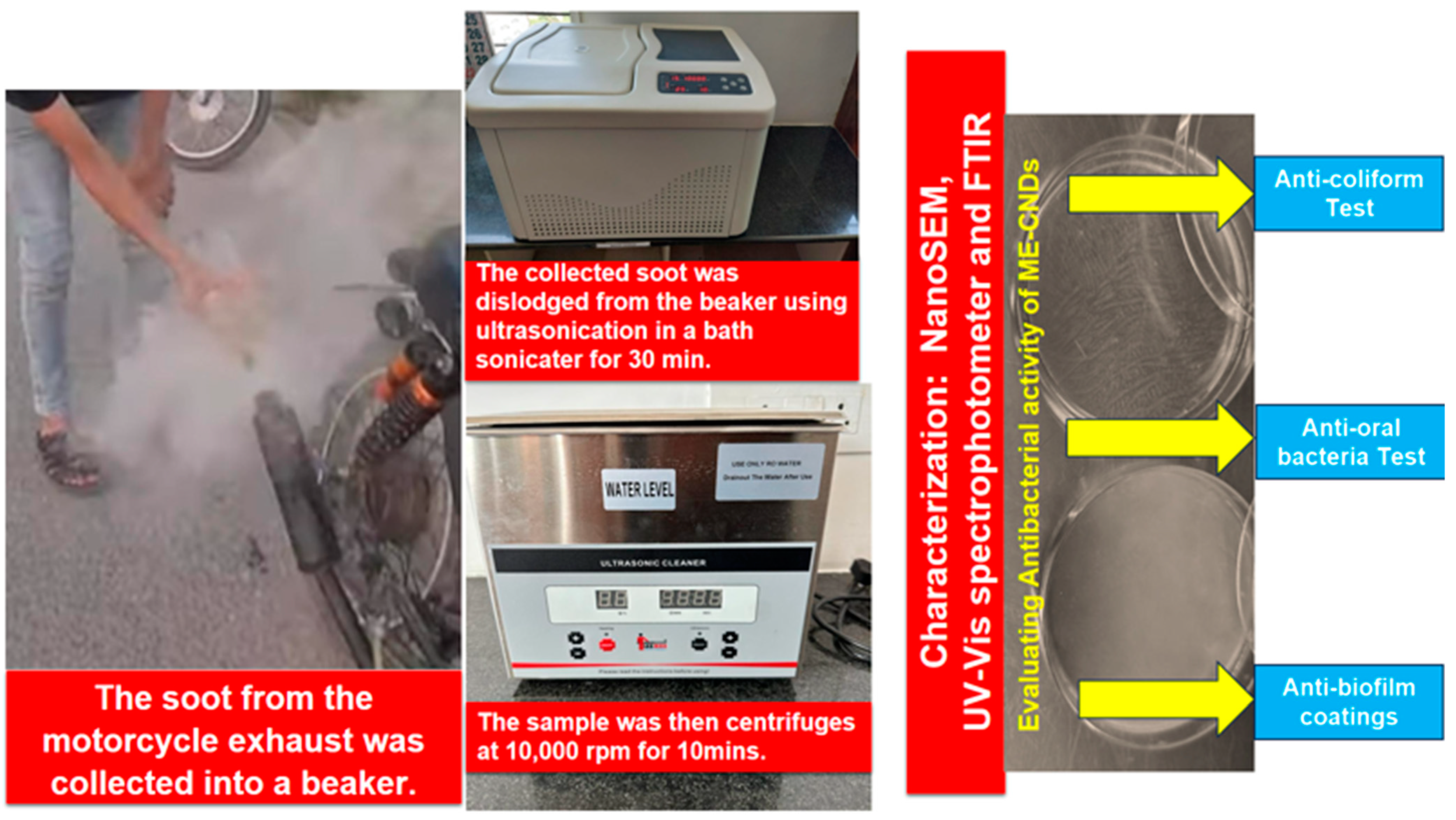
2.1. Collection of Exhaust Soot

Placing a sterile beaker next to the exhaust outlet allowed soot from the motorbike exhaust to be captured. The soot particles settled on the glass beaker were dislodged into 50 mL of water via sonication for one hour. The goal of sonication is to suspend carbon particles from soot in water; however, it can also break down bigger particles into smaller particles. The soot suspension was sonicated and then centrifuged for 10 min at 10,000 rpm to extract carbon nanodots [10]. The carbon nanodot-containing supernatant was separated from the precipitate. The supernatant was used for further research, and the precipitate was cast out [19].

2.2. Characterization of Carbon Nanodots

The isolated nanomaterial was morphologically characterized using a scanning electron microscope. Using a Fourier-transform infrared spectroscopy (FTIR), the functional groups and chemical composition of carbon nanodots (CNDs) were evaluated. and their optical characteristics and absorbance were examined using a UV–visible spectrophotometer.

2.3. Anti-Coliform Assay

Motorcycle exhaust carbon nanodots (ME-CNDs) were tested against coliforms such as E. faecalis and E. coli. The ME-CNDs were incubated with three different concentrations (0.2 g/L, 0.5 g/L, and 1 g/L) with E. faecalis and E. coli. The bacteria surviving the interaction of the CNDs were quantified by plating on nutrient agar at 37 °C in nutrient broth. Colony-forming unit/mL, or TVC, was enumerated, and the data were displayed as the mean ± standard deviation from three independent experimental replicates [20]. Version 12.0 of IBM SPSS for Windows (SPSS Inc., Chicago, IL, USA) was used to conduct the statistical analysis. A p-value of 0.05, 0.01, and 0.001 correspondingly was used to define statistics.

2.4. Anti-Oral Bacterial Assay

One of the authors provided the oral samples. Utilizing a sterilized brush, samples were collected. The oral microorganisms were then suspended in 20 mL of phosphate buffer and kept in falcon tubes in the refrigerator until use.
The antibacterial assay was carried out by mixing oral samples containing 10 mL with different quantities of carbon nanomaterial (0.2 g/L, 0.5 g/L, and 1 g/L). All experiments were carried out using three duplicates. The medium Mitis Salivarius Agar Base (HiMedia M259, HiMedia Laboratories Private Ltd., Maharashtra, India), which is specifically intended for oral bacterial growth, was used to enumerate the surviving bacteria post-incubation with the ME-CNDs. This process was carried out overnight in a shaker cum incubator. Using standard microbiological methods, the colonies were enumerated and the TVC was determined following a 48-h incubation period.

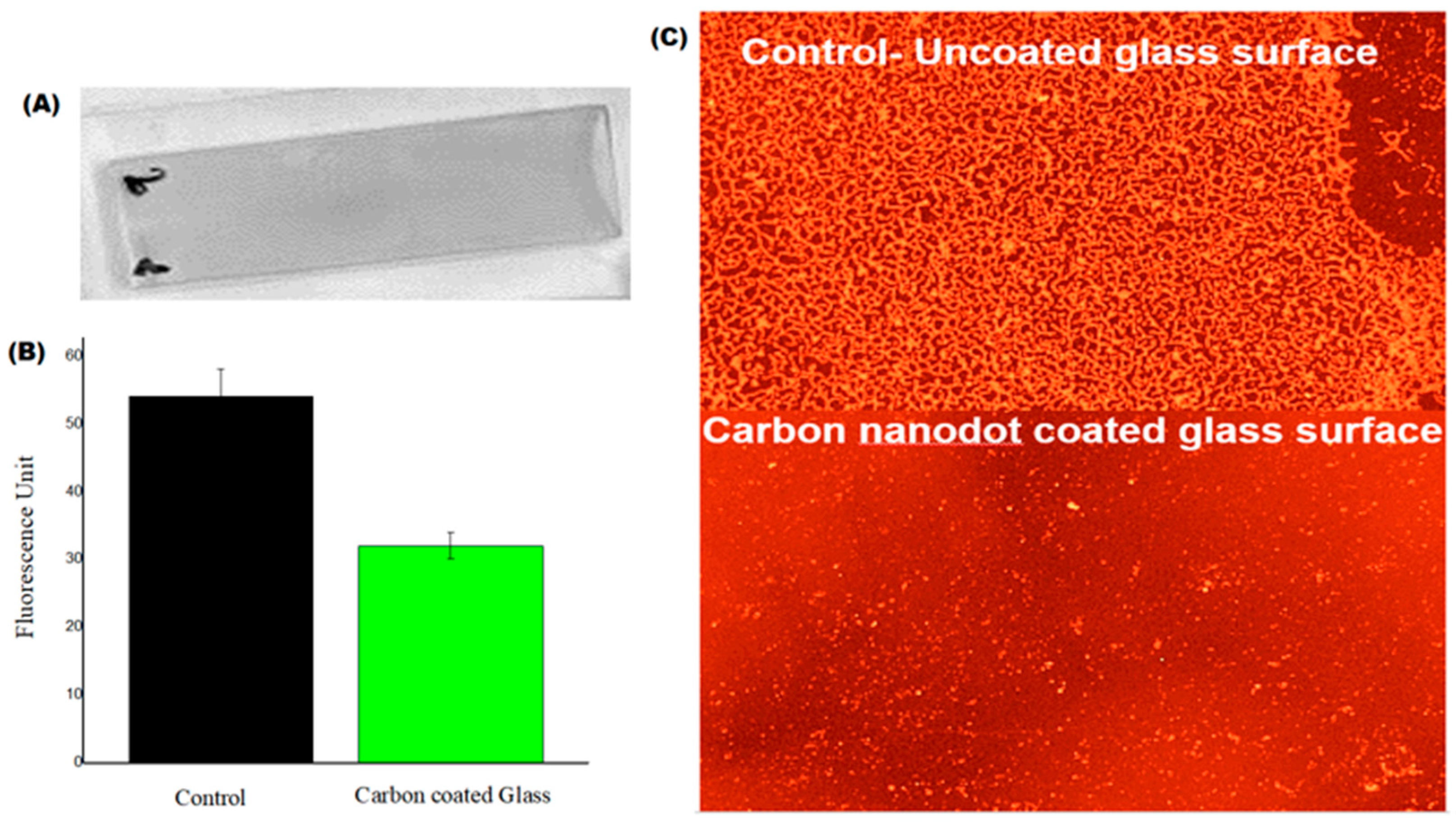
2.5. Anti-Biofilm Assay

Glass surfaces were coated with carbon nanoparticles using a straightforward dip-coating method, which involved dipping glass slides in a saturated ME-CNDs solution for 24 h. Glass slides were withdrawn and dried in a hot air oven overnight, and then exposed to S. aureus cultures for an entire day. After the slides were removed, the weakly adhering biofilm was gently cleaned off, 0.1% acridine orange was added, and the slides were incubated for two minutes [9]. After washing off unbound dye, the slides were allowed to air dry. A Fluorescence microscope Olympus CKX53 (Olympus Life Science, Waltham, MA, USA), was used to image the biofilm on coated and untreated glass surfaces of ME-CNDs. The fluorescent dye Acridine Orange exhibits differential staining of double-stranded DNA and single-stranded RNA. The orange-fluorescing cells on the surface represent the cells that are actively metabolizing. Using ImageJ (Version 1.45k), the fluorescence was quantified. The average of the data obtained from imaging five locations on the same surface using three samples was reported. The schematic workflow used in this paper is shown in Figure 1.

3. Results

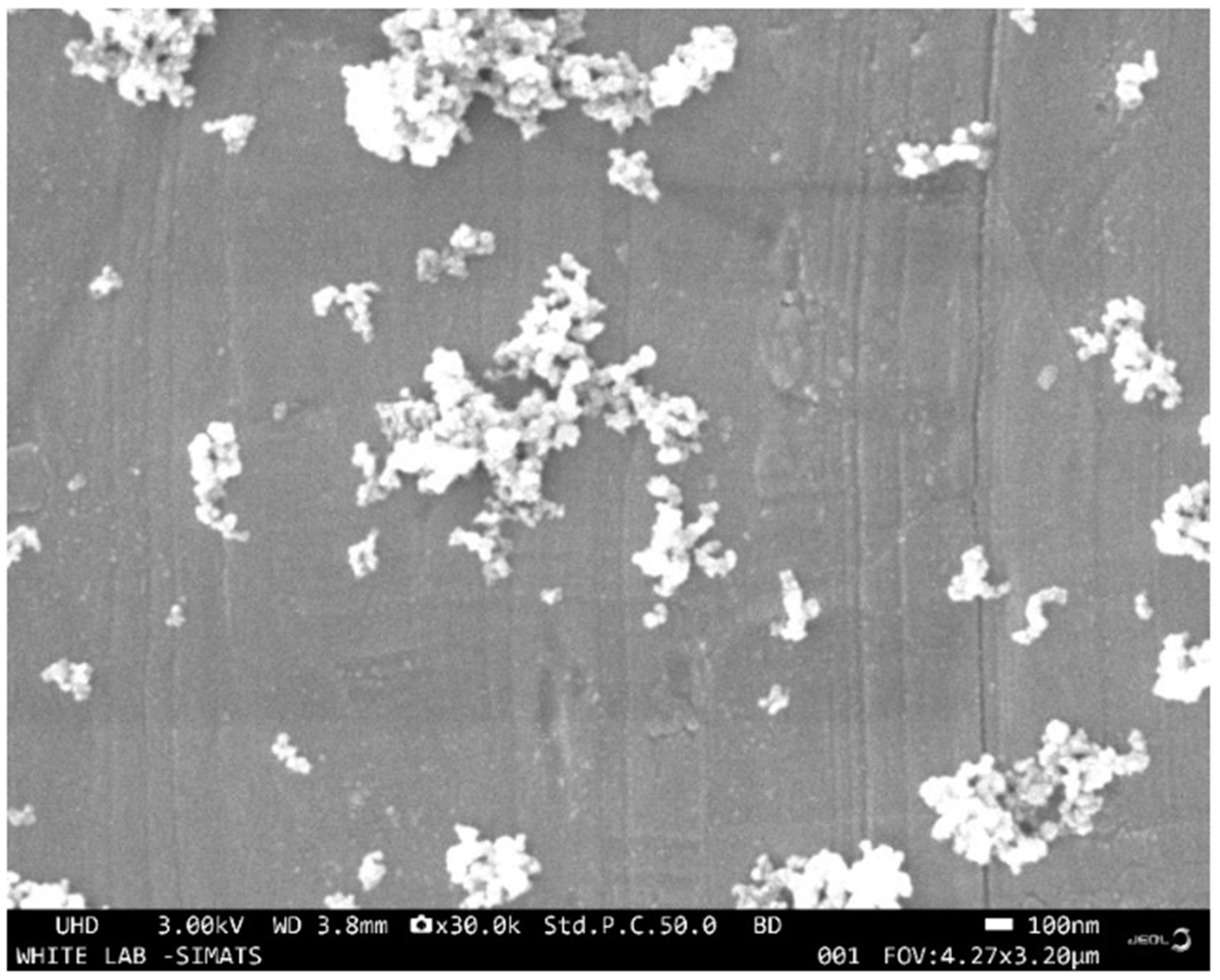
3.1. Characterization of Motorcycle Exhaust Carbon Nanodots (ME-CNDs)

NanoSEM was used to confirm the size and shape of the ME-CNDs. The existence of carbon nanodots, 8–13 nm sized, from the exhaust soot was visualized (Figure 2).
The extracted nanocarbon was chemically characterized to determine the chemical composition of nanodots using UV-Vis and FTIR spectroscopy. The carbonyl (C=O) and hydroxyl (−OH) group’s stretching vibrations were linked to the occurrence of FTIR absorption bands with peak locations at 1795 and 3410 cm−1, respectively [21,22]. The vibrations of sp2 and sp3 C–H are responsible for the distinctive peaks at 1330 and 829 cm−1, while C=C stretching vibration is responsible for the peak at 1593 cm−1. The NO3 stretched vibration is attributed to the peak at 2437 cm−1 [23]. The FT-IR data confirmed the presence of oxygen-rich groups (hydroxyl, carbonyl, and carboxyl) on CND surfaces (Figure 3A). In their UV–visible absorption spectra, the CNDs carbon core has a strong peak at 295 nm, which is associated with the π–π* transitions in the aromatic C=C bond. This is shown in Figure 3B.

3.2. Antibacterial Activity of ME-CNDs

The CNDs produced from motorbike exhausts exhibited anti-coliform activity, as verified by the results of the antibacterial tests. The plate count method findings are shown in (Figure 4). It was noticed that E. faecalis coliform bacteria showed statistically extremely significant inhibition, while E. coli showed significantly lower but still significant inhibition rates compared to E. faecalis. Since not much significant difference based on the ME-CNDs concentration gradients beyond 0.2 g/L concentrations (0.2 g/L, 0.5 g/L, and 1 g/L) (Figure S1), especially within 0.5 g/L and 1 g/L concentrations, was evident, graphs portrayed in the results below have been depicted using data generated at 0.5 g/L concentrations for all antibacterial studies.
The antibacterial tests against oral bacteria provided evidence of the antibacterial activity of carbon nanodots produced from smoke. Figure 5 illustrates the inhibition of oral germs that results from the anti-oral bacterial effect.
Carbon coatings on glass were subjected to S. aureus cultures, and their anti-biofilm potential was assessed. The glass slide’s carbon coating is seen in Figure 6A. The control surface is the one that is not coated. Figure 6B shows that the carbon-coated surfaces’ ability to inhibit biofilm formation was validated by epifluorescence imaging of bacterial culture-exposed surfaces. By employing image analysis to quantify the biofilm, it was also possible to confirm that the carbon-coated surfaces significantly inhibited the production of biofilm as compared with the control (Figure 6C).

4. Discussion

The outcome of the investigation showed the effectiveness that ME-CNDs exhibited against coliforms, especially E. faecalis, due to their potent antibacterial properties. Inhibition of oral microorganisms is another possible effect of CNDs. Likewise, the carbon coatings generated from ME-CNDs demonstrated significant antibiofilm activity. The antibacterial action of carbon compounds, which were isolated from a redundant source like chimney soot, is confirmed by these data. It has been observed that CNPs have antibacterial activity via traversing cell membranes, infiltrating the interior of a cell, and engaging with intracellular locations. The bacteria’s ability to divide and propagate can be inhibited by CNDs. They can also cause phospholipid peroxidation, which may result in cell death, and they can generate reactive oxygen species to break down the outer membranes of bacteria [24,25,26].
Vehicle exhaust waste soot has been reported to be converted into water-soluble fluorescent carbon dots by Thulasi et al., 2020 [3] using a straightforward acid reflux technique. Their carbon material had a spherical form and an approximate particle size of 4 nm. The emissive character of the car exhaust waste soot-derived CNDs was confirmed by spectroscopic experiments, which also revealed that their emission was excitation-dependent.
In 2018, Song et al. [27] tested carbon dots (CDs) against both common and drug-resistant bacteria. At 1000 μg/mL, CDs suppressed E. coli and dramatically reduced S. aureus. At 1000 μg/mL, drug-resistant strains AREC and KREC were effectively inhibited, while full inhibition was achieved at 1200 μg/mL. Other bacteria such as P. vulgaris, B. subtilis, and P. aeruginosa were still susceptible to CD’s effectiveness. In contrast to ampicillin, CDs continuously slowed down the development of bacteria without endangering HeLa cells. Tests using CDs obtained from different plants revealed no antibacterial impact, highlighting the special qualities of CDs derived from cigarettes.
Liu et al., 2017 [28] reported that a green hydrothermal approach utilizing metronidazole was used to create highly photoluminescent carbon nanodots (CNDs). These 2.9 nm nanodots, known as CNDs-250, demonstrated specific antibacterial efficacy against obligate anaerobes such as Porphyromonas gingivalis. Their varied light emission and 28.1% quantum yield make them suitable for a variety of bioimaging applications.
This work’s demonstration by Manikandan et al., 2020 [29] of the ME-CND’s antibacterial activity is another significant milestone. There is a continuous quest for alternative antibacterial agents due to the present rise in antibiotic resistance. The synthesis of antibacterial nanomaterials is typically not environmentally friendly; thus, while one problem (antibacterial) is solved, another problem (nanotoxicity) is exacerbated. The current study employed CNDs to combat pathogenic bacteria by deriving them from a redundant resource, such as motorbike exhausts. As demonstrated by the experiments, ME-CND coatings on glass may also prevent the production of biofilms. Thus, there is no denying that this is an extremely creative, economical, and environmentally feasible implementation.
The speculated mechanism behind the antibacterial activity of the carbon nanodots is projected in Figure 7. CNDs’ tiny size and high membrane permeability enable them to interact with and destroy vital cellular components, allowing them to effectively permeate bacterial cell membranes [30,31]. The processes of bacterial pumping mechanisms, which are in charge of transferring antibiotic and other antimicrobial compounds from cells, can be inhibited by CNDs [31]. This increases CNDs’ antimicrobial effectiveness. CNDs can interact with and affect the lipid bilayer of bacteria, leading to damage to the membrane and eventual cell death [31]. Some CNDs can produce reactive oxygen species (ROS), including hydrogen peroxide and superoxide radicals, which can harm bacterial cells and cause oxidative stress [32,33]. All CND kinds do not, however, use the same ROS pathway [33]. Under light, certain CNDs can function as photosensitizers and produce ROS, which can have antibacterial properties that are both photothermal and photodynamic [32,33]. Further in-depth studies are needed to arrive at the exact mechanism operating in the case of the ME-CNDs.

5. Conclusions

The results revealed that the nanomaterial recovered from motorbike exhausts were carbon nanodots. The antibacterial properties of the ME-CNDs were confirmed. E. faecalis was more susceptible to the antibacterial action than E. coli. Staphylococcus biofilms were inhibited by the ME-CNDs. This work demonstrates the effectiveness of separating nanomaterial from motorcycle exhausts and shows that ME-CNDs have antibacterial properties. However, no matter how green the approach may be, there will always be toxic effluents or chemicals released in the process. The present work takes the CND synthesis to the next level, where we use a totally chemical-free method for extracting CNDs from a useless and redundant source such as motorcycle exhaust.

Supplementary Materials

The following supporting information can be downloaded at: https://www.mdpi.com/article/10.3390/microbiolres15030115/s1, Figure S1 Graph showing antibacterial activity of 0.2 g/L, 0.5 g/L and 1g/L ME-CNDs against E.coli and E. faecalis.

Author Contributions

S.S.: data curation and writing—original draft; J.-W.O.: writing—review and editing, funding support, and resources; P.V.: writing—original draft, visualization, and writing—review and editing; M.M.: conceptualization, methodology, and writing—review and editing; J.G.: conceptualization, methodology, formal analysis, writing—original draft, visualization, writing—review and editing, data curation, supervision, and resources. All authors have read and agreed to the published version of the manuscript.

Funding

This research received no external funding.

Institutional Review Board Statement

Not applicable.

Informed Consent Statement

Not appliable.

Data Availability Statement

Data will be made available on request.

Conflicts of Interest

The authors declare no conflict of interest.

References

  1. Hardy, J.T. Climate Change: Causes, Effects, and Solutions; John Wiley & Sons: Hoboken, NJ, USA, 2003. [Google Scholar]
  2. Devi, S.; Gupta, R.K.; Paul, A.K.; Kumar, V.; Sachdev, A.; Gopinath, P.; Tyagi, S. Ethylenediamine mediated luminescence enhancement of pollutant derivatized carbon quantum dots for intracellular trinitrotoluene detection: Soot to shine. RSC Adv. 2018, 8, 32684–32694. [Google Scholar] [CrossRef]
  3. Thulasi, S.; Kathiravan, A.; Asha Jhonsi, M. Fluorescent Carbon Dots Derived from Vehicle Exhaust Soot and Sensing of Tartrazine in Soft Drinks. ACS Omega 2020, 5, 7025–7031. [Google Scholar] [CrossRef]
  4. Sun, Y.P.; Zhou, B.; Lin, Y.; Wang, W.; Fernando, K.S.; Pathak, P.; Xie, S.Y. Quantum-sized carbon dots for bright and colorful photoluminescence. J. Am. Chem. Soc. 2006, 128, 7756–7757. [Google Scholar] [CrossRef] [PubMed]
  5. Xu, X.; Ray, R.; Gu, Y.; Ploehn, H.J.; Gearheart, L.; Raker, K.; Scrivens, W.A. Electrophoretic analysis and purification of fluorescent single-walled carbon nanotube fragments. J. Am. Chem. Soc. 2004, 126, 12736–12737. [Google Scholar] [CrossRef] [PubMed]
  6. Sciortino, L.; Sciortino, A.; Popescu, R.; Schneider, R.; Gerthsen, D.; Agnello, S.; Messina, F. Tailoring the emission color of carbon dots through nitrogen-induced changes of their crystalline structure. J. Phys. Chem. C 2018, 122, 19897–19903. [Google Scholar] [CrossRef]
  7. Zhu, S.; Meng, Q.; Wang, L.; Zhang, J.; Song, Y.; Jin, H.; Yang, B. Highly photoluminescent carbon dots for multicolor patterning, sensors, and bioimaging. Angew. Chem. Int. Ed. 2013, 125, 4045–4049. [Google Scholar] [CrossRef]
  8. Makvandi, P.; Wang, C.Y.; Zare, E.N.; Borzacchiello, A.; Niu, L.N.; Tay, F.R. Metal-based nanomaterials in biomedical applications: Antimicrobial activity and cytotoxicity aspects. Adv. Funct. Mater. 2020, 30, 1910021. [Google Scholar] [CrossRef]
  9. Zhang, J.; Yu, S.H. Carbon dots: Large-scale synthesis, sensing and bioimaging. Mater. Today 2016, 19, 382–393. [Google Scholar] [CrossRef]
  10. Shah, M.S.A.S.; Nag, M.; Kalagara, T.; Singh, S.; Manorama, S.V. Silver on PEG-PU-TiO2 polymer nanocomposite films: An excellent system for antibacterial applications. Chem. Mater. 2008, 20, 2455–2460. [Google Scholar] [CrossRef]
  11. Esmeryan, K.D.; Castano, C.E.; Chaushev, T.A.; Mohammadi, R.; Vladkova, T.G. Silver-doped superhydrophobic carbon soot coatings with enhanced wear resistance and anti-microbial performance. Colloids Surf. A Physicochem. Eng. Asp. 2019, 582, 123880. [Google Scholar] [CrossRef]
  12. Esmeryan, K.D.; Stamenov, G.S.; Chaushev, T.A. An innovative approach for in-situ detection of postejaculatory semen coagulation and liquefaction using superhydrophobic soot coated quartz crystal microbalances. Sens. Actuators A Phys. 2019, 297, 111532. [Google Scholar] [CrossRef]
  13. Sahu, S.; Behera, B.; Maiti, T.K.; Mohapatra, S. Simple one-step synthesis of highly luminescent carbon dots from orange juice: Application as excellent bio-imaging agents. Chem. Commun. 2012, 48, 8835–8837. [Google Scholar] [CrossRef]
  14. Lai, C.W.; Hsiao, Y.H.; Peng, Y.K.; Chou, P.T. Facile synthesis of highly emissive carbon dots from pyrolysis of glycerol; gram scale production of carbon dots/mSiO2 for cell imaging and drug release. J. Mater. Chem. 2012, 22, 14403–14409. [Google Scholar] [CrossRef]
  15. Park, S.Y.; Lee, H.U.; Park, E.S.; Lee, S.C.; Lee, J.W.; Jeong, S.W.; Lee, J. Photoluminescent green carbon nanodots from food-waste-derived sources: Large-scale synthesis, properties, and biomedical applications. ACS Appl. Mater. Interfaces 2014, 6, 3365–3370. [Google Scholar] [CrossRef] [PubMed]
  16. Liu, H.; Ye, T.; Mao, C. Fluorescent carbon nanoparticles derived from candle soot. Angew. Chem. 2007, 119, 6593–6595. [Google Scholar] [CrossRef]
  17. Yang, S.T.; Cao, L.; Luo, P.G.; Lu, F.; Wang, X.; Wang, H.; Sun, Y.P. Carbon dots for optical imaging in vivo. J. Am. Chem. Soc. 2009, 131, 11308–11309. [Google Scholar] [CrossRef]
  18. Venkatesan, S.; Mariadoss, A.J.; Arunkumar, K.; Muthupandian, A. Fuel waste to fluorescent carbon dots and its multifarious applications. Sens. Actuators B Chem. 2019, 282, 972–983. [Google Scholar] [CrossRef]
  19. Guo, Y.; Zhang, L.; Cao, F.; Leng, Y. Thermal treatment of hair for the synthesis of sustainable carbon quantum dots and the applications for sensing Hg2+. Sci. Rep. 2016, 6, 35795. [Google Scholar] [CrossRef]
  20. Gopal, J.; George, R.P.; Muraleedharan, P.; Khatak, H.S. Photocatalytic inhibition of microbial adhesion by anodized titanium. Biofouling 2004, 20, 167–175. [Google Scholar] [CrossRef]
  21. Gopal, J.; Muthu, M.; Chun, S. Autochthonous self-assembly of nature’s nanomaterials: Green, parsimonious and antibacterial carbon nanofilms on glass. Phys. Chem. Chem. Phys. 2016, 18, 18670–18677. [Google Scholar] [CrossRef]
  22. Nautiyal, C.S.; Chauhan, P.S.; Nene, Y.L. Medicinal smoke reduces airborne bacteria. J. Ethnopharmacol. 2007, 114, 446–451. [Google Scholar] [CrossRef] [PubMed]
  23. Chun, S.; Muthu, M.; Gansukh, E.; Thalappil, P.; Gopal, L. The ethanopharmacological aspect of carbon nanodots in turmeric smoke. Sci. Rep. 2016, 6, 35586. [Google Scholar] [CrossRef] [PubMed]
  24. Cho, K.H.; Park, J.E.; Osaka, T.; Park, S.G. The study of antimicrobial activity and preservative effects of nanosilver ingredient. Electrochim. Acta 2005, 51, 956–960. [Google Scholar] [CrossRef]
  25. Shiraishi, Y.; Hirai, T. Selective organic transformations on titanium oxide-based photocatalysts. J. Photochem. Photobiol. C Photochem. Rev. 2008, 9, 157–170. [Google Scholar] [CrossRef]
  26. Limbach, L.K.; Li, Y.; Grass, R.N.; Brunner, T.J.; Hintermann, M.A.; Muller, M.; Stark, W.J. Oxide nanoparticle uptake in human lung fibroblasts: Effects of particle size, agglomeration, and diffusion at low concentrations. Environ. Sci. Technol. 2005, 39, 9370–9376. [Google Scholar] [CrossRef]
  27. Song, Y.; Lu, F.; Li, H.; Wang, H.; Zhang, M.; Liu, Y.; Kang, Z. Degradable carbon dots from cigarette smoking with broad-spectrum antimicrobial activities against drug-resistant bacteria. ACS Appl. Bio Mater. 2018, 1, 1871–1879. [Google Scholar] [CrossRef]
  28. Liu, J.; Lu, S.; Tang, Q.; Zhang, K.; Yu, W.; Sun, H.; Yang, B. One-step hydrothermal synthesis of photoluminescent carbon nanodots with selective antibacterial activity against Porphyromonas gingivalis. Nanoscale 2017, 9, 7135–7142. [Google Scholar] [CrossRef]
  29. Manikandan, M.; Chun, S.; Kazibwe, Z.; Gopal, J.; Singh, U.B.; Oh, J.W. Phenomenal bombardment of antibiotic in poultry: Contemplating the environmental repercussions. Int. J. Environ. Res. Public Health 2020, 17, 5053. [Google Scholar] [CrossRef]
  30. Fang, M.; Lin, L.; Zheng, M.; Liu, W.; Lin, R. Antibacterial functionalized carbon dots and their application in bacterial infections and inflammation. J. Mater. Chem. B 2023, 11, 9386–9403. [Google Scholar] [CrossRef]
  31. Yu, M.; Li, P.; Huang, R.; Xu, C.; Zhang, S.; Wang, Y.; Xing, X. Antibacterial and antibiofilm mechanisms of carbon dots: A review. J. Mater. Chem. B 2023, 11, 734–754. [Google Scholar] [CrossRef]
  32. Zhao, W.B.; Liu, K.K.; Wang, Y.; Li, F.K.; Guo, R.; Song, S.Y.; Shan, C.X. Antibacterial Carbon Dots: Mechanisms, Design, and Applications. Adv. Healthc. Mater. 2023, 12, e2300324. [Google Scholar] [CrossRef]
  33. Dong, X.; Liang, W.; Meziani, M.J.; Sun, Y.P.; Yang, L. Carbon Dots as Potent Antimicrobial Agents. Theranostics 2020, 10, 671–686. [Google Scholar] [CrossRef]
Figure 1. Diagram illustrating the study’s work process.
Microbiolres 15 00115 g001
Figure 2. NanoSEM micrographs of the 8–13 nm sized ME-CNDs.
Microbiolres 15 00115 g002
Figure 3. Characterization of ME-CNDs (A) FTIR spectra and (B) UV-Vis spectra.
Microbiolres 15 00115 g003
Figure 4. Graph showing results of anti-coliform activity of ME-CNDs (0.5 g/L). Inset shows the distinct inhibition of viable colonies on agar plates owing to the carbon nanodot interaction. * significant; ** highly significant based on statistical analysis using paired t-tests.Error bars = standard deviation mean.
Microbiolres 15 00115 g004
Figure 5. Results of (A) TVC counts showing inhibition of oral bacteria following incubation with 0.5 g/L ME-CNDs and (B) epifluorescence images showing fluorescing live oral bacteria post-interaction with ME-CNDs. Error bars = standard deviation of mean.
Microbiolres 15 00115 g005
Figure 6. (A) Photograph showing carbon coating on glass. (B) Graph showing results of ImageJ analysis of the fluorescence on the acquired images on the control and carbon-coated glass surfaces. Epifluorescence images of biofilm on (C) carbon-coated and control glass surfaces. Error bars = standard deviation of mean.
Microbiolres 15 00115 g006
Figure 7. Schematic representation of the plausible mechanism behind the antibacterial activity of the CNDs. Reactive oxygen species are referred to as ROS and carbon nanodots as CNDs.
Microbiolres 15 00115 g007
Disclaimer/Publisher’s Note: The statements, opinions and data contained in all publications are solely those of the individual author(s) and contributor(s) and not of MDPI and/or the editor(s). MDPI and/or the editor(s) disclaim responsibility for any injury to people or property resulting from any ideas, methods, instructions or products referred to in the content.

