Article

Exploring the Antibacterial Potential of Bile Salts: Inhibition of Biofilm Formation and Cell Growth in Pseudomonas aeruginosa and Staphylococcus aureus

1
Department of Biosciences, School of Liberal Arts and Sciences, Mody University of Science and Technology, Lakshmangarh 332311, Rajasthan, India
2
Department of Medicine, Pennsylvania State University, Hershey Medical Centre, Hershey, PA 17033, USA
*
Authors to whom correspondence should be addressed.
Microbiol. Res. 2024, 15(3), 1269-1279; https://doi.org/10.3390/microbiolres15030085
Submission received: 7 June 2024 / Revised: 30 June 2024 / Accepted: 19 July 2024 / Published: 21 July 2024

Abstract

:
Chronic infections often involve notorious pathogens like Pseudomonas aeruginosa and Staphylococcus aureus, demanding innovative antimicrobial strategies due to escalating resistance. This investigation scrutinized the antibacterial prowess of bile salts, notably taurocholic acid (TCA), ursodeoxycholic acid (UDCA), and ox bile salt (OBS), against these pathogens. Evaluations encompassed minimum inhibitory concentration (MIC) determination, scrutiny of their impact on biofilm formation, and anti-virulence mechanisms. UDCA exhibited the highest efficacy, suppressing S. aureus and P. aeruginosa biofilms by 83.5% and 78%, respectively, at peak concentration. TCA also significantly reduced biofilm development by 81% for S. aureus and 75% for P. aeruginosa. Microscopic analysis revealed substantial disruption of biofilm architecture by UDCA and TCA. Conversely, OBS demonstrated ineffectiveness against both pathogens. Mechanistic assays elucidated UDCA and TCA’s detrimental impact on the cell membrane, prompting the release of macromolecular compounds. Additionally, UDCA and TCA inhibited protease and elastase synthesis in P. aeruginosa and staphyloxanthin and lipase production in S. aureus. These results underscore the potential of UDCA and TCA in impeding biofilm formation and mitigating the pathogenicity of S. aureus and P. aeruginosa.

1. Introduction

Various disciplines within current healthcare rely on the accessibility of potent antibiotic medications. Antibiotics selectively kill or suppress susceptible bacteria, while naturally or acquired antibiotic-resistant microorganisms survive and multiply. Resistance to antibiotics presents a substantial and pressing threat to global public health. Microorganisms have acquired resistance to nearly all available antibiotics, resulting in a significant economic burden associated with these multidrug-resistant microorganisms. In addition, there has been a significant decrease in the number of newly introduced compounds in the market [1,2,3]. According to analysis conducted by the World Health Organization (WHO), the number of new antibiotics being developed for priority infections in 2021 decreased to 27, compared to 31 products in 2017 [4].
Among various mechanisms of antibiotic resistance, the formation of biofilms by microorganisms is one of the modes which presents a challenge. Biofilms are structured collections of microorganisms that are embedded in a matrix made up of polysaccharides, extracellular DNA, proteins, and/or lipids [5]. Practically all bacteria possess the capacity to create biofilms under specific circumstances. Bacteria inhabiting a biofilm can exhibit a 10 to 1000 times increase in antibiotic resistance compared to similar bacteria living in a planktonic state [6]. Planktonic bacteria, which are already resistant to numerous antimicrobials, see a significant increase in resistance when they form biofilms. Biofilms can develop on various surfaces, such as living tissues, indwelling medical devices, and contact lenses. Biofilms are resistant to elimination and contribute to the worsening of conditions, persistent infections, repeated infections, and failures of medical devices or implants [7]. Biofilms have a capricious behaviour and morphology, which frequently leads to ineffective treatment or outcomes that are difficult to anticipate [8]. The combination of a slow pace of growth, modified metabolism, persister cells, varying levels of oxygen, extracellular enzymes that deactivate antibiotics, and a dense biofilm structure collectively contribute to a significant level of resistance against antibiotic treatment. Studies have shown that biofilm-forming bacteria hinder the healing process in 60% of chronic wounds and 6% of acute wounds [9]. In addition to impeding wound healing, biofilms stimulate an inflammatory response in the host that has limited ability to penetrate the biofilm, resulting in damage to cells in the vicinity [10]. Hence, it is important to discover the new therapeutics in order to enhance the success rate against persistent and opportunistic illnesses and to generate an immediate effect in the present therapeutic field.
Bile salts are a significant group of organic compounds that are synthesized in the liver of all vertebrates. They are combined with taurine or glycine to form Na+ and K+ salts and then released into bile. Bile salt derivatives possessing long steroid backbones and an amphiphilic shape can effectively permeate bacterial membranes and exhibit antibacterial properties due to their favorable biocompatibility [11,12,13]. Bile salts have the ability to influence immune cells in the mucosal lining [14], activate the immunological response in the intestines [15], and preserve the integrity of the intestinal barrier to inhibit the passage of pathogens [16]. Bile salt remained a distinct part of Traditional Chinese Medicine where the utilization of bile from 44 distinct animal species for the treatment of various ailments has been documented [17]. Low concentrations of bile acids in the gastrointestinal tract have been found to cause an excessive development of bacteria and potential pathogens, leading to an increased likelihood of inflammation and bacterial translocation [13].
In view of all the mentioned characteristics of bile salt, it is evident that bile salt may provide an advantage in combating against microbes associated with chronic infections. However, its deleterious effect on pathogenic microbes needs to be validated. Staphylococcus aureus and Pseudomonas aeruginosa are the most often found microorganisms in chronic ulcers generally in the form of a biofilm that is resistant to antimicrobial treatment [18]. Thus, the aim of the current study is to evaluate the effect of different bile salts, namely taurocholic acid and ursodeoxycholic acid on biofilm forming microorganism (P. aeruginosa and S. aureus) and to elucidate its mechanism of action. Taurocholic acid and ursodeoxycholic acid are secondary bile salts that are produced as a result of the metabolism of intestinal flora. Additionally, ox bile extract was also evaluated against the pathogen as a traditional crude bile salt.

2. Materials and Methods

2.1. Chemicals

Taurocholic acid (TCA), Dimethyl sulfoxide (DMSO), 3-(4,5-dimethyl-2-thiazolyl)-2,5-diphenyl2H-tetrazolium bromide (MTT), Dulbecco’s modified Eagle’s medium and fetal bovine serum (FBS), ox bile salt (OBS) were purchased from Sigma-Aldrich, St. Louis, MO, USA. Ursodeoxycholic acid (UDCA) was purchased from Anant Pharmaceuticals Pvt. Ltd., Ambernath, India. Crystal violet and all microbiological culture medium was obtained from Himedia, Mumbai, India.

2.2. Bacterial Cultures

P. aeruginosa MTCC 1688 and S. aureus ATCC 25923 were procured from Microbial Type Culture Collection, India and American Type Culture Collection, USA, respectively, and were preserved at −20 °C as stock cultures. The bacterial cultures were grown in brain heart infusion broth in a shaker incubator overnight at 37 °C for all in vitro investigations.

2.3. Determination of Minimum Inhibitory Concentration (MIC)

The minimum inhibitory concentration of different bile salts was determined using a broth microdilution method [19]. Two-fold serial dilutions of each bile salt were added to the wells of sterile 96-well plates containing nutrient broth (100 µL) with a bacterial concentration (P. aeruginosa MTCC 1688 and S. aureus ATCC 25923) of 106 CFU/mL. The bile salts were dissolved in the solvent (either distilled water or dimethyl sulfoxide, depending upon their solubility) to a final concentration of 10 mg/mL and added to the wells. Following an incubation period of 24 h at 37 °C, the MIC was determined as the lowest concentration that completely inhibited the growth of the bacteria by measuring the optical density (OD) at 600 nm using a microplate reader (Power Wave XS 2, BioTek, Winooski, VT, USA). The growth inhibition for each compound was determined by the formula
Percentage of Inhibition = (OD of Control − OD of Test)/(OD of Control) × 100%

2.4. Colony Forming Unit (CFU) Analysis

CFU analysis was conducted to determine the variation in cell count between control and bile-salt-treated bacterial cells of P. aeruginosa and S. aureus. Briefly, P. aeruginosa and S. aureus were grown in the absence and presence of bile salts for 24 h at 37 °C under aerobic conditions. After completion of the incubation period, the control and treated bacterial cells were serially diluted and spread on tryptic soy agar and incubated overnight at 37 °C. Thereafter, the number of bacterial colonies was counted and represented as CFU.

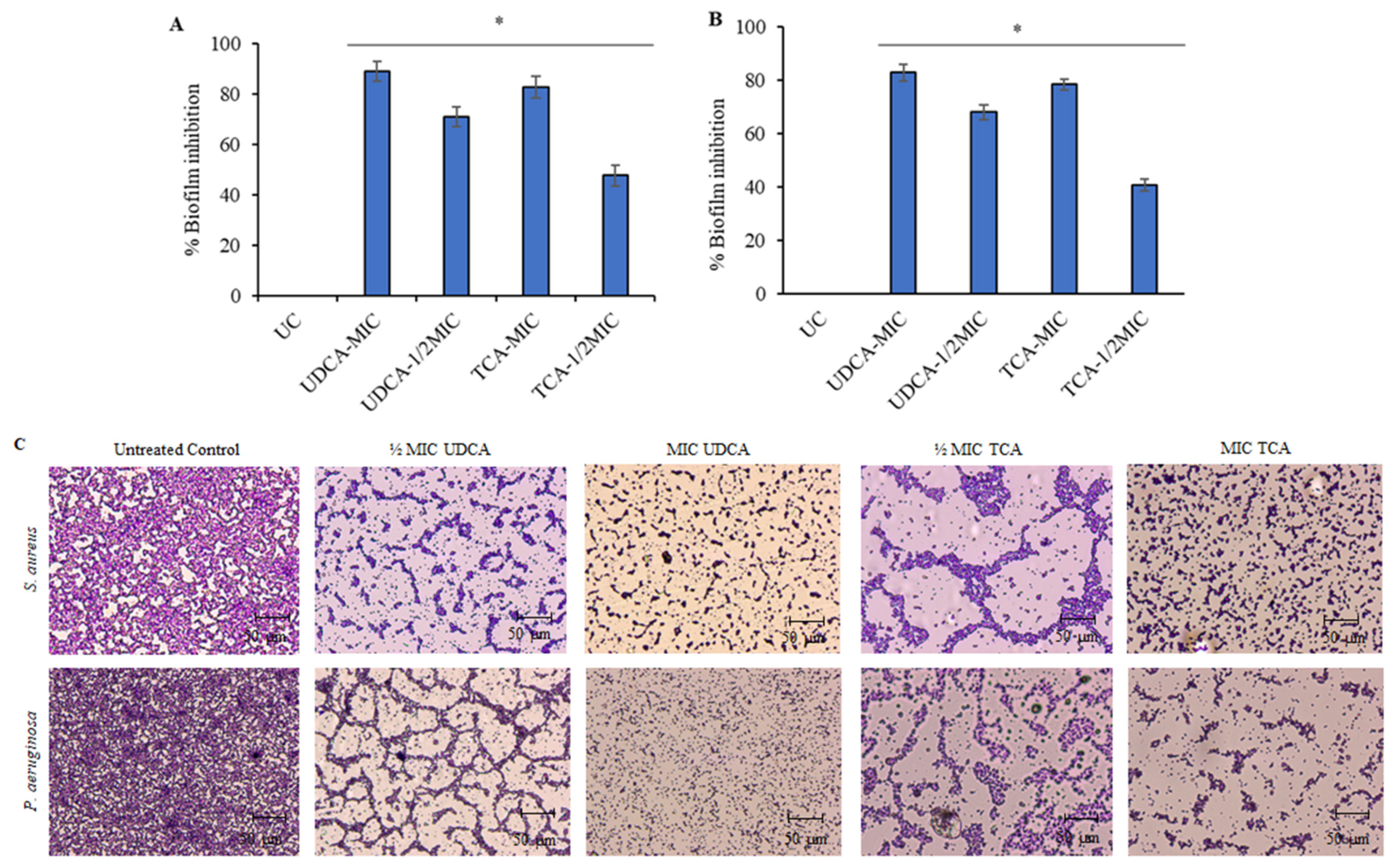
2.5. Biofilm Inhibition

Crystal violet staining assays was used to measure the anti-biofilm activity of bile salts against P. aeruginosa and S. aureus. Overnight cultures of microbes were diluted in fresh tryptone soy broth supplemented with 2% sucrose to obtain a bacterial suspension at a concentration of 106 cells/mL. Then, 50 µL of bacterial suspension was added to 96-well plates in triplicate and statically incubated at 37 °C for 24 h with different bile salts. The unattached microorganisms were rinsed away with sterile distilled water three times. The biofilm formed was stained with 0.1% crystal violet at room temperature for 15 min. After discarding the excess dye from the 96-well plates, the plate was washed again with distilled water three times. The crystal violet stained biofilm was dissolved in 95% ethanol prior to air drying at 55 °C for 10 min and quantified by determining the absorbance at OD 570 nm.

2.6. Change in the Biofilm Structure

The effect of bile salts on biofilm formation was analyzed as described previously [20]. Overnight culture of P. aeruginosa and S. aureus was suspended in fresh culture medium and adjusted to 106 CFU/mL. The bacterial suspension was added to fresh media containing sterilized cover slips along with different concentration of bile salts in a 6-well culture plate and incubated for 24 h at 37 °C. After incubation, the cover slips were rinsed with distilled water to remove loosely adhered cells. The biofilms that adhered to the coverslips were then stained with a 0.1% crystal violet solution and visualized under a light microscope (Olympus) at 400× magnification.

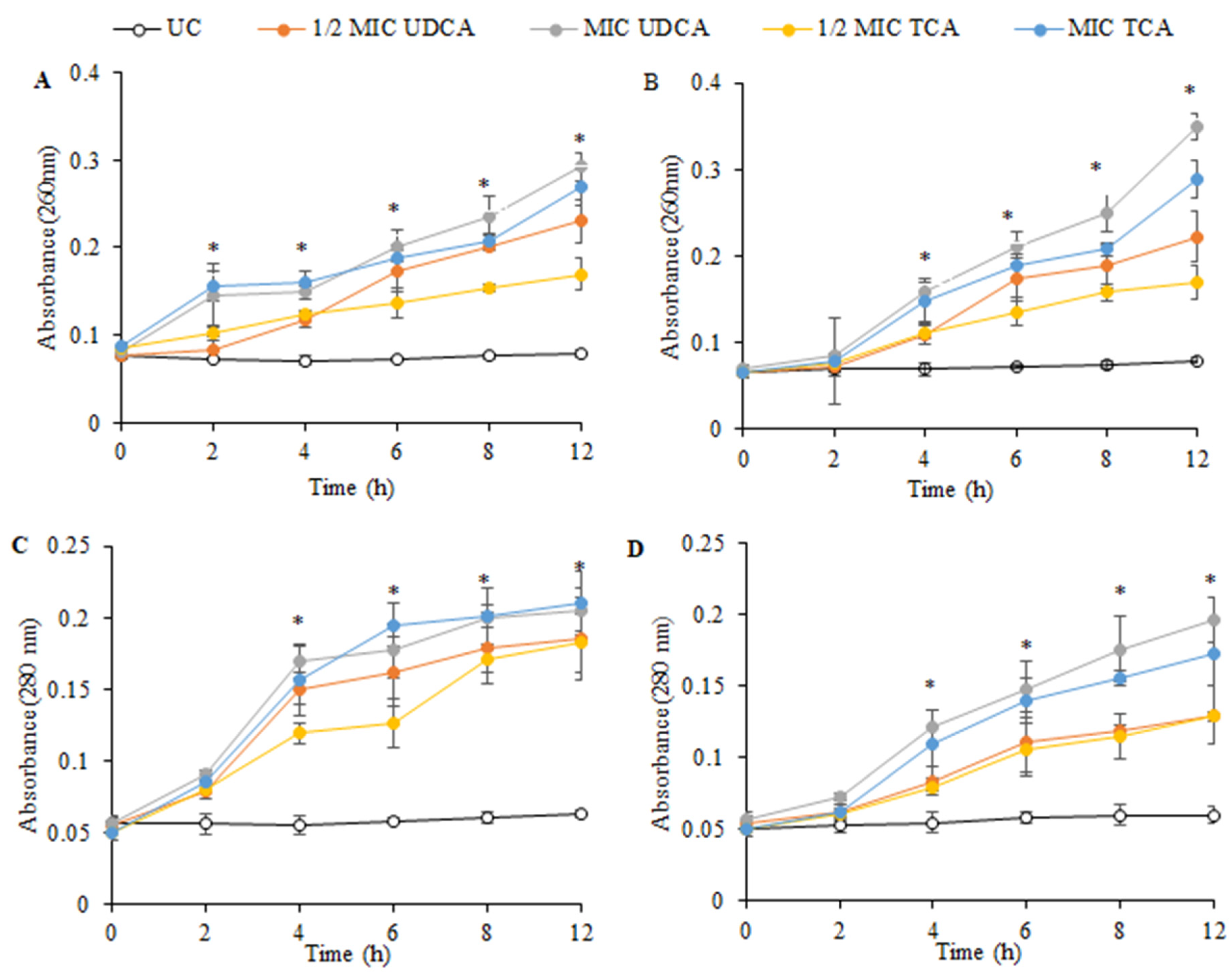
2.7. Effect on Nucleic Acid and Protein Leakage

The release of intracellular constituents from bacterial cells was analyzed by determining the content of DNA and proteins in culture medium. Briefly, 250 µL of overnight-grown cell suspension of P. aeruginosa and S. aureus (106 CFU/mL) was added along with bile salts to a total volume of 1 mL. The cultures were incubated for 24 h at 37 °C. After completion of the incubation period, the bacterial cultures were centrifuged at 20,000 rpm and 30,000 rpm for protein and nucleic acid estimation, respectively. The protein content in the bacterial supernatant was measured using the method of Bradford [21]. The nucleic acid content was estimated by measuring absorbance at 260 nm.

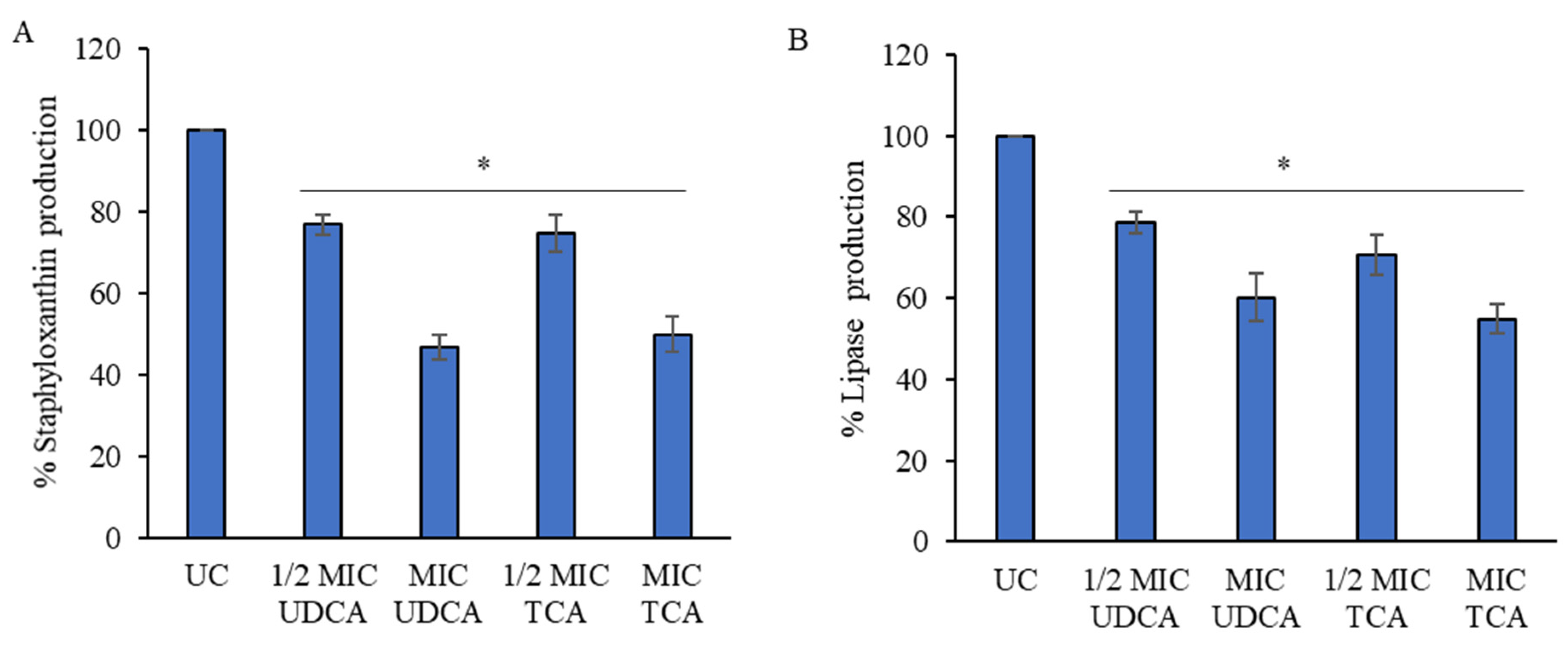
2.8. Inhibition of S. aureus Biofilm Virulence

The impact of bile salt treatment on the virulence factors of S. aureus was evaluated by measuring the levels of staphyloxanthin and lipase synthesis. The estimation of Staphyloxanthin production was conducted using the procedure established by Lee et al. [22]. In brief, 20 µL of overnight-cultured S. aureus was added to 2 mL of new LB medium. The culture was then incubated for 24 h at 37 °C in a tube with constant shaking at 250 rpm, in the presence of bile salts. After incubation, the culture was centrifuged at 8000× g for 10 min. Following this, the pellet was washed with sterile phosphate-buffered saline (PBS) and resuspended in pure ethanol and incubated at 40 °C for of 20 min. Cells were discarded by centrifugation at 10,000× g for 10 min, and supernatant absorbance was measured at 450 nm.
Lipase production by S. aureus was investigated using the method of Lee et al. [22]. Briefly, 20 μL of overnight cultured S. aureus was inoculated into 2 mL of LB and incubated for 24 h at 37 °C with or without bile salts. The incubated culture was centrifuged at 8000× g for 10 min, and 0.1 mL aliquots were mixed with 0.9 mL of substrate (10% (v/v) of buffer A having 3 mg/mL of p-nitrophenyl palmitate in isopropyl alcohol and 90% (v/v) of buffer B having 1 mg/mL of gum arabic and 2 mg/mL sodium deoxycholate in Na2PO4 buffer (50 mM, pH 8.0) and heated at 40 °C in the dark for 30 min. Lipase reactions were then stopped by adding 1 M Na2CO3, and mixtures were centrifuged at 10,000× g for 10 min. The absorbances were measured at 405 nm.

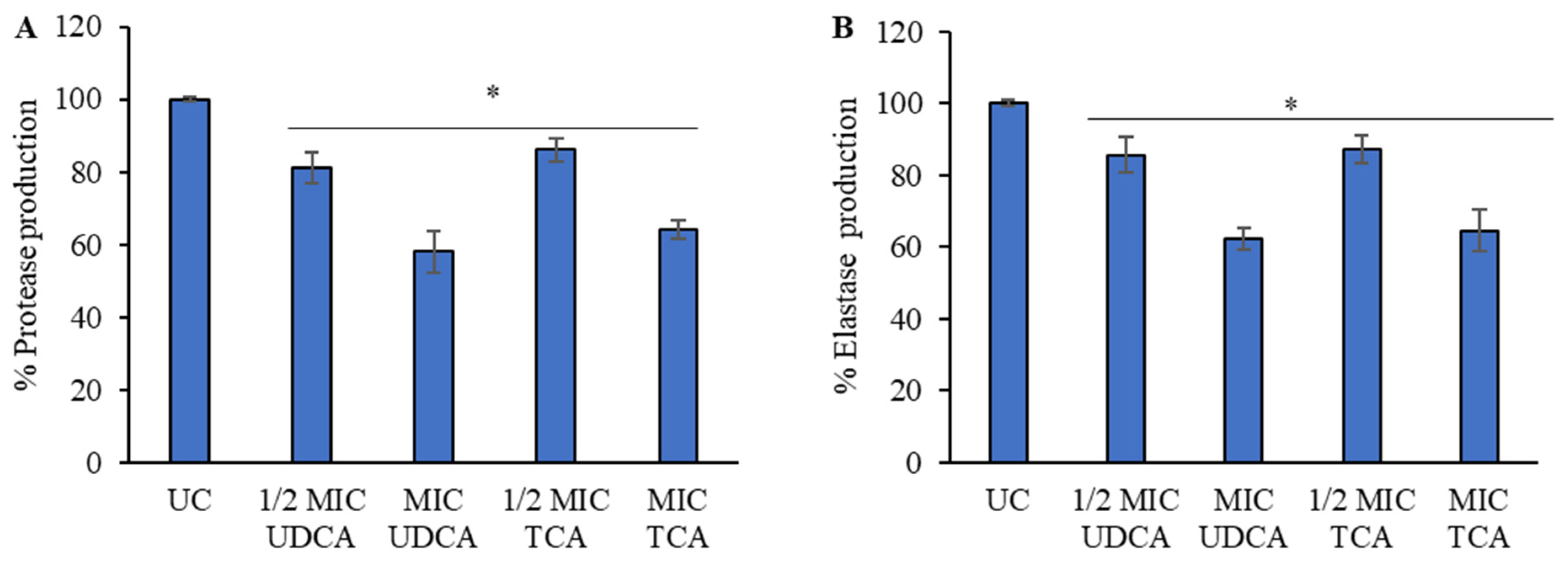
2.9. Evaluation of P. aeruginosa Virulence Factors

The effect of bile salt on P. aeruginosa virulence factors was evaluated by quantifying elastase and protease production [23]. Initially, 5 µL of a 24 h culture of P. aeruginosa was added along with 5 µL of bile salt to make a final volume of 500 µL and incubated for 24 h. Subsequently, the tubes were centrifuged at 9503× g for 10 min and the supernatant was used to quantify the production of proteases and elastases. For elastase quantification, 25 µL of supernatant was taken and 225 µL of 100 mM tris buffer pH 7.5, supplemented with 10 mg/mL elastin Congo red (ERC), was added. Subsequently, this mixture was incubated with shaking for 3 h at 37 °C. After that, PBS buffer at pH 6.0 was added and allowed to cool for 2 min to stop the reaction. Finally, the mixture was centrifuged at 9503× g for 10 min to separate the insoluble ERC, and an OD of 495 nm was recorded to determine elastase production.
For the quantification of proteases, first, 37.5 µL of supernatant was taken and 250 µL of a solution of 100 mM tris buffer pH 8.0 supplemented with 0.3% (w/v) azocasein was added and incubated for one hour at 37 °C statically. Next, trichloroacetic acid (TCA 10% (w/v)) was added to stop the reaction and insoluble azocasein was centrifuged at 9503× g for 10 min. Finally, the OD 400 nm of the supernatant was recorded to determine protease production.

2.10. Statistical Analysis

All the experiments were conducted thrice and the data were presented as mean ± standard deviation (SD). Student’s t-test was utilized to conduct statistical analysis in order to establish the significance of the difference between means. A p-value < 0.05 indicated a statistically significant difference. The statistical analysis was performed using GraphPad Prism (version 9.4.0).

3. Results

3.1. Antimicrobial Effect of Bile Salts

The antimicrobial effect of bile salts was studied via recognizing the MIC (Table 1) and a reduction in the colony forming units (CFUs) (Figure 1) of P. aeruginosa and S. aureus. UDCA exhibited the lowest MIC against both the microorganisms followed by TCA. The minimum MIC of UDCA against S. aureus was determined to be 1300 µg/mL, whereas the MIC against P. aeruginosa was discovered to be 1500 µg/mL. TCA exhibited an MIC of 2000 µg/mL and 3000 µg/mL against S. aureus and P. aeruginosa, respectively. Furthermore, both UDCA and TCA showed a significant (p < 0.05) impact on reducing CFU against both bacteria. OBS was found to be ineffective against both microbes; thus, it was dropped for further analysis.

3.2. Effect on Biofilm

The impact of bile salts on bacterial biofilm formation was evaluated using the crystal violet method and is illustrated in Figure 2A. Both UDCA and TCA were discovered to inhibit the formation of biofilm mass in P. aeruginosa and S. aureus. An evident decrease in biofilm formation was found at half the MIC of UDCA and TCA against both microorganisms, with a substantial statistical significance (p < 0.05). UDCA demonstrated a biofilm inhibition rate of 89.26% and 82.98% against S. aureus and P. aeruginosa, respectively. TCA showed the highest inhibition rates of 82.98% and 78.51% against S. aureus and P. aeruginosa, respectively.
In addition, the impact of bile salts on the structure of biofilms was also examined. Performing light microscopic analysis is a proven method for obtaining valuable information about the action of anti-biofilm compounds by directly observing the suppression of a biofilm after exposure to bile salts. Both S. aureus and P. aeruginosa displayed a dense layer of biofilm in the untreated control samples. However, the application of bile salts caused a degradation in the structure of the biofilm (Figure 2B). The suppression of biofilm development in the microbe’s biofilm structure exhibited a distinct dose-dependent relationship with the increasing concentration of bile salts, specifically UDCA and TCA.

3.3. Effect on Cellular Constituents

Efficient antibacterial properties can provoke the release of cellular macromolecules from a bacterial cell. To evaluate this, the discharge of nucleic acid and protein upon the action of bile salts in culture medium was studied. Both the test compounds (UDCA and TCA) exhibited similar effects on bacterial cells (Figure 3). After treatment with either UDCA or TCA, a time-dependent discharge of nucleic acid (OD 260 nm) and protein (OD 280 nm) was observed. Treated cultures of S. aureus and P. aeruginosa indicated a significant increase in absorbance in a dose-dependent manner when compared to untreated cells (p < 0.05). The discharge of these macromolecules from bacterial cells might have caused deteriorative effects on bacterial cells, leading to their low survival as indicated previously by the low appearance of CFU upon treatment.

3.4. Effect on Virulence Factors

S. aureus is recognized for its ability to produce many virulence factors, including lipase and staphyloxanthin, which contribute to the formation of biofilms [24]. Staphloxanthin specifically plays a crucial role in preserving the structural integrity of the bacterial membrane and is linked to the ability of bacteria to survive under challenging circumstances. Both UDCA and TCA exhibited a reduction in staphyloxanthin synthesis in a dose-dependent manner, as seen in Figure 4. A reduction of over 50% was seen at the MIC of UDCA. The treatment with TCA at the MIC resulted in a 55% inhibition. A dose-dependent inhibitory effect on lipase synthesis was reported, with similar effects shown for both UDCA and TCA. The application of TCA resulted in a significant (p < 0.05) decrease in lipase production, surpassing the reductions caused by other bile salts tested, with a reduction rate of 54.83%.
To elucidate the effect of UDCA and TCA on virulence factors of P. aeruginosa, protease and elastase were quantified. Treatment with even a sub-inhibitory concentration of bile salt viz. UDCA and TCA resulted in a significant (p < 0.05) negative effect on the production of protease (Figure 5). A 42% reduction in the production of protease was observed at the highest concentration of UDCA utilized. Additionally, TCA also resulted in only 64% protease production in comparison to the untreated control. Similarly, an adverse effect was observed for elastase production upon bile salt treatment. UDCA resulted in a 37.8% reduction in elastase production in P. aeruginosa, whereas TCA caused a decrease of 35.6%. The virulence factors of both S. aureus and P. aeruginosa play a crucial role in host colonization, avoiding the host’s immune response, and facilitating quorum sensing. Both UDCA and TCA, at their minimum inhibitory concentration (MIC), affected the production of virulence factors, aligning with their ability to suppress biofilm formation as described in the previous section.

4. Discussion

According to a WHO report, the rise in antimicrobial resistance is a matter of great concern in healthcare systems. The occurrence of biofilms among the bacterial community further enhances the problem. Biofilms can impact antimicrobial efficacy, as well as immune responses, contributing to antimicrobial resistance, resulting in the establishment of persistent chronic infections. Thus, the evolvement of new molecules/therapies is always needed to overcome these challenges. In the current study, we evaluated the effect of bile salts UDCA and TCA on the biofilm-forming microbes, i.e., S. aureus and P. aeruginosa. Both the microbes are capable of causing nosocomial infections and have been in detected in chronic wound aggregates [18]. Biles salts are known to have variety of biological functions [14,15,16].
Besides being an integral part of cholesterol metabolism in the body, bile salts also possess antibacterial functions through various mechanisms. Thus, the effect of bile salts, mainly UDCA and TCA, on planktonic bacterial growth was observed. Both the pathogens became susceptible to the action of bile salts. Such an effect was also reported by others where pathogens were unable to survive in the presence of high levels of bile salts [25,26]. However, researchers also indicated that microbes can easily tolerate the low physiological concentration of bile. This could be reason behind the high MIC of bile salt evaluated against the microbes. Despite being an intricate part of many traditional medicine systems, OBS was unable to exhibit any reduction in the growth of microorganisms at the highest concentration used in the study. Shekarfouoush et al. [27] also noticed a similar response and stated that ox bile does not have any inhibitory activity against several pathogenic Gram-positive and -negative bacteria in vitro. This could be due to the presence of trihydroxyconjugated bile salts, in ox bile, which have been demonstrated to have a low level of effectiveness [28]. It was observed that S. aureus was comparatively more sensitive towards the action of bile salts than the Gram-negative P. aeruginosa. A similar effect on sensitivity of Gram-positive bacteria towards bile salts was reported by Tian et al. [29].
In terms of biofilm inhibition, both the bile salts exhibited dose-dependent action in countering the biofilm formation by reducing the biofilm biomass and distorting the biofilm structure. This could be due to the detergent action of bile salts [30] causing a change in the hydrophobicity of the bacterial cell membrane. This feature was evident in our study as macromolecules in the form of nucleic acid and proteins were being discharged in the culture medium. According to reports, even modest amounts of bile can harm the integrity of cell membranes by affecting their permeability and fluidity. This can be caused by changes in the activity of important enzymes located in the membrane, an increase in the movement of certain ions across the membrane and a change in the hydrophobicity [31,32]. Staphyloxanthin provides S. aureus resistance to phagocytosis [33] and can enhance the cell membrane stability of bacteria by ordering the alkyl chains of membrane lipids, thus enhancing the survival of S. aureus [34]. The virulence factors of the pathogens play a crucial role in biofilm formation and also help in evading host innate response mechanisms. By countering the virulence factors such as staphyloxanthin, lipase, protease and elastase production, bile salt assisted in countering the resistance. The production of bacterial protease significantly influences biological functions, including the infection process, by hydrolyzing peptide bonds and degrading proteins that are essential for basic biological functions in hosts [35]. Thus, the reduction in protease production caused by bile salts helps in reducing pathogenicity. Overall, the reduction in the production of key virulence factors of pathogenic microorganisms resulted in reduced colonization and impaired growth when treated with bile salts such as UDCA and TCA.

5. Conclusions

In conclusion, bile salts (UDCA and TCA) showed potent antibacterial effects against S. aureus and P. aeruginosa. However, UDCA demonstrated a non-significant superiority against both microbes in comparison to TCA. Following the administration of bile salts, the cell membranes of both bacteria sustained irreversible harm, as evidenced by the heightened liberation of nucleic acids and proteins. The utilization of bile salt led to the inhibition of biofilm formation, as demonstrated by changes in the biofilm’s structure. The findings of this study confirm that bile salts can penetrate the bacterial barrier and engage with the cell membrane, leading to the eradication of S. aureus and P. aeruginosa cells. Furthermore, both UDCA and TCA led to the inhibition of virulence factors as evidenced by the retarded colonization of bacterial cells. This illustrates the potential of bile salts as a natural antibacterial agent and could offer opportunities for treating chronic illnesses caused by biofilm formation.

Author Contributions

Conceptualization, A.T., N.J., and H.K.D.; methodology, A.T., V.K., and H.K.D.; experimentation, A.T.; data interpretation, A.T. and V.K.; writing original draft, A.T.; reviewing, H.K.D. All authors have read and agreed to the published version of the manuscript.

Funding

This research work is supported by “CURIE Core Grants for Women Universities”, Dated 23 January 2023, File No.: DST/CURIE-01/2023/MU.

Institutional Review Board Statement

Not applicable.

Informed Consent Statement

Not applicable.

Data Availability Statement

All data are available on request from the corresponding author.

Conflicts of Interest

The authors declare no conflicts of interest.

References

  1. Diarmaid, H.; Anders, K. Discovery and preclinical development of new antibiotics. Upsala J. Med. Sci. 2014, 119, 162–169. [Google Scholar]
  2. Renwick, M.J.; Brogan, D.M.; Mossialos, E. A systematic review and critical assessment of incentive strategies for discovery and development of novel antibiotics. J. Antibiot. 2016, 69, 73–88. [Google Scholar] [CrossRef] [PubMed]
  3. Renwick, M.J.; Simpkin, V.; Mossialos, E. Targeting Innovation in Antibiotic Drug Discovery and Development: The Need for a One-Health, One-Europe, One-World Framwork; Health Policy Series No. 45; European Observatory on Health Systems and Policies: London, UK, 2016; pp. 1–133. ISBN 9789289050401. [Google Scholar]
  4. 2021 Antibacterial Agents in Clinical and Preclinical Development: An Overview and Analysis; World Health Organization: Geneva, Switzerland, 2022; ISBN 9789240047655.
  5. Sutherland, I.W. The biofilm matrix—An immobilized but dynamic microbial environment. Trends Microbiol. 2001, 9, 222–227. [Google Scholar] [CrossRef] [PubMed]
  6. Wu, H.; Moser, C.; Wang, H.-Z.; Høiby, N.; Song, Z.-J. Strategies for combating bacterial biofilm infections. Int. J. Oral Sci. 2014, 7, 1–7. [Google Scholar] [CrossRef] [PubMed]
  7. Bryers, J.D. Medical Biofilms. Biotechnol. Bioeng. 2008, 100, 1–18. [Google Scholar] [CrossRef] [PubMed]
  8. Bjarnsholt, T. The role of bacterial biofilms in chronic infections. APMIS 2013, 121 (Suppl. 136), 1–51. [Google Scholar] [CrossRef] [PubMed]
  9. James, G.A.; Swogger, E.; Wolcott, R.; deLancey Pulcini, E.; Secor, P.; Sestrich, J.; Costerton, J.W.; Stewart, P.S. Biofilms in chronic wounds. Wound Repair. Regen. 2008, 16, 37–44. [Google Scholar] [CrossRef] [PubMed]
  10. Seth, A.K.; Geringer, M.R.; Hong, S.J.; Leung, K.P.; Mustoe, T.A.; Galiano, R.D. In vivo modeling of biofilm-infected wounds: A review. J. Surg. Res. 2012, 178, 330–338. [Google Scholar] [CrossRef]
  11. Scholmerich, J.; Becher, M.S.; Schmidt, K.; Schubert, R.; Kremer, B.; Feldhaus, S.; Gerok, W. Influence of hydroxylation and conjugation of bile salts on their membrane-damaging properties–studies on isolated hepatocytes and lipid membrane vesicles. Hepatology 1984, 4, 661–666. [Google Scholar] [CrossRef]
  12. Blanchet, M.; Brunel, J.M. Bile Acid Derivatives: From Old Molecules to a New Potent Therapeutic Use: An Overview. Curr. Med. Chem. 2018, 25, 3613–3636. [Google Scholar] [CrossRef]
  13. di Gregorio, M.C.; Cautela, J.; Galantini, L. Physiology and Physical Chemistry of Bile Acids. Int. J. Mol. Sci. 2021, 22, 1780. [Google Scholar] [CrossRef] [PubMed]
  14. Keating, N.; Keely, S.J. Bile acids in regulation of intestinal physiology. Curr. Gastroenterol. Rep. 2009, 11, 375–382. [Google Scholar] [CrossRef]
  15. Soroka, C.J.; Boyer, J.L. Biosynthesis and trafficking of the bile salt export pump, BSEP: Therapeutic implications of BSEP mutations. Mol. Asp. Med. 2014, 37, 3–14. [Google Scholar] [CrossRef]
  16. Sun, R.; Xu, C.; Feng, B.; Gao, X.; Liu, Z. Critical roles of bile acids in regulating intestinal mucosal immune responses. Therap. Adv. Gastroenterol. 2021, 14, 17562848211018098. [Google Scholar] [CrossRef] [PubMed]
  17. Wang, D.Q.; Carey, M.C. Therapeutic uses of animal biles in traditional Chinese medicine: An ethnopharmacological, biophysical chemical and medicinal review. World J. Gastroenterol. 2014, 20, 9952–9975. [Google Scholar] [CrossRef] [PubMed]
  18. Serra, R.; Grande, R.; Butrico, L.; Rossi, A.; Settimio, U.F.; Caroleo, B.; Amato, B.; Gallelli, L.; de Franciscis, S. Chronic wound infections: The role of Pseudomonas aeruginosa and Staphylococcus aureus. Expert Rev. Anti-Infect. Ther. 2015, 13, 605–613. [Google Scholar] [CrossRef] [PubMed]
  19. M07-A10; Methods for Dilution Antimicrobial Susceptibility Tests for Bacteria That Grow Aerobically—Tenth Edition. Clinical and Laboratory Standards Institute: Wayne, PA, USA, 2015.
  20. Wilson, C.; Lukowicz, R.; Merchant, S.; Valquier-Flynn, H.; Caballero, J.; Sandoval, J.; Okuom, M.; Huber, C.; Brooks, T.D.; Wilson, E.; et al. Quantitative and Qualitative Assessment Methods for Biofilm Growth: A Mini-review. Res. Rev. J. Eng. Technol. 2017, 6, 4. [Google Scholar]
  21. Bradford, M.M. A rapid and sensitive method for the quantitation of microgram quantities of protein utilizing the principle of protein-dye binding. Anal. Biochem. 1976, 72, 248–254. [Google Scholar] [CrossRef] [PubMed]
  22. Lee, J.-H.; Cho, H.S.; Kim, Y.; Kim, J.; Banskota, S.; Cho, M.H.; Lee, J. Indole and 7-benzyloxyindole attenuate the virulence of Staphylococcus aureus. Appl. Microbiol. Biotechnol. 2013, 97, 4543–4552. [Google Scholar] [CrossRef] [PubMed]
  23. Hernández-Moreno, L.V.; Pabón-Baquero, L.C.; Prieto-Rodriguez, J.A.; Patiño-Ladino, O.J. Bioactive Compounds from P. pertomentellum That Regulate QS, Biofilm Formation and Virulence Factor Production of P. aeruginosa. Molecules 2023, 28, 6181. [Google Scholar] [CrossRef]
  24. Nguyen, M.T.; Luqman, A.; Bitschar, K.; Hertlein, T.; Dick, J.; Ohlsen, K.; Broker, B.; Schittek, B.; Gotz, F. Stapphylococcal (phosphor)lipases promote biofilm formation and host cell invasion. Int. J. Med. Microbiol. 2018, 308, 653–663. [Google Scholar] [CrossRef] [PubMed]
  25. Mateus, C.; Maia, C.J.; Domingues, F.; Bücker, R.; Oleastro, M.; Ferreira, S. Evaluation of Bile Salts on the Survival and Modulation of Virulence of Aliarcobacter butzleri. Antibiotics 2023, 12, 1387. [Google Scholar] [CrossRef] [PubMed]
  26. Nickerson, K.P.; Chanin, R.B.; Sistrunk, J.R.; Rasko, D.A.; Fink, P.J.; Barry, E.M.; Nataro, J.P.; Faherty, C.S. Analysis of Shigella flexneri resistance, biofilm formation, and transcriptional profile in response to bile salts. Infect. Immun. 2017, 85, e01067-16. [Google Scholar] [CrossRef] [PubMed]
  27. Shekarforoush, S.; Jaladat, A.M.; Hadadi, M.; Salehi, Z.; Shakib, N.H.; Parvizi, M.M.; Motamedifar, M. In Vitro Anti-bacterial Effect of Ox-bile Against Some Important Gram-positive and Gram-negative Bacteria. Trends Med. Sci. 2022, 2, e136719. [Google Scholar] [CrossRef]
  28. Grill, J.P.; Cayuela, C.; Antoine, J.M.; Schneider, F. Isolation and characterization of a Lactobacillus amylovorus mutant depleted in conjugated bile salt hydrolase activity: Relation between activity and bile salt resistance. J. Appl. Microbiol. 2000, 89, 553–563. [Google Scholar] [CrossRef] [PubMed]
  29. Tian, Y.; Gui, W.; Koo, I.; Smith, P.B.; Allman, E.L.; Nichols, R.G.; Rimal, B.; Cai, J.; Liu, Q.; Patterson, A.D. The microbiome modulating activity of bile acids. Gut Microbes 2020, 11, 979–996. [Google Scholar] [CrossRef] [PubMed]
  30. Herold, B.C.; Kirkpatrick, R.; Marcellino, D.; Travelstead, A.; Pilipenko, V.; Krasa, H.; Bremer, J.; Dong, L.J.; Cooper, M.D. Bile salts: Natural detergent for the prevention of sexually transmitted diseases. Antimicrob. Agents Chemother. 1999, 43, 745–751. [Google Scholar] [CrossRef] [PubMed]
  31. Heuman, D.M.; Bajaj, R.S.; Lin, Q. Adsorption of mixtures of bile salt taurine conjugates to lecithin-cholesterol membranes: Implications for bile salt toxicity and cytoprotection. J. Lipid Res. 1996, 37, 562–573. [Google Scholar] [CrossRef]
  32. Coleman, R.; Lowe, P.J.; Billington, D. Membrane lipid composition and susceptibility to bile salt damage. Biochim. Biophys. Acta 1980, 588, 294–300. [Google Scholar] [CrossRef]
  33. Abbas, H.A.; Elsherbini, A.M.; Shaldam, M.A. Glyceryl trinitrate blocks staphyloxanthin and biofilm formation in Staphylococcus aureus. Afr. Health Sci. 2019, 19, 1376–1384. [Google Scholar] [CrossRef]
  34. Mishra, N.N.; Liu, G.Y.; Yeaman, M.R.; Nast, C.C.; Proctor, R.A.; Mckinnell, J.; Bayer, A.S. Carotenoid-related alteration of cell membrane fluidity impacts Staphylococcus aureus susceptibility to host defence peptides. Antimicrob. Agents Chemother. 2011, 55, 526–531. [Google Scholar] [CrossRef] [PubMed]
  35. Ramírez-Larrota, J.S.; Eckhard, U. An Introduction to bacterial biofilms and their proteases, and their roles in host infection and immune evasion. Biomolecules 2022, 12, 306. [Google Scholar] [CrossRef] [PubMed]
Figure 1. Effect of bile salt treatment on colony forming units (CFU) of: (A) S. aureus and (B) P. aeruginosa. * p < 0.05 Significant in comparison to untreated control (UC).
Microbiolres 15 00085 g001
Figure 2. Effect of bile salt treatment on biofilm formation of (A) S. aureus and (B) P. aeruginosa and (C) on biofilm structure. * p < 0.05 significant in comparison to untreated control (UC).
Microbiolres 15 00085 g002
Figure 3. Effect of bile salts on release of intracellular constituents- nucleic acid in (A) S. aureus and (B) P. aeruginosa and protein in (C) S. aureus and (D) P. aeruginosa. * p < 0.05 Significant compared to the untreated control (UC).
Microbiolres 15 00085 g003
Figure 4. Effect of bile salts (UDCA and TCA) on the production of staphyloxanthin (A) and lipase (B) in S. aureus ATCC 25923. * p < 0.05 significant in comparison to untreated control (UC).
Microbiolres 15 00085 g004
Figure 5. Effect of bile salts (UDCA and TCA) on the production of protease (A) and elastase (B) in P. aeruginosa MTCC 1688. * p < 0.05 significant in comparison to untreated control (UC).
Microbiolres 15 00085 g005
Table 1. MIC of bile acids on pathogenic microorganisms.
Test CompoundS. aureus ATCC 25923P. aeruginosa MTCC 1688
MIC (µg/mL)MIC (µg/mL)
UDCA13001500
TCA20003000
Ox bile saltNDND
ND: not detected.
Disclaimer/Publisher’s Note: The statements, opinions and data contained in all publications are solely those of the individual author(s) and contributor(s) and not of MDPI and/or the editor(s). MDPI and/or the editor(s) disclaim responsibility for any injury to people or property resulting from any ideas, methods, instructions or products referred to in the content.

