Article

Exploring Gut Microbiota in Red Palm Weevil (Rhynchophorus ferrugineus): Effects on Pest Management, Pesticide Resistance, and Thermal Stress Tolerance

by
Omnia Abdullah Elkraly
1,2,
Tahany Abd Elrahman
1,
Mona Awad
3,*,
Hassan Mohamed El-Saadany
2,
Mohamed A. M. Atia
4,
Noura S. Dosoky
5,
El-Desoky S. Ibrahim
3 and
Sherif M. Elnagdy
1,*
1
Botany and Microbiology Department, Faculty of Science, Cairo University, Giza 12613, Egypt
2
Bio-Insecticides Production Unit, Plant Protection Research Institute (PPRI), Agricultural Research Center (ARC), Ministry of Agriculture, Dokki, Giza 12611, Egypt
3
Department of Economic Entomology and Pesticides, Faculty of Agriculture, Cairo University, Giza 12613, Egypt
4
Genome Mapping Department, Agricultural Genetic Engineering Research Institute (AGERI), Agricultural Research Center (ARC), Giza 12619, Egypt
5
Aromatic Plant Research Center, Lehi, UT 84043, USA
*
Authors to whom correspondence should be addressed.
Microbiol. Res. 2024, 15(3), 1359-1385; https://doi.org/10.3390/microbiolres15030092
Submission received: 2 June 2024 / Revised: 15 July 2024 / Accepted: 24 July 2024 / Published: 28 July 2024

Abstract

:
The red palm weevil (RPW), Rhynchophorus ferrugineus, poses a significant threat to date palms globally, heavily relying on symbiotic microbes for various physiological and behavioral functions. This comprehensive study delves into the intricate dynamics of RPW gut microbiota, revealing a diverse microbial community consisting of seven genera and eight species from Proteobacteria, Firmicutes, and Actinobacteria. The stability of gut bacteria across different life stages was observed, with notable impacts on larval metabolism attributed to shifts in bacterial composition. Bacillus subtilis emerged as a key player, producing a spectrum of metabolic enzymes. Furthermore, the gut bacteria exhibited remarkable pesticide degradation capabilities, suggesting a potential role in the host’s resistance to pesticides. The Arthrobacter sp. was identified as a promising candidate for eco-friendly pest biocontrol and biodegradation strategies. Investigating the influence of thermal stress on two groups of RPW larvae (conventional-fed and antibiotic-fed) at varying temperatures (15, 27, and 35 °C) unveiled potential survival implications. This study highlights the pivotal role of bacterial symbionts in enabling larvae adaptation and thermal stress tolerance. In essence, this research contributes crucial insights into the diversity and functions of RPW gut bacteria, emphasizing their prospective applications in pest control strategies.

1. Introduction

Insects rely on symbiotic interactions with microorganisms for their evolutionary success and diversification [1]. Bacterial partnerships are the most common endosymbionts in insects, found intracellularly and extracellularly, including in specialized cells such as bacteriocytes, reproductive tissues, and gut lumen [2]. These endosymbiotic microbes have a major impact on their host, changing its physiology, behavior, fitness, and a range of other characteristics [1,3,4,5]. Bacterial symbionts play crucial roles in the survival of their insect hosts by providing nutrients, boosting fitness, or fending off biotic and abiotic stresses [6,7]. These symbionts can be facultative or obligate in associating with their hosts [5,8]. Gut bacteria play various roles in invertebrates, including providing nutrition, aiding digestion, protecting against infections, detoxifying, communicating, and reproducing [1,9,10,11]. Plant-eating insects rely on gut bacteria since their diets are typically high in chemical defenses and low in nutrients [12]. The role of symbiotic bacteria in breaking down cellulose components of plant sources is well-recognized in termites and grasshoppers [13]. Commensal bacteria have been shown to significantly impact the development, health, and disease of the honeybee Apis mellifera L. (Hymenoptera: Apidae) [14].
The Rhynchophorus ferrugineus Olivier (Coleoptera: Curculionidae), also known as the red palm weevil (RPW), is a significant threat to palm trees worldwide and is responsible for palm mortality [15,16,17]. The larvae of RPW bore into the soft tissues of the trunk, making it the most significant stem and trunk borer of palm trees [17]. In the early stages of infection, infested palm trees are challenging to detect since there are no outward signs of infestation [18,19,20]. To control R. ferrugineus, a combination of synthetic pesticides, pheromone trapping, and the introduction of biological control agents like entomopathogenic fungi, bacteria, and nematodes are employed [21,22]. However, these methods are not sustainable, environmentally friendly, or cost-effective. The RPW field populations have developed resistance to pesticides causing economic harm in Egypt, the Gulf region, and the Mediterranean region (a loss of EUR 483 million) [23]. Therefore, adopting sustainable and environmentally friendly management measures to protect palm trees from this destructive insect pest is crucial.
Understanding insect–symbiont interaction is very effective in pest control strategies such as integrated pest management (IPM) programs. The gut microbiota of notorious pests such as R. ferrugineus was extensively studied to understand the physiological mechanisms that can be used in IPM programs [23,24], which makes it a viable candidate for establishing eco-friendly pest management strategies [25]. Many insects, including the RPW, rely on bacterial symbionts to break down cellulose [26,27,28]. Obligate or facultative associations with gut bacteria are also essential for the evolution of wood-feeding insects [26,29,30]. The gut bacteria families of RPW contain Enterobacteriaceae, Streptococcaceae, Lactobacillaceae, and Entomoplasmataceae bacteria, which can break down polysaccharides and sucrose to modulate metabolism [28,31,32]. Alterations in the gut microbiota can significantly affect how pests metabolize their nutrients [23,28,33]. Due to the diverse bacterial species present in the RPW gut microbiota, it is challenging to determine the impact of a single bacterial species on host fitness [24].
Several studies found a correlation between insecticide resistance and gut microbiota, which adds complexity to the resistance mechanisms [34,35,36,37,38,39]. Bacteria were shown to directly degrade organic insecticides such as chlorpyrifos (CP), ethoprophos, and dimethoate [40,41], and agricultural pests frequently ingest these bacteria [42]. The gut microbiota may potentially aid in detoxification by modulating the host immune system [36]. CP is widely used as an organophosphorus insecticide in agriculture worldwide, but concerns have been raised regarding its potential health risks to humans due to food contamination. The hazardous impact of CP on humans, animals, and the ecosystem has led to research on developing and implementing efficient removal strategies. Using native microorganisms, particularly bacteria, has gained interest due to their effectiveness, economy, and environmental friendliness [11,36].
Different insect species have different thermal needs and thresholds, including development rates, activity levels, and minimum temperatures needed for movement and flight. It is well known that in gregarious conditions, R. ferrugineus can modify the climatic conditions at the interior of the palm trees independently from what would be the outside climatic conditions [43]. Lethal and optimal temperatures were well established for R. ferrugineus in previous studies. The optimum temperature range for R. ferrugineus larvae is 26.6–29.5 °C [22,44], with calculated lower lethal threshold temperatures of 10.3 °C for neonate larvae and 4.5 °C for older instars, and higher lethal threshold temperatures of 40 °C [45]. The developmental threshold is between 15 and 38 °C [46].
This study aimed to examine the diversity of gut bacteria in RPW adults and larvae, investigate their ability to degrade cellulose (polysaccharides), evaluate their potential in degrading pesticides, and examine thermal stress tolerance.

2. Materials and Methods

2.1. Insect Sampling and Rearing

Fifty R. ferrugineus adults and about 950 larvae, mostly from the fifth to seventh instar, were collected from about 30 infested date palm fields (Phoenix dactylifera L. (Arecales: Arecaceae)) Zaghloul and Barhy in (30°33′58.1″ N 31°55′54.6″ E) El Kassasin District, Ismailia Governorate, Egypt. The fields were not treated with pesticides. Rhynchophorus ferrugineus was identified using morphology identification keys [47,48]. The insects were reared in plastic boxes (30 × 20 × 15 cm) with sugarcane (Saccharum officinarum L., (poales: poaceae)) [49] in the dark at 27 °C and 75% relative humidity [28].

2.2. Bacterial Isolation and Identification

Ten seventh instar larvae and twenty adults (ten females and ten males; morphologically determined [50]) of field-collected RPW were immobilized in a deep freezer at −20 °C for half an hour. The samples were surface-sterilized by submerging in 70% ethanol for 90 s and then rinsed in sterile distilled water. The samples were dissected in sterile conditions in a Petri dish with a sterile 0.85% NaCl solution, using dissecting scissors and fine-tipped forceps under a stereomicroscope to recover fully intact viscera [39,51]. The experiment was performed in triplicates. The intestinal tracts of each larva and adult were homogenized and vortexed in 2 mL centrifuge tubes with 1 mL of 0.85% NaCl. The resulting homogenate was then serially diluted in sterile saline (104 to 106), and 100 µL of the gut homogenate was placed on nutrient agar (NA) media and then incubated for 7 days at 30 °C in aerobic conditions [52] to isolate slow-growing bacteria [25,28,53]. Eight single colonies were selected based on color, size, and morphology, and they were purified on NA plates [54]. Pure single colonies were preserved in glycerol at −80 °C until identification and screening for various activities.

2.3. Molecular Identification of Bacterial Isolates

2.3.1. Bacterial Genomic DNA Extraction

To extract the genomic DNA of bacterial isolates, the GeneJET Genomic DNA Purification Kits #K0701 and #K0702 were used, following the instructions provided by the manufacturer (Thermo Scientific, Waltham, MA, USA). A single colony was inoculated into 5 mL of nutrient broth (NB) and incubated at 37 °C while being shaken at 150 rpm for 24 h. The isolated DNA was visualized after undergoing gel electrophoresis and subsequently stored at −20 °C in aliquots.

2.3.2. Bacterial 16S rRNA Amplification

The identification of bacterial isolates was carried out by analyzing their 16S rRNA gene sequences. The genomic DNA was utilized as a template to amplify about 123 base pairs of the 16S rRNA gene with the aid of bacterial primers Bact1369F (5′ CGGTGAATACGTTCYCGG 3′) and Prok1492R (5′ GGWTACCTTGTTACGACTT 3′) [25,55,56,57]. The PCRs were performed using the amaR OnePCR Master Mix (GeneDireX, Taiwan, China) and 10 pmol of each primer was obtained. The thermocycling conditions comprised of an initial denaturation, 35 cycles of denaturation, annealing, extension, and a final extension at 95 °C for 5 min, 95 °C for 20 s, 50 °C for 30 s, 72 °C for 30 s, followed by 72 °C for 5 min, respectively. The amplicons were purified through the application of GeneJET PCR purification kits #K0701 and #K0702 (Thermo Scientific, Waltham, MA, USA) and then Sanger sequenced from both directions with the aid of the Bact1369 forward and Prok1492 reverse primers. The 16S rRNA gene sequences were assembled using DNA Baser Assembler v5.15 (Bucharest, Romania). The taxonomy of the strains was determined using BLAST against the NCBI database based on the top BLAST hit. The partial 16S rRNA gene sequences for isolated bacteria were submitted to NCBI under accession numbers. Statistically significant, highly similar sequences were obtained by aligning the 16S partial sequences against the NCBI 16S ribosomal RNA (Bacteria- and Archaea-type strains) database using BLASTN (v2.13.0+) [58]. Multiple sequence alignment was performed with the top closest species or genera in the similarity using MUSCLE (v3.8.425, https://www.ebi.ac.uk/Tools/msa/muscle/ (accessed on 28 March 2023)) [59]. A maximum-likelihood phylogenetic tree was reconstructed using IQ-TREE (v1.6.12) by combining ModelFinder, tree search, ultrafast bootstrap, and SH-aLRT test (“-alrt 1000 -bb 1000”) [60], and it was visualized using ETE3 [61].

2.4. Screening for the Production of Digestive Enzymes

Each gut bacterial isolate was streaked on Berg’s agar media and aerobically incubated at 30 °C for 7 days [52] to assess their ability to produce digestive enzymes (cellulase, xylanase, pectinase, and amylase). Berg’s agar [62] was prepared using the following ingredients (in g/100 mL): 0.2 g NaNO3, 0.05 g MgSO4∙7H2O, 0.005 g K2HPO4, 1 mg FeSO4, 2 mg CaCl2, 0.2 mg MnSO4, and 2% agar, without affecting the composition of the minimal medium. Carbohydrate substrates of 0.1% carboxymethylcellulose (CMC), 1% oat-spelled xylan, 1% pectin, and 1% starch were added to respective plates (n = 3). The clear zone around the colonies were assessed after growth by flooding the plates with 0.2 g/L potassium iodide for 5 min. The ratio of the diameters of the clear zone to the bacterial colony represented the index [63,64,65].
For the lipolytic enzymes, the composition of Tween 80 medium included (per liter) peptone, 10.0 g; NaCl, 5.0 g; CaCl2.2H2O, 0.1 g; agar, 18.0 g; Tween 80, 10 mL (v/v); pH 7.4 [66]. The medium was inoculated in triplicates with the isolates and incubated at 30 °C for 7 days [67,68]. Positive results were indicated by forming a visible precipitation zone around the colony, resulting from the formation of crystals of the liberated calcium salt of the fatty acid [68]. The lipolytic index was calculated as explained above. Additionally, the isolated bacterial strains were screened for protease production on an agar medium containing 10 g/L gelatin and 20 g/L agar. Inoculated plates (n = 3) were incubated at 30 °C for 3 days, and the formation of a hydrolysis index was observed [64].

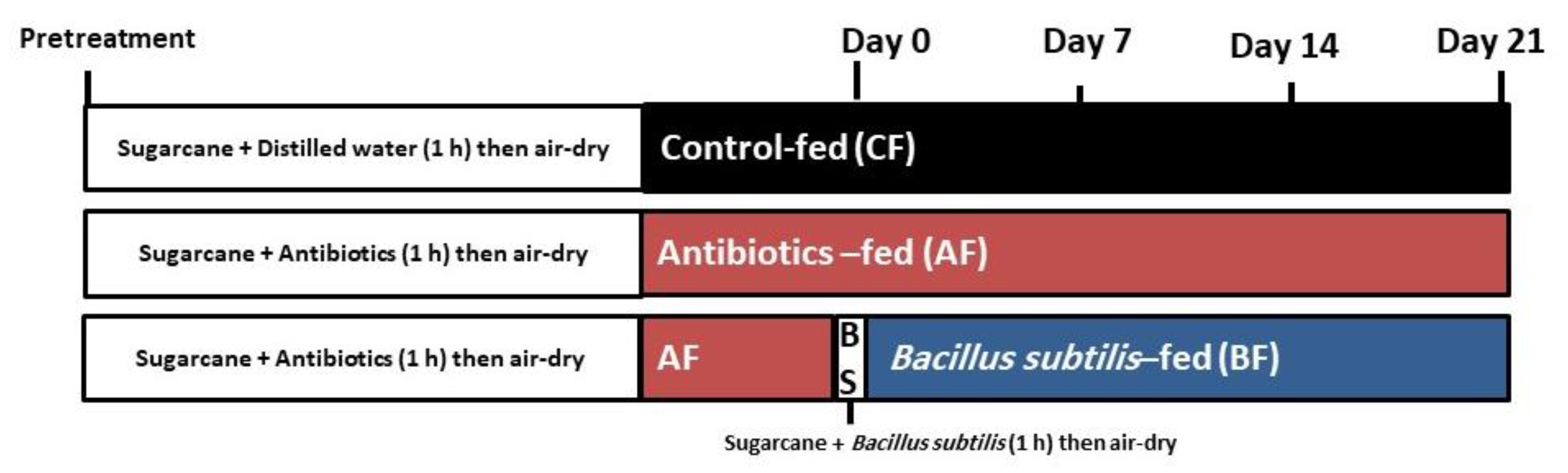
2.5. Effect of Gut Microbiota on the Nutrition Metabolism of RPW Larvae In Vivo

The role of gut microbiota on host metabolism was evaluated using an antibiotic cocktail containing kanamycin, tetracycline, gentamicin, and erythromycin (at a final concentration of 600 mg/L about 150 mg/L of each, using sterilized distilled water) [28,69]. The chosen gut isolate from enzyme screening experiments, especially cellulose degradation, Bacillus subtilis, was cultured in liquid Luria–Bertani (LB) medium at 30 °C overnight, then centrifuged (Centurion VS1283 LED, west Sussex, UK) at 5000 rpm, washed with sterile water, and reconstituted in water to achieve an OD600 of 1.8 [70]. Under aseptic conditions, 90 larvae (7th instar) from a field-collected population were randomly assigned to three treatment groups: a control group fed dry, sterilized regular sugarcane pieces soaked in sterilized distilled water (100 mL) for an hour (CF); a group fed sterilized sugarcane pieces soaked in the antibiotic cocktail (100 mL) for an hour (AF); and a group of AF after a week of antibiotic treatment were fed sterilized sugarcane soaked in the bacterial suspension (AF + B. subtilis, 100 mL) for an hour (BF), as shown in (Figure 1). The sugarcane pieces were air-dried before eating and changed daily for each group [24,38,52,70]. Each specimen’s body weight (mass) was measured with an electronic microbalance (Sartorius R180D, Gottingen, Germany) before treatment. All treatments started in parallel and lasted for 21 days. Five samples from each group were collected on the seventh, fourteenth, and twenty-first days of the treatment [28]. Hemolymph was extracted to examine metabolic indicators such as glucose, protein, and triglyceride (TAG) concentrations. Before collecting the hemolymph, larvae were cleaned under running water to remove excreta and food particles and then frozen on ice for 5 min. The cuticles of the larvae were punctured with a blunt fine needle, and 50 µL of hemolymph was collected from each larva in clean labeled 1.5 mL microcentrifuge tubes containing 2 μL of 0.2% phenylthiourea (PTU) to suppress hemolymph pigmentation; tubes were immersed in ice. Hemolymph samples were collected after 7, 14, and 21 days following feeding [28]. Glucose, protein, and TAG concentrations were detected using a glucose measurement kit, a protein (Biuret method) kit, and a TAG assay kit, respectively (all kits, Biodiagnostic, enzymatic colorimetric method, Giza, Egypt). These tests were carried out using a spectrometric reader (Spex® Jenway® 7205 Series Spectrophotometers, London, UK), according to the manufacturer’s protocol. Each experiment consisted of three replicates. At the end of the treatment, three larvae were dissected to isolate gut bacteria and assess the count in the gut bacterial community for each group, as previously mentioned in Section 2.2. [25].

2.6. Screening of Pesticide-Degrading Bacteria

In vitro screening of three different pesticides from distinct pesticide groups, including pyrethroid (λ-cyhalothrin), organophosphate (chlorpyrifos ethyl), and biopesticides (emamectin benzoate), was carried out. All 8 isolates were streaked on 1/10 diluted NA plates, supplemented with 100 ppm of each pesticide (n = 3), and then incubated for 7 days at 30 °C [25,38]. Isolates that showed growth or degradation of pesticides and formed a clear zone index were detected, as described in Section 2.5.

2.7. Chlorpyrifos Biodegradation Assay In Vitro

From the results of pesticide-degrading screening, chlorpyrifos (Pyriban A®48 % EC) was selected for the bacterial biodegradation experiment due to it being hardly degraded by isolates. The selected bacterial isolate, Arthrobacter sp., was adjusted to 1.5 × 10⁸ CFU/mL (a cell density of 0.5 McFarland) and grown in triplicates with chlorpyrifos minimal salt liquid medium containing 0.0012 g/L ammonium molybdate tetrahydrate, 0.003 g/L manganese sulfate monohydrate, 0.004 g/L ferrous sulfate heptahydrate, 0.1 g/L calcium chloride dehydrate, 0.4 g/L magnesium sulfate heptahydrate, 0.7 g/L monopotassium phosphate, 0.9 g/L sodium hydrogen phosphate, and 2 g/L sodium nitrate, pH 6.7 ± 0.2. Samples were collected after 4 and 10 days of incubation on a rotary water bath shaker (RSB-12 Water Bath Shaker, Mumbai, India) at 150 rpm and 35 °C to determine the pesticide content. Additionally, samples were re-cultured on NA plates to confirm bacterial viability [38].
Chlorpyrifos was extracted from samples using the QuEChERS method [71]. Ten grams of samples were extracted with 10 mL acetonitrile, and the mixture was vortexed for one minute. The mixture was then agitated rapidly for one minute and centrifuged (Centurion VS1283 LED, West Sussex, UK) at 4000 rpm for five minutes. Next, 4.0 g of MgSO4 anhydrous, 1.0 g of NaCl, 1.0 g of trisodium citrate dehydrate, and 0.5 g of disodium hydrogen citrate sesquihydrate were added. Pesticide reference standards were purchased from Dr Ehrenstorfer (Augsburg, Germany) with purities > 95% [25,72,73]. The concentration of the pesticide was determined using an Agilent HPLC system with the following chromatographic conditions: Agilent HPLC 1260 Infinity II autosampler, detector: DAD, wavelength: 200 nm, column temperature: 30 °C, Ascentis apelco, C18 column (150 × 4.6 mm × 5 μm), isocratic mobile phase: acetonitrile: methanol (90:10), flow rate: 0.6 mL/min, and injector volume: 20 µL [25].

2.8. Effect of Gut Microbiota on Chlorpyrifos Biodegradation of RPW Larvae In Vivo

To investigate the role of gut microbiota in RPW larvae in chlorpyrifos resistance in vivo, the bioassay of chlorpyrifos was carried out to measure the concentration of the LC15 value in conventionally fed larvae for 24 h as a control. As stated in Section 2.6, three sets of RPW larvae (CF, AF, and AF + Arthrobacter sp.) were produced for a week, and Arthrobacter sp. density was noted at an OD600 of 1.85. Following each group of larvae being treated with the LC15 value of chlorpyrifos, the survival rate percentage for each larval group was measured compared to the control.
Using the dipping food technique, chlorpyrifos was exposed to the seventh larval instar of field-collected RPW [74,75]. The sterilized sugarcane pieces (about 3 cm long and 1.5 cm in diameter) were dipped in the prepared solutions for one minute. After immersion, the treated pieces were air-dried and fed to the starving larvae for over six hours. In addition to these treatments, the untreated control was made up of sugarcane pieces submerged in distilled water. To identify the chlorpyrifos concentration range that causes mortality, several serial dilutions of the chemical utilizing five concentrations (3, 6, 12, 24, and 48 μg/mL) were freshly made in distilled water, each concentration containing 10 larvae, in triplicates (10 larvae, in triplicates, the total number of larvae used = 150). After a 24 h exposure, the larval mortalities were recorded to calculate the median lethal concentration (LC50) and (LC15) values.
After computing the control, the three RPW larval groups were exposed to the estimated LC15 values for the tested survival rate in each group (10 larvae, in triplicates, the total number of larvae used = 90) [38,70,76]. At the end of the experiment, some larvae of each group were dissected to isolate gut bacteria and assess the count in the gut bacterial community, as previously mentioned in Section 2.2 [52,70,77].

2.9. Thermal Stress Treatment

The 6th instar of field-collected RPW was divided into two groups (250 larvae each), antibiotic-fed larvae group (AF) and control larvae group (CF), for a week, and then each group was incubated at three temperatures: (a) low-temperature conditions (15.0 ± 1 °C), (b) high-temperature conditions (35.0 ± 1 °C), and (c) control group: no thermal stress treatment (27 ± 1 °C) [44,78].
Fitness parameters were used to study the effect of temperature on insect hosts and the endosymbiotic relationship. Morphological parameters (survival rate, activity, and body weight difference) (the total number of larvae used = 180) were examined. Also, physiological parameters (biochemical analysis of hemolymph including glucose, protein, and TAG concentrations in hemolymph) and oxidative stress (lipid peroxide and catalase activity) (both the total number of larvae used = 320) were examined. The sample observation and collection duration for all tested parameters were after 1, 7, and 14 days. At the end of each treatment, three larvae were selected for gut bacteria isolation to detect the count and distribution.

2.9.1. Fitness Parameter Tests

The survival rate, activity, and body weight difference of 90 larvae of AF and 90 larvae of CF groups were divided into 10 individuals, in triplicates, and incubated at (15, 35, and 27 °C). Each treatment monitored the survival rate, larval activity, and the weight difference under thermal stress in triplicates. The initial body weight was determined using an electronic microbalance before thermal treatment. The survival rate of the tested larvae was calculated. Observations were recorded to estimate larval behavior, development, and mortality.

2.9.2. Biochemical Analysis of Hemolymph (Hemolymph Index)

The glucose, protein, and TAG concentrations were measured, as previously mentioned in Section 2.6. Each experiment consisted of five replicates.

2.9.3. Antioxidant Assays

After weight and hemolymph, the larval samples were collected and transferred to clean and sterile tubes and stored immediately at −80 °C until further analysis. Each treatment and control were replicated five times. The treated larvae were homogenized in a potassium phosphate buffer (50 mM, pH 7.0) at 30 μL buffer per 1 mg of body weight. The homogenate was centrifuged for 15 min at 7000× g at 4 °C, and the supernatants were used for analysis [79,80].
The level of lipid peroxidation was determined indirectly by monitoring the formation of malondialdehyde (MDA) at 534 nm (Spex® Jenway® 7205 Series Spectrophotometers -, London, UK). MDA concentration was expressed as nmol of MDA produced per mg protein (nmol mg−1 protein). The catalase (CAT) enzyme activity was estimated by measuring the rate of H2O2 consumption via absorbance at 510 nm (Spex® Jenway® 7205 Series Spectrophotometers, London, UK). One unit of CAT activity was defined as the amount that decomposes 1 μmol of H2O2 per minute per mg protein (U mg−1 protein). The total protein concentration of all samples was measured spectrophotometrically using a Protein Biuret Kit (Biodiagnostic, Giza, Egypt) [79,80,81].

2.9.4. Gut Bacterial Isolation and Identification

At the end of the treatment, three larvae of each treatment were selected for gut bacteria isolation to detect the count and distribution, as previously mentioned in Section 2.2. The experiment was performed in triplicates. The dominant single colonies were selected based on their color, size, and morphology and were purified on NA plates. Pure single colonies were conventionally morphologically and biochemically identified [82,83] only to confirm with the gut bacterial molecular identification results. The biochemical experiments were performed using API 20E test strips (bioMérieux, Craponne, France) and confirmed by the API web program (bioMérieux) to compare the results with molecular identification.

2.10. Statistical Analysis

All statistical analyses were performed using Proc ANOVA in SAS, and significant differences between means were compared at p = 0.05. Two-way analysis of variance (ANOVA) was used to analyze the metabolic indices (larval body weight, glucose, protein, and TAG concentrations) with three treatment groups (CF, AF, and AF + B. subtilis) and different exposure times (after 7, 14, and 21 days of treatment). Statistical analyses of the survival rate of three groups of RPW larvae (CF, AF, and AF + Arthrobacter sp.) treated with the concentration LC15 value of chlorpyrifos were calculated using a separate one-way ANOVA. One-way ANOVA was used to compare between two treatment groups (AF and CF). In each group, we used two-way ANOVA, with thermal stress at three different temperatures (15, 27, and 35 °C) and different exposure times (after 1, 7, and 14 days of treatment) to test for differences between the average of different fitness parameters (survival rate, larval body weight differences, glucose, protein, and TAG concentrations in hemolymph, catalase, and lipid peroxide). The gut bacteria counts were expressed as colony-forming units per RPW gut (CFU/gut). The count results were log10 transformed to normalize the data.

3. Results

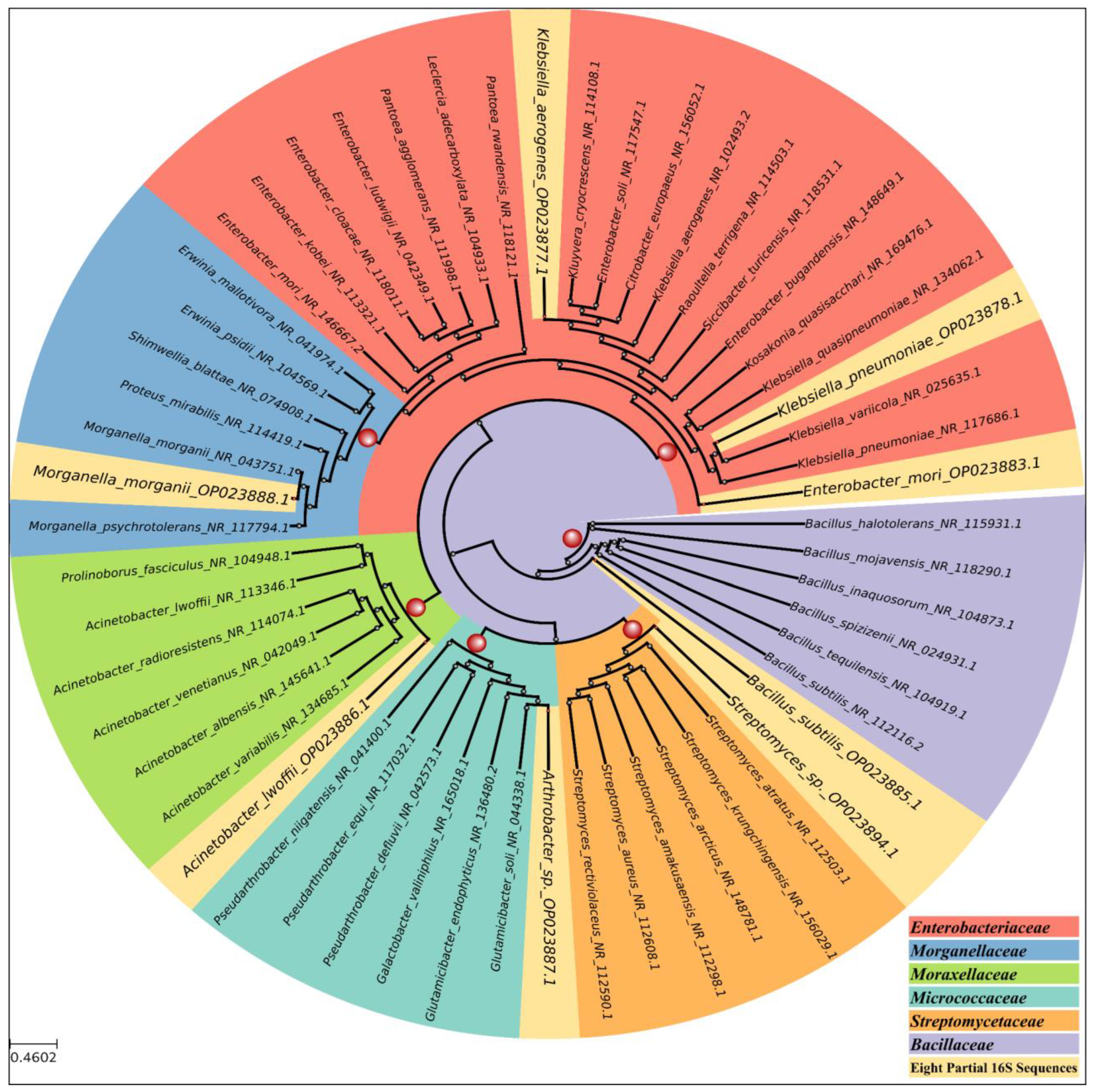
3.1. Bacterial Isolation and Identification

The number of gut bacteria in RPW larvae ranged from 8 × 10⁵ to 12 × 105 CFU gut−1, while in adults (both female and male) it ranged from 4 × 10⁵ to 7 × 10⁵ CFU gut−1. However, statistical analysis revealed no significant differences in bacterial counts between life stages (p = 0.184) or sexes (p = 0.188). A total of 70 bacterial isolates were obtained from 9 larval and 18 adult guts of RPW grown under aerobic conditions. These isolates were identified by 16S rRNA gene sequence analysis and assigned to seven genera and eight species belonging to three phyla, with Proteobacteria accounting for 61.4% of the total bacteria, followed by Firmicutes (21.5%) and Actinobacteria (17.1%) (Table 1).
Enterobacter aerogenes, Acinetobacter lwoffii, and Bacillus subtilis were the most dominant bacterial species across the tested life stages, with Enterobacter aerogenes, Acinetobacter lwoffi, Morganella morganii, and Bacillus subtilis being present in the gut of RPW throughout the tested life stages. Enterobacter mori and Arthrobacter sp. were detected in the larval gut, while Streptomyces sp. was detected in the adult gut. Klebsiella pneumonia was detected in the gut of larvae and female adults.
Based on the alignment results against the NCBI 16S ribosomal RNA database, the six best subject sequences of non-repeated genera and species were selected. The average query coverage was 99%, and the average percentage of identity was 97.5% (Table 1). The phylogenetic analysis results (Figure 2) showed that each partial 16S sequence belonged to the family descended from it. Enterobacter aerogenes (Acc. No. OP023877), Klebsiella pneumoniae (Acc. No. OP023878), and Enterobacter mori (Acc. No. OP023883) belonged to the Enterobacteriaceae family, Bacillus subtilis (Acc. No. OP023885) belonged to the Bacillaceae family, Acinetobacter lwoffii (Acc. No. OP023886) belonged to the Moraxellaceae family, Arthrobacter_sp. (Acc. No. OP023887) belonged to the Micrococcaceae family, Morganella_morganii (Acc. No. OP023888) belonged to the Morganellaceae family, and Streptomyces sp. (Acc. No. OP023894) belonged to the Streptomycetaceae family (Table 2).

3.2. Screening for the Production of Digestive Enzymes

In this study, B. subtilis and Streptomyces sp. were capable of producing all the tested enzymes to degrade CMC, xylan, pectin, starch, protease, and lipolytic enzymes (Figure 3). All eight identified isolates were able to produce amylase to degrade starch. However, out of the eight isolates, only some could degrade CMC (four isolates), xylan (four isolates), pectin (three isolates), and starch (seven isolates). The clear zone indices for each isolate are shown in Table 2 and Figure 3. Given that B. subtilis could produce all the tested enzymes, it was chosen for the in vivo study. Additionally, five isolates could degrade Tween 80, and two isolates (Arthrobacter sp. and B. subtilis) could degrade gelatin.

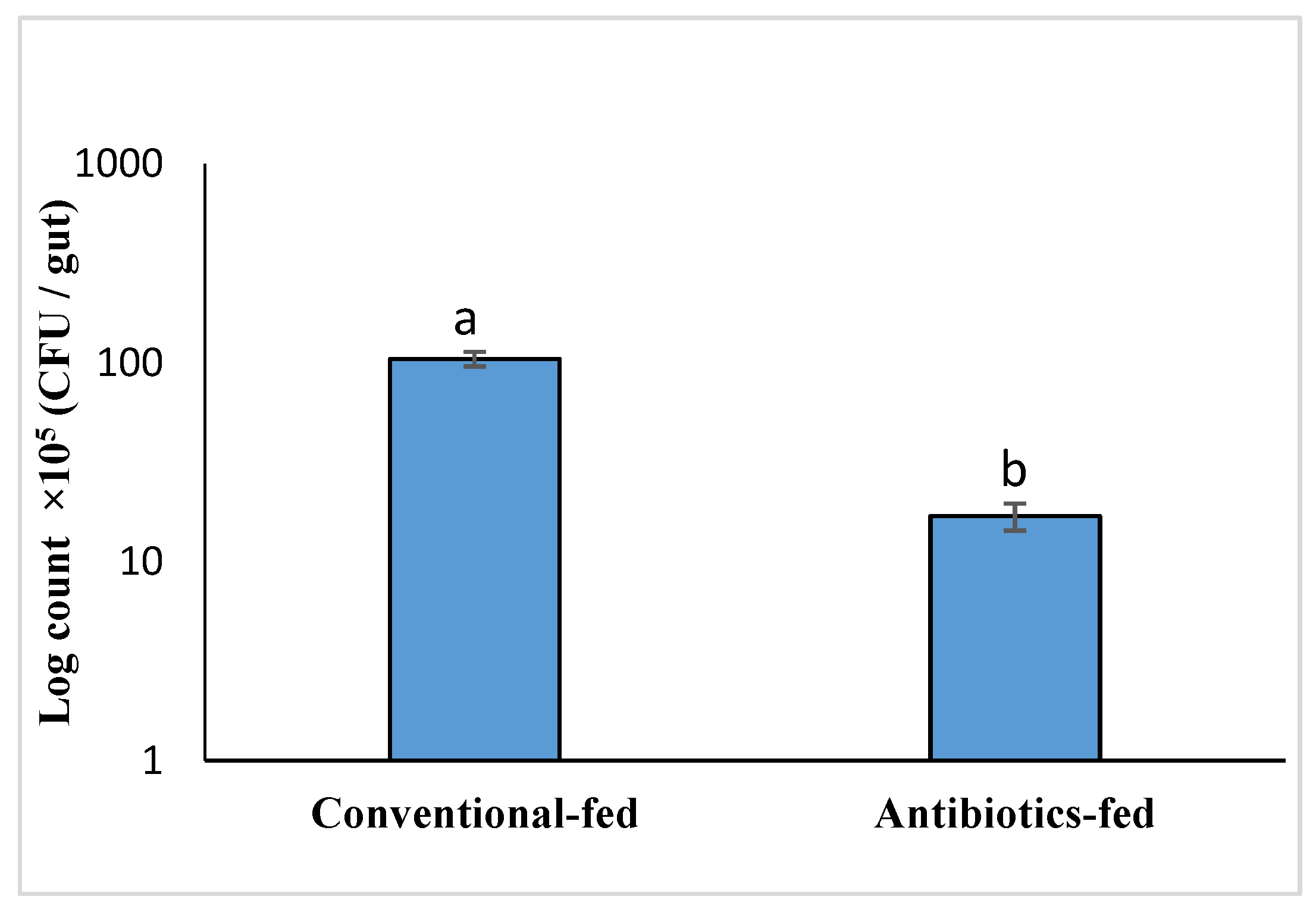
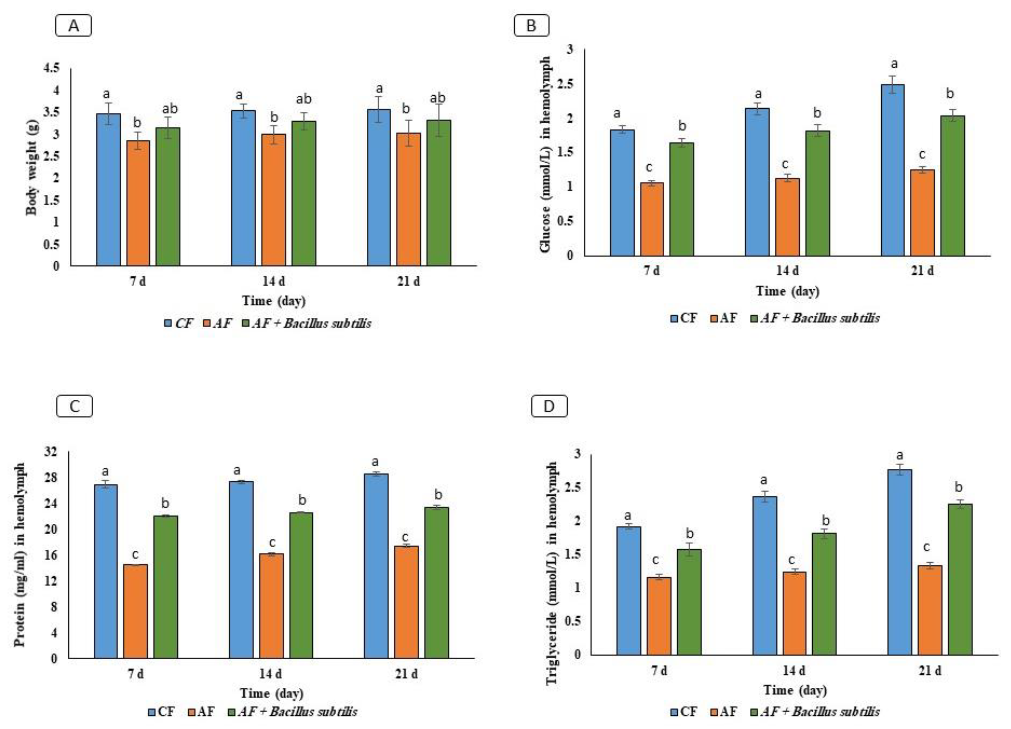
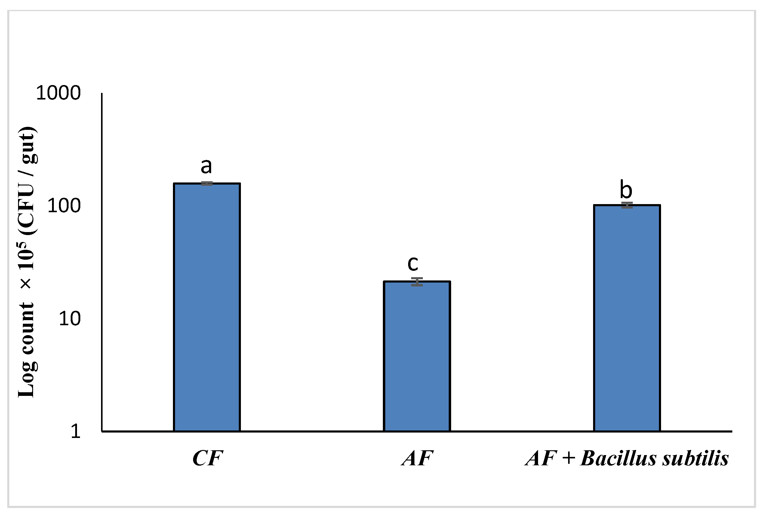
3.3. Effect of Gut Microbiota on RPW Larvae’s Nutrient Metabolism In Vivo

To examine the efficiency of the antibiotic cocktail in the enumeration and diversity of gut bacteria, RPW larvae were fed on sterilized sugarcane soaked in the antibiotic cocktail for one hour a day for seven days before the experiment. The results showed highly significant differences in gut bacterial count between the conventional-fed and antibiotic-fed larvae groups, as depicted in Figure 4.
Glucose (F = 480, LSD = 0.07, p < 0.001), protein (F = 2712.9, LSD = 0.32, p < 0.001), and TAG (F = 132.9, LSD = 0.14, p < 0.001) concentrations in hemolymph were measured and compared between the conventional-fed (CF), antibiotic-fed (AF), and bacteria-fed (AF + B. subtilis) treatments over time to assess the impact of gut bacteria on host nutrition metabolism and larval body weight (F = 9.86, LSD = 0.31, p = 0.002) (Figure 5). During the experiment, the net weight gain of AF larvae was significantly lower than that of CF larvae with normal microbiomes. However, RPW larvae fed (AF + B. subtilis) increased the net weight gain of AF but did not show a significant difference compared to AF and CF larvae (Figure 5A). Glucose, protein, and TAG concentrations in hemolymph were significantly higher in CF than (AF + B. subtilis) but lower in the antibiotic-fed group (Figure 5A–C). Exposure time affected glucose (F = 78.98, LSD = 0.07, p < 0.001), protein (F = 80.12, LSD = 0.32, p = 0.004), and TAG concentrations (F = 34.08, LSD = 0.142, p < 0.001). At the end of the experiment, highly significant differences (F = 59.82, LSD = 21.6, p < 0.001) in gut bacterial count were found in each feeding larvae group (Figure 6).

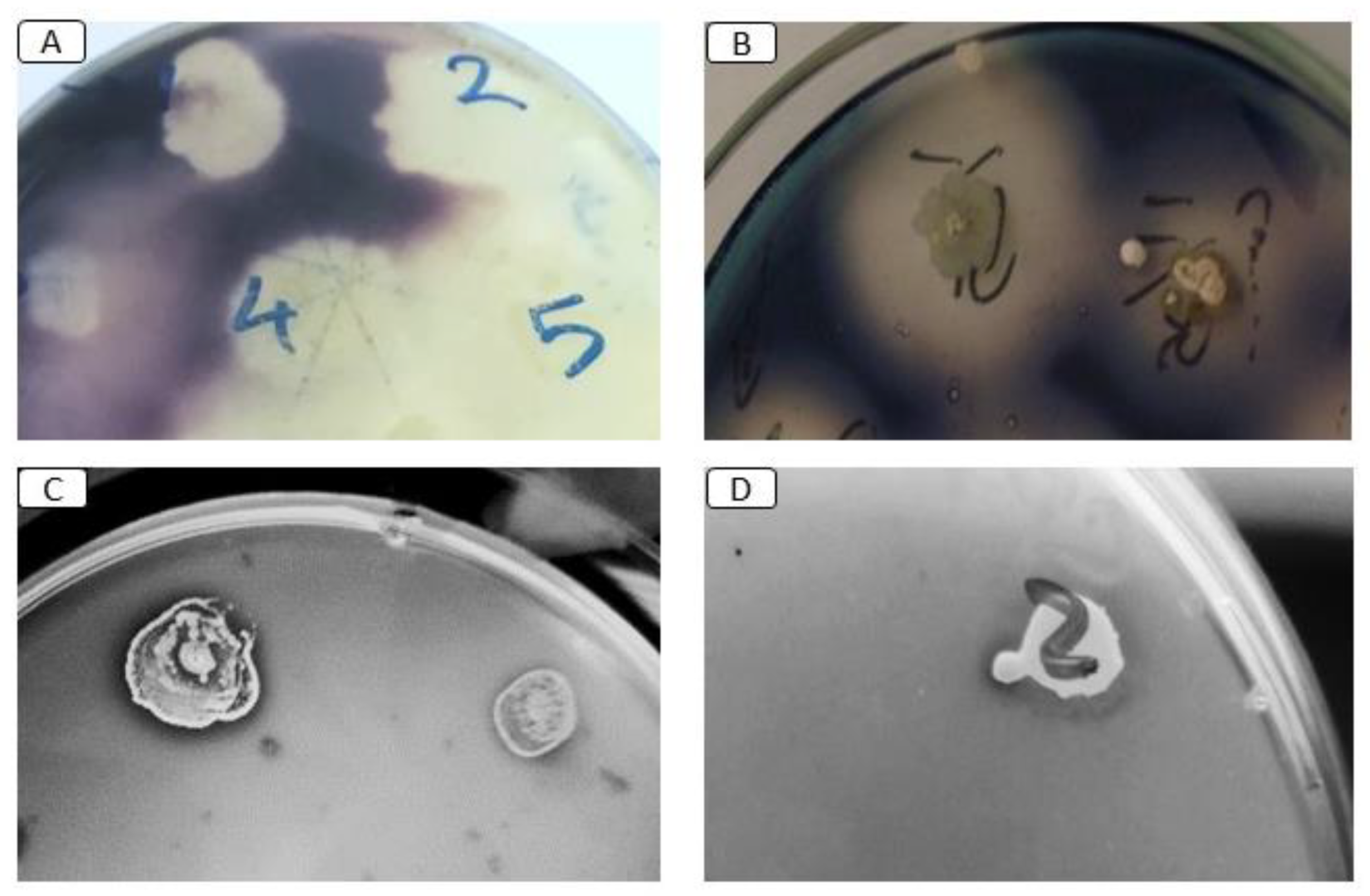
3.4. Screening of Pesticide-Degrading Bacteria

Table 2 shows that all isolates could grow on both Emamectin benzoate and λ-cyhalothrin media. The Arthrobacter sp., Streptomyces sp., and E. aerogenes isolates were able to degrade Emamectin benzoate and λ-cyhalothrin media with a clear zone. In contrast, M. morganii and E. aerogenes could only degrade λ-cyhalothrin, forming a clear zone. While Arthrobacter sp. isolate was able to grow well in chlorpyrifos medium, E. aerogenes, B. subtilis, and M. morganii could not. However, only Arthrobacter sp. was able to degrade chlorpyrifos, forming a clear zone.
Based on the obtained results and considering the health and environmental impacts of chlorpyrifos, Arthrobacter sp. was selected as a promising isolate for in vitro chlorpyrifos biodegradation. The Arthrobacter sp. isolate was able to grow on a minimal medium supplemented with 15 ppm chlorpyrifos, and the count after incubation for 10 days increased to 2.8 × 10⁸ CFU/mL. Chlorpyrifos was quantified by the HPLC method, and Arthrobacter sp. was able to degrade approximately 44.7% after incubation for 4 days. After 10 days of incubation, the isolates were able to completely degrade the chlorpyrifos pesticide compared to the control under the same conditions (Table 3).
The bioassay results 24 h post bioassay of chlorpyrifos on 7th instar larvae of RPW show that the LC15 and LC50 values were 8.347 and 20.736 mg/L, respectively (Table 4). According to the LC15 value, when treated with three different groups of larval (CF, AF, and AF + Arthrobacter sp.) and calculating the survival rate to understand the role of gut bacteria in vivo, the results in Figure 7 show a significance in the survival rate% of (AF, and AF + Arthrobacter sp.) compared with CF as a control (p = 0.0012). This result indicates the role of gut bacteria such as Arthrobacter sp. in host protection by degrading pesticides as an insect technique toward pesticide resistance. At the end of the experiment, highly significant differences in gut bacterial count were found in each feeding larvae group (Figure 8).

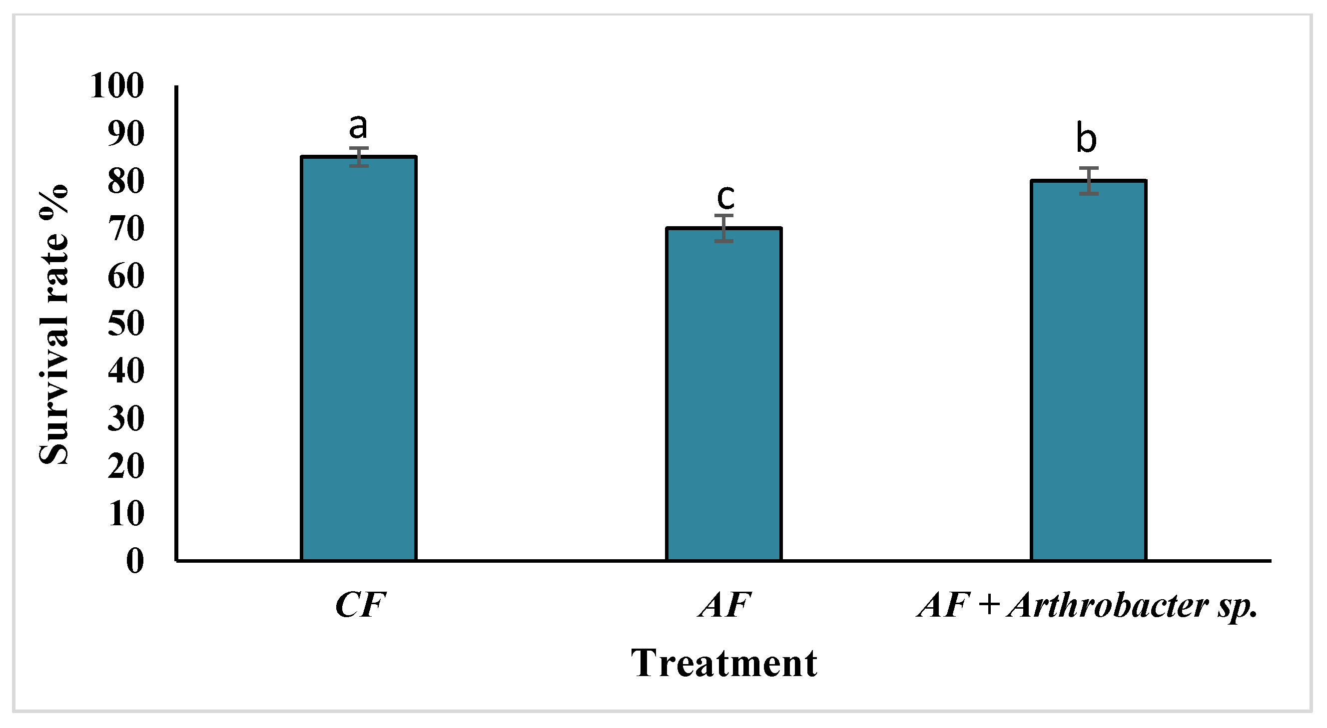
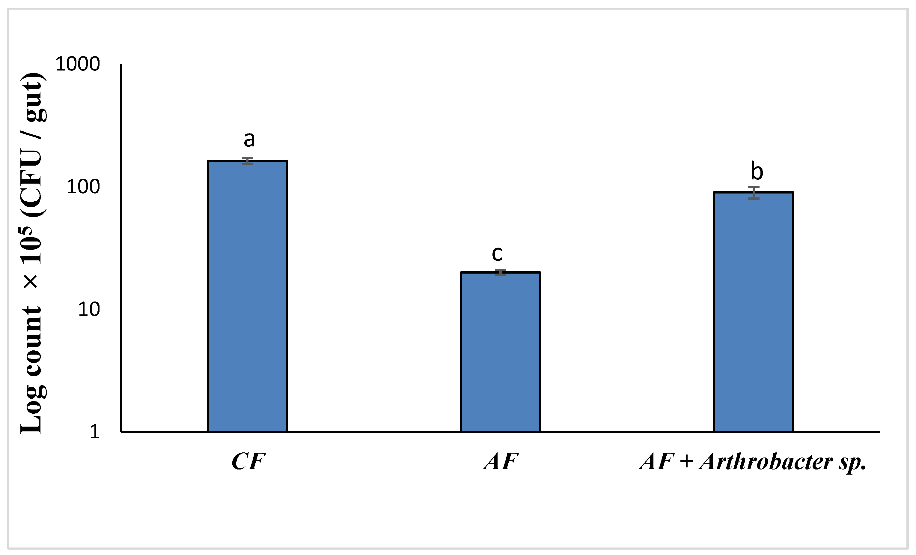
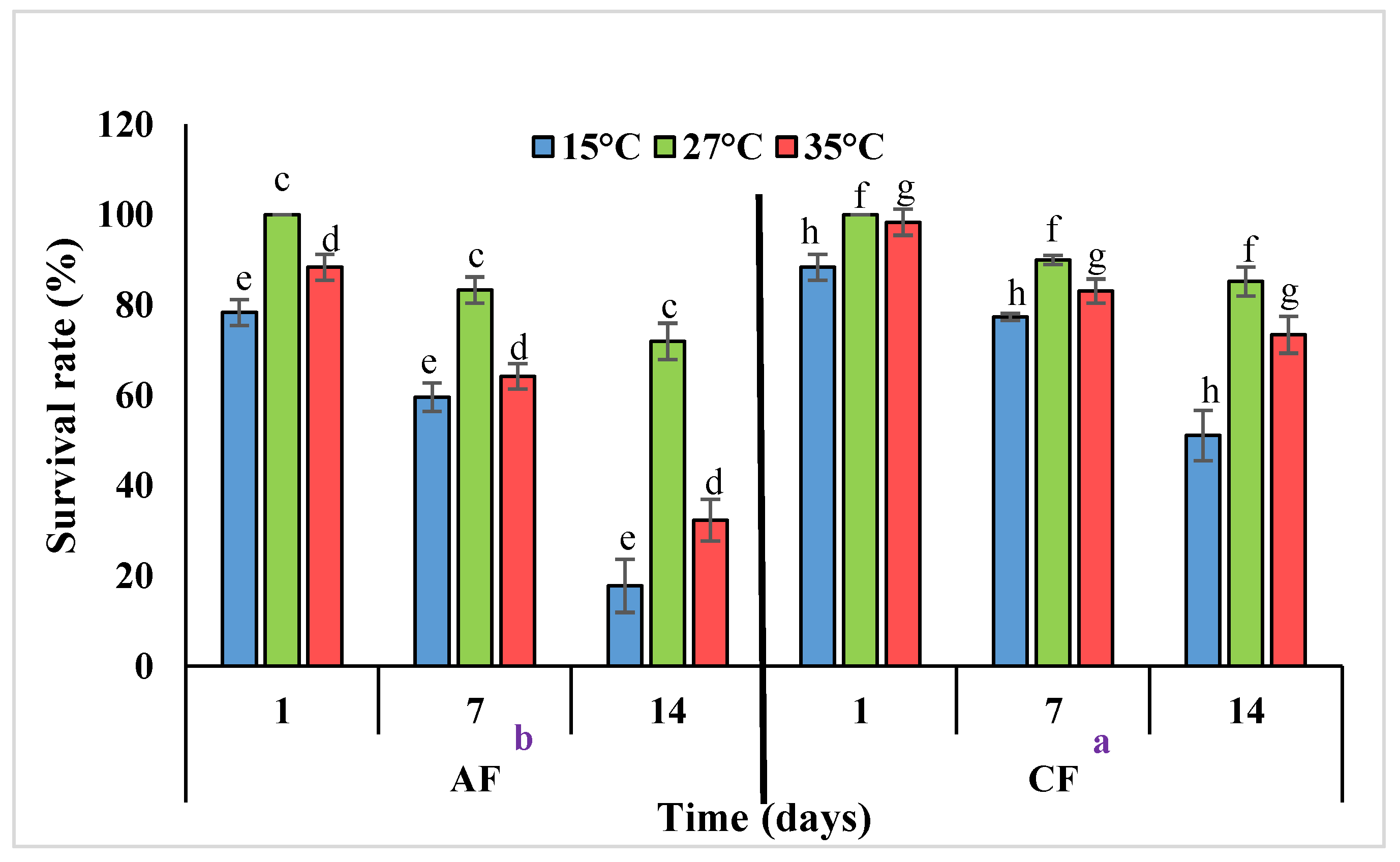
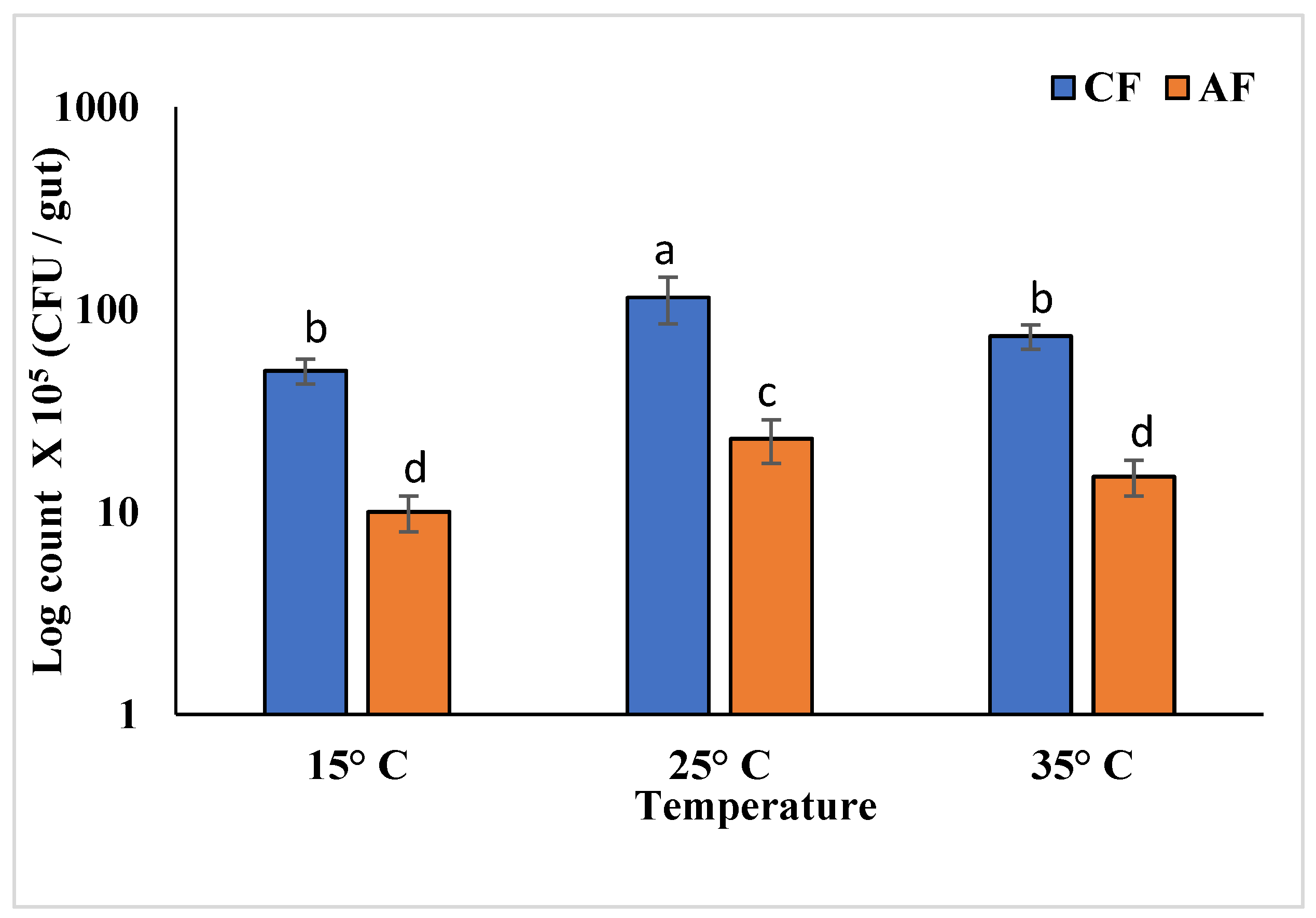
3.5. Thermal Stress Treatment

Comparing AF and CF larvae after 14 days of incubation at three different temperatures (15, 27, and 35 °C), the result indicates that the survival rate of the RPW larvae was generally decreased by temperature change (F = 46.28, LSD = 5, p < 0.001). Changes in the survival rate of RPW after cold hardening or heat shock for different durations are presented in Figure 9. The two-way ANOVA showed that the survival rate was significantly affected by temperature in AF groups (F = 42.40, LSD = 7.69, p < 0.001), duration (F = 85.46, LSD = 7.69, p < 0.001), and their interaction (F = 63.9, p < 0.001; in the CF groups, the survival rate was significantly affected by temperature (F = 28.20, LSD = 5.46, p < 0.001), duration (F = 47.59, LSD = 5.46, p < 0.001), and their interaction (F = 37.9, p < 0.001). The highest survival rate was at 27 °C without thermal stress in both treatment groups (AF and CF), 71.9% and 85.2%, respectively. However, the survival rate decreased significantly in thermal stress. In response to cold hardening (15 °C), the survival rate was 72.3% and 51.9% in CF and AF, respectively, while under heat shock (35 °C) it was 84.9 and 61.6% in CF and AF, respectively. It is worth noting that the survival rate of the AF decreased significantly compared to CF larvae.
Figure 10A shows a significant difference in the RPW larval weight of AF and CF groups after 14 days of thermal stress (F = 5.77, LSD = 0.09, p < 0.001). In the AF group, the larvae’s body weight became smaller compared with the CF group. At 15 °C, all larvae shrunk and lost weight. The weight difference in both conventional and antibiotic-fed larvae decreased significantly during the treatment period compared to larvae incubated without stress at 27 °C. However, at 27 °C, the larvae weight increased, and then at 35 °C, it increased non-significantly (Figure 10A).
It is worth mentioning that while observing the RPW larval activity, the CF larvae groups were more active than the AF groups incubated at different temperatures. The RPW larvae became less active, shrunk at 15 °C, and became active at 35 °C. The findings show that both examined insects’ hemolymph concentrations significantly changed in response to thermal stress, as shown in Figure 10B–D. Statistical analysis showed that treatment temperature in the AF groups was dramatically reduced compared to CF larvae hemolymph glucose (F = 107.69, LSD = 0.14, p < 0.001), protein (F = 102.62, LSD = 0.22, p < 0.001), and TAG (F = 453.5, LSD = 0.77, p < 0.001) concentrations. Also, two-way ANOVA demonstrated that glucose, protein, and TAG concentrations were significantly affected by treatment temperature, duration, and their interaction, as described in Figure 10B–D.
RPW larvae dramatically increased their MDA concentrations—a marker of lipid peroxidation (LPO) level—in response to temperature stress in AF compared to CF larvae (F = 88. 9, LSD = 1.26, p < 0.001) (Figure 10E). Two-way ANOVA indicates that the MDA concentration in RPW tissues was strongly impacted by temperature in CF (two-way ANOVA, F = 158.7, LSD = 1.26, p < 0.001) and AF groups (two-way ANOVA, F = 525.1, LSD = 1.29, p < 0.001). Moreover, the duration of exposure had a significant impact on CF (two-way ANOVA, F = 19.53, LSD = 1.26, p < 0.001) and AF (two-way ANOVA, F = 20.12, LSD = 1.29, p < 0.001). The interaction between temperature and time had a substantial impact on MDA activity in CF and AF as well, (two-way ANOVA, F = 89.11, p < 0.001) and (two-way ANOVA, F = 272.6, p < 0.001), respectively. In the CF group, LPO increased after one day of incubating RPW larvae at 15 °C then at 35 °C and decreased for the following 7 days, but after two weeks, a considerable rise was seen again in LPO at 15 °C and 35 °C (Figure 10E). Additionally, in the RPW AF, the cell damage was significantly higher than in the CF group with the normal microbiome. Figure 10E shows that the highest LPO damage occurred after a 14-day incubation at 15 °C.
In larval tissue, the temperature had a significant effect on the CAT activities (Figure 10F) in CF and AF tissues (F = 262.74, LSD = 0.3, p < 0.001). Two-way ANOVA indicates that temperature and time had major effects on enzyme activity, and the interaction between temperature and exposure time in CAT activity was similarly significant in CF (two-way ANOVA, F = 578.4, LSD = 0.36, p < 0.001), (two-way ANOVA, F = 24.5, LSD = 0.36, p < 0.001), (two-way ANOVA, F = 301.5, p < 0.001) and AF tissues (two-way ANOVA, F = 757.2, LSD = 0.195, p < 0.001), (two-way ANOVA, F = 16.59, LSD = 0.195, p < 0.001), (two-way ANOVA, F = 386.9, p < 0.001), respectively. On the first day in the CF group, RPW tissues incubated at 15 °C showed increased CAT activity. After 7 days, activity at these temperatures gradually dropped, but activity remained higher for up to 14 days than it did at 27 °C. The highest CAT activity was observed after 14 days of exposure (Figure 10F). However, in the AF group, the CAT activity significantly decreased in the disturbed microbiome compared with larvae with a normal microbiome. The CAT activity decreased with exposure time.
At the end of the experiment, the gut bacterial count showed highly significant differences in each group and at different studied temperatures (Figure 11). The gut bacterial count decreased in larvae incubated at 35 °C by about 38.4% in CF and about 40% in AF, and it decreased in larvae incubated at 15 °C by about 62.5% in CF and about 80% in AF compared to CF and AF larvae incubated at 27 °C. Also, the distribution of gut bacteria was affected by thermal stress compared with the RPW larvae reared at 27 °C as control based on 16S rRNA sequencing (as shown in Section 3.1). Bacterial gut isolates were identified after thermal treatment by using API 20E. Table 5 shows that K. pneumonia and M. morganii were often found among the isolates in all three temperature treatments in both AF and CF groups. In addition, B. subtilis was mainly present in the gut of larvae in all CF treatments but was absent from AF larvae. However, E. mori, Arthrobacter sp., and Streptomyces sp. were not present at 15 °C and 35 °C in both AF and CF groups, but E. aerogenes and A. lwoffii were present in the CF group at 35 °C.

4. Discussion

Chemical insecticides have long been the backbone of insect pest management. However, there is a push to use fewer chemicals in sustainable agriculture due to growing worries about the environment, human health, and pesticide resistance. This has sparked interest in innovative methods of controlling insect pests. There is a wealth of information showing that bacteria in symbiotic partnerships have a major impact on an insect’s characteristics. Insect bacterial symbioses have gained attention recently as potent sources of long-term solutions for pest management without the use of insecticides [4]. Indeed, the vast array of symbiotic relationships between bacteria and insects is now widely understood, as is their significant influence in dictating an enormous rise in the metabolic and ecological capacities of insects [23,24].
Previous studies revealed that a single insect gut can harbor 105–109 prokaryotic cells [1,25]. In this study, the RPW gut bacterial isolates belonged to Proteobacteria, Firmicutes, and Actinobacteria. Proteobacteria (Enterobacter, Klebsiella, Acinetobacter, and Morganella) were the dominant population in the RPW gut, matching previous reports [24,26,28,30,32]. The presence of Spiroplasma and Rickettsia in RPW collected from six different locations in Egypt was reported in our previous study by Awad et al. (2021), and the results reported the presence of Spiroplasma in all sites; there was a coexistence of Rickettsia in only three locations, including the same location studied in Ismailia, and all RPW females contained Rickettsia [84]. Across the tested life stages, Enterobacter (E. aerogenes and E. mori), particularly E. aerogenes, was the most abundant species. The same abundant species were found in the genus Bacillus (B. subtilis) [32,85,86]. Actinobacteria were also detected in the RPW gut lumen [26,28,31,87,88,89]. Enterobacter aerogenes, A. lwoffii, M. morganii, and B. subtilis were present in the RPW gut throughout the life cycle, suggesting a potential impact on their host fitness. Enterobacter, Acinetobacter, Morganella, and Bacillus were identified as the most predominant microorganisms within the insect gut microbiota [25,26,28,35,88,90,91,92]. However, K. pneumonia was found across females and was not detected in males (Table 1). Our results vindicate the finding that the genus Klebsiella is associated with females only [5].
Some bacterial isolates in this work degraded polysaccharides and hydrolyzed cellulose, hemicelluloses, pectin, and starch via proteolytic and lipolytic enzymes. These enzymes are typically absent in insects [93]. Rhynchophorus ferrugineus was reported to maintain facultative or obligate symbioses with bacteria that can break down cellulose [28]. Similar to a previous study [28], A. lwoffii, B. subtilis, Streptomyces sp., and E. aerogenes, were capable of degrading cellulose. Phylogenetic analysis indicated that most RPW gut cellulolytic isolates belonged to the phylum Proteobacteria, with genera such as Pseudomonas, Cellvibrio, and Enterobacter [87]. Furthermore, E. cloacae, which produces various carbohydrate-modifying and glycolytic enzymes like cellulases, trehalases, and other glucosidases to facilitate insect nutrition acquisition, was the most abundant gut bacteria in Plutella xylostella L. [23,24]. Additionally, Acinetobacter demonstrated cellulose and lignin degradation activity [53,94,95,96], while a Bacillus isolate from Anoplophora chinensis gut showed cellulose and/or aromatics degradation capabilities [96]. Bacillus subtilis produces a variety of enzymes including cellulase, xylanase, pectinase, amylase, and proteolytic and lipolytic enzymes [97]. Streptomyces sp. isolated from wood wasps produces both endo- and exoglucanases, among others [98,99]. Streptomyces flavogriseus demonstrated the ability to degrade plant lignocellulosic substrate [97,99,100,101] and produce proteolytic and lipolytic enzymes [102,103]. Streptomyces flavogriseus also produces lipolytic enzymes, specifically lipase/esterase [68]. Plutella xylostella L. (Lepidoptera: Plutellidae) gut bacterial isolates of Bacillus, Enterobacter, and Morganella produce esterase, with B. cereus (KC985225) demonstrating high esterase activity [92].
Antibiotics are commonly used to disrupt the relationship between gut bacteria and their host to reveal the potential benefits of gut bacteria to the host [24,28,69]. Antibiotics significantly reduced the bacterial count in the gut of RPW larvae and dramatically affected the metabolic indices. The weight of AF larvae was not significantly decreased, while the weight gain of AF + B. subtilis larvae was significantly increased compared to the control. In the AF group, the glucose and TAG levels in the hemolymph were significantly reduced, while increasing significantly in the AF + B. subtilis group. Introducing Lactococcus lacti and Enterobacter cloacae to germ-free RPW larvae improved the protein, glucose, and TAG contents in the hemolymph [23,24]. Similarly, in Bactrocera dorsalis Hendel (Diptera: Tephritidae) larvae from axenic flies, reintroducing gut bacteria (Citrobacter, Klebsiella, Providencia, and Enterobacter) increased the protein and TAG content [104]. Compared to the control, the gut flora was altered in the AF, leading to a significant decrease in hemolymph protein. This could be explained by increasing protein absorption by accelerating the digestion of protein-rich diets. These results show that antibiotic-induced alterations in the gut bacteria of RPW larvae significantly affected host nutrition metabolism, either by manufacturing particular amino acids or by controlling nutrient allocation [105,106]. In contrast, the protein concentration in the AF + B. subtilis group hemolymph increased but not significantly compared to CF, suggesting that B. subtilis is unable to improve protein uptake. The Tenebrio molitor (Coleoptera: Tenebrionidae) larvae treated with three probiotic bacteria (B. subtilis, B. toyonensis, and E. faecalis) exhibited significant improvements in terms of growth, time to pupation, protein, and total saturated fatty acids content of the larval cells [107].
Certain bacteria can break down organic insecticides such as chlorpyrifos, ethoprophos, and dimethoate [41,108], and these bacteria are commonly ingested by agricultural pests from numerous sources in food and the environment [40,41,42,108]. Studies on RPW gut bacterial isolates have shown that Proteobacterial families such as Enterobacteria, Pseudomonada, and Burkholderia can degrade chlorpyrifos, λ-cyhalothrin, acephate, trichlorfon, and spinosad [39,42,76,109]. Actinobacteria and Firmicutes bacteria play a role in the removal of toxins from the environment [92]. In the resistant strain of Spodoptera frugiperda (Lepidoptera: Noctuidae), the gut bacteria Enterococcus (Firmicutes) was found to break down pesticides such as chlorpyrifos, λ-cyhalothrin, deltamethrin, spinosad, and lufenuron [39]. Previous studies have also shown that different gut symbionts of insects from various orders can detoxify pesticides, such as Benzoylurea, Carbamate, Methoprene, Neonicotinoid, Organochloride, and Organophosphate, through the different species of genera Acetobacter, Actinobacteria, Aeromonas, Arsenphonus, Burkholderia, Citrobacter, Clostridium, Enterococcus, Exiguobacterium, Lachnospiracease, Lactobacillus, Lysinibacillus, Microbacterium, Pseudomonas, Staphylococcus, Symbiotaphrina, and Wolbachia [110]. According to the HPLC results of this study, Arthrobacter sp. isolates completely degraded chlorpyrifos after 10 days compared to the control, which is in accordance with previous studies [111,112,113]. Arthrobacter has also been shown to efficiently degrade deltamethrin and spinosyn in vitro, along with Pseudomonas isolated from insecticide-resistant S. frugiperda larvae [76]. Actinobacteria, which include genera such as Arthrobacter, Bacillus, Microbacterium, Micrococcus, Nocardioides, Rhodococcus, and Streptomyces, could degrade various classes of pesticides [114,115,116]. The survival rate indicated the vital role of gut bacteria in vivo by feeding the larval groups the same amount of chlorpyrifos. The findings point to the importance of gut bacteria like Arthrobacter sp. in protecting the host. When exposed to chlorpyrifos, microbes harbored by P. xylostella promote host survival [36]. In the brown plant hopper, Nilaparvata lugens (Stål) (Hemiptera: Delphacidae), antibiotics decreased detoxifying metabolism and enhanced host insecticide susceptibility [117].
Morphological, behavioral, ecological, physiological, and biochemical adaptations are among insect strategies of survival to withstand extreme temperatures [78,118]. RPW larvae were changed morphologically, physiologically, and biochemically by changing temperatures [81,119]. RPW quickly produced a variety of substances including glucose, glycerol, and numerous amino acids. The fat body content decreased, and a thick and dense cuticle was created to decrease chilling damage [78,120]. Heat shock proteins and sugar alcohols (polyols) are produced as a response to both cold and heat shock, and high-temperature pretreatment increases the resistance to chilling stress [121]. The results showed that the insects can withstand high temperatures. Compared to the AF group, the normal gut bacteria in the CF group were more active and adaptable. This indicates that endosymbiotic gut bacteria have an important role in giving the insect the ability to live at high temperatures efficiently and helping it raise the temperatures of the host (palm trees) above its natural temperature as a result of increased insect metabolism, activity, and fermentation processes, as consistent with the Mozib and El-Shafie (2013) study [43].
Under thermal stress, the gut microbiome improves survival rate, and hosts can survive in otherwise harsh environments by acting as a buffer against the deadly consequences of heat shock [122,123,124]. The gut bacteria of B. dorsalis flies were removed via antibiotic treatment, which significantly reduced the median survival duration after exposure to a temperature stress of 10 °C to approximately 68% of that in conventional flies, and the relative levels of proline and arginine metabolites were significantly downregulated by 34- and 10-, respectively, in the hemolymph [7]. However, when a crucial symbiotic bacterium, Klebsiella michiganensis, was recolonized, the median survival time significantly increased to 160% of those antibiotic treatment flies and restored their lifespan to that of regular flies, significantly upregulating the levels of proline and arginine by 13- and 10-fold, respectively [7]. Also, the B. dorsalis endosymbiotic microbiota shows a significant role in aiding host survival under thermal stress at three different temperatures (18, 35, and 27 °C) compared with antibiotic-treated flies [5]. The gut microbiome and temperature had a significant impact on the survival curves of Drosophila subobscura (Diptera: Drosophilidae) when compared to axenic flies [6].
In cold-climate overwintering Coleoptera, hymolymph glucose levels were up to six-times higher than in control populations [125,126]. Hemolymph glucose levels in RPW significantly increased (6–10 times more than the control) when they were exposed to cold (5 °C) [46,78,120,127]. Glucose is an endogenous cryoprotectant, enabling them to react to and survive a sudden and rapid change in temperature [78,120]. When silkworm larvae and pupae larvae were exposed to high-temperature stress, the protein levels of hemolymph significantly decreased while the glucose levels increased significantly; despite greater trehalose concentrations, many somatic tissues may easily access glucose as a fuel source [128]. To protect themselves against low-temperature damage, Agonoscena pistaciae Burc. and Laut. (Hemiptera: Psyllidae), Osmia rufa L. (Hymenoptera: Megachilidae), Ectomyelois ceratoniae Zeller (Lepidoptera: Pyralidae), and Spodoptera litura Fab. (Lepidoptera: Noctuidae) have all been found to accumulate lipids [129,130,131,132]. Adult Myzus persicae Sulzer (Hemiptera: Aphididae) exposed to high temperatures had a markedly lower triacylglycerol concentration. These findings imply that lipids are essential for behavioral thermoregulation and thermal stress-related healing mechanisms [121,133].
According to recent studies, bacteria in a healthy gut microbiome create antioxidative metabolites [134,135]. In this study, LPO and CAT activity considerably changed in RPW larvae under various thermal stressors. Compared to the AF group, the normal gut bacteria in the CF group were more active and adaptable. Thermal stress led to a significantly increased amount of MDA concentration in RPW larvae. This proves unequivocally that thermal stress is connected to the LPO process and other oxidative stress responses in arthropods [136,137]. MDA concentrations increased significantly in response to thermal exposure, but after prolonged exposure, they gradually dropped before rising again. The antioxidant system likely eliminated oxidative damage as the MDA decreased with increased exposure; a similar phenomenon was observed in citrus red mites Panonychus citri McGregor (Acari: Tetranychidae) and Neoseiulus cucumeris Oudemans (Acari: Phytoseiidae) [137]. However, the AF differed from the CF in that LPO remained elevated, indicating that the antioxidant system may be poor at removing ROS brought on by thermal stress, emphasizing the importance of gut bacteria.
CAT is thought to be the main H2O2-scavenging enzyme in arthropods [138]. Evidently, there was a noticeable increase in CAT activity, which improved H2O2 elimination and reduced oxidative stress-related damage. Furthermore, elevated CAT activities produced by temperature stress were seen in the fat body of the 5th instar silkworm, Bombyx mori L. (Lepidoptera, Bombycidae), and Bactrocera dorsalis Hendel (Diptera: Tephritidae) [81,139]. The findings showed that the CF insects were more responsive to antioxidants than in the AF group. In AF larvae, the MDA concentration was significantly higher, and CAT activity was significantly lower, which suggests it could not repair the oxidative stress damage when compared to CF larvae. There was no change in the CAT activity of the control reared at 27 °C. A varied and temperature-dependent bacterial population inhabiting the gut of RPW, K. pneumonia, M. morganii, and B. subtilis, were observed in the three different temperatures; however, in the AF larvae, B. subtilis disappeared. Among the B. dorsalis gut bacteria, 88% varied in temperature when raised at 27, 18, and 35 °C, but 12% (Klebsiella oxytoca, Providencia rettigeri, Morgonella morganii, and Bacillus cereus) were observed to be stable, while Actinobacter disappeared [5].

5. Conclusions

This study investigated which gut bacteria in the red palm weevil formed a complex microbial community of seven genera and eight species belonging to three phyla. The gut bacteria remained stable throughout different life stages and demonstrated the capability to improve metabolism. Bacillus subtilis was confirmed to produce a variety of metabolic enzymes including cellulase, xylanase, pectinase, amylase, and proteolytic and lipolytic enzymes. Moreover, the gut bacteria showed a capability to degrade different classes of pesticides. This study highlights the critical role that gut bacteria play in the host’s fitness and tolerance. This study also investigated how cold and heat stress affected RPW larvae, finding that present bacterial symbionts played a part in the larvae’s ability to adapt and tolerate thermal stress. Therefore, gut bacteria represent a potential candidate for developing eco-friendly strategies to manage insect pests. Arthrobacter sp. was identified as a potential candidate for developing eco-friendly pest biocontrol strategies. However, further investigation is needed before incorporating this technique into IPM programs. For example, adding bactericidal compounds with pesticides to reduce symbiotic gut bacteria in insects could be a potential approach. Additionally, gut bacterial isolates that can degrade pesticides could be utilized in the environment. Furthermore, gut bacteria could also be helpful in mass-rearing programs. It is necessary to consider the importance of endosymbionts as part of prediction models when assessing the ecological effects of global climate change.
Additionally, this study also investigated the gut bacterial isolates’ potential to degrade chlorpyrifos, a model pesticide, which could be helpful as an environmentally friendly biodegrading agent. The studied insects were reared at three different temperature regimes (15, 35, and 27 °C), and comparisons were made according to fitness parameters to examine thermal stress tolerance. The findings of this study could confirm that gut bacteria are a crucial factor in insect pest fitness, insecticide resistance, and adaptation to climate change, which also serves as a basis for symbiosis-dependent pest management strategies that could be implemented in IPM programs as a new management technique. Therefore, it serves as a basis for symbiosis-dependent pest management strategies that could be implemented in IPM programs as a new management technique.

Author Contributions

Conceptualization, O.A.E., M.A., T.A.E., and S.M.E.; methodology, O.A.E.; software, O.A.E.; validation, O.A.E., M.A., T.A.E., and S.M.E.; formal analysis, O.A.E., M.A.M.A., and E.-D.S.I.; investigation, O.A.E.; resources, M.A., T.A.E., and S.M.E.; data curation, O.A.E. and M.A.M.A.; writing—original draft preparation, O.A.E. and N.S.D.; writing—review and editing, O.A.E., T.A.E., N.S.D., and S.M.E.; visualization, O.A.E.; supervision, M.A., T.A.E., H.M.E.-S., and S.M.E.; project administration, T.A.E. and S.M.E.; funding acquisition, M.A., T.A.E., N.S.D., and S.M.E. All authors have read and agreed to the published version of the manuscript.

Funding

This research received no external funding.

Institutional Review Board Statement

Not applicable.

Informed Consent Statement

Not applicable.

Data Availability Statement

Data are contained within the article.

Conflicts of Interest

The authors declare no conflicts of interest.

References

  1. Engel, P.; Moran, N.A. The Gut Microbiota of Insects—Diversity in Structure and Function. FEMS Microbiol. Rev. 2013, 37, 699–735. [Google Scholar] [CrossRef]
  2. Kikuchi, Y. Endosymbiotic Bacteria in Insects: Their Diversity and Culturability. Microbes Environ. 2009, 24, 195–204. [Google Scholar] [CrossRef]
  3. Masson, F.; Lemaitre, B. Growing Ungrowable Bacteria: Overview and Perspectives on Insect Symbiont Culturability. Microbiol. Mol. Biol. Rev. 2020, 84, 10–1128. [Google Scholar] [CrossRef] [PubMed]
  4. Li, G.; Zheng, X.; Zhu, Y.; Long, Y.; Xia, X. Bacillus Symbiont Drives Alterations in Intestinal Microbiota and Circulating Metabolites of Lepidopteran Host. Environ. Microbiol. 2022, 24, 4049–4064. [Google Scholar] [CrossRef]
  5. Ayyasamy, A.; Kempraj, V.; Pagadala Damodaram, K.J.; Jayanthi, K.; Damodaram, P. Endosymbiotic Bacteria Aid to Overcome Temperature Induced Stress in the Oriental Fruit Fly, Bactrocera dorsalis. Microb. Ecol. 2021, 82, 783–792. [Google Scholar] [CrossRef]
  6. Jaramillo, A.; Castañeda, L.E. Gut Microbiota of Drosophila subobscura Contributes to Its Heat Tolerance and Is Sensitive to Transient Thermal Stress. Front. Microbiol. 2021, 12, 654108. [Google Scholar] [CrossRef] [PubMed]
  7. Raza, M.F.; Wang, Y.; Cai, Z.; Bai, S.; Yao, Z.; Awan, A.; Zhang, Z.; Zheng, W.; Id, H.Z. Gut Microbiota Promotes Host Resistance to Low-Temperature Stress by Stimulating Its Arginine and Proline Metabolism Pathway in Adult Bactrocera dorsalis. PLoS Pathog. 2020, 16, e1008441. [Google Scholar] [CrossRef]
  8. Gil, R.; Latorre, A.; Moya, A. Bacterial Endosymbionts of Insects: Insights from Comparative Genomics. Environ. Microbiol. 2004, 6, 1109–1122. [Google Scholar] [CrossRef] [PubMed]
  9. Storelli, G.; Defaye, A.; Erkosar, B.; Hols, P.; Royet, J.; Leulier, F. Lactobacillus plantarum Promotes Drosophila Systemic Growth by Modulating Hormonal Signals through TOR-Dependent Nutrient Sensing. Cell Metab. 2011, 14, 403–414. [Google Scholar] [CrossRef]
  10. Wang, H.; Jin, L.; Zhang, H. Comparison of the Diversity of the Bacterial Communities in the Intestinal Tract of Adult Bactrocera dorsalis from Three Different Populations. J. Appl. Microbiol. 2011, 110, 1390–1401. [Google Scholar] [CrossRef] [PubMed]
  11. Jaffar, S.; Ahmad, S.; Lu, Y. Contribution of Insect Gut Microbiota and Their Associated Enzymes in Insect Physiology and Biodegradation of Pesticides. Front. Microbiol. 2022, 13, 979383. [Google Scholar] [CrossRef]
  12. Visôtto, L.E.; Oliveira, M.G.A.; Ribon, A.O.B.; Mares-Guia, T.R.; Guedes, R.N.C. Characterization and Identification of Proteolytic Bacteria from the Gut of the Velvetbean Caterpillar (Lepidoptera: Noctuidae). Environ. Entomol. 2009, 38, 1078–1085. [Google Scholar] [CrossRef] [PubMed]
  13. Wang, J.J.J.M.; Bai, J.; Zheng, F.Y.; Ling, Y.; Li, X.X.J.; Wang, J.J.J.M.; Zhi, Y.C.; Li, X.X.J. Diversity of the Gut Microbiome in Three Grasshopper Species Using 16S RRNA and Determination of Cellulose Digestibility. PeerJ 2020, 8, e10194. [Google Scholar] [CrossRef] [PubMed]
  14. Zheng, H.; Powell, J.E.; Steele, M.I.; Dietrich, C.; Moran, N.A. Honeybee Gut Microbiota Promotes Host Weight Gain via Bacterial Metabolism and Hormonal Signaling. Proc. Natl. Acad. Sci. USA 2017, 114, 4775–4780. [Google Scholar] [CrossRef]
  15. FAO. Current Situation of Red Palm Weevil in the NENA Region [Current Situation of Management Practices, Challenges/Weaknesses and Available Research and Technologies for Its Improvement]. 2017, pp. 29–31. Available online: http://Www.Fao.Org/3/a-Ms664e.Pdf (accessed on 25 April 2021).
  16. El-Shafie, A.F.H.; Faleiro, R.J. Red Palm Weevil Rhynchophorus ferrugineus (Coleoptera: Curculionidae): Global Invasion, Current Management Options, Challenges and Future Prospects. In Invasive Species—Introduction Pathways, Economic Impact, and Possible Management Options; El-Shafie, H., Ed.; Intechopen: London, UK, 2020; pp. 1–29. [Google Scholar]
  17. Al-Dosary, N.M.N.; Al-Dobai, S.; Faleiro, J.R. Review on the Management of Red Palm Weevil Rhynchophorus ferrugineus Olivier in Date Palm Phoenix Dactylifera L. Emir. J. Food Agric. 2016, 28, 34–44. [Google Scholar] [CrossRef]
  18. Rochat, D.; Dembilio, O.; Jaques, J.A.; Suma, P.; Alessandra, L.; Pergola;Hamidi, R.; Kontodimas, D.; Soroker, V. Rhynchophorus ferrugineus: Taxonomy, Distribution, Biology, and Life Cycle. In Handbook of Major Palm Pests; Soroker, V., Colazza, S., Eds.; Wiley-Blackwell: Chichester, UK, 2017; pp. 69–104. [Google Scholar]
  19. Kontodimas, D.; Soroker, V.; Pontikakos, C.; Suma, P.; Beaudoin-Ollivier, L.; Karamaouna, F.; Riolo, P. Visual Identification and Characterization of Rhynchophorus ferrugineus and Paysandisia Archon Infestation. In Handbook of Major Palm Pests: Biology and Management; Soroker, V., Colazza, S., Eds.; John Wiley & Sons: Chichester, UK, 2017; pp. 187–208. [Google Scholar]
  20. Abdel-Baky, N.F.; Aldeghairi, M.A.; Motawei, M.I.; Al-Shuraym, L.A.M.; Al-Nujiban, A.A.S.; Alharbi, M.T.M.; Rehan, M. Monitoring of Infestation Percentages of the Invasive Red Palm Weevil, Rhynchophorus ferrugineus (Coleoptera: Curculionidae), and Management Tactics: A Six-Year Study. Braz. J. Biol. 2022, 82, e263707. [Google Scholar] [CrossRef] [PubMed]
  21. Mazza, G.; Francardi, V.; Simoni, S.; Benvenuti, C.; Cervo, R.; Faleiro, J.R.; Llácer, E.; Longo, S.; Nannelli, R.; Tarasco, E.; et al. An Overview on the Natural Enemies of Rhynchophorus Palm Weevils, with Focus on R. ferrugineus. Biol. Control. 2014, 77, 83–92. [Google Scholar] [CrossRef]
  22. Peng, L.; Miao, Y.; Hou, Y. Demographic Comparison and Population Projection of Rhynchophorus ferrugineus (Coleoptera: Curculionidae) Reared on Sugarcane at Different Temperatures. Sci. Rep. 2016, 6, 31659. [Google Scholar] [CrossRef]
  23. Liu, Q.; Su, Z.; Liu, H.; Lu, S.; Ma, B.; Zhao, Y.; Hou, Y.; Shi, Z. The Effect of Gut Bacteria on the Physiology of Red Palm Weevil, Rhynchophorus ferrugineus Olivier and Their Potential for the Control of This Pest. Insects 2021, 12, 594. [Google Scholar] [CrossRef]
  24. Habineza, P.; Muhammad, A.; Ji, T.; Xiao, R.; Yin, X.; Hou, Y.; Shi, Z. The Promoting Effect of Gut Microbiota on Growth and Development of Red Palm Weevil, Rhynchophorus ferrugineus (Olivier) (Coleoptera: Dryophthoridae) by Modulating Its Nutritional Metabolism. Front. Microbiol. 2019, 10, 1212. [Google Scholar] [CrossRef]
  25. ElKraly, O.A.; Awad, M.; El-Saadany, H.M.; Hassanein, S.E.; Elrahman, T.A.; Elnagdy, S.M. Impact of Gut Microbiota Composition on Black Cutworm, Agrotis ipsilon (Hufnagel) Metabolic Indices and Pesticide Degradation. Anim. Microbiome 2023, 5, 44. [Google Scholar] [CrossRef] [PubMed]
  26. Jia, S.; Zhang, X.; Zhang, G.; Yin, A.; Zhang, S.; Li, F.; Wang, L.; Zhao, D.; Yun, Q.; Wang, J.; et al. Seasonally Variable Intestinal Metagenomes of the Red Palm Weevil (Rhynchophorus ferrugineus). Environ. Microbiol. 2013, 15, 3020–3029. [Google Scholar] [CrossRef] [PubMed]
  27. Shi, W.; Xie, S.; Chen, X.; Sun, S.; Zhou, X.; Liu, L.; Gao, P.; Kyrpides, N.C.; No, E.G.; Yuan, J.S. Comparative Genomic Analysis of the Endosymbionts of Herbivorous Insects Reveals Eco-Environmental Adaptations: Biotechnology Applications. PLoS Genet. 2013, 9, e1003131. [Google Scholar] [CrossRef]
  28. Muhammad, A.; Fang, Y.; Hou, Y.; Shi, Z. The Gut Entomotype of Red Palm Weevil Rhynchophorus ferrugineus Olivier (Coleoptera: Dryophthoridae) and Their Effect on Host Nutrition Metabolism. Front. Microbiol. 2017, 8, 2291. [Google Scholar] [CrossRef] [PubMed]
  29. Ceja-Navarro, J.A.; Karaoz, U.; Bill, M.; Hao, Z.; White, R.A.; Arellano, A.; Ramanculova, L.; Filley, T.R.; Berry, T.D.; Conrad, M.E.; et al. Gut Anatomical Properties and Microbial Functional Assembly Promote Lignocellulose Deconstruction and Colony Subsistence of a Wood-Feeding Beetle. Nat. Microbiol. 2019, 4, 864–875. [Google Scholar] [CrossRef] [PubMed]
  30. Jing, T.; Qi, F.; Wang, Z. Most Dominant Roles of Insect Gut Bacteria: Digestion, Detoxification, or Essential Nutrient Provision? Microbiome 2020, 8, 38. [Google Scholar] [CrossRef] [PubMed]
  31. Valzano, M.; Achille, G.; Burzacca, F.; Ricci, I.; Damiani, C.; Scuppa, P.; Favia, G. Deciphering Microbiota Associated to Rhynchophorus ferrugineus in Italian Samples: A Preliminary Study. J. Entomol. Acarol. Res. 2012, 44, 85–89. [Google Scholar] [CrossRef]
  32. Tagliavia, M.; Messina, E.; Manachini, B.; Cappello, S.; Quatrini, P. The Gut Microbiota of Larvae of Rhynchophorus ferrugineus Oliver (Coleoptera: Curculionidae). BMC Microbiol. 2014, 14, 136. [Google Scholar] [CrossRef] [PubMed]
  33. Dawadi, B.; Wang, X.; Xiao, R.; Muhammad, A.; Hou, Y.; Shi, Z. PGRP-LB Homolog Acts as a Negative Modulator of Immunity in Maintaining the Gut-Microbe Symbiosis of Red Palm Weevil, Rhynchophorus ferrugineus Olivier. Dev. Comp. Immunol. 2018, 86, 65–77. [Google Scholar] [CrossRef]
  34. Gracy, R.G.; Malathi, V.M.; Jalali, S.K.; Jose, V.L.; Thulasi, A. Variation in Larval Gut Bacteria between Insecticide-Resistant and -Susceptible Populations of Helicoverpa armigera (Hübner) (Lepidoptera: Noctuidae). Phytoparasitica 2016, 44, 477–490. [Google Scholar] [CrossRef]
  35. Cheng, D.; Guo, Z.; Riegler, M.; Xi, Z.; Liang, G.; Xu, Y. Gut Symbiont Enhances Insecticide Resistance in a Significant Pest, the Oriental Fruit Fly Bactrocera dorsalis (Hendel). Microbiome 2017, 5, 1–12. [Google Scholar] [CrossRef] [PubMed]
  36. Xia, X.; Sun, B.; Gurr, G.G.M.G.; Vasseur, L.; Xue, M.; You, M.; Yu, Z.; Newcombe, G.; Xu, Y.; You, M.; et al. Gut Microbiota Mediate Insecticide Resistance in the Diamondback Moth, Plutella xylostella (L.). Front. Microbiol. 2018, 9, 25. [Google Scholar] [CrossRef]
  37. Gressel, J. Microbiome Facilitated Pest Resistance: Potential Problems and Uses. Pest Manag. Sci. 2018, 74, 511–515. [Google Scholar] [CrossRef] [PubMed]
  38. Chen, B.; Zhang, N.; Xie, S.; Zhang, X.; He, J.; Muhammad, A.; Sun, C.; Lu, X.; Shao, Y.; Sunb, C.; et al. Gut Bacteria of the Silkworm Bombyx mori Facilitate Host Resistance against the Toxic Effects of Organophosphate Insecticides. Environ. Int. 2020, 143, 105886. [Google Scholar] [CrossRef]
  39. Gomes, A.F.F.; Omoto, C.; Cônsoli, F.L. Gut Bacteria of Field-Collected Larvae of Spodoptera frugiperda Undergo Selection and Are More Diverse and Active in Metabolizing Multiple Insecticides than Laboratory-Selected Resistant Strains. J. Pest Sci. 2020, 93, 833–851. [Google Scholar] [CrossRef]
  40. Yang, C.; Liu, N.; Guo, X.; Qiao, C. Cloning of Mpd Gene from a Chlorpyrifos-Degrading Bacterium and Use of This Strain in Bioremediation of Contaminated Soil. FEMS Microbiol. Lett. 2006, 265, 118–125. [Google Scholar] [CrossRef] [PubMed]
  41. Li, X.; He, J.; Li, S. Isolation of a Chlorpyrifos-Degrading Bacterium, Sphingomonas Sp. Strain Dsp-2, and Cloning of the Mpd Gene. Res. Microbiol. 2007, 158, 143–149. [Google Scholar] [CrossRef]
  42. Kikuchi, Y.; Hayatsu, M.; Hosokawa, T.; Nagayama, A.; Tago, K.; Fukatsu, T. Symbiont-Mediated Insecticide Resistance. Proc. Natl. Acad. Sci. USA 2012, 109, 8618–8622. [Google Scholar] [CrossRef]
  43. Mozib, M.E.; El-Shafie, H.A. Effect of Red Palm Weevil, Rhynchophorus ferrugineus (Olivier) Infestation on Temperature Profiles of Date Palm Tree. J. Entomol. Nematol. 2013, 5, 77–83. [Google Scholar] [CrossRef]
  44. Salama, H.S.; Hamdy, M.K.; El-Din, M.M. The Thermal Constant for Timing the Emergence of the Red Palm Weevil, Rhynchophorus ferrugineus (Oliv.) (Coleoptera, Curculionidae). Anz. Fur Schadlingskunde 2002, 75, 26–29. [Google Scholar] [CrossRef]
  45. Dembilio, Ó.; Jacas, J.A. Basic Bio-Ecological Parameters of the Invasive Red Palm Weevil, Rhynchophorus ferrugineus (Coleoptera: Curculionidae), in Phoenix canariensis under Mediterranean Climate. Bull. Entomol. Res. 2011, 101, 153–163. [Google Scholar] [CrossRef] [PubMed]
  46. Martín, M.M.; García, T.C. Manejo de La Cría Del Picudo Rojo de La Palmera, Rhynchophorus ferrugineus (Olivier, 1790) (Coleoptera, Dryophthoridae), En Dieta Artificial y Efectos En Su Biometría y Biología. Bol. Sanid. Veg. Plagas 2006, 32, 631–642. [Google Scholar]
  47. Mahmud, A.I.; Viez, E.R.A.; Bruxelles, U.L.D.; Brussel, V.U.; Degli, U.; Firenze, S.; Terengganu, K. Red Palm Weevil (Rhynchophorus ferrugineus Olivier, 1790): Threat of Palms. J. Biol. Sci. 2015, 15, 56–67. [Google Scholar] [CrossRef]
  48. Sukirno, S.; Tufail, M.; Rasool, K.G.; Aldawood, A.S. Palm Weevil Diversity in Indonesia: Description of Phenotypic Variability in Asiatic Palm Weevil, Rhynchophorus vulneratus (Coleoptera: Curculionidae). J. Entomol. Res. Soc. 2018, 20, 1–22. [Google Scholar]
  49. Kaakeh, W.; Abou-Nour, M.M.; Khamis, A.A. Mass Rearing of the Red Palm Weevil, Rhynchophorus ferrugineus Oliv., on Sugarcane and Artificial Diets for Laboratory Studies: Illustration of Methodology. In Proceedings of the Second International Conference on Date Palm, Al-Ain, United Arab Emirates, 25–27 March 2001; pp. 344–357. [Google Scholar]
  50. Food and Agriculture Organization of the United Nations (FAO). Red Palm Weevil: Guidelines on Management Practices; FAO: Rome, Italy, 2020. [Google Scholar]
  51. Regev, A.; Keller, M.; Strizhov, N.; Sneh, B.; Prudovsky, E.; Chet, I.; Ginzberg, I.; Koncz-Kalman, Z.; Koncz, C.; Schell, J.; et al. Synergistic Activity of a Bacillus thuringiensis δ-Endotoxin and a Bacterial Endochitinase against Spodoptera littoralis Larvae. Appl. Environ. Microbiol. 1996, 62, 3581–3586. [Google Scholar] [CrossRef] [PubMed]
  52. Muhammad, A.; Habineza, P.; Hou, Y.; Shi, Z. Preparation of Red Palm Weevil Rhynchophorus ferrugineus (Olivier) (Coleoptera: Dryophthoridae) Germ-Free Larvae for Host-Gut Microbes Interaction Studies. Bio Protoc. 2019, 9, e3456. [Google Scholar] [CrossRef]
  53. Rojas-Jiménez, K.; Hernández, M. Isolation of Fungi and Bacteria Associated with the Guts of Tropical Wood-Feeding Coleoptera and Determination of Their Lignocellulolytic Activities. Int. J. Microbiol. 2015, 2015, 285018. [Google Scholar] [CrossRef]
  54. Indiragandhi, P.; Anandham, R.; Madhaiyan, M.; Poonguzhali, S.; Kim, G.H.; Saravanan, V.S.; Sa, T. Cultivable Bacteria Associated with Larval Gut of Prothiofos-Resistant, Prothiofos-Susceptible and Field-Caught Populations of Diamondback Moth, Plutella xylostella and Their Potential for, Antagonism towards Entomopathogenic Fungi and Host Insect Nutriti. J. Appl. Microbiol. 2007, 103, 2664–2675. [Google Scholar] [CrossRef]
  55. Suzuki, M.T.; Taylor, L.T.; DeLong, E.F. Quantitative Analysis of Small-Subunit RRNA Genes in Mixed Microbial Populations via 5′-Nuclease Assays. Appl. Environ. Microbiol. 2000, 66, 4605–4614. [Google Scholar] [CrossRef]
  56. Shigeno, Y.; Toyama, M.; Nakamura, M.; Niimi, K.; Takahashi, E.; Benno, Y. Comparison of Gut Microbiota Composition between Laboratory-Bred Marmosets (Callithrix jacchus) with Chronic Diarrhea and Healthy Animals Using Terminal Restriction Fragment Length Polymorphism Analysis. Microbiol. Immunol. 2018, 62, 702–710. [Google Scholar] [CrossRef]
  57. Abdelmoneim, T.K.; Mohamed, M.S.M.; Abdelhamid, I.A.; Wahdan, S.F.M.; Atia, M.A.M. Development of Rapid and Precise Approach for Quantification of Bacterial Taxa Correlated with Soil Health. Front. Microbiol. 2023, 13, 1095045. [Google Scholar] [CrossRef] [PubMed]
  58. Zhang, Z.; Schwartz, S.; Wagner, L.; Miller, W. A Greedy Algorithm for Aligning DNA Sequences. J. Comput. Biol. 2000, 7, 203–214. [Google Scholar] [CrossRef] [PubMed]
  59. Madeira, F.; Pearce, M.; Tivey, A.R.N.; Basutkar, P.; Lee, J.; Edbali, O.; Madhusoodanan, N.; Kolesnikov, A.; Lopez, R. Search and Sequence Analysis Tools Services from EMBL-EBI in 2022. Nucleic Acids Res. 2022, 50, W276–W279. [Google Scholar] [CrossRef] [PubMed]
  60. Huerta-Cepas, J.; Serra, F.; Bork, P. ETE 3: Reconstruction, Analysis, and Visualization of Phylogenomic Data. Mol. Biol. Evol. 2016, 33, 1635–1638. [Google Scholar] [CrossRef]
  61. Nguyen, L.; Schmidt, H.; von Haeseler, A.; Minh, B. IQ-TREE: A Fast and Effective Stochastic Algorithm for Estimating Maximum-Likelihood Phylogenies. Mol. Biol. Evol. 2015, 32, 268–274. [Google Scholar] [CrossRef] [PubMed]
  62. Berg, B.; Hofsten, B.V.; Pettersson, G. Growth and Cellulase Formation by Cellvibrio fulvus. J. Appl. Bacteriol. 1972, 35, 201–214. [Google Scholar] [CrossRef] [PubMed]
  63. Anand, A.A.P.; Vennison, S.J.; Sankar, S.G.; Prabhu, D.I.G.; Vasan, P.T.; Raghuraman, T.; Geoffrey, C.J.; Vendan, S.E. Isolation and Characterization of Bacteria from the Gut of Bombyx mori That Degrade Cellulose, Xylan, Pectin and Starch and Their Impact on Digestion. J. Insect Sci. 2010, 10, 107. [Google Scholar] [CrossRef] [PubMed]
  64. De Veras, B.O.; Dos Santos, Y.Q.; Diniz, K.M.; Carelli, G.S.C.; Dos Santos, E.A. Screening of Protease, Cellulase, Amylase and Xylanase from the Salt-Tolerant and Thermostable Marine Bacillus subtilis Strain Sr60 [Version 1; Peer Review: 1 Approved with Reservations]. F1000Res 2018, 7, 1704. [Google Scholar] [CrossRef]
  65. Rehman, U.H.; Naveed, N.; Aman, A.; Asif, M.; Hameed, A.; Ali, S.; Qader, U. Morphological and Molecular Based Identification of Pectinase Producing Bacillus licheniformis from Rotten Vegetable. J. Genet. Eng. Biotechnol. 2015, 13, 139–144. [Google Scholar] [CrossRef]
  66. Sierra, G. A Simple Method for the Detection of Lipolytic Activity of Micro-Organisms and Some Observations on the Influence of the Contact between Cells and Fatty Substrates. Antonie Van Leeuwenhoek 1957, 23, 15–22. [Google Scholar] [CrossRef]
  67. Willerding, L.; Oliveira, L.A.D.; Moreira, F.W.; Germano, M.G.; Chagas, A.F., Jr. Lipase Activity among Bacteria Isolated from Amazonian Soils. Enzym. Res. 2011, 2011, 1–5. [Google Scholar] [CrossRef]
  68. Kumar, D.; Kumar, L.; Nagar, S.; Raina, C.; Parshad, R.; Gupta, V.K. Screening, Isolation and Production of Lipase/Esterase Producing Bacillus sp. Strain DVL2 and Its Potential Evaluation in Esterification and Resolution Reactions. Arch. Appl. Sci. Res. 2012, 4, 1763–1770. [Google Scholar]
  69. Chu, C.C.; Spencer, J.L.; Curzi, M.J.; Zavala, J.A.; Seufferheld, M.J. Gut Bacteria Facilitate Adaptation to Crop Rotation in the Western Corn Rootworm. Proc. Natl. Acad. Sci. USA 2013, 110, 11917–11922. [Google Scholar] [CrossRef] [PubMed]
  70. Xia, X.; Lan, B.; Tao, X.; Lin, J.; You, M. Characterization of Spodoptera litura Gut Bacteria and Their Role in Feeding and Growth of the Host. Front. Microbiol. 2020, 11, 1492. [Google Scholar] [CrossRef]
  71. Anastassiades, M.; Lehotay, S.J.; Štajnbaher, D.; Schenck, F.J. Fast and Easy Multiresidue Method Employing Acetonitrile Extraction/Partitioning and “Dispersive Solidphase Extraction” for the Determination of Pesticide Residues in Produce. J. AOAC Int. 2003, 86, 412–431. [Google Scholar] [CrossRef] [PubMed]
  72. Ibrahima, M.A.; Belal, M.H.; Abdallah, I.S.; El-Sawi, S.A.M. Monitoring and Risk Assessment of Pesticide Residues in Some Locally Produced Vegetables and Fruits. Egypt. J. Chem. 2022, 65, 429–439. [Google Scholar] [CrossRef]
  73. Salama, M.; Barakat, D.; Ibrahim, E.-D. Effectiveness and Persistence of Some Synthetic Insecticides and Their Nanoformulation Against Whitefly (Bemisia tabaci) and Aphids (Aphis craccivora) on Fennel Plants and Soil. Egypt. J. Chem. 2022, 66, 235–246. [Google Scholar] [CrossRef]
  74. Shawir, M.S.; Abbassy, M.A.; Salem, Y.M. Laboratory Evaluation of Some Insecticides against Larval and Adult Stages of Red Palm Weevil’s Rhynchophorus ferrugineus (Olivier). Alex. Sci. Exch. J. Int. Q. J. Sci. Agric. Environ. 2014, 35, 75–79. [Google Scholar] [CrossRef]
  75. Ahmed, F.; El-Sobki, A. Biochemical and Histological Responses of Red Palm Weevil, Rhynchophorus ferrugineus Exposed to Sub-Lethal Levels of Different Insecticide Classes. Egypt. Acad. J. Biol. Sci. F Toxicol. Pest Control. 2021, 13, 293–308. [Google Scholar] [CrossRef]
  76. De Almeida, L.G.; De Moraes, L.A.B.; Trigo, J.R.; Omoto, C.; Cônsoli, F.L. The Gut Microbiota of Insecticide-Resistant Insects Houses Insecticide-Degrading Bacteria: A Potential Source for Biotechnological Exploitation. PLoS ONE 2017, 12, e0174754. [Google Scholar] [CrossRef]
  77. Chen, H.; Zhang, M.; Hochstrasser, M. The Biochemistry of Cytoplasmic Incompatibility Caused by Endosymbiotic Bacteria. Genes 2020, 11, 852. [Google Scholar] [CrossRef] [PubMed]
  78. León-Quinto, T.; Fimia, A.; Madrigal, R.; Serna, A. Morphological Response of the Red Palm Weevil, Rhynchophorus ferrugineus, to a Transient Low Temperature Analyzed by Computer Tomography and Holographic Microscopy. J. Therm. Biol. 2020, 94, 102748. [Google Scholar] [CrossRef] [PubMed]
  79. Awad, M.; Ibrahim, E.D.S.; Osman, E.I.; Elmenofy, W.H.; Mahmoud, A.W.M.; Atia, M.A.M.; Moustafa, M.A.M. Nano-Insecticides against the Black Cutworm Agrotis ipsilon (Lepidoptera: Noctuidae): Toxicity, Development, Enzyme Activity, and DNA Mutagenicity. PLoS ONE 2022, 17, e0254285. [Google Scholar] [CrossRef] [PubMed]
  80. Moustafa, M.A.M.; Awad, M.; Amer, A.; Hassan, N.N.; Ibrahim, E.D.S.; Ali, H.M.; Akrami, M.; Salem, M.Z.M. Insecticidal Activity of Lemongrass Essential Oil as an Eco-Friendly Agent against the Black Cutworm Agrotis ipsilon (Lepidoptera: Noctuidae). Insects 2021, 12, 737. [Google Scholar] [CrossRef] [PubMed]
  81. Zhang, G.; Liu, H.; Wang, J.; Wang, Z. Effects of Thermal Stress on Lipid Peroxidation and Antioxidant Enzyme Activities of the Predatory Mite, Neoseiulus cucumeris (Acari: Phytoseiidae). Exp. Appl. Acarol. 2014, 64, 73–85. [Google Scholar] [CrossRef] [PubMed]
  82. Kandler, O.; Weiss, N.; Sneath, P.H.A.; Mair, N.S.; Sharpe, M.E.; Holt, J.G. Bergey’s Manual of Systematic Bacteriology; Williams Williams Baltim: Philadelphia, PA, USA, 1986; Volume 1, pp. 1208–1234. [Google Scholar]
  83. Palleroni, N.J. Gram-Negative Aerobic Rods and Cocci, Family I. Pseudomonadaceae. Bergey’s Man. Syst. Bacteriol. 1984, 1, 140–219. [Google Scholar]
  84. Awad, M.; Sharaf, A.; Elrahman, T.A.; El-Saadany, H.M.; ElKraly, O.A.; Elnagdy, S.M.; Abd, T.; Hassan, E.; El, M.; Elrahman, T.A.; et al. The First Report for the Presence of Spiroplasma and Rickettsia in Red Palm Weevil Rhynchophorus ferrugineus (Coleoptera: Curculionidae) in Egypt. Acta Parasitol. 2021, 66, 593–604. [Google Scholar] [CrossRef] [PubMed]
  85. Khiyami, M.; Alyamani, E. Aerobic and Facultative Anaerobic Bacteria from Gut of Red Palm Weevil (Rhynchophorus ferrugineus). Afr. J. Biotechnol. 2008, 7, 1432–1437. [Google Scholar]
  86. Mohamed, A.M.A.; Farooq, M.; Pathak, M.R. Identification and Characterization of Bacterial Community Associated with the Chewed Feeding Waste of Red Palm Weevil in Infested Date Palm Trees. Adv. Biosci. Biotechnol. 2020, 11, 80–93. [Google Scholar] [CrossRef]
  87. Butera, G.; Ferraro, C.; Colazza, S.; Alonzo, G.; Quatrini, P. The Culturable Bacterial Community of Frass Produced by Larvae of Rhynchophorus ferrugineus Olivier (Coleoptera: Curculionidae) in the Canary Island Date Palm. Lett. Appl. Microbiol. 2012, 54, 530–536. [Google Scholar] [CrossRef]
  88. Montagna, M.; Chouaia, B.; Mazza, G.; Prosdocimi, E.M.; Crotti, E.; Mereghetti, V.; Vacchini, V.; Giorgi, A.; De Biase, A.; Longo, S.; et al. Effects of the Diet on the Microbiota of the Red Palm Weevil (Coleoptera: Dryophthoridae). PLoS ONE 2015, 10, e0117439. [Google Scholar] [CrossRef]
  89. Osman, B.M.A.; Magdy, M.; Elsharabasy, S.F.; Rashed, M.A.; Ibrahim, S.A. Molecular Analysis of Gut Bacterial Endosymbionts Diversity in Red Palm Weevil Larvae, Rhynchophorus ferrugineus (Coleoptera; Curculionidae). Arab Univ. J. Agric. Sci. 2018, 26, 1213–1221. [Google Scholar] [CrossRef]
  90. Zhang, N.; He, J.; Shen, X.; Sun, C.; Muhammad, A.; Shao, Y. Contribution of Sample Processing to Gut Microbiome Analysis in the Model Lepidoptera, Silkworm Bombyx mori. Comput. Struct. Biotechnol. J. 2021, 19, 4658–4668. [Google Scholar] [CrossRef]
  91. Chen, B.; Yu, T.; Xie, S.; Du, K.; Liang, X.; Lan, Y.; Sun, C.; Lu, X.; Shao, Y. Data Descriptor: Comparative Shotgun Metagenomic Data of the Silkworm Bombyx mori Gut Microbiome. Sci. Data 2018, 5, 180285. [Google Scholar] [CrossRef] [PubMed]
  92. Ramya, S.L.; Venkatesan, T.; Srinivasa Murthy, K.S.; Jalali, S.K.; Verghese, A. Detection of Carboxylesterase and Esterase Activity in Culturable Gut Bacterial Flora Isolated from Diamondback Moth, Plutella xylostella (Linnaeus), from India and Its Possible Role in Indoxacarb Degradation. Braz. J. Microbiol. 2016, 47, 327–336. [Google Scholar] [CrossRef] [PubMed]
  93. Gandotra, S.; Bhuyan, P.M.; Gogoi, D.K.; Kumar, A.; Subramanian, S. Screening of Nutritionally Important Gut Bacteria from the Lepidopteran Insects Through Qualitative Enzyme Assays. Proc. Natl. Acad. Sci. India Sect. B Biol. Sci. 2018, 88, 329–337. [Google Scholar] [CrossRef]
  94. Shil, R.K.; Mojumder, S.; Sadida, F.F.; Uddin, M.; Sikdar, D. Isolation and Identification of Cellulolytic Bacteria from the Gut of Three Phytophagus Insect Species. Braz. Arch. Biol. Technol. 2014, 57, 927–932. [Google Scholar] [CrossRef]
  95. Ghodake, G.S.; Kalme, S.D.; Jadhav, J.P.; Govindwar, S.P. Purification and Partial Characterization of Lignin Peroxidase from Acinetobacter calcoaceticus NCIM 2890 and Its Application in Decolorization of Textile Dyes. Appl. Biochem. Biotechnol. 2009, 152, 6–14. [Google Scholar] [CrossRef] [PubMed]
  96. Rizzi, A.; Crotti, E.; Borruso, L.; Jucker, C.; Lupi, D.; Colombo, M.; Daffonchio, D. Characterization of the Bacterial Community Associated with Larvae and Adults of Anoplophora chinensis Collected in Italy by Culture and Culture-Independent Methods. Biomed. Res. Int. 2013, 2013, 420287. [Google Scholar] [CrossRef]
  97. Yang, J.; Zhao, J.; Wang, B.; Yu, Z. Unraveling Aerobic Cultivable Cellulolytic Microorganisms within the Gastrointestinal Tract of Sheep (Ovis aries) and Their Evaluation for Cellulose Biodegradation. Can. J. Microbiol. 2021, 68, 237–248. [Google Scholar] [CrossRef]
  98. Takasuka, T.E.; Book, A.J.; Lewin, G.R.; Currie, C.R.; Fox, B.G. Aerobic Deconstruction of Cellulosic Biomass by an Insect-Associated Streptomyces. Sci. Rep. 2013, 3, srep01030. [Google Scholar] [CrossRef] [PubMed]
  99. Castañeda-Cisneros, Y.E.; Mercado-Flores, Y.; Anducho-Reyes, M.A.; Álvarez-Cervantes, J.; Ponce-Lira, B.; Evangelista-Martínez, Z.; Téllez-Jurado, A. Isolation and Selection of Streptomyces Species from Semi-arid Agricultural Soils and Their Potential as Producers of Xylanases and Cellulases. Curr. Microbiol. 2020, 77, 3472. [Google Scholar] [CrossRef] [PubMed]
  100. Saini, A.; Aggarwal, N.K.; Sharma, A.; Yadav, A.; Ventorino, V.; Ionata, E.; Birolo, L.; Montella, S.; Marcolongo, L.; de Chiaro, A.; et al. Actinomycetes: A Source of Lignocellulolytic Enzymes. Enzym. Res. 2015, 2015, 279381. [Google Scholar] [CrossRef]
  101. Ventorino, V.; Ionata, E.; Birolo, L.; Montella, S.; Marcolongo, L.; de Chiaro, A.; Espresso, F.; Faraco, V.; Pepe, O. Lignocellulose-Adapted Endo-Cellulase Producing Streptomyces Strains for Bioconversion of Cellulose-Based Materials. Front. Microbiol. 2016, 7, 2061. [Google Scholar] [CrossRef]
  102. Korayem, A.S.; Abdelhafez, A.A.; Zaki, M.M.; Saleh, E.A. Optimization of Biosurfactant Production by Streptomyces Isolated from Egyptian Arid Soil Using Plackett–Burman Design. Ann. Agric. Sci. 2015, 60, 209–217. [Google Scholar] [CrossRef]
  103. Ghorbel, S.; Kammoun, M.; Soltana, H.; Nasri, M.; Hmidet, N. Streptomyces flavogriseus HS1: Isolation and Characterization of Extracellular Proteases and Their Compatibility with Laundry Detergents. Biomed. Res. Int. 2014, 2014, 345980. [Google Scholar] [CrossRef] [PubMed]
  104. Hassan, B.; Siddiqui, J.A.; Xu, Y. Vertically Transmitted Gut Bacteria and Nutrition Influence the Immunity and Fitness of Bactrocera dorsalis Larvae. Front. Microbiol. 2020, 11, 596352. [Google Scholar] [CrossRef]
  105. Wong, A.C.N.; Dobson, A.J.; Douglas, A.E. Gut Microbiota Dictates the Metabolic Response of Drosophila to Diet. J. Exp. Biol. 2014, 217, 1894–1901. [Google Scholar] [CrossRef]
  106. Douglas, A.E. Multiorganismal Insects: Diversity and Function of Resident Microorganisms. Annu. Rev. Entomol. 2015, 60, 17–34. [Google Scholar] [CrossRef]
  107. Rizou, E.; Kalogiouri, N.; Bisba, M.; Papadimitriou, A.; Kyrila, G.; Lazou, A. Amelioration of Growth, Nutritional Value, and Microbial Load of Tenebrio molitor (Coleoptera: Tenebrionidae ) through Probiotic Supplemented Feed. Eur. Food Res. Technol. 2022, 248, 727–739. [Google Scholar] [CrossRef]
  108. Singh, B.K.; Walker, A.; Morgan, J.A.W.; Wright, D.J. Biodegradation of Chlorpyrifos by Enterobacter Strain B-14 and Its Use in Bioremediation of Contaminated Soils. Appl. Environ. Microbiol. 2004, 70, 4855–4863. [Google Scholar] [CrossRef] [PubMed]
  109. Itoh, H.; Tago, K.; Hayatsu, M.; Kikuchi, Y. Detoxifying Symbiosis: Microbe-Mediated Detoxification of Phytotoxins and Pesticides in Insects. Nat. Prod. Rep. 2018, 35, 434–454. [Google Scholar] [CrossRef] [PubMed]
  110. Siddiqui, J.A.; Khan, M.M.; Bamisile, B.S.; Hafeez, M.; Qasim, M.; Rasheed, M.T.; Rasheed, M.A.; Ahmad, S.; Shahid, M.I.; Xu, Y. Role of Insect Gut Microbiota in Pesticide Degradation: A Review. Front. Microbiol. 2022, 13, 870462. [Google Scholar] [CrossRef] [PubMed]
  111. Akbar, S.; Sultan, S. Soil Bacteria Showing a Potential of Chlorpyrifos Degradation and Plant Growth Enhancement. Braz. J. Microbiol. 2016, 47, 563–570. [Google Scholar] [CrossRef] [PubMed]
  112. Pino, N.; Peñuela, G. Simultaneous Degradation of the Pesticides Methyl Parathion and Chlorpyrifos by an Isolated Bacterial Consortium from a Contaminated Site. Int. Biodeterior. Biodegrad. 2011, 65, 827–831. [Google Scholar] [CrossRef]
  113. Mallick, K.; Bharati, K.; Banerji, A.; Shakil, N.A.; Sethunathan, N. Bacterial Degradation of Chlorpyrifos in Pure Cultures and in Soil. Bull. Environ. Contam. Toxicol. 1999, 62, 48–54. [Google Scholar] [CrossRef]
  114. El-Helow, E.R.; Badawy, M.E.I.; Mabrouk, M.E.M.; Mohamed, E.A.H.; El-Beshlawy, Y.M. Biodegradation of Chlorpyrifos by a Newly Isolated Bacillus subtilis Strain, Y242. Bioremediat J. 2013, 17, 113–123. [Google Scholar] [CrossRef]
  115. Kim, H.; Kim, D.U.; Lee, H.; Yun, J.; Ka, J.O. Syntrophic Biodegradation of Propoxur by Pseudaminobacter sp. SP1a and Nocardioides Sp. SP1b Isolated from Agricultural Soil. Int. Biodeterior. Biodegrad. 2017, 118, 1–9. [Google Scholar] [CrossRef]
  116. Kumar, M.; Yadav, A.N.; Saxena, R.; Paul, D.; Tomar, R.S. Biodiversity of Pesticides Degrading Microbial Communities and Their Environmental Impact. Biocatal. Agric. Biotechnol. 2021, 31, 101883. [Google Scholar] [CrossRef]
  117. Tang, T.; Zhang, Y.; Cai, T.; Deng, X.; Liu, C.; Li, J.; He, S.; Li, J.; Wan, H. Antibiotics Increased Host Insecticide Susceptibility via Collapsed Bacterial Symbionts Reducing Detoxification Metabolism in the Brown Planthopper, Nilaparvata lugens. J. Pest Sci. 2021, 94, 757–767. [Google Scholar] [CrossRef]
  118. González-Tokman, D.; Córdoba-Aguilar, A.; Dáttilo, W.; Lira-Noriega, A.; Sánchez-Guillén, R.A.; Villalobos, F. Insect Responses to Heat: Physiological Mechanisms, Evolution and Ecological Implications in a Warming World. Biol. Rev. 2020, 95, 802–821. [Google Scholar] [CrossRef]
  119. Li, W.Z.; Zhu, T.; Zhou, J.J.; Shang, S.Q. Effects of Short-Term Heat Stress on the Activity of Three Antioxidant Enzymes of Predatory Mite Neoseiulus barkeri (Acari, Phytoseiidae). Front. Physiol. 2022, 13, 937033. [Google Scholar] [CrossRef] [PubMed]
  120. León-Quinto, T.; Serna, A. Cryoprotective Response as Part of the Adaptive Strategy of the Red Palm Weevil, Rhynchophorus ferrugineus, against Low Temperatures. Insects 2022, 13, 246. [Google Scholar] [CrossRef] [PubMed]
  121. Chen, H.; Solangi, G.S.; Zhao, C.; Yang, L.; Guo, J.; Wan, F.; Zhou, Z. Physiological Metabolic Responses of Ophraella communa to High Temperature Stress. Front. Physiol. 2019, 10, 1053. [Google Scholar] [CrossRef] [PubMed]
  122. Fontaine, S.S.; Kohl, K.D. The Microbiome Buffers Tadpole Hosts from Heat Stress: A Hologenomic Approach to Understand Host–Microbe Interactions under Warming. J. Exp. Biol. 2023, 226, jeb245191. [Google Scholar] [CrossRef] [PubMed]
  123. Tougeron, K.; Iltis, C.; Rampnoux, E.; Goerlinger, A.; Dhondt, L.; Hance, T. Still Standing: The Heat Protection Delivered by a Facultative Symbiont to Its Aphid Host Is Resilient to Repeated Thermal Stress. Curr. Res. Insect Sci. 2023, 3, 100061. [Google Scholar] [CrossRef] [PubMed]
  124. Fontaine, S.S.; Mineo, P.M.; Kohl, K.D. Experimental Manipulation of Microbiota Reduces Host Thermal Tolerance and Fitness under Heat Stress in a Vertebrate Ectotherm. Nat. Ecol. Evol. 2022, 6, 405–417. [Google Scholar] [CrossRef] [PubMed]
  125. Feng, Y.; Xu, L.; Li, W.; Xu, Z.; Cao, M.; Wang, J.; Tao, J.; Zong, S. Seasonal Changes in Supercooling Capacity and Major Cryoprotectants of Overwintering Asian Longhorned Beetle (Anoplophora glabripennis) Larvae. Agric. For. Entomol. 2016, 18, 302–312. [Google Scholar] [CrossRef]
  126. Saeidi, M.; Moharramipour, S. Physiology of Cold Hardiness, Seasonal Fluctuations, and Cryoprotectant Contents in Overwintering Adults of Hypera postica (Coleoptera: Curculionidae). Environ. Entomol. 2017, 46, 960–966. [Google Scholar] [CrossRef]
  127. Martin, M.M.; Cabello, T. Biología y Ecología Del Curculiónido Rojo de La Palmera, Rhynchophorus ferrugineus; Universidad de Almería: Almería, Spain, 2005; ISBN 84-689-5292-3. [Google Scholar]
  128. Malik, M.A.; Malik, F.A. Ontogenic Changes in Haemolymph Biochemical Composition in the Silkworm, Bombyx mori L. under Thermal Stress Division of Sericulture; Sher-e-Kashmir University of Agricultural Sciences and Technology, Department of Studies in Sericultural Science: Kashmir, India, 2009; Volume 2, pp. 16–21. [Google Scholar]
  129. Zhu, W.; Zhang, H.; Meng, Q.; Wang, M.; Zhou, G.; Li, X.; Wang, H.; Miao, L.; Qin, Q.; Zhang, J. Metabolic Insights into the Cold Survival Strategy and Overwintering of the Common Cutworm, Spodoptera litura (Fabricius) (Lepidoptera: Noctuidae). J. Insect Physiol. 2017, 100, 53–64. [Google Scholar] [CrossRef]
  130. Heydari, M.; Izadi, H. Effects of Seasonal Acclimation on Cold Tolerance and Biochemical Status of the Carob Moth, Ectomyelois ceratoniae Zeller, Last Instar Larvae. Bull. Entomol. Res. 2014, 104, 592–600. [Google Scholar] [CrossRef] [PubMed]
  131. Dmochowska, K.; Giejdasz, K.; Fliszkiewicz, M.; Żółtowska, K. Prolonged Postdiapause: Influence on Some Indicators of Carbohydrate and Lipid Metabolism of the Red Mason Bee, Osmia Rufa. J. Insect Sci. 2013, 13, 77. [Google Scholar] [CrossRef] [PubMed]
  132. Sadeghi, R.; Izadi, H.; Mahdian, K. Energy Allocation Changes in Overwintering Adults of the Common Pistachio Psylla, Agonoscena pistaciae Burckhardt & Lauterer (Hemiptera: Psyllidae). Neotrop. Entomol. 2012, 41, 493–498. [Google Scholar] [CrossRef]
  133. Ghaedi, B.; Andrew, N.R. The Physiological Consequences of Varied Heat Exposure Events in Adult Myzus persicae: A Single Prolonged Exposure Compared to Repeated Shorter Exposures. PeerJ 2016, 4, e2290. [Google Scholar] [CrossRef]
  134. Feng, T.; Wang, J. Oxidative Stress Tolerance and Antioxidant Capacity of Lactic Acid Bacteria as Probiotic: A Systematic Review. Gut Microbes 2020, 12, 1801944. [Google Scholar] [CrossRef]
  135. Rajoka, M.S.R.; Thirumdas, R.; Mehwish, H.M.; Umair, M.; Khurshid, M.; Hayat, H.F.; Phimolsiripol, Y.; Pallarés, N.; Martí-Quijal, F.J.; Barba, F.J. Role of Food Antioxidants in Modulating Gut Microbial Communities: Novel Understandings in Intestinal Oxidative Stress Damage and Their Impact on Host Health. Antioxidants 2021, 10, 1563. [Google Scholar] [CrossRef]
  136. Jena, K.; Kumar Kar, P.; Kausar, Z.; Babu, C.S. Effects of Temperature on Modulation of Oxidative Stress and Antioxidant Defenses in Testes of Tropical Tasar Silkworm Antheraea mylitta. J. Therm. Biol. 2013, 38, 199–204. [Google Scholar] [CrossRef]
  137. Yang, L.-H.; Huang, H.; Wang, J.-J. Antioxidant Responses of Citrus Red Mite, Panonychus citri (McGregor) (Acari: Tetranychidae), Exposed to Thermal Stress. J. Insect Physiol. 2010, 56, 1871–1876. [Google Scholar] [CrossRef] [PubMed]
  138. Sohal, R.S.; Arnold, L.; Orr, W.C. Effect of Age on Superoxide Dismutase, Catalase, Glutathione Reductase, Inorganic Peroxides, TBA-Reactive Material, GSH/GSSG, NADPH/NADP+ and NADH/NAD+ in Drosophila Melanogaster. Mech. Ageing Dev. 1990, 56, 223–235. [Google Scholar] [CrossRef]
  139. Nabizadeh, P.; Jagadeesh Kumar, T.S. Fat Body Catalase Activity as a Biochemical Index for the Recognition of Thermotolerant Breeds of Mulberry Silkworm, Bombyx mori L. J. Therm. Biol. 2011, 36, 1–6. [Google Scholar] [CrossRef]
Figure 1. Summary of the experimental design showing RPW larvae treatments during the course of the study.
Microbiolres 15 00092 g001
Figure 2. The phylogenetic tree for 56 of the 16S sequences. The names with the light-yellow background are the eight 16S sequences of our study. The rest of the sequences are from the database used to select the six best results from the pairwise alignment of the eight 16S against the NCBI database of 16S sequences. The database names formed in colors distinguish each family from the other.
Microbiolres 15 00092 g002
Figure 3. Activities of RPW gut isolates’ (A) cellulase activity, indicated by the visualization of clear halos around the colonies; (B) amilolytic activity, indicated by the visualization of clear halos around the colonies; (C) protease activity, indicated by the clear zone around the growth; (D) chlorpyrifos degradation, indicated by the growth and clearance of the media surrounding the colony of Arthrobacter sp.
Microbiolres 15 00092 g003
Figure 4. Gut bacterial count of RPW larvae feeding on antibiotics cocktail compared to control. Means with the same letter within each factor and character are not significantly different (p ≤ 0.01).
Microbiolres 15 00092 g004
Figure 5. Impact of the gut bacterial community in R. ferrugineus larvae after feeding on B. subtilis bacterium and antibiotics: the body weight (A) and the concentration of hemolymph glucose (B), protein (C), and triglyceride (D). Different letters indicate the significance across groups. Means with the same letter within each factor and character are not significantly different (p ≤ 0.01).
Microbiolres 15 00092 g005
Figure 6. The gut bacterial count of conventional-fed (CF), antibiotic-fed (AF), and bacteria-fed (AF + Bacillus subtilis) treatments after 21 days of treatment. Means with the same letter within each factor and character are not significantly different (p ≤ 0.01).
Microbiolres 15 00092 g006
Figure 7. The survival rate of (AF and AF + Arthrobacter sp.) larvae compared to CF as control larvae when treated with the LC15 value of chlorpyrifos in each group. Different letters indicate the significance across groups. Means with the same letter within each factor and character are not significantly different (p ≤ 0.01).
Microbiolres 15 00092 g007
Figure 8. The gut bacterial count of conventional-fed (CF), antibiotic-fed (AF), and bacteria-fed (AF + Arthrobacter sp.) treatments after 8 days of treatment. Means with the same letter within each factor and character are not significantly different (p ≤ 0.01).
Microbiolres 15 00092 g008
Figure 9. The survival rate of antibiotic-fed (AF) larvae compared to conventional-fed (CF) control larvae when incubated at different temperatures for 2 weeks. Different letters indicate the significance across groups. Means with the same letter within each factor and character are not significantly different (p ≤ 0.01).
Microbiolres 15 00092 g009
Figure 10. Impact of the RPW gut bacterial community in AF and CF following a two-week incubation period at varying temperatures to compare body weight differences (A) and hemolymph glucose, protein, and triglyceride concentrations (BD); the activities of antioxidant capacity malondialdehyde (MDA) (E), catalase (CAT) (F). The values (n = 5) are presented as mean ± SD. Different letters indicate the significance across groups. Means with the same letter within each factor and character are not significantly different (p ≤ 0.01).
Microbiolres 15 00092 g010
Figure 11. The RPW gut bacterial count after incubating in different temperatures for 2 weeks in CF and AF larvae groups. Different superscripts indicate significant differences between treatments. Means with the same letter within each factor and character are not significantly different (p ≤ 0.01).
Microbiolres 15 00092 g011
Table 1. Taxonomic positioning, distribution, and frequency (%) of gut bacterial isolates at the phylum, genus, and species level.
PhylumFamilyGeneraSpeciesNumber (n = 70)DistributionFrequency (%)
Proteobacteria
(61.4%)
Enterobacteriaceae (30%)Enterobacter
(25.7%)
Enterobacter aerogenes15L, F, M21.4
Enterobacter mori3L4.3
KlebsiellaKlebsiella pneumonia3L, F4.3
MoraxellaceaeAcinetobacterAcinetobacter lwoffii14L, F, M20
MorganellaceaeMorganellaMorganella morganii8L, F, M11.4
Firmicutes
(21.5%)
BacillaceaeBacillusBacillus subtilis15L, F, M21.4
Actinobacteria
(17.1%)
StreptomycetaceaeStreptomycesStreptomyces sp. 8F, M11.4
MicrococcaceaeArthrobacterArthrobacter sp.4L5.8
L = larvae, F = female, and M = male.
Table 2. Screening of the activity of bacterial isolates in vitro for digestive enzyme production and the ability to grow and degrade different pesticides (100 ppm). The numbers represent the average values of the clear zone indices; however, the signs (−, +, and ++) denote whether there has been no growth, growth, or good growth of bacterial colonies in media supplemented with pesticides.
IsolatesGenBank Accession Number(s)The Mean of Degradation Enzymes Clear Zone IndicesGrowth and Mean Clear Zone Index in Media Supplemented with Pesticides (100 ppm)
CMCXylanStarchPectinTween 80%GelatinChlorpyrifosEmamectin Benzoateλ-Cyhalothrin
Enterobacter aerogenesOP0238771.2 ± 0.101.1 ± 0.101.1 ± 0.10++
(1.1±0.1)
++
(1.1 ± 0.1)
Klebsiella pneumoniaeOP0238780001.1 ± 0.100+++
(1.1 ± 0.1)
Enterobacter moriOP023883003.5 ± 0.3000++++
(1.1 ± 0.1)
Bacillus subtilisOP0238854.0 ± 0.11.2 ± 0.12.7 ± 0.11.2 ± 0.11.2 ± 0.81.1 ± 0.1+++++
(1.2 ± 0.1)
Acinetobacter lwoffiiOP0238863.5 ± 0.31.1 ± 0.11.4 ± 0.201.3 ± 0.20++++
(1.2 ± 0.0)
Arthrobacter sp. OP02388701.2 ± 0.11.2 ± 0.1001.2 ± 0.1++
(1.3 ± 0.2)
++
(1.2 ± 0.1)
++
(1.1 ± 0.5)
Morganella morganiiOP023888004.0 ± 0.401.2 ± 0.60+++
Streptomyces sp.OP0238941.35 ± 0.11.3 ± 0.11.4 ± 0.11.7 ± 0.32.0 ± 0.20+++
(1.2 ± 0.1)
++
(1.1 ± 0.7)
Table 3. Effect of Arthrobacter sp. isolate on the metabolism of chlorpyrifos against control in liquid medium.
NameExposure Time (Days)Amount Recovered (ppm)Amount Recovered (%)Loss (%)
Arthrobacter sp.Zero time151000
After 4 days5.8344.755.29
After 10 days0.00.0100
ControlZero time151000
After 4 days13.041000
After 10 days12.051000
Table 4. Toxicity of chlorpyrifos on the 7th instar larvae of field-collected RPW.
LC15 (mg/L)
(95% Confidence Limit)
LC50 (mg/L)
(95% Confidence Limit)
Slope ± SEχ2
8.347 (5.685–10.798)20.736 (16.624–26.717)2.6225 ± 0.3793.265
χ2: Chi-square.
Table 5. Distribution of gut isolates after thermal treatment in conventional-fed (CF) and antibiotic-fed (AF) larvae, the signs (− and +) denote whether there has been detected, or not detected in different temperatures.
IsolatesGenBank Accession Number (s)15 °C27 °C35 °C
AFCFAFCFAFCF
Enterobacter aerogenesOP023877++
Klebsiella pneumoniaeOP023878++++++
Enterobacter moriOP023883+
Bacillus subtilisOP023885+++
Acinetobacter lwoffiiOP023886++
Arthrobacter sp.OP023887+
Morganella morganiiOP023888++++++
Streptomyces sp.OP023894+
Disclaimer/Publisher’s Note: The statements, opinions and data contained in all publications are solely those of the individual author(s) and contributor(s) and not of MDPI and/or the editor(s). MDPI and/or the editor(s) disclaim responsibility for any injury to people or property resulting from any ideas, methods, instructions or products referred to in the content.

